Communication

Rapid Identification of HSA Genetically Modified Goats by Combining Recombinase Polymerase Amplification (RPA) with Lateral Flow Dipstick (LFD)

1
Key Laboratory of Agricultural Animal Genetics, Breeding, and Reproduction of Ministry of Education & Key Laboratory of Swine Genetics and Breeding of Ministry of Agriculture, Huazhong Agricultural University, Wuhan 430070, China
2
Hongshan Laboratory, Huazhong Agricultural University, Wuhan 430070, China
*
Author to whom correspondence should be addressed.
Agriculture 2022, 12(7), 927; https://doi.org/10.3390/agriculture12070927
Submission received: 18 May 2022 / Revised: 13 June 2022 / Accepted: 16 June 2022 / Published: 27 June 2022
(This article belongs to the Special Issue Detection and Identification of Transgenic Organisms in Agriculture)

Abstract

:
Genetically modified (GM) animals have attracted considerable attention and faced challenges. Human serum albumin (HSA) GM goats have been used to produce goat milk with serum albumin from humans, which has shown great potential in the market. Establishing an accurate method to distinguish goats with a genome modified by HSA has become necessary. Here, we established a platform to detect HSA GM goats by combining the advantages of recombinase polymerase amplification (RPA) and lateral flow dipstick (LFD) strategies. The whole detection process could be completed within 1 h, obtaining a direct result that could be visualized by a characteristic red band after a quick amplification under a constant temperature of 42 °C in the RPA experiments. Moreover, the GM goats could be identified with a detection limit of 0.1 ng using our method. Therefore, this study provided a rapid and convenient RPA-LFD method for the immediate detection of HSA GM goats. This will be useful for the identification of HSA GM goats, which may be used to distinguish the mixture of GM mutton and normal mutton.

1. Introduction

Human serum albumin (HSA) exhibits a wide spectrum of biological functions. Over 500 tons of HSA is demanded annually worldwide, and the market value exceeds $1.5 billion [1,2]. Currently, the main commercial supply of HSA comes from human plasma [3]. However, this approach is limited by production capacity and increases the risk of viral infections [4,5]. The propagation of genetically engineered specimens in various mammalian species as bioreactors is the best choice to solve this problem [6]. Genetically modified animals, including domestic goats, are produced by applying the somatic cell nuclear transfer (SCNT)-based cloning strategy with the use of in vitro-transfected cells, SCNT-mediated multiplication, and/or the subsequent assisted crossbreeding of existing transgenic animals that had been formerly generated using the standard technique of intrapronuclear microinjection of zygotes with gene constructs [7,8,9,10,11]. Currently, transgenic goats are produced using the somatic cell nuclear transfer strategy, and their udders are bioreactors that synthesize recombinant human therapeutic proteins, including β-lactoglobulin protein, recombinant human antithrombin, and recombinant human lactoferrin [12,13,14]. However, there are still many controversial issues concerning genetically modified organisms (GMOs), with safety assessments being the most critical issue [15]. Detection technology, with reliable, sensitive, and convenient nucleic acid detection characteristics, is the key to ensuring the success of the safety evaluations of GMOs and their products [16].
Isothermal amplification is a promising method in genetically modified animal identification because of its rapid and effective amplification at a constant temperature without the need for thermal cycling [17,18,19]. Recombinase polymerase amplification (RPA) is a recent isothermal nucleic acid amplification technique that simulates the process of DNA replication in cells. It can complete nucleic acid amplification in less than 1 h in a temperature range from 37 to 42 °C [20,21,22]. Through the RPA strategy, researchers have developed many novel techniques for nucleic acid amplification and detection [23,24,25,26]. Because the lateral flow dipstick (LFD) is highly sensitive, convenient, rapid, and does not require any equipment, it has great application prospects in the field of detection [27,28]. To date, LFDs have been used for the detection of genetically modified crops [29,30], nucleic acids [31], and viruses [32], etc. In addition, RPA products can be further detected using LFDs in only 5 min, and they do not require any special equipment. Moreover, the results are visible with the naked eye [33]. The RPA method, combined with a lateral flow dipstick (RPA-LFD), has been widely used, and it has the advantages of high sensitivity and simplicity in detection and operation. Only a few minutes are required to carry out the whole test process, there is no need for any special equipment, and the results can be visualized directly [34,35]. Therefore, RPA-LFD has become a promising method to be applied in transgenic nucleic acid detection.
In this study, RPA-LFD was applied for the rapid, sensitive, and visual detection of HSA transgenic goats. The RPA-LFD reaction can be completed within 1 h, and the results can be observed visually without using additional instruments. Transgenic ingredients as low as 0.1 ng could be detected in the HSA transgenic goats using RPA-LFD, whose detection results were comparable to the results of ordinary PCR. Therefore, our new method holds great promise in the identification of HSA transgenic goat samples.

2. Materials and Methods

2.1. Sample Preparation and DNA Extraction

The blood samples of transgenic and non-transgenic goats were supplied by Shanghai Transgenic Research Center. The genomic DNA (eight DNA samples for transgenic goats and seven DNA samples for non-transgenic animals) was extracted using the proteinase K-SDS-phenol/chloroform protocol [36]. A NanoDrop 2000 Spectrophotometer was used to measure DNA concentration and purity (Thermo Fisher Scientific, Waltham, MA, USA).

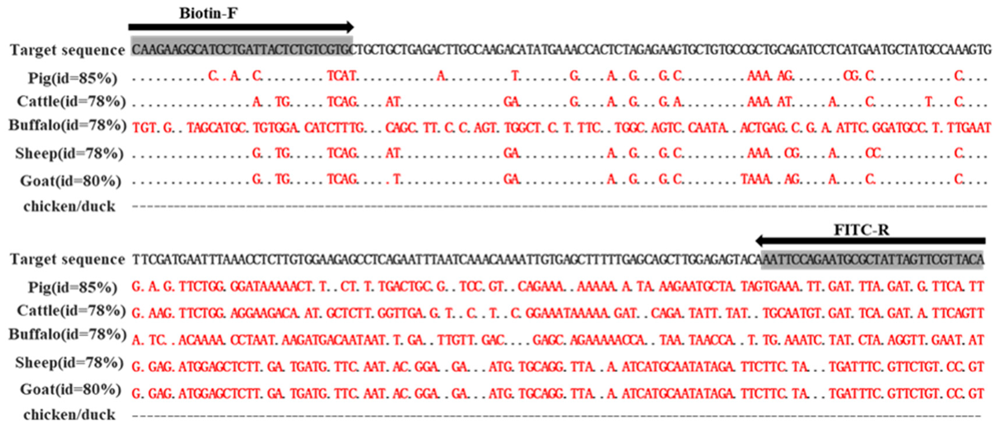
2.2. RPA Primer Design

To identify HSA GM goats, we first screened the genome to find a conservative sequence of HSA as a candidate sequence. The sequence of the HSA gene was critically evaluated against that of different species (pigs, cattle, buffalos, sheep, goats, chickens, and ducks) using BLAST software. To further analyze the homology of the sequence, target sequences of different species were downloaded from the Ensembl database (http://asia.ensembl.org/index.html, accessed on 3 February 2019) and aligned with the HSA sequence using ClustalW software. As shown in Figure 1, the candidate sequence showed high specificity in humans compared with the sequences of other species. It had a low identity with pig (85%), cattle (78%), buffalo (78%), sheep (78%), goat (80%), chicken (0%), and duck (0%) sequences. On the basis of the instruction manuals of the TwistAmp®nfo kits, Primer Premier 5.0 software was used to design species-specific RPA primers (30–35 bp, GC content 30–70%) without probes for RPA detection in the selected HSA-specific sequence. Figure 1 shows primer mismatches with sequences from other species in the primer binding region. A pair of preferred primers were selected for RPA analysis by screening the candidate primers. The primers were chemically modified for LFD detection. A normal forward primer was labeled Biotin at the 5′end (Biotin-F: 5′-Biotin-CAAGAAGGCATCCTGATTACTCTGTCGTGC-3′), and FITC was labeled at the 5′end of the reverse primer (FITC-R: 5′-FITC-TGTAACGAACTAATAGCGCATTCTGGAATT-3′). Specific primers were applied to detect HSA transgenic goats using RPA (Table 1).

2.3. RPA Reaction System Optimization

RPA amplification was conducted according to the operation manual of the TwistAmp® Basic kit. Each RPA reaction was conducted in an RPA tube of 50 μL reaction mixture: FITC-R/Biotin-F (10 μM), a DNA template of 2 μL (50 ng/µL), ddH2O, and a rehydration buffer of 29.5 μL. All components were added to the freeze-dried pellet and thoroughly mixed. To initiate the reaction, 2.5 μL of 280 mM magnesium acetate (MgAc) was added. Amplification results were detected by approximately 1.5% agarose gel electrophoresis (AGE).
To establish the RPA assay, initial investigations were carried out to explore primer concentrations and to test different amplification temperatures. Four concentrations, 0.1 µmol/L, 0.15 µmol/L, 0.2 µmol/L, and 0.25 µmol/L, were selected to determine the optimal primer concentration. Then, RPA amplification was performed at a 38–42 °C (2 °C per gradient interval) reaction temperature under the conditions of the optimized primer concentration.

2.4. Preparation of Antibody-Modified Gold Nanoparticles

Gold nanoparticles were prepared following previously reported methods [37,38]. Briefly, 2 mL of 1% HAuCl4 and 1% trisodium citrate (3.6 mL) were slowly added to 200 mL of boiled ddH2O. Then, the solution was boiled to become a stable wine-red color. Then, 0.1 mol/L K2CO3 (150 µL) and 10 mM monoclonal FITC antibody (60 µL) were added to the AuNPs solution (10 mL) to enable the coupling of the AuNPs and antibody. After that, 10% BSA solution (1.2 mL) and 2% PEG2000 (292 µL) were added to the coupling solution. Then, the AuNP antibody conjugate was centrifuged for 30 min and suspended in 2 mL borate buffer solution. The conjugates were stored at 4 °C for further use.

2.5. Preparation of Lateral Flow Dipsticks

We assembled LFDs with four different types of functional components, namely, a sample pad, conjugate pad, nitrocellulose membrane, and absorbent pad, by partial superimposition. The sample pad was used for sample loading. The conjugate pad was made of a cellulose film for the release of AuNP-FITC conjugates. For signal generation, monoclonal Biotin antibody solution (1 mg/mL) was sprayed on the nitrocellulose membranes to generate test lines (T line), and FITC secondary antibody solution (1 mg/mL) was sprayed on the nitrocellulose membranes as the control line (C line). The sample pad, conjugate pad, nitrocellulose membrane, and absorbent were assembled on a support plate to form the LFD.

2.6. Detection of GM Samples with RPA-LFD

For PRA amplification product detection, 48 μL of running buffer was used to dilute 2 μL amplification products, and the mixture was added to the sample pad of the LFD for 5–10 min. The results were determined by observing the colors of the test lines.

3. Results

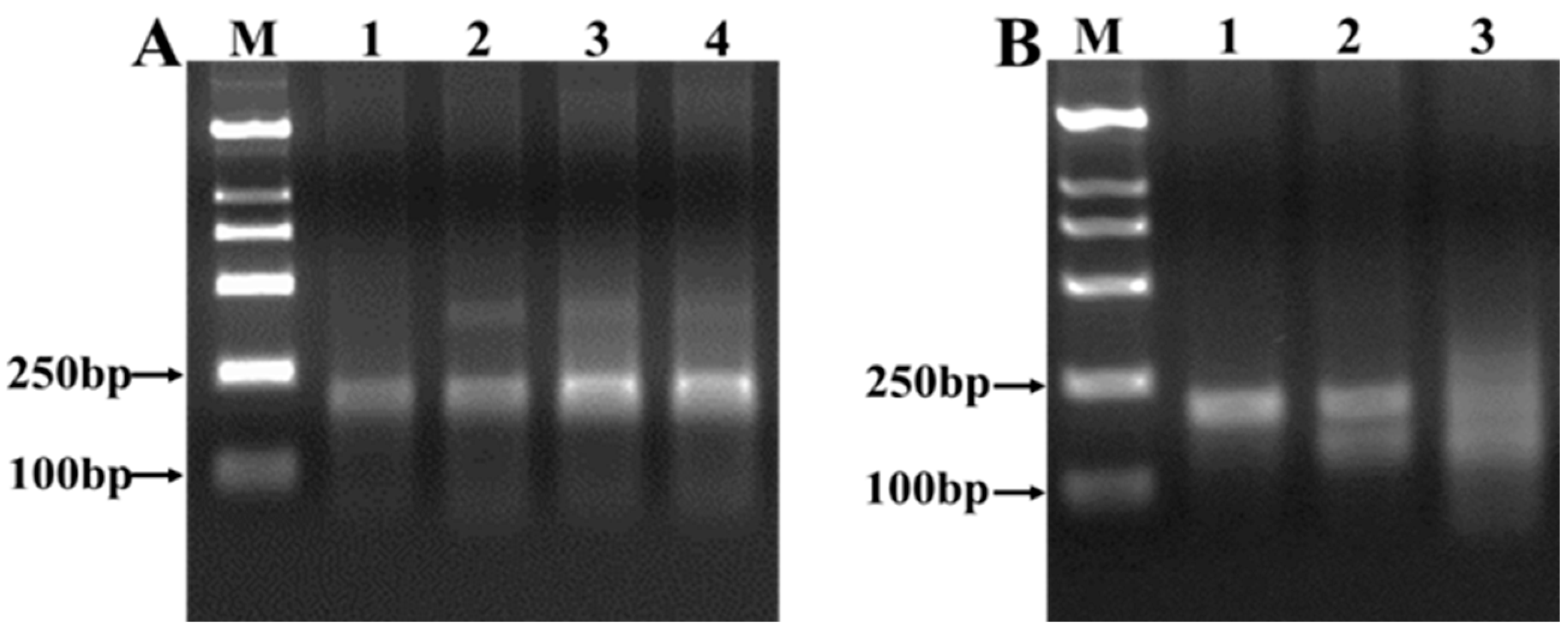
3.1. Optimization of RPA-LFD Assay

To improve the performance of RPA-LFD, different culture temperatures and times were selected to explore the appropriate conditions for RPA-LFD detection. Numerous factors affect RPA amplification reactions. To improve the performance of RPA-LFD, we reduced the number of members of the reaction system without probes to decrease the complexity of the reaction. Primer dimers have been found to cause false positives due to the accumulation of AuNPs on the test line [19] and to strongly influence the efficiency of RPA. In our study, the concentrations of the primers were optimized to avoid primer dimers. The suitable primer concentrations for the RPA-LFD assay were explored by selecting four different primer concentrations (1 µmol/L, 0.15 µmol/L, 0.2 µmol/L, and 0.25 µmol/L). The transgenic goat DNA was used as a template to determine the suitable RPA-LFD conditions, which amplified the 231bp DNA product. RPA amplification was carried out at a suitable reaction primer concentration of 0.1 µmol/L, because there is no non-specific amplification and primer dimer at this primer concentration for RPA amplification (Figure 2A).
The RPA reaction mixture was used at three temperatures (38–42 °C) for 30 min at the optimized primer concentration to explore the suitable reaction temperature, and the reaction time was chosen based on the previous experiments of our research team [30]. The detection result showed that the suitable reaction temperature was 42 °C (Figure 2B). RPA can only be amplified with one pair of primers. Compared with Loop-mediated isothermal amplification (LAMP), this design is relatively simple and convenient [39]. The established method is suitable for the detection of transgenic goats, which is fully demonstrated by these advantages.

3.2. Specificity of RPA-LFD Assay

The specificity of primers plays critical role in the detection of genetically modified animals. To assess the specificity of the established RPA-LFD method, the genomic DNAs extracted from seven non-transgenic animals, namely, goats, pigs, cattle, buffalos, sheep, chickens, and ducks, were tested using RPA-LFD as shown in Figure 3. Consistent with the expected results, LFB only showed a positive signal in the positive control (PC) samples of the transgenic goats, and a control line and a test line were simultaneously observed in the detection zone. Moreover, the other non-transgenic samples only showed a control line, with no color in the test line. These results indicate that the RPA-LFD assay can successfully distinguish its target DNA from that of non-transgenic animals without cross-reaction or false positive signals were observed, and that the HSA transgenic RPA-LFD assay is specific for the detection of its corresponding target.

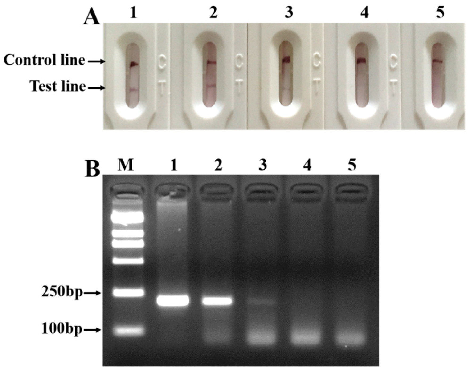
3.3. Sensitivity of RPA-LFD

The RPA-LFD system showed effective performance, including sensitivity and stability. To determine the detection threshold of the RPA-LFD assay, a dilution series of transgenic goat DNA with concentrations of 0.01, 0.1, 10, and 50 ng were performed for the RPA-LFD sensitivity analysis. The template for the negative control was sterile ultrapure water. As shown in Figure 4, as the concentration of the template decreased, the red band in the test line gradually weakened until it faded, and the amplification products decreased. When the concentration was reduced to 0.1 ng, a light red band still appeared on the test line. However, when the content was lower than 0.1 ng, the detection signal could not be observed (Figure 4A). Therefore, the detection limit of the RPA-LFD method is 0.1 ng, which means it is highly sensitive.
The sensitivity of the RPA-LFD assay was further compared with that of conventional PCR, followed by agarose gel electrophoresis (PCR-AGE). A conventional PCR was performed using the same DNA template and RPA primers as described above. The results indicate that the sensitivity of the RPA-LFD assay was equivalent to that of a conventional PCR of 0.1 ng (Figure 4B).

3.4. Application of RPA-MLFD in GM Goats

Finally, to test the efficiency of the RPA-LFD assay screening of genetically modified samples, the DNAs of nine animals (transgenic goats and non-transgenic goats) were selected as templates to validate the reliability of RPA-LFD. All samples were tested according to the optimized RPA-LFD assay reaction system, and the result can be observed in Figure 5. According to the results, the test line was present in eight transgenic samples. However, the test line was not observed in the negative and blank controls, and the LFD worked normally. The RPA-LFD test results are consistent with the information provided by the sample developer.

4. Discussion

In this study, we propose a high-sensitivity, easy-to-use, less time-consuming, and cost-efficient RPA combined with the LFD analysis method for the detection of HSA in transgenic goats. A pair of primers targeting the conserved region of HSA was designed, and the target product was successfully amplified in less than 1 h under isothermal conditions. The specificity of this method was confirmed by the fact that no amplification products were observed in non-transgenic samples. Therefore, the RPA method is an effective and proper method with high credibility for the detection of exogenous nucleic acid in HSA transgenic goats.
However, the RPA-LFD method was performed in isothermal conditions without thermal cyclers. The RPA method only took 30 min, while PCR took 2 h in our study. Furthermore, we compared the reported methods for the detection of GMOs (Table 2). Compared with PCR methods, the RPA-LFD method does not require an expensive thermal cycler, and it is rapid and more convenient. The RPA-LFD method is faster and more convenient than other isothermal nucleic acid amplification methods (such as the LAMP method) because it only takes tens of minutes to complete the entire reaction time and the primer design is relatively simple. Therefore, the RPA-LFD method has great advantages for the detection of transgenic animals.

5. Conclusions

In this study, we established a novel RPA-LFD assay for HSA detection. The whole RPA-LFD reaction process can be completed within 1 h, and the results can be determined by the naked eye without using additional instruments. The detection limits of the RPA-LFD and PCR were as low as 0.1 ng in our study. This meets the national threshold of transgenic detection. In addition, we also tested real transgenic samples, and the detection rate achieved 100%, indicating that the method has good repeatability. In this study, the RPA-LFD method for the detection of GMO events was tested and demonstrated in terms of specificity, stability, and reproducibility. In conclusion, this method offers significant advantages, not only because it is rapid, has a low-cost, and is easy to use, but also because it is highly specific, sensitive, visual, and credible. Thus, it might be a rapid, convenient, and accurate detection system for GMO.

Author Contributions

Conceptualization and methodology, Q.S., Y.Z. and B.L.; experiments, Q.S.; writing—original draft preparation, Q.S. and Y.Z.; writing—review and editing, Y.Z., B.L., K.G. and X.Z. All authors have read and agreed to the published version of the manuscript.

Funding

This work was supported by the National Science and Technology Major Project of China (2018ZX08012-001).

Institutional Review Board Statement

Not applicable.

Informed Consent Statement

Not applicable.

Data Availability Statement

The data presented in this study are available in this article.

Conflicts of Interest

The authors declare no conflict of interest.

Abbreviations

GM, genetically modified; HSA, human serum albumin; RPA, recombinase polymerase amplification; LFD, lateral flow dipstick; SCNT, somatic cell nuclear transfer; GMOs, genetically modified organisms; LAMP, loop-mediated isothermal amplification.

References

  1. Huang, L.F.; Liu, Y.K.; Lu, C.A.; Hsieh, S.L.; Yu, S.M. Production of human serum albumin by sugar starvation induced promoter and rice cell culture. Transgenic Res. 2005, 14, 569–581. [Google Scholar] [CrossRef] [PubMed]
  2. Peters, T. The albumin molecule. In All about Albumin: Biochemistry, Genetics and Medical Applications Chapter; Academic Press: Cambridge, MA, USA, 1995; Volume 6, pp. 251–284. [Google Scholar]
  3. Qian, Q.; You, Z.; Ye, L.; Che, J.; Wang, Y.; Wang, S.; Zhong, B. High-efficiency production of human serum albumin in the posterior silk glands of transgenic silkworms, Bombyx mori L. PLoS ONE 2018, 13, e0191507. [Google Scholar] [CrossRef] [PubMed] [Green Version]
  4. Chamberland, M.E.; Alter, H.J.; Busch, M.P.; Nemo, G.; Ricketts, M. Emerging infectious disease issues in blood safety. Emerg. Infect. Dis. 2001, 7, 552–553. [Google Scholar] [CrossRef] [PubMed]
  5. MacLennan, S.; Barbara, J.A.J. Risks and side effects of therapy with plasma and plasma fractions. Best Pract. Res. Clin. Haematol. 2006, 19, 169–189. [Google Scholar] [CrossRef]
  6. Tao, C.; Zhang, Q.; Feng, N.; Shi, D.; Liu, B. Development of a colloidal gold immunochromatographic strip assay for simple and fast detection of human α-lactalbumin in genetically modified cow milk. J. Dairy Sci. 2016, 99, 1773–1779. [Google Scholar] [CrossRef]
  7. Luo, Y.; Wang, Y.; Liu, J.; Lan, H.; Shao, M.; Yu, Y.; Quan, F.; Zhang, Y. Production of transgenic cattle highly expressing human serum albumin in milk by phiC31 integrase-mediated gene delivery. Transgenic Res. 2015, 24, 875–883. [Google Scholar] [CrossRef]
  8. Samiec, M.; Skrzyszowska, M. Transgenic mammalian species, generated by somatic cell cloning, in biomedicine, biopharmaceutical industry and human nutrition/dietetics—Recent achievements. Pol. J. Vet. Sci. 2011, 14, 317–328. [Google Scholar] [CrossRef] [Green Version]
  9. He, Z.; Lu, R.; Zhang, T.; Jiang, L.; Zhou, M.; Wu, D.; Cheng, Y. A novel recombinant human plasminogen activator: Efficient expression and hereditary stability in transgenic goats and in vitro thrombolytic bioactivity in the milk of transgenic goats. PLoS ONE 2018, 13, e0201788. [Google Scholar] [CrossRef]
  10. Skrzyszowska, M.; Samiec, M. Generating cloned goats by somatic cell nuclear transfer—Molecular determinants and application to transgenics and biomedicine. Int. J. Mol. Sci. 2021, 22, 7490. [Google Scholar] [CrossRef]
  11. Zhang, Q.; Chen, J.Q.; Lin, J.; Yu, Q.H.; Yu, H.Q.; Xu, X.J.; Liu, G.H.; Yang, Q. Production GH transgenic goat improving mammogenesis by somatic cell nuclear transfer. Mol. Biol. Rep. 2014, 41, 4759–4768. [Google Scholar] [CrossRef]
  12. Zhu, H.; Hu, L.; Liu, J.; Chen, H.; Cui, C.; Song, Y.; Jin, Y.; Zhang, Y. Generation of β-lactoglobulin-modified transgenic goats by homologous recombination. FEBS J. 2016, 283, 4600–4613. [Google Scholar] [CrossRef] [PubMed]
  13. Baguisi, A.; Behboodi, E.; Melican, D.T.; Pollock, J.S.; Destrempes, M.M.; Cammuso, C.; Williams, J.L.; Nims, S.D.; Porter, C.A.; Midura, P.; et al. Production of goats by somatic cell nuclear transfer. Nat. Biotechnol. 1999, 17, 456–461. [Google Scholar] [CrossRef] [PubMed]
  14. Yuan, Y.G.; Song, S.Z.; Zhu, M.M.; He, Z.Y.; Lu, R.; Zhang, T.; Mi, F.; Wang, J.Y.; Cheng, Y. Human lactoferrin efficiently targeted into caprine beta-lactoglobulin locus with transcription activator-like effector nucleases. Asian Australas. J. Anim. Sci. 2017, 30, 1175–1182. [Google Scholar] [CrossRef] [PubMed] [Green Version]
  15. Tao, C.; Zhang, Q.; Zhai, S.; Liu, B. Detection of HbsAg and hATIII genetically modified goats (Caprahircus) by loop-mediated isothermal amplification. Mol. Biol. Rep. 2013, 40, 6177–6182. [Google Scholar] [CrossRef]
  16. Singh, R.K.; Singh, V.K.; Raghavendrarao, S.; Phanindra, M.L.; Venkat Raman, K.; Solanke, A.U.; Kumar, P.A.; Sharma, T.R. Expression of finger millet EcDehydrin7 in transgenic tobacco confers tolerance to drought stress. Appl. Biochem. Biotechnol. 2015, 177, 207–216. [Google Scholar] [CrossRef]
  17. Deng, H.; Gao, Z. Bioanalytical applications of isothermal nucleic acid amplification techniques. Anal. Chim. Acta 2015, 853, 30–45. [Google Scholar] [CrossRef]
  18. Notomi, T.; Mori, Y.; Tomita, N.; Kanda, H. Loop-mediated isothermal amplification (LAMP): Principle, features, and future prospects. J. Microbiol. 2015, 53, 1–5. [Google Scholar] [CrossRef]
  19. Zhai, S.; Liu, C.; Zhang, Q.; Tao, C.; Liu, B. Detection of two exogenous g enes in transgenic cattle by loop-mediated isothermal amplification. Transgenic Res. 2012, 21, 67–73. [Google Scholar] [CrossRef]
  20. Piepenburg, O.; Williams, C.H.; Stemple, D.L.; Armes, N.A. DNA detection using recombination proteins. PLoS Biol. 2006, 4, e204. [Google Scholar] [CrossRef]
  21. Safenkova, I.V.; Ivanov, A.V.; Slutskaya, E.S.; Samokhvalov, A.V.; Zherdev, A.V.; Dzantiev, B.B. Key significance of DNA-target size in lateral flow assay coupled with recombinase polymerase amplification. Anal. Chim. Acta 2020, 1102, 109–118. [Google Scholar] [CrossRef]
  22. Lobato, I.M.; O’Sullivan, C.K. Recombinase polymerase amplification: Basics, applications and recent advances. TRAC Trends Anal. Chem. 2018, 98, 19–35. [Google Scholar] [CrossRef] [PubMed]
  23. Santiago-Felipe, S.; Tortajada-Genaro, L.A.; Puchades, R.; Maquieira, A. Recombinase polymerase and enzyme-linked immunosorbent assay as a DNA amplification-detection strategy for food analysis. Anal. Chim. Acta 2014, 811, 81–87. [Google Scholar] [CrossRef] [PubMed]
  24. Loo, J.F. An aptamer-based bio-barcode assay with isothermal recombinase polymerase amplification for cytochrome-c detection and anti-cancer drug screening. Talanta 2013, 115, 159–165. [Google Scholar] [CrossRef] [PubMed]
  25. Babu, B.; Washburn, B.K.; Miller, S.H.; Poduch, K.; Sarigul, T.; Knox, G.W.; Ochoa-Corona, F.M.; Paret, M.L. A rapid assay for detection of rose rosette virus using reverse transcription-recombinase polymerase amplification using multiple gene targets. J. Virol. Methods 2017, 240, 78–84. [Google Scholar] [CrossRef] [PubMed] [Green Version]
  26. Londoño, M.A.; Harmon, C.L.; Polston, J.E. Evaluation of recombinase polymerase amplification for detection of begomviruses by plant diagnostic clinics. Virol. J. 2016, 13, 48. [Google Scholar] [CrossRef] [PubMed] [Green Version]
  27. Li, K.; Luo, Y.; Huang, K.; Yang, Z.; Wan, Y.; Xu, W. Single universal primer recombinase polymerase amplification-based lateral flow biosensor (SUP-RPA-LFB) for multiplex detection of genetically modified maize. Anal. Chim. Acta 2020, 1127, 217–224. [Google Scholar] [CrossRef]
  28. Li, J.; Macdonald, J. Advances in isothermal amplification: Novel strategies inspired by biological processes. Biosens. Bioelectron. 2015, 64, 196–211. [Google Scholar] [CrossRef]
  29. Cheng, N.; Shang, Y.; Xu, Y.; Zhang, L.; Luo, Y.; Huang, K.; Xu, W. On-site detection of stacked genetically modified soybean basedon event-specific TM-LAMP and a DNAzyme-lateral flow biosensor. Biosens. Bioelectron. 2017, 91, 408–416. [Google Scholar] [CrossRef]
  30. Cui, D.; Zhai, S.; Yang, Y.; Wu, Y.; Li, J.; Yan, X.; Shen, P.; Gao, H.; Wu, G. A Label-Free electrochemical impedance genosensor coupled with recombinase polymerase amplification for genetically modified maize detection. Agriculture 2022, 12, 454. [Google Scholar] [CrossRef]
  31. Gomez-Martinez, J.; Silvy, M.; Chiaroni, J.; Fournier-Wirth, C.; Roubinet, F.; Bailly, P.; Brès., J.C. Multiplex lateral flow assay for rapid visual blood group genotyping. Anal. Chem. 2018, 90, 7502–7509. [Google Scholar] [CrossRef]
  32. Anfossi, L.; D’Arco, G.; Calderara, M.; Baggian, C.; Giovannoli, C.; Giraudi, G. Development of a quantitative lateral flow immunoassay for the detection of aflatoxins in maize. Food Addit. Contam. Part A 2011, 28, 226–234. [Google Scholar] [CrossRef] [PubMed]
  33. Gao, W.; Huang, H.; Zhu, P.; Yan, X.; Fan, J.; Jiang, J.; Xu, J. Recombinase polymerase amplification combined with lateral flow dipstick for equipment-free detection of salmonella in shellfish. Bioprocess Biosyst. Eng. 2018, 4, 603–611. [Google Scholar] [CrossRef] [PubMed]
  34. Nair, G.; Rebolledo, M.; White, A.C.; Jr Crannell, Z.; Richards-Kortum, R.R.; Pinilla, A.E.; Ramírez, J.D.; López, M.C.; Castellanos-Gonzalez, A. Detection of Entamoeba histolytica by Recombinase polymerase amplification. Am. J. Trop. Med. Hyg. 2015, 93, 591–595. [Google Scholar] [CrossRef] [PubMed]
  35. Zhao, M.; Wang, B.; Xiang, L.; Xiong, C.; Shi, Y.; Wu, L.; Meng, X.; Dongd, G.; Xie, Y.; Sun, W. A novel onsite and visual molecular technique to authenticate saffron (Crocus sativus) and its adulterants based on recombinase polymerase amplification. Food Control 2019, 100, 117121. [Google Scholar] [CrossRef]
  36. Ebbehoj, K.F.; Thomsen, P.D. Species differentiation of heated meat products by DNA hybridization. Meat Sci. 1991, 30, 221–234. [Google Scholar] [CrossRef]
  37. Frens, G. Controlled nucleation for regulation of particle-size in monodisperse gold suspensions. Nat. Phys. Sci. 1973, 241, 20–22. [Google Scholar] [CrossRef]
  38. Fu, M.; Zhang, Q.; Zhou, X.; Liu, B. Recombinase polymerase amplification based multiplex lateral flow dipstick for fast identification of duck ingredient in adulterated beef. Animal 2020, 10, 1765. [Google Scholar] [CrossRef]
  39. Deb, R.; Sengar, G.S.; Singh, U.; Kumar, S.; Raja, T.V.; Alex, R.; Alyethodi, R.R.; Prakash, B. LAMP assay for rapid diagnosis of cow DNA in goat milk and meat samples. Iran. J. Vet. Res. 2017, 18, 134–137. [Google Scholar]
  40. Rott, M.E.; Lawrence, T.S.; Wall, E.M.; Green, M.J. Detection and quantification of roundup ready soy in foods by conventional and real-time polymerase chain reaction. J. Agric. Food Chem. 2004, 52, 5223–5232. [Google Scholar] [CrossRef]
  41. Košir, A.B.; Spilsberg, B.; Holst-Jensen, A.; Žel, J.; Dobnik, D. Development and inter-laboratory assessment of droplet digital PCR assays for multiplex quantification of 15 genetically modified soybean lines. Sci. Rep. 2017, 9, 8601. [Google Scholar] [CrossRef] [Green Version]
  42. Berdal, K.G.; Holst-Jensen, A. Event-specific qualitative and quantitative PCR detection of the the practical detection and quantification limits in GMO analyses. Eur. Food Res. Technol. 2001, 213, 432–438. [Google Scholar] [CrossRef]
  43. Guan, X.; Guo, J.; Shen, P.; Yang, L.; Zhang, D. Visual and rapid detection of two genetically modified soybean events using loop-mediated isothermal amplification method. Food Anal. Methods 2010, 3, 313–320. [Google Scholar] [CrossRef]
Figure 1. Specificity analysis of target sequence (transgenic goats) and location of the RPA primers. BLAST and ClustalW results of target sequence (transgenic goats) with genomic sequences of other species (non-transgenic): pigs (85%), cattle (78%), buffalos (78%), sheep (78%), goats (80%), chickens, and ducks (0%). Biotin-F: forward primer, FITC-R: reverse primer.
Agriculture 12 00927 g001
Figure 2. Optimized conditions of RPA: (A) Optimization of the concentration of primers. Lane M represents Marker 2000. Lanes 1–4 represent 0.1 µmol/L, 0.15 µmol/L, 0.2 µmol/L, and 0.25 µmol/L, respectively. (B) Optimization of the reaction temperature. Lane M represents Marker 2000. Lanes 1–3 represent 42 °C, 40 °C, and 38 °C, respectively.
Agriculture 12 00927 g002
Figure 3. Specificity assay of RPA-LFD: PC, positive control, transgenic goat DNA; NC, negative control, non-transgenic goat DNA; lines 1–6 represent DNA of non-transgenic animals, namely, pigs, cattle, buffalo, sheep, chickens, and ducks, respectively; BC, blank control, water.
Agriculture 12 00927 g003
Figure 4. Comparison of the sensitivity of RPA-LFD and conventional PCR: (A) sensitivity of RPA-LFD, (B) sensitivity of conventional PCR. M, Marker 2000; lines 1–5 represent 50 ng, 10 ng, 0.1 ng, 0.01 ng, and blank control, respectively.
Agriculture 12 00927 g004
Figure 5. Detection of transgenic samples: NC, negative control, non-transgenic goat DNA; BC, blank control, water. Lines 1–8 represent transgenic goat DNA.
Agriculture 12 00927 g005
Table 1. Oligonucleotide primers for the RPA-LFD assay in this study.
Primer NameSequence (5′–3′)
Biotin-FBiotin-CAAGAAGGCATCCTGATTACTCTGTCGTGC
FITC-RFITC-TGTAACGAACTAATAGCGCATTCTGGAATT
Table 2. Comparison of RPA-LFD with other methods for detection of GMOs.
Analytical MethodInstrument
(Thermal Cycler)
Time for Detection aSuitability for On-Site TestingReference
Conventional PCRNeedAbout 2 hUnsuitable[40]
ddPCRNeed>3 hUnsuitable[41]
Real-time PCRNeed>2.5 hUnsuitable[42]
LAMPNo needAbout 2 hSuitable[43]
RPA-LFDNo needAbout 30 minSuitableThis study
a It does not include DNA extraction time.
Publisher’s Note: MDPI stays neutral with regard to jurisdictional claims in published maps and institutional affiliations.

