 Bo Zhang1,2†
 Geng Qin
 Zelin Chen
 Qiang Lin
- 1Key Laboratory of South China Sea Fishery Resources Exploitation & Utilization, Ministry of Agriculture and Rural Affairs, South China Sea Fisheries Research Institute, Chinese Academy of Fishery Sciences, Guangzhou, China
- 2Key Laboratory of Aquatic Product Processing, Ministry of Agriculture and Rural Affairs, South China Sea Fisheries Research Institute, Chinese Academy of Fishery Sciences, Guangzhou, China
- 3CAS Key Laboratory of Tropical Marine Bio-Resources and Ecology, South China Sea Institute of Oceanology, Chinese Academy of Sciences, Guangzhou, China
- 4Southern Marine Science and Engineering Guangdong Laboratory (Guangzhou), South China Sea Institute of Oceanology, Chinese Academy of Sciences, Guangzhou, China
- 5Sanya Institute of Ocean Eco-Environmental Engineering, Sanya, China
Male pregnancy in syngnathids (seahorses, pipefishes, and sea dragons) is an evolutionary innovation in the animal kingdom. Paternal immune resistance to the fetus is a critical challenge, particularly in seahorses with fully enclosed brood pouches and sophisticated placentas. In this study, comparative genomic analysis revealed that all syngnathid species lost three vertebrate-conserved Toll-like receptors (TLR1, TLR2, and TLR9), of which all play essential roles in immune protection and immune tolerance in the uterus and placenta. Quantitative real-time PCR (qRT-PCR) analysis showed that the TLR paralog genes including TLR18, TLR25, and TLR21 were highly expressed in the placenta inside the seahorse brood pouch and changed dynamically during the breeding cycle, suggesting the potentially important role of the TLRs during male pregnancy. Furthermore, the immune challenge test in vitro showed a remarkable expression response from all three TLR genes to specific pathogenic antigens, confirming their immune function in seahorse brood pouches. Notably, the altered antigen recognition spectrum of these genes appeared to functionally compensate in part for the lost TLRs, in contrast to that observed in other species. Therefore, we suggest that gene loss and co-option of TLRs may be a typical evolutionary strategy for facilitating paternal immunological adaptation during male pregnancy.
1 Introduction
Pregnancy is one of the most significant achievements in vertebrate evolution (1). Offspring survival rate is remarkably improved via pregnancy by providing an ideal environment for embryo development and protecting the embryos from adverse external conditions (1). Pregnant mothers usually develop adaptive immune functions in the uterus, which protect the fetus via different immune pathways (2). However, a fundamental problem in the evolution of pregnancy among organisms is that pregnant mothers must avoid non-self-embryo rejection (3). Thus, appropriate maternal–fetal immune tolerance adjustments are vital for a successful pregnancy in viviparous animals (4).
The Syngnathidae family comprises seahorses, pipefish, and sea dragons, which are well known for their unique male pregnancy (5, 6). Males of most syngnathid species have evolved a brood pouch that function similarly to the mammalian uterus (7, 8). Seahorse brood pouch exhibits the most sophisticated morphological structures, a pocket-like structure in which eggs are embedded. The structures of seahorse brood pouch can be roughly divided into two layers: a folded inner pseudostratified columnar epithelium (termed placenta) and a smooth outer stratified cuboidal epithelium. The placenta serves as the site of embryo attachment (7, 9). During breeding cycle, the structure of the placenta exhibited a comparable physiological cycle to mammals uterus (9). It can be divided into three sequential cycle stages: the normal stage (non-pregnant stage), pregnant stage and the repair stage (10). The brood pouch not only provides shelter, nutrition, and immune protection for the embedded embryos, but also aids in avoiding rejection of the non-self-embryo from the pregnant father (8, 11–13). Previous studies have shown that the syngnathid immune system evolved via gene loss, mutation (14, 15), or expansion (13), thereby achieving a balance between immunological protection and embryo tolerance (10, 16). Thus, pregnant male syngnathids provide an excellent model for investigating the evolution of immunological adaptations during pregnancy (17).
The Toll-like receptor (TLR) gene family encodes pathogen recognition receptors (PRRs) of the immune system and plays vital roles in host immune responses (18, 19). As typical PRRs, TLRs discern invading microorganisms by recognizing pathogen-associated molecular patterns, leading to the activation of innate immune response or the development of antigen-specific acquired immunity (20, 21). Compared to that of other invertebrates, the TLR gene family in vertebrates evolved relatively conservatively (20). In addition, duplication, pseudogenization, loss, and positive selection of TLR members have been observed, particularly in species that have adapted to unique pathogenic environments (18, 22). TLRs within a subfamily usually recognize similar pathogen-associated molecular patterns (20). For example, members of the TLR1 subfamily (including TLR1, TLR2, TLR18, and TLR25) have markedly similar ligand recognition profiles, including bacterial lipopolysaccharides (LPS), lipoteichoic acid (LTA), and peptidoglycan (PGN), etc. Although TLR9 and TLR21 belong to different subfamilies, both recognize bacterial and viral CpG-deoxynucleotides containing DNA (CpG-DNA) (23, 24). Therefore, compensatory effects are common among existing TLR members when certain TLRs lose their function (24).
TLR proteins are involved in immune activation and play important roles in mammalian pregnancy (25) by balancing host resistance and immune tolerance in the uterus and placenta during pregnancy (26, 27). Several TLRs such as TLR1, TLR2, and TLR9 are expressed in placental immune cells (T cells and regulatory T (TReg)), which directly participate in maternal-fetal immune tolerance during pregnancy (28, 29) and non-immune cells (such as trophoblasts and decidual cells) of the mammalian uterus and placenta (30). To guarantee conception and pregnancy, TLRs undergo remarkable expression changes (25, 31), which regulate maternal tolerance to allogenic fetuses and maintain innate immune responses to microorganisms (32).
To understand the potential function of the TLR gene family in paternal immunological adaptation in male pregnant syngnathids, we analyzed TLR families among syngnathids and other teleost species based on genome comparison. The expression patterns of TLRs with potential functions were detected in the placentas of seahorse brood pouches during the breeding cycle. Moreover, seahorse TLR ligands were recognized via an immune challenge in vitro to verify their immune function during pregnancy. Consequently, the immunological adaptive mechanisms caused by the TLR gene family evolution were successfully identified in syngnathids.
2 Results
2.1 TLR phylogenetic and comparative analyses
Through genomic and transcriptome data analysis, TLR gene family members were identified in five Syngnathidae species: nine TLR genes were identified in lined and tiger tail seahorses, and 10 TLRs were identified in weedy, alligator, and manado pipefish (Figure 1). The phylogenetic tree divided Syngnathidae TLRs into five subfamilies: TLR1, TLR3, TLR5, TLR7, and TLR11 (Figure 2A). Similar to most teleosts, Syngnathidae lack the TLR4 subfamily. The TLR1 subfamily comprises TLR18 and TLR25, in addition to TLR1, TLR2, and TLR14. The TLR3 and TLR5 subfamilies include only one gene member each; namely TLR3 and TLR5, respectively. TLR7, TLR8, and TLR 9 formed the TLR7 subfamily; TLR21 and TLR22 were clustered in a clade under the TLR11 subfamily. Compared with that of other teleosts containing at least 12 TLR members, the TLR gene family was contracted in Syngnathidae fishes. In all five Syngnathidae fishes TLR1 subfamily, TLR1 and TLR2 were lost except TLR25, which was present only in a few species. Notably, TLR25 even showed an additional copy in weedy and alligator pipefish. Of the five Syngnathidae species, TLR9 was absent in four excluding manado pipefish.
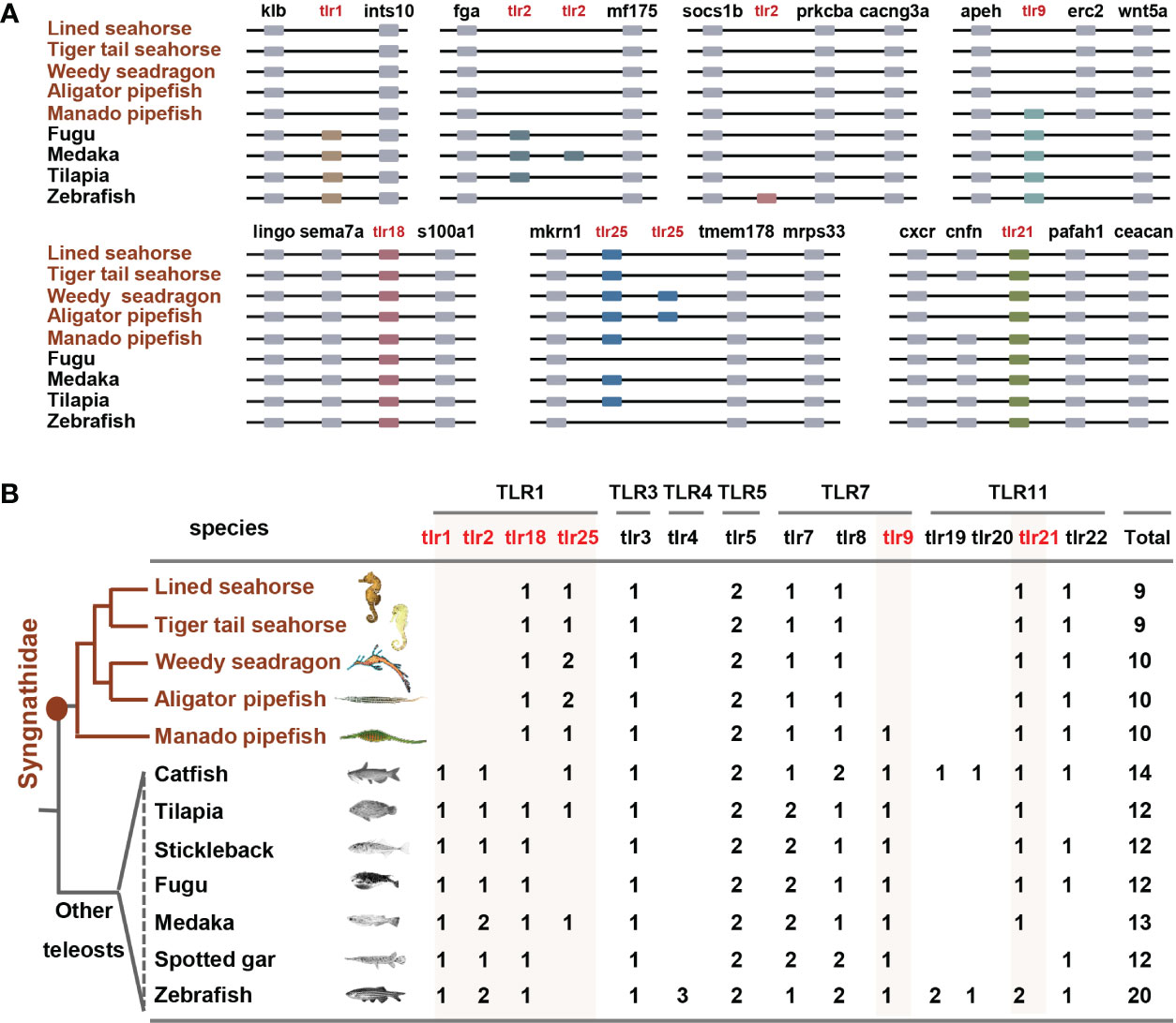
Figure 1 TLR gene family contracted in Syngnathidae fishes. (A) syntenic analysis plots show TLRs and their upstream and downstream genes, which are sequentially arranged and connected by black lines. Partial synteny map of the genomic region surrounding TLR family genes. (B) Statistical plot of TLR gene family in teleosts. TLR1, TLR2 and TLR9 were lost in Syngnathidae. Syngnathidae fishes and the focus TLRs are highlighted in red.
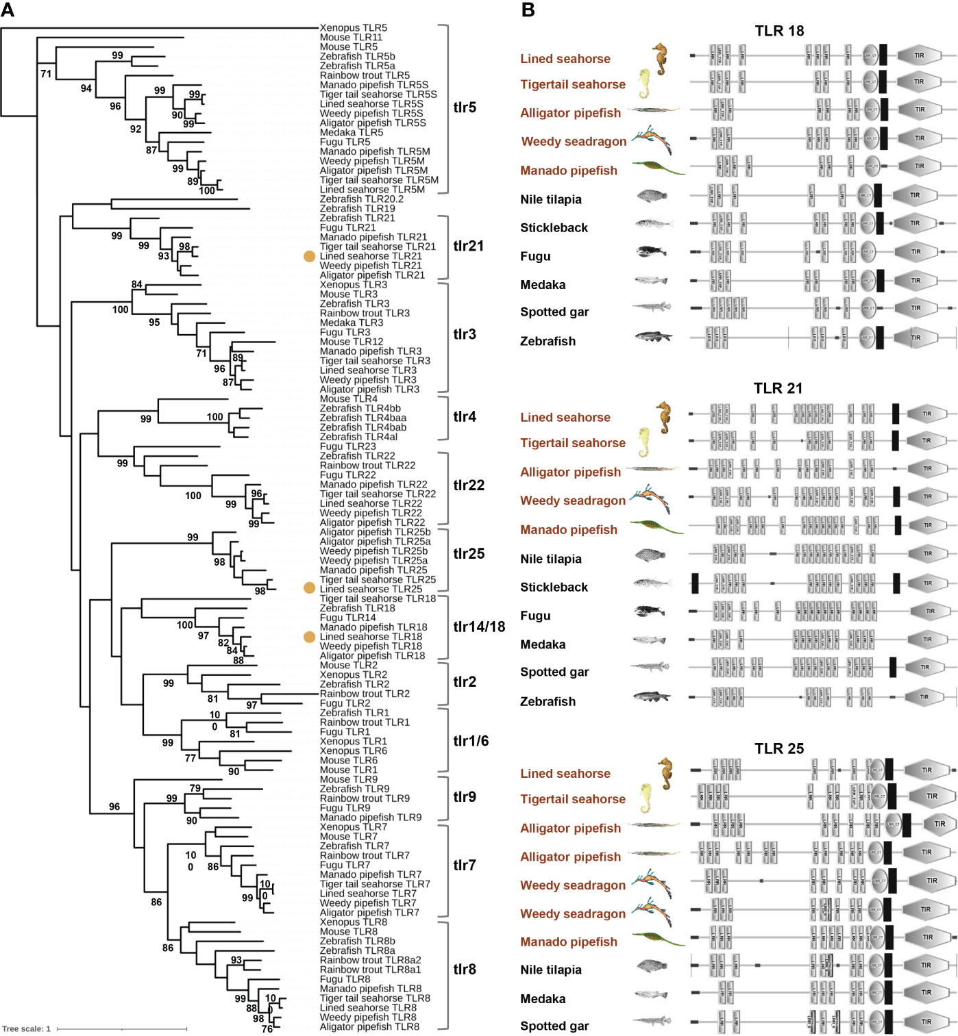
Figure 2 Phylogenetic analysis of teleost TLRs. (A) TLR18, TLR21, and TLR25 of lined seahorse were note in colored dot. (B) Protein domain structures of TLR18, TLR21 and TLR25 in teleosts. LRR, leucine-rich repeat; LRR-TYP, leucine-rich repeat typical subfamily; TIR, Toll/IL-1 receptor; NT, N-(nitrogen) terminal; CT, C-(carboxyl) terminal.
Further, TLR genes syntenic analyses revealed the different mechanisms of TLRs lost in Syngnathidae fish. The loss of TLR1 in five Syngnathidae species (lined and tiger tail seahorses, weedy, alligator, and manado pipefish) was caused by a genomic fragment insertion. TLR2 loss in the five studied Syngnathidae fish species was completely eliminated during genomic evolution. Although TLR25 showed a tandom (localized) duplication at the adjacent locus in weedy and alligator pipefish, TLR25 and TLR21 showed conserved syntenies. Moreover, our results revealed different TLR constructions in the five Syngnathidae species. Similar to other vertebrate homologs, the domain architecture of these TLRs presented typical features of the TLR family, including a multiple-LRR domain at the N-terminal, central TM region, and Toll/IL-1 receptor (TIR) domain at the C-terminal (Figure 2B). However, the structures of the same TLR orthologs were different in different species, or even within the same species (Figure 2B). In general, TLR copy numbers and structures are variable, thus suggesting their potential for rapid mutation and possibility for the evolution of seahorse immune adaptation.
2.2 Expression profiles of TLR18, TLR25 and TLR21 in lined seahorse
Due to the loss of conserved TLR1 subfamily members TLR1 and TLR2 in Syngnathidae, we analyzed the spatiotemporal expression patterns of paralogs from the same subfamily (TLR18 and TLR25). As TLR9 is also absent in Syngnathidae, we examined the expression level of the seahorse TLR21 gene, which is thought to recognize similar ligands, using RT-PCR (24). The expression profiles showed that TLR18, TLR25, and TLR21 were ubiquitously expressed in all eleven examined tissues (brain, gill, liver, intestine, kidney, blood, skin, muscle, testis, placenta and out layer of brood pouch) in healthy lined seahorses and were highly expressed in immune and immune-related tissues (Figure 3). The highest TLR18 expression was observed in the inner placenta of brood pouches. In addition, TLR21 and TLR25 were highly expressed in the placenta, with expression levels in the pouch second to the highest in tissues. At different breeding stages (pre-pregnancy, early-pregnancy, mid-pregnancy, late-pregnancy, and post-pregnancy), the expression patterns of TLR18, TLR25, and TLR21 differed in the placenta; however, the expression of TLR25 did not change significantly. TLR18 was highly expressed in the early pregnancy stage and remained at a steady level during the subsequent stages. TLR21 was initially highly expressed in the pre-pregnancy stages and subsequently decreased to a relatively low level, with a slight increase in the concluding stages. As well as, the genes expression of placenta T and Treg cell surface marker molecules (33) significantly down-regulated at the mid- and later pregnancy stages (p<0.05) (Supplementary Figure S2). Taken together, all the three TLRs were highly expressed in the seahorse placenta, of which their expression patterns differed in the placenta during the pregnancy cycle. This suggested that the TLRs may function in placenta and their action modes may correspond to the placenta’s immunological demands at different breeding stages.
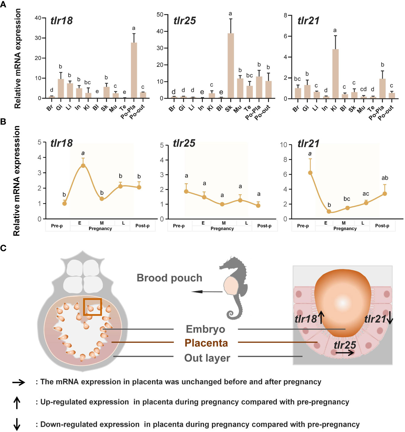
Figure 3 Temporal and spatial expression of seahorse TLRs. (A) TLR18, TLR25, and TLR21 were highly expressed in placenta of seahorse brood pouch (mean ± SEM, n = 3). Br, brain; Gi, gill; Li, liver; In, intestine; Ki, kidney; Bl, blood; Sk, skin; Mu, muscle; Te, testis; Po-Pla, placenta of brood pouch; Po-out, out layer of brood pouch. Different letters indicate significant differences (p < 0.05). (B) The expression patterns of seahorse TLRs varied in the placenta during pregnancy cycle (mean ± SEM, n = 11). Different letters indicate significant differences (p < 0.05). (C) Schematic of expression pattern of TLRs in the seahorse placenta.
2.3 Ligand recognition profiles of TLR18, TLR25, and TLR21 in lined seahorse
We detected the ligand cognitive profiles of lined seahorse TLR18, TLR25, and TLR21 (Figure 4). The result showed that the expressions of TLR18 and TLR25 were significantly up-regulated after the LTA challenge. In addition, TLR18 and TLR25 expression were significantly upregulated after LPS and PGN stimulation, and synthetic diacylated lipopeptides (Pam2CSK4) stimulation, respectively. Seahorse TLR21 was significantly upregulated after the CpG-ODNs challenge, particularly after the CpG-2006, CpG-202 and CpG-2007 challenges, which all contained the “GTCGTT” motif. In summary, these three TLRs responded to different antigens, indicating their involvement in seahorse immune defense. Different TLRs responded in different patterns, suggesting diversity in seahorse immune protection.
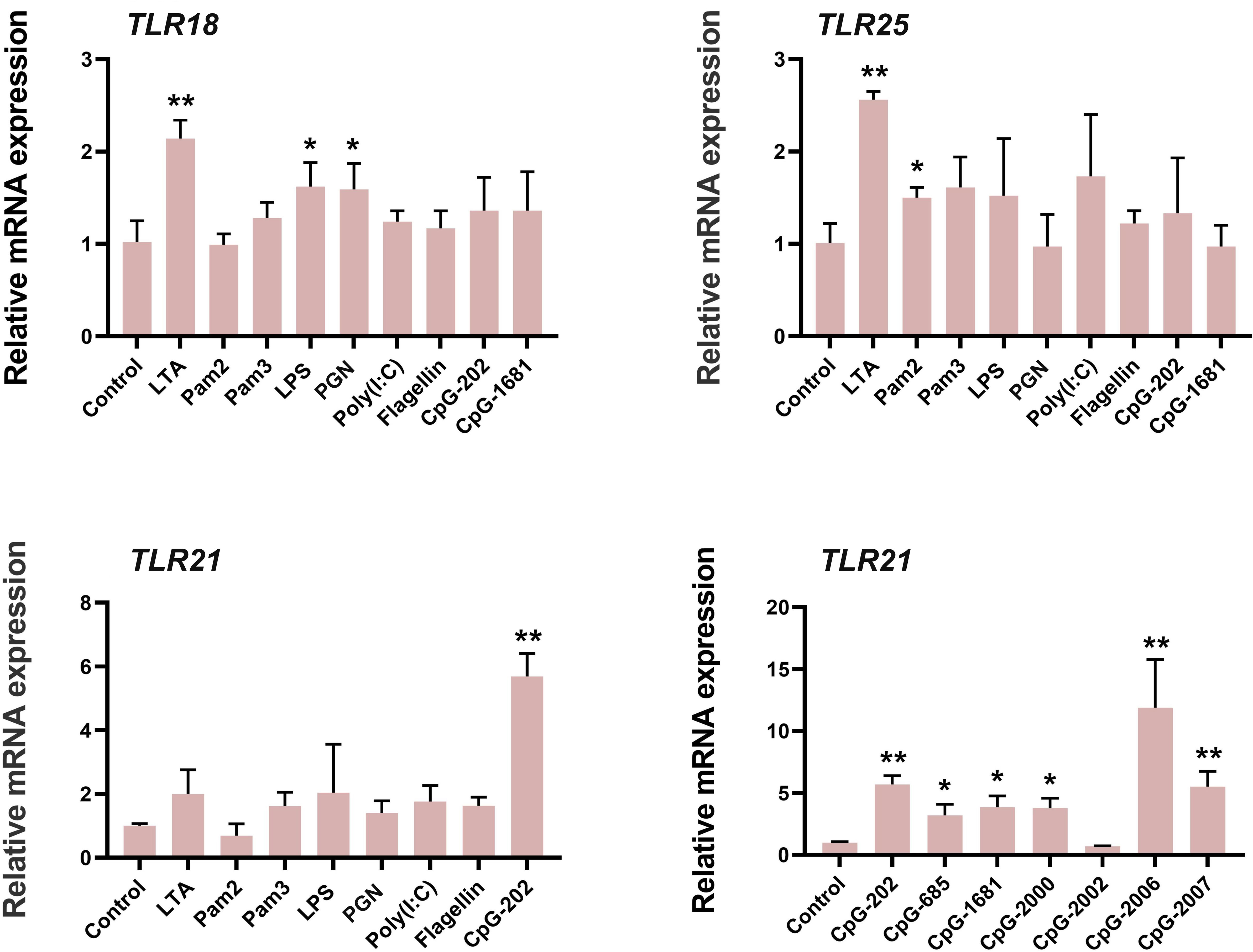
Figure 4 Ligand recognition profiles of TLR18, TLR25 and TLR21 in lined seahorse. Relative expression of TLR paralogues after stimulation with pathogen-associated molecular patterns are presented as the mean ± SEM (n = 5), and significant differences between control and treated groups are indicated with *(p < 0.05, Student’s t-test) or ** (p < 0.01, Student’s t-test).
3 Discussion
Toll-like receptors (TLRs) are types of pathogen recognition receptors (PRRs) that play important roles in both vertebrate innate and acquired immunity (20, 34). The TLR gene family is an evolutionarily ancient family that has been widely retained in organisms ranging from cnidarians to humans (18). To date, 10 TLRs have been identified in humans (TLR1–TLR10), 12 in mice (TLR1–9 and TLR11-13), and 10 in birds (TLR1a, TLR1b, TLR2a, TLR2b, TLR3–TLR5, TLR7, TLR15, and TLR21) (18, 35). The repertoire of TLRs in fish is complex and varies among species owing to dynamic gene gain/loss during genome duplication events (36). In general, seven TLRs (TLR1-3, TLR5 and TLR7-9) are orthologous to their mammalian and bird counterparts, which has been commonly conserved in teleosts (36). In addition, teleosts contain fish-specific TLRs: namely, TLR14/18–27. However, fish-specific TLRs vary among species (37). According to current research, the Atlantic cod (Gadus morhua L.) is the only known species lacking TLR1, TLR2, and TLR5, whereas an expansion of TLR9 is present (five copies) (38). The unique TLR repertoires of fish are considered to be independent co-options during organismal evolution for adaption to specific immune demands (18). In the present study, we found that three conserved TLRs (TLR1, TLR2, and TLR9) were lost in syngnathids, which is unique among bony fish, thus indicating specific immune adaptation evolution in syngnathids. Male pregnancy in syngnathids is mainly carried out by the brood pouch, which is a unique trait in vertebrates (39). Similar to the mammalian uterus, the brood pouch provides a site for embryo implantation and is involved in both maternal immune tolerance and host resistance (8).
To elucidate the immunological adaptive mechanism of syngnathid male pregnancy, two hypotheses have been proposed by different researchers based on current data. One hypothesis is that a trade-off between immunological tolerance and embryo rejection accompanies the evolution of unique male pregnancy, with the loss of several MHC II pathway genes in the pipefishes and a highly divergent invariant chain (CD74) in seahorses (14). The second hypothesis is that immunogenetic losses co-occurred with male seahorse pregnancy (5). In this study, we comprehensively analyzed the TLR gene family in teleosts and identified three conserved TLRs that were uniquely lost in pregnant male syngnathid fishes, suggesting that specific adaptive evolution commonly exists in syngnathids. Additionally, studies have confirmed that TLR1, TLR2, and TLR9 are closely related to immune regulation during mammalian pregnancies (18, 40). The findings showed that TLR1 variant ‘S248N’ influenced placental malaria during pregnancy (41) and that variants of TLR2 were positively associated with recurrent pregnancy loss (42). TLR9 activation coupled with IL-10 deficiency has been shown to induce adverse pregnancy outcomes (43). Kang et al. (44) found that excessive TLR9 signaling contribute to the pathogenesis of spontaneous abortion by impairing Treg cell survival via the activation of Caspase 8/3. Many studies have shown that activation of the TLR signaling pathway by exogenous and endogenous ligands can drive the induction of autoreactive T cells (28) and effector T cells to express TLRs (including but not limited to: TLR1, TLR2, and TLR9) (28, 45). The binding of TLR agonists to T cells contributes to their activation, which applies to both effector T cells and regulatory T (TReg) cells (28). Maternal-fetal immune tolerance during pregnancy depends on the balance between effector T cells and TReg cells. TReg cells are among the most important cell types involved in establishing immune tolerance to self-antigens and antigens encountered in foreign grafts (29). In this study, the loss of three conserved TLRs (TLR1, TLR2, and TLR9) in syngnathids may influence the formation or maturation of T cells and TReg cells, which help to block paternal immune rejection. We hypothesized that the loss of TLR1, TLR2, and TLR9 in syngnathids might be an adaptive evolution of paternal-fetal immune tolerance during pregnancy (Figure 5). Our findings provide new insight into the immune balance of male pregnancy in syngnathids.
In addition to their involvement in the adaptive immune regulation during pregnancy, TLRs are critical innate immune molecular defenses in mammalian uterine immune protection against infections by exogenous pathogens. By recognizing different types of pathogen-associated molecular patterns (PAMPs), TLRs activate downstream cascades as part of the innate immune response (20, 40). The expression of multiple TLRs in a temporal and spatial manner in the mammalian uterus has been reported in previous studies, which are believed to play an important role in the resistance of the uterus to pathogen infection and thus are necessary for embryo development (25, 30). In humans, TLR1, TLR4, TLR7, and TLR8 have shown statistically significant increases in expression during the second trimester compared to that in the first trimester, which is believed to be conducive to pregnancy (25). Stimulation of TLR2 and TLR4 with zymosan and LPS induces IL-6 and IL-8 production in placental cultures, indicating that these placental TLRs can recognize pathogenic PAMPs and induce the innate immune responses (46). In the present study, seahorse TLR18, TLR25, and TLR21 were highly expressed in the placenta and were varied. TLRs were significantly responsive to various PAMP agonists after stimulation. Thus, we concluded that these three TLRs play vital roles in the innate immune responses of seahorse brood pouches against infections by exogenous pathogens. Considering that the highest TLR18 expression was observed in early pregnancy stages, this may indicate a strong immune response to eliminate pathogenic bacteria from the brood pouch, and thus provide a sterile environment for subsequent embryo development. When the brood pouch is open to the outside environment during non-pregnancy stages, the immune protection function mainly depends on TLR21; therefore, TLR21 expression also increases.
Vertebrates exhibit immune redundancy or immune compensation effects. In zebrafish, both TLR9 and TLR21 recognize CpG-DNAs with partial functional redundancy (24). In chickens, the TLR9 is lost from the genome and instead a functional institution by TLR21 (23). To detect the potential immunological compensatory mechanism for the loss of the three conserved TLRs (TLR1, TLR2, and TLR9) in syngnathids, we examined the ligand recognition profiles of three other fish-specific TLRs (TLR18, TLR21, and TLR25) in the lined seahorse. We found that both TLR18 and TLR25 showed significant immune activation following the LTA challenge. In addition, TLR18 showed an immune response to the LPS and PGN challenges, similar to TLR25 in response to Pam2CSK4. TLR21 showed an immune response only to CpG-DNAs, indicating strong immune specificity. Based on phylogenetic analyses, the vertebrate TLR1, TLR2, TLR18, and TLR25 genes belonged to the TLR1 subfamily and should therefore recognize the general class of associated PAMPs (20). In mammals, the bacterial cell-wall components the lipoproteins and lipopeptides are predominantly recognized by the TLR1 subfamily (47). TLR2 recognizes LTA, a characteristic component of the bacterial cell wall. In addition, TLR2 recognizes various other ligands from bacteria by forming heterodimers with TLR1 or TLR6. Synthetic synthetic triacylated lipopeptides (Pam3CSK4) is recognized by TLR2-TLR1 heterodimers, whereas Pam2CSK4 is a ligand for TLR2-TLR6 heterodimers (48). In European common carp (Cyprinus carpio L.), TLR2 can sense both PGN and LTA from Gram-positive Staphylococcus aureus and is less sensitive to the stimulation with Pam3CSK4 (49). In a study by Wei et al. (50), TLR1 and TLR2 expression in the spleen of the orange-spotted grouper (Epinephelus coioide) was up-regulated after LPS treatment. As found in our previous study (in which TLR18 was mistermed as TLR2) (51) and in grass carp (Ctenopharyngodon idella), TLR18 responds to LPS challenging. Our results supported the hypothesis that TLRs within a subfamily recognize a general class of associated PAMPs. Moreover, our study showed that TLR21 was significantly responsive to CpG-DNAs challenging, consistent with previous observations in zebrafish and chickens (23, 24). Thus, we suggest that the immune function of the lost vertebrate-conserved LTRs (TLR1, TLR2, and TLR9) is at least partially compensated for by TLR18, TLR21, and TLR25 (Figure 5).
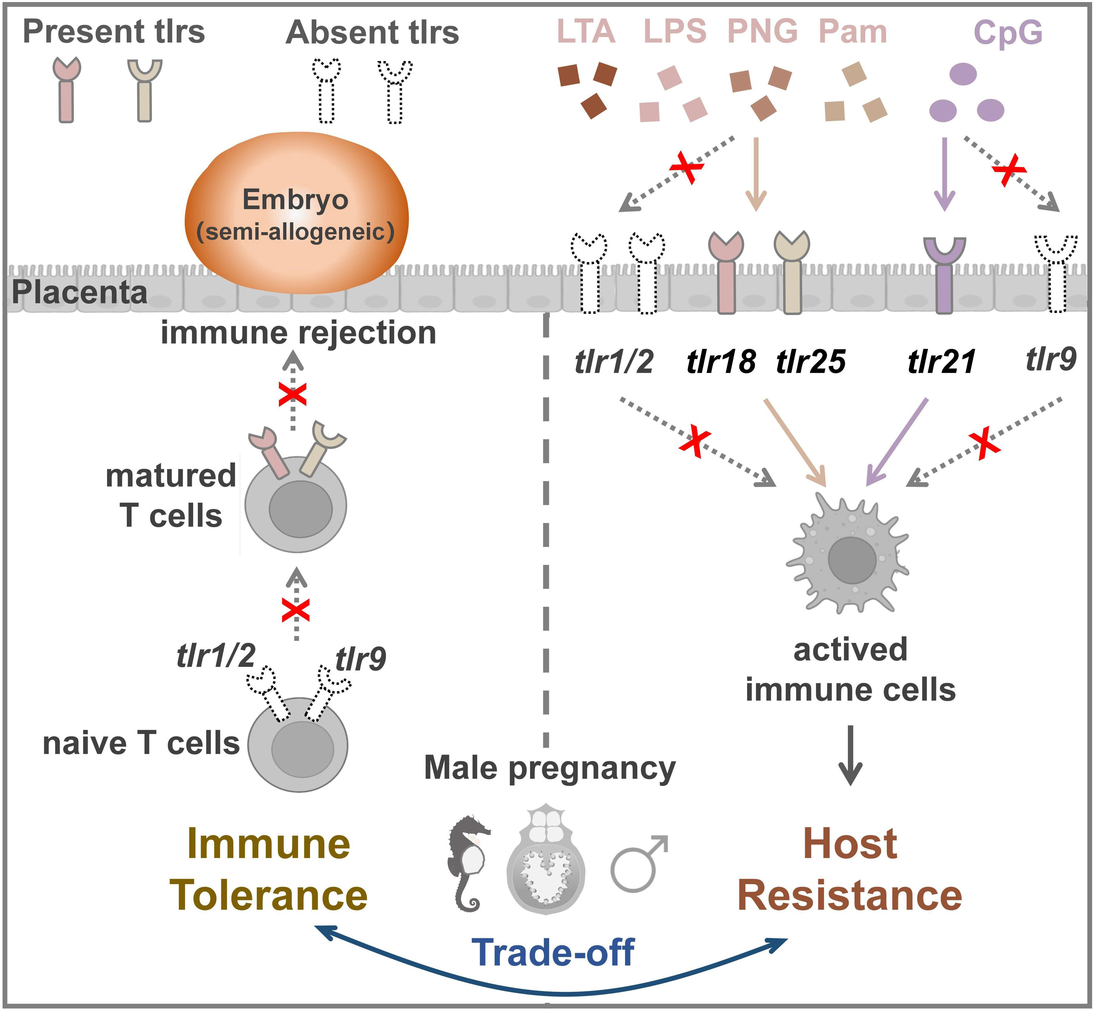
Figure 5 The co-option schedule of TLR adaptation to male pregnancy evolution. The loss of TLR1, TLR2, and TLR9 might block immune rejection to facilitate the implantation of the embryo. The three TLRs (TLR18, TLR25, and TLR21) that were highly expressed in placenta partially and functionally compensated for the loss of the three conserved TLRs in host resistance.
4 Materials and methods
4.1 Experimental animals and tissue sampling
Healthy lined seahorses (Hippocampus erectus) were collected from a seahorse breeding farm in Zhangzhou City, Fujian Province, China. All seahorses were kept temporarily in the seawater aquaculture system of the South China Sea Institute of the Chinese Academy of Sciences (Guangzhou, China) for two weeks. Seahorses were maintained in filtered circulating water under a 16:8 h light: dark cycle. Salinity, temperature, pH, and dissolved oxygen indices were 25 ± 1.0 ‰, 28 ± 0.5 °C, 7.5 ± 0.5, and 6.5 ± 0.5 mg·L−1 (mean ± SD), respectively.
For sample collection, seahorses were anesthetized with 0.05% tricaine methane sulfonate (MS222) before dissection. Eleven tissues (brain, gill, liver, intestine, kidney, gonad, muscle, skin, blood, and placenta and out layer of brood pouch) were surgically removed. According to the method of Zhang and Whittington (10, 52), the reproductive stages of brood pouch were judged by the placenta morphology and the embedded embryos development stage. In this study, five different reproductive stages (including pre-pregnancy, early-pregnancy, mid-pregnancy, late-pregnancy, and post-pregnancy) were divided. In brief, pre-pregnancy stage, no embryos embedded, thin and small blood vessels placenta; early-pregnancy stage, early-developed embryos (1-2 d) attached, vascularized with abundant blood vessels; mid-pregnancy stage, mid-developed embryos (8-12 d) attached, vascularized with abundant blood vessels; late-pregnancy stage, later-developed embryos (>15 d) attached, vascularized with abundant blood vessels; post-pregnancy stage, embryos released, blood vessels gradually resume the normal stage. To avoid RNA degradation, the collected samples were immediately frozen in liquid nitrogen and stored at −80°C. All experiments were conducted in accordance with the regulations of the Animal Research and Ethics Committee of the Chinese Academy of Sciences (approval number: SCSIO-IACUC-2019-000137).
4.2 Genome-wide identification of TLR genes in syngnathidae
Five representative fish of the Syngnathidae family were used for comparative TLR gene analysis. To identify the TLR genes, the TLR proteins of several representative vertebrates (including human (Homo sapiens), mouse (Mus musculus), chicken (Gallus gallus), channel catfish (Ictalurus punctatus), miiuy croaker (Miichthys miiuy), and zebrafish (Danio rerio)) were downloaded from the National Center for Biotechnology Information (NCBI) database as a query for native blast analysis in five Syngnathidae whole genomes and transcriptome databases (e-value ≤0.00001) (8, 10, 11, 53–55). To verify the TLR genes, the deduced TLRs’ architecture was analyzed using the SMART online prediction tool (http://smart.embl-heidelberg.de/) and sequences were identified using the NCBI Basic Local Alignment Search Tool (BLAST).
4.3 TLR genes evolution and compare
To elucidate the evolution of seahorse TLRs, phylogenetic analysis was performed based on 107 amino acid sequences obtained from 11 teleost fish species (including five Syngnathidae fish, five well-studied teleosts, and mice; see Supplementary Table S1). Multiple sequences were aligned using ClawstW and a phylogenetic tree was constructed using the maximum likelihood method in MEGA 6 with 1000 bootstrap replicates.
Syntenic analyses of TLRs were performed by comparing the locus position of TLR genes and upstream and downstream gene types. The genomes of five Syngnathidae fish species and other four representative teleosts (including zebrafish (D. rerio), tilapia (Oreochromis niloticus), fugu (Takifugu rubripes), and medaka (Oryzias latipes)) were utilized. Data for the five Syngnathidae and four representative teleosts were obtained from a previous study (55), and genomicus databases (http://www.genomicus.biologie.ens.fr/genomicus), respectively.
A genome-wide comparison of TLR construction in teleosts was conducted by comparing the genomic databases of five Syngnathidae fish with seven representative teleosts, including zebrafish (D. rerio), tilapia (Oreochromis niloticus), fugu (Takifugu rubripes), and medaka (Oryzias latipes), channel catfish (Ictalurus punctatus), spotted gar (Lepisosteus oculatus), and three-spined stickleback (Gasterosteus aculeatus)).
4.4 Tissue expression profile of TLR18, TLR25 and TLR21 in lined seahorse
Extracted seahorse tissues were ground in liquid nitrogen and total RNA was isolated using Trizol Reagent (Ambion, USA) according to manufacturer instructions (56). RNA was reverse transcribed into cDNA using the ReverTra Ace qPCR RT Master Mix kit (Toyobo, Japan) for different tissues. Gene expression across tissues for lined seahorse TLR18, TLR21, and TLR25 was determined by quantitative real-time PCR (qRT-PCR). The primers (Supplementary Table S2) were designed using Primer 5.0 software (Plymouth, UK) and the specificity of the primers was detected by melting curve analysis. Primer amplification efficiency was evaluated using a standard curve (Supplementary Figure S1). The β-actin gene was used as an internal reference gene for relative quantification analysis. Amplification was carried out in Light Cycler 480 thermocycler (Roche, USA), and qRT-PCR was performed in 10 μl volumes (Supplementary Table S3) using SYBR Premix Ex-Taq™ reagent (Takara, Japan). The amplification reaction conditions and qRT-PCR parameters were as follows: denaturation for 3 min at 95°C; 40 cycles of 20 s at 95°C, 20 s at 58°C, and 20 s at 72°C; followed by 30 s at 95°C and 1 min at 60°C. CT values were calculated with a fluorescence threshold of 0.5 to calculate relative gene expression. The relative mRNA expression of each gene was calculated using the 2−ΔΔCt method (57). All data from the qRT-PCRs were presented as the mean ± standard error (SE). Statistical differences were estimated using unpaired Student’s t-tests or one-way ANOVAs followed by Tukey’s tests. All data in this study included three biological repeats in each group.
4.5 Expression profile of TLR18, TLR25, and TLR21 in seahorse breeding cycle
To detect whether specially evolved TLRs were involved in seahorse pregnancy, the expression profiles of TLR18, TLR25, and TLR21 were detected in the placenta at five different reproductive stages (including pre-pregnancy, early-pregnancy, mid-pregnancy, late-pregnancy, and post-pregnancy). The detection was conducted by qRT-PCR as above.
4.6 Ligands recognition of seahorse TLR genes
To characterize the immune ligand recognition of TLR18, TLR21, and TLR25 in lined seahorses, eight typical TLR ligands (including 30 μg/mL LPS, 50 μg/mL LTA, 10 μg/mL Pam2CSK4, 10 μg/mL Pam3CSK4, 50 μg/mL polyinosinic:polycytidylic acid (Poly(I:C)), 10 μg/mL phosphorothioate-modified CpG-oligodeoxynucleotides (CpG-ODNs), 0.2 μg/mL Flagellin, and 50 μg/mL PGN) were selected to conduct a challenge experiment in vitro. The challenge experiment was conducted using a lined seahorse embryonic cell line (Chinese patent: ZL 2017 1 1050527.5) established in our on-site laboratory (52). Briefly, the cells were maintained in 6-well cell culture plates containing 5 mL Dulbecco’s modified eagle medium per well: Ham’s nutrient mixture F-12 (1:1) medium (DMEM/F12) supplemented with fetal bovine serum (FBS, 20%) at 28°C and 5% CO2 atmosphere. To address these challenges, cells were incubated with pathogenic ligands for 6 h and subsequently collected for TLR detection.
Data availability statement
The original contributions presented in the study are included in the article/Supplementary Material. Further inquiries can be directed to the corresponding authors.
Ethics statement
The animal study was reviewed and approved by the Animal Research and Ethics Committee of the Chinese Academy of Sciences (approval number: SCSIO-IACUC-2019-000137).
Author contributions
QL and GQ supervised the project and designed the research. BZ and WX performed the genome and genetic analyses. BZ and WX performed qPCR & biological function detection. BZ, WX, ZC, LQ, and XW performed original draft writing and drawing figures. QL and GQ reviewed the writing. All authors contributed to the article and approved the submitted version.
Funding
This project was supported by the National Natural Science Foundation of China (41825013), the Marine Economic Development Project (GDNRC [2022] 36), the National Natural Science Foundation of China (32000350, 42076131), the Key Research and Development Program of Hainan Province (ZDYF2021SHFZ109), and the Central Public-interest Scientific Institution Basal Research Fund of South China Sea Fisheries Research Institute of CAFS (2021TS03).
Conflict of interest
The authors declare that the research was conducted in the absence of any commercial or financial relationships that could be construed as a potential conflict of interest.
Publisher’s note
All claims expressed in this article are solely those of the authors and do not necessarily represent those of their affiliated organizations, or those of the publisher, the editors and the reviewers. Any product that may be evaluated in this article, or claim that may be made by its manufacturer, is not guaranteed or endorsed by the publisher.
Supplementary material
The Supplementary Material for this article can be found online at: https://www.frontiersin.org/articles/10.3389/fimmu.2023.1224698/full#supplementary-material
References
1. Bainbridge DRJ. The evolution of pregnancy. Early Hum Dev (2014) 90(11):741–5. doi: 10.1016/j.earlhumdev.2014.08.013
2. Bonney EA. Immune regulation in pregnancy: a matter of perspective? Obstetrics Gynecol Clinics (2016) 43(4):679–98. doi: 10.1016/j.ogc.2016.07.004
3. Thellin O, Heinen E. Pregnancy and the immune system: between tolerance and rejection. Toxicology (2003) 185(3):179–84. doi: 10.1016/S0300-483X(02)00607-8
4. Parker J, Dubin A, Schneider R, Wagner KS, Jentoft S, Böhne A, et al. Immunological tolerance in the evolution of male pregnancy. Mol Ecol (2023) 32(4):819–40. doi: 10.1111/mec.16333
5. Liu Y, Qu M, Jiang H, Schneider R, Qin G, Lou W, et al. Immunogenetic losses co-occurred with seahorse male pregnancy and mutation in tlx1 accompanied functional asplenia. Nat Commun (2022) 13(1):7610. doi: 10.1038/s41467-022-35338-7
6. Qu M, Zhang Y, Gao Z, Zhang Z, Liu Y, Wan S, et al. The genetic basis of the leafy seadragon’s unique camouflage morphology and avenues for its efficient conservation derived from habitat modeling. Sci China Life Sci (2023) 66:1213–30. doi: 10.1007/s11427-022-2317-6
7. Kawaguchi M, Okubo R, Harada A, Miyasaka K, Takada K, Hiroi J, et al. Morphology of brood pouch formation in the pot-bellied seahorse Hippocampus abdominalis. Zool Lett (2017) 3:19. doi: 10.1186/s40851-017-0080-9
8. Zhang YH, Ravi V, Qin G, Dai H, Zhang HX, Han FM, et al. Comparative genomics reveal shared genomic changes in syngnathid fishes and signatures of genetic convergence with placental mammals. Natl Sci Rev (2020) 7(6):964–77. doi: 10.1093/nsr/nwaa002
9. Laksanawimol P, Damrongphol P, Kruatrachue M. Alteration of the brood pouch morphology during gestation of male seahorses, Hippocampus kuda. Mar Freshw Res (2006) 57:497–502. doi: 10.1071/MF05112
10. Whittington CM, Griffith OW, Qi W, Thompson MB, Wilson AB, Notes A. Seahorse brood pouch transcriptome reveals common genes associated with vertebrate pregnancy. Mol Biol Evol (2015) 32(12):3114–31. doi: 10.1093/molbev/msv177
11. Lin Q, Fan S, Zhang Y, Xu M, Zhang H, Yang Y, et al. The seahorse genome and the evolution of its specialized morphology. Nature (2016) 540(7633):395–9. doi: 10.1038/nature20595
12. Wu Y, Zhang H, Zhang B, Lin Q, Liu Y. Molecular characterization of TLR22 and its role in immunological modification of the brood pouch of the lined seahorse, Hippocampus erectus. Aquaculture (2021) 539:736628. doi: 10.1016/j.aquaculture.2021.736628
13. Xiao W, Chen Z, Zhang Y, Wu Y, Jiang H, Zhang H, et al. Hepcidin gene Co-option balancing paternal immune protection and Male pregnancy. Front Immunol (2022) 13:884417–7. doi: 10.3389/fimmu.2022.884417
14. Roth O, Solbakken MH, Tørresen OK, Bayer T, Matschiner M, Baalsrud HT, et al. Evolution of male pregnancy associated with remodeling of canonical vertebrate immunity in seahorses and pipefishes. Proc Natl Acad Sci (2020) 117(17):9431–9. doi: 10.1073/pnas.1916251117
15. Wang X, Qu M, Liu Y, Schneider RF, Song Y, Chen Z, et al. Genomic basis of evolutionary adaptation in a warm-blooded fish. Innovation (2022) 3(1):100185. doi: 10.1016/j.xinn.2021.100185
16. Parker J. Male Pregnancy and the evolutionary importance of immunological tolerance in syngnathid fishes. (2021).
17. Stölting KN, Wilson AB. Male Pregnancy in seahorses and pipefish: beyond the mammalian model. BioEssays (2007) 29(9):884–96. doi: 10.1002/bies.20626
18. Leulier F, Lemaitre B. Toll-like receptors–taking an evolutionary approach. Nat Rev Genet (2008) 9(3):165–78. doi: 10.1038/nrg2303
19. Mukherjee S, Huda S, Sinha Babu SP. Toll-like receptor polymorphism in host immune response to infectious diseases: a review. Scandinavian J Immunol (2019) 90(1):e12771. doi: 10.1111/sji.12771
20. Roach JC, Glusman G, Rowen L, Kaur A, Purcell MK, Smith KD, et al. The evolution of vertebrate toll-like receptors. Proc Natl Acad Sci (2005) 102(27):9577–82. doi: 10.1073/pnas.0502272102
21. Pietretti D, Wiegertjes GF. Ligand specificities of toll-like receptors in fish: indications from infection studies. Dev Comp Immunol (2014) 43(2):205–22. doi: 10.1016/j.dci.2013.08.010
22. Sundaram AYM, Kiron V, Dopazo J, Fernandes JMO. Diversification of the expanded teleost-specific toll-like receptor family in Atlantic cod, gadus morhua. BMC Evolutionary Biol (2012) 12(1):1–17. doi: 10.1186/1471-2148-12-256
23. Brownlie R, Zhu J, Allan B, Mutwiri GK, Babiuk LA, Potter A, et al. Chicken TLR21 acts as a functional homologue to mammalian TLR9 in the recognition of CpG oligodeoxynucleotides. Mol Immunol (2009) 46(15):3163–70. doi: 10.1016/j.molimm.2009.06.002
24. Yeh DW, Liu YL, Lo YC, Yuh CH, Yu GY, Lo JF, et al. Toll-like receptor 9 and 21 have different ligand recognition profiles and cooperatively mediate activity of CpG-oligodeoxynucleotides in zebrafish. Proc Natl Acad Sci (2013) 110(51):20711–6. doi: 10.1073/pnas.1305273110
25. Pudney J, He X, Masheeb Z, Kindelberger DW, Kuohung W, Ingalls RR. Differential expression of toll-like receptors in the human placenta across early gestation. Placenta (2016) 46:1–10. doi: 10.1016/j.placenta.2016.07.005
26. Patrascan CC. The role of toll-like receptors in the development of immunological tolerance in neonates. (2010). doi: 10.17192/z2010.0376
27. Koga K, Izumi G, Mor G, Fujii T, Osuga Y. Toll-like receptors at the maternal-fetal interface in normal pregnancy and pregnancy complications. Am J Reprod Immunol (2014) 72(2):192–205. doi: 10.1111/aji.12258
28. Mills KHG. TLR-dependent T cell activation in autoimmunity. Nat Rev Immunol (2011) 11(12):807–22. doi: 10.1038/nri3095
29. Gobert M, Lafaille JJ. Maternal-fetal immune tolerance, block by block. Cell (2012) 150(1):7–9. doi: 10.1016/j.cell.2012.06.020
30. Koga K, Mor G. Toll-like receptors at the maternal–fetal interface in normal pregnancy and pregnancy disorders. Am J Reprod Immunol (2010) 63(6):587–600. doi: 10.1111/j.1600-0897.2010.00848.x
31. Atli MO, Kose M, Hitit M, Kaya MS, Bozkaya F. Expression patterns of toll-like receptors in the ovine corpus luteum during the early pregnancy and prostaglandin F2α-induced luteolysis. Theriogenology (2018) 111:25–33. doi: 10.1016/j.theriogenology.2018.01.010
32. Abrahams VM, Mor G. Toll-like receptors and their role in the trophoblast. Placenta (2005) 26(7):540–7. doi: 10.1016/j.placenta.2004.08.010
33. Tsang JCH, Vong JSL, Ji L, Poon LCY, Jiang P, Lui KO, et al. Integrative single-cell and cell-free plasma RNA transcriptomics elucidates placental cellular dynamics. PNAS (2017) 114(37):E7786–95. doi: 10.1073/pnas.1710470114
34. Fitzgerald KA, Kagan JC. Toll-like receptors and the control of immunity. Cell (2020) 180(6):1044–66. doi: 10.1016/j.cell.2020.02.041
35. Temperley ND, Berlin S, Paton IR, Griffin DK, Burt DW. Evolution of the chicken toll-like receptor gene family: a story of gene gain and gene loss. BMC Genomics (2008) 9(1):1–12. doi: 10.1186/1471-2164-9-62
36. Khan I, Maldonado E, Silva L, Almeida D, Johnson WE, O'Brien SJ, et al. The vertebrate TLR supergene family evolved dynamically by gene gain/loss and positive selection revealing a host–pathogen arms race in birds. Diversity (2019) 11(8):131. doi: 10.3390/d11080131
37. Palti Y. Toll-like receptors in bony fish: from genomics to function. Dev Comp Immunol (2011) 35(12):1263–72. doi: 10.1016/j.dci.2011.03.006
38. Solbakken MH, Tørresen OK, Nederbragt AJ, Seppola M, Gregers TF, Jakobsen KS, et al. Evolutionary redesign of the Atlantic cod (Gadus morhua L.) toll-like receptor repertoire by gene losses and expansions. Sci Rep (2016) 6(1):25211. doi: 10.1038/srep25211
39. Li C, Li Y, Qin G, Chen Z, Qu M, Zhang B, et al. Regulatory role of retinoic acid in male pregnancy of the seahorse. Innovation (2020) 1(3):100052. doi: 10.1016/j.xinn.2020.100052
40. Pasare C, Medzhitov R. Toll-like receptors: balancing host resistance with immune tolerance. Curr Opin Immunol (2003) 15(6):677–82. doi: 10.1016/j.coi.2003.09.002
41. Hamann L, Bedu-Addo G, Eggelte TA, Schumann RR, Mockenhaupt FP. The toll-like receptor 1 variant S248N influences placental malaria. Infect Genet Evol (2010) 10(6):785–9. doi: 10.1016/j.meegid.2010.05.005
42. Bahia W, Soltani I, Haddad A, Radhouani A, Mahdhi A, Ferchi S, et al. Links between SNPs in TLR-2 and TLR-4 and idiopathic recurrent pregnancy loss. Br J Biomed Sci (2020) 77(2):64–8. doi: 10.1080/09674845.2019.1687151
43. Thaxton JE, Romero R, Sharma S. TLR9 activation coupled to IL-10 deficiency induces adverse pregnancy outcomes. J Immunol (2009) 183(2):1144–54. doi: 10.4049/jimmunol.0900788
44. Kang X, Zhang X, Liu Z, Xu H, Wang T, He L, et al. Excessive TLR9 signaling contributes to the pathogenesis of spontaneous abortion through impairment of treg cell survival by activation of caspase 8/3. Int Immunopharmacol (2015) 29(2):285–92. doi: 10.1016/j.intimp.2015.11.004
45. Reynolds JM, Pappu BP, Peng J, Martinez GJ, Zhang Y, Chung Y, et al. Toll-like receptor 2 signaling in CD4(+) T lymphocytes promotes T helper 17 responses and regulates the pathogenesis of autoimmune disease. Immunity (2010) 32(5):692–702. doi: 10.1016/j.immuni.2010.04.010
46. Holmlund U, Cebers G, Dahlfors AR, Sandstedt B, Bremme K, EkstrÖm EVAS, et al. Expression and regulation of the pattern recognition receptors toll-like receptor-2 and toll-like receptor-4 in the human placenta. Immunology (2002) 107(1):145–51. doi: 10.1046/j.1365-2567.2002.01491.x
47. Manukyan M, Triantafilou K, Triantafilou M, Mackie A, Nilsen N, Espevik T, et al. Binding of lipopeptide to CD14 induces physical proximity of CD14, TLR2 and TLR1. Eur J Immunol (2005) 35(3):911–21. doi: 10.1002/eji.200425336
48. Zhang J, Kong X, Zhou C, Nie G, Li X. Toll-like receptor recognition of bacteria in fish: ligand specificity and signal pathways. Fish Shellfish Immunol (2014) 41(2):380–8. doi: 10.1016/j.fsi.2014.09.022
49. Ribeiro C, Hermsen T, Taverne-Thiele AJ, Savelkoul HFJ, Wiegertjes GF. Evolution of recognition of ligands from gram-positive bacteria: similarities and differences in the TLR2-mediated response between mammalian vertebrates and teleost fish. J Immunol (2010) 184(5):2355–68. doi: 10.4049/jimmunol.0900990
50. Wei YC, Pan TS, Chang MX, Huang B, Xu Z, Lou TR, et al. Cloning and expression of toll-like receptors 1 and 2 from a teleost fish, the orange-spotted grouper Epinephelus coioides. Vet Immunol Immunopathol (2011) 141(3-4):173–82. doi: 10.1016/j.vetimm.2011.02.016
51. Zhang B, Zhang H, Qin G, Liu Y, Han X, Yin J, et al. TLR2 gene in seahorse brood pouch plays key functional roles in LPS-induced antibacterial responses. J Fish Dis (2019) 42(7):1085–9. doi: 10.1111/jfd.13006
52. Zhang B, Qin G, Qu L, Zhang Y, Li C, Cang C, et al. Wnt8a is one of the candidate genes that play essential roles in the elongation of the seahorse prehensile tail. Mar Life Sci Technol (2021) 3(4):416–26. doi: 10.1007/s42995-021-00099-7
53. Lin Q, Qiu Y, Gu R, Xu M, Li J, Bian C, et al. Draft genome of the lined seahorse, Hippocampus erectus. GigaScience (2017) 6:1–6. doi: 10.1093/gigascience/gix030
54. Qin G, Zhang Y, Zhang B, Zhang Y, Liu Y, Lin Q. Environmental estrogens and progestins disturb testis and brood pouch development with modifying transcriptomes in male-pregnancy lined seahorse Hippocampus erectus. Sci Total Environ (2020) 715:136840. doi: 10.1016/j.scitotenv.2020.136840
55. Li C, Olave M, Hou Y, Qin G, Schneider RF, Gao G, et al. Genome sequences reveal global dispersal routes and suggest convergent genetic adaptations in seahorse evolution. Nat Commun (2021) 12:1094. doi: 10.1038/s41467-021-21379-x
56. Qu M, Liu Y, Zhang Y, Wan S, Ravi V, Qin G, et al. Seadragon genome analysis provides insights into its phenotype and sex determination locus. Sci Adv (2021) 7(34):eabg5196. doi: 10.1126/sciadv.abg5196
Keywords: seahorse, pregnancy, immune tolerance, host resistance, toll-like receptor
Citation: Zhang B, Xiao W, Qin G, Chen Z, Qiu L, Wang X and Lin Q (2023) Gene loss and co-option of toll-like receptors facilitate paternal immunological adaptation in the brood pouch of pregnant male seahorses. Front. Immunol. 14:1224698. doi: 10.3389/fimmu.2023.1224698
Received: 18 May 2023; Accepted: 04 July 2023;
Published: 31 July 2023.
Edited by:
Jianmin Ye, South China Normal University, ChinaReviewed by:
Xiuzhen Sheng, Ocean University of China, ChinaJing Xing, Ocean University of China, China
Copyright © 2023 Zhang, Xiao, Qin, Chen, Qiu, Wang and Lin. This is an open-access article distributed under the terms of the Creative Commons Attribution License (CC BY). The use, distribution or reproduction in other forums is permitted, provided the original author(s) and the copyright owner(s) are credited and that the original publication in this journal is cited, in accordance with accepted academic practice. No use, distribution or reproduction is permitted which does not comply with these terms.
*Correspondence: Geng Qin, qingeng@scsio.ac.cn; Qiang Lin, linqiang@scsio.ac.cn
†These authors have contributed equally to this work