Growth pattern of Fortunian scalidophoran sclerites
 Jiachen Qin
 Yunhuan Liu*
  Mingjin Liu
- School of Earth Science and Resources, Chang’an University, Xi’an, China
Fortunian scalidophoran worms have shown high diversity, with 7 genera and species and 10 indeterminate forms. Current studies have mainly focused on morphology as well as early evolution, and studies on ontogeny have not been carried out due to the limited number of specimens. Here, we report new material of an Orsten-type preserved Indeterminate Form 3 from the Zhangjiagou section. Collected specimens of Indeterminate Form 3 with different annulus widths indicate the presence of several ontogenetic stages. We found newly formed sclerites on the annulus of Indeterminate Form 3 at different ontogenetic stages, suggesting that the sclerites of Indeterminate Form 3 become more numerous in addition to increasing in size during growth. The size of the large sclerites may also increase as the worms grow, however, their number may not change.
1 Introduction
As the earliest unambiguous fossil record of ecdysozoans, scalidophorans showed high diversity during the early Cambrian Fortunian stage (−535 Ma), with 7 genera and species and 10 indeterminate forms (Liu et al., 2014a; 2019; Zhang et al., 2015; 2018; Shao et al., 2018a; 2020a; 2020b; Zhang, 2021; Qin et al., 2023). This is consistent with the hypothesis of molecular clock studies, which posits cycloneuralians may have originated in the Ediacaran and radiated during the early Cambrian period (Rota-Stabelli et al., 2013; dos Reis et al., 2016; Erwin. 2020).
These fossils have gone through three-dimensional phosphatization, i.e., Orsten-type preservation (Maas et al., 2006). Orsten-type fossils preserve the most primitive three-dimensional structures of organisms. This facilitates the reconstruction of body structures and means Orsten-type fossils are more conducive to the discussion of morphological functions, affinities, and paleoecology. In addition, the preservation of Orsten-types can preserve successive developmental stages of a species, thus facilitating the reconstruction of ontogeny (Maas et al., 2006). Contemporaneous cnidarians (Olivooides, Quadrapyrgites, Qinscyphus, and Hexaconularia) have been found in a large number of fossils at different developmental stages, and more complete developmental sequences have been established on this basis (Dong et al., 2013; Han et al., 2013; Liu et al., 2014b; 2017; Steiner et al., 2014; Wang et al., 2017; Shao et al., 2018b; Zhang et al., 2023). In more recent strata, the developmental patterns of some paleoscolecids have also been discussed (Brock and Cooper, 1993; Müller and HinzSchallreuter, 1993; Zhang and Pratt, 1996; Duan and Dong, 2013; Xian et al., 2023).
In contrast to those, most Fortunian scalidophoran fossils are fragmented, and except for a little preserved introvert information, most of them only preserve the trunk part. The fossils of Fortunian scalidophorans are also relatively rare, and most of the species are only represented by a single specimen. Thus, the previous studies mainly focus on morphology, while a study of ontogenetic development has not been carried out.
Here, we describe a new specimen of Indeterminate Form 3 sensu Liu et al., 2019 (hereafter referred to as Form 3), which has narrower annuli and is probably at a younger developmental stage than the specimens already reported. This specimen also has a smaller number of sclerites and preserves some sclerites that are not yet fully developed. It is suggested that Form 3’s armor will change as it grows, mainly by appearing larger and having more sclerites as it grows.
2 Material and methods
The studied specimens are now deposited at the University Museum of Chang’an University (UMCU), Xi’an, China. They were collected from the bottom of the Kuanchuanpu Formation in the Zhangjiagou section, Xijiang Country, Southern Shaanxi. This fossil-rich horizon falls within the Anabarites trisulcatus—Protohertzina anabarica Assemblage Zone, with an estimated age of 531.8–536.4 Ma (Steiner et al., 2007; 2014), and belongs to the Fortunian Stage (Peng et al., 2020).
Rock samples were processed through standard acetic acid etching methods. Fossils were obtained from the undissolvable residues and picked out under a binocular microscope. Selected microfossils were mounted on stubs and coated with gold for observation under a Quanta 650 field-emission environmental scanning electron microscope at Chang’an University.
The specimens were measured using the ruler tool in Adobe Photoshop CS5, and the annuli with less deformation were mainly selected for the measurement.
3 Systematic paleontology
Ecdysozoa Aguinaldo et al., 1997.
Cycloneuralia Ahlrichs, 1995.
Scalidophora Lemburg, 1995.
Indeterminate Form 3 sensu Liu et al., 2019.
(Figure 1Figure 2; Figure 3A–H)
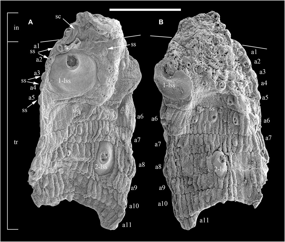
FIGURE 1. SEM images of Indeterminate Form 3 sensu Liu et al., 2019 from the Fortunian Zhangjiagou Section. (A and B) Two opposite sides of UMCU 23XX01. Abbreviations: in, introvert; tr, trunk; sc, scalid; ss, spinose sclerite; 1-lss, first large spinose sclerite; 2-lss, second large spinose sclerite; (a1–a11), the 1st to 11th annuli. The scale bar represents 500 μm.
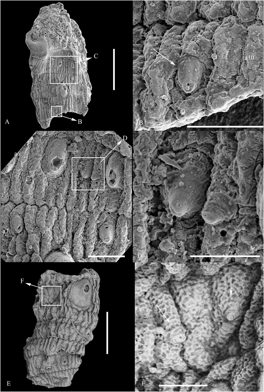
FIGURE 2. Newly formed sclerites of Indeterminate Form 3 sensu Liu et al., 2019. (A–D) UMCU 23XX01; (E–F) UMCU 22XX02; (B–C) close-up of A; (D) close-up of C; (F) close-up of (E). The scale bars represent 500 μm (A and E), 100 μm (B, C, and (F), and 50 μm (D).
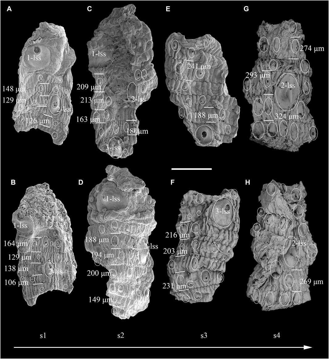
FIGURE 3. The possible developmental sequence of Indeterminate Form 3 sensu Liu et al., 2019. (A,B) UMCU 23XX01; (C,D) UMCU 22XX01; (E,F) UMCU 22XX02; (G,H) UMCU 16CHD0409-004. (C, D, E, and F) as cited in Qin et al., 2023; (G and H) as cited in Liu et al., 2019. The outlines of sclerites are marked by circles. Abbreviations: 1-lss, first large spinose sclerite; 2-lss, second large spinose sclerite; 3-lss, third large spinose sclerite; (s1–s4), developmental stages 1-4. The scale bar represents 500 μm.
2018 Form B Zhang et al., Fig. 5.4.
2019 Indeterminate Forms 3 Liu et al., Fig 9D.
2023 Indeterminate Form 3 Qin et al., Figures 1–3.
3.1 Material
UMCU 23XX01 (Figure 1; Figure 2A–D; Figure 3A,B), UMCU 22XX01 (Figures 3C,D), UMCU 22XX02 (Figures 2E,F; Figure 3E,F), UMCU 16CHD0409-004 (Figures 3G,H).
3.2 Description
UMCU 23XX01 has two parts; the anterior part is the introvert and the posterior part is the trunk. The introvert has long, spine-link, internally hollow scalds. The trunk has 11 annuli with spinose sclerites, each with an enlarged base and a spine. The spinose sclerites on the same annulus vary in size and are randomly and sparsely distributed. A pair of rounded large spinose sclerites are located at the anterior part of the trunk, and a pair of large oval spinose sclerites are located on the eighth and ninth annuli.
UMCU 22XX01 is described in Qin et al., 2023; UMCU 16CHD0409-004 is described in Liu et al., 2019. The description of UMCU 22XX02 follows Qin et al., 2023, with only minor revisions to the sclerites. Two small sclerites appear on its second annulus (Figure 3B in Qin et al., 2023; Figures 2E,F); they seem to be newly formed.
3.3 Occurrence
Kuanchuanpu Formation (Fortunian Stage), Zhangjiagou section, Xixiang County, Shaanxi Province, South China; Anabarites trisulcatus-Protohertzina anabarica Assemblage zone.
3.4 Comparison
The main part of the trunk of these specimens is very similar, upon which similarly shaped sclerites are randomly and sparsely distributed. Both UMCU 23XX01 and UMCU 22XX01 have two pairs of large sclerites, and the first pair of large sclerites are both found in the anterior part of the trunk, and the second pair of large sclerites are both found in the middle of the trunk. This suggests that these specimens are from the same species, and the position of the large sclerites on the trunk may be fixed. There are at least five large sclerites (lss, Figure 1; Figure 3) on the trunk, in order from anterior to posterior: the first pair of large sclerites (1-lss), the second pair of large sclerites (2-lss), and the third large single sclerite (3-lss). Comparisons of Form 3 with other worms can be found in Liu et al., 2019 and Qin et al., 2023 and will not be repeated here.
Because the position of the large sclerites on the trunk may be fixed, when using UMCU 22XX01 as a reference, UMCU 23XX01 may represent the first two-thirds of it. UMCU 16CHD0409-004 has a pair of oval large spinose sclerites that may represent the middle part of UMCU 22XX01. The large rounded sclerites in UMCU 22XX02 may not correspond to the first large paired sclerites in UMCU 23XX01 and UMCU 22XX01: 1) the large sclerites in UMCU 22XX02 are not paired (also possibly due to incomplete preservation); 2) the first large paired sclerites in UMCU 22XX01 are next to triangular sclerites rather than oval sclerites as in UMCU 22XX02. Therefore, the large sclerite in UMCU 22XX02 may correspond to the single large sclerite located at the posterior part of the trunk in UMCU 22XX01.
3.5 Remarks
It is generally accepted that the annulus widens as the worm grows, both in extant scalidophorans (Neuhaus, 1995) and in paleoscolecids, which are considered to be early scalidophorans (cycloneuralians) (Brock and Cooper, 1993; Müller and HinzSchallreuter, 1993; Zhang and Pratt, 1996; Duan and Dong, 2013; Xian et al., 2023), so we divided these four specimens into four developmental stages according to the width of the annulus (Table 1; Figure 3s1-s4). According to these specimens, the size of the sclerites also gradually increases with growth (Table 1).
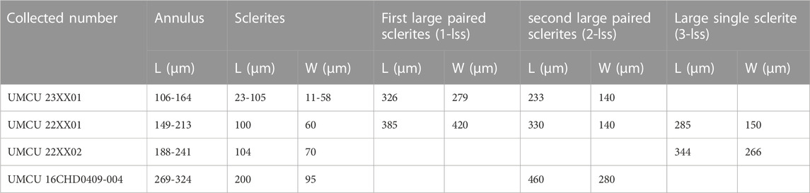
TABLE 1. Measurements.
4 Discussion
Among the previously reported Fortunian armored scalidophorans, most of them have similar morphology and size to the sclerites within the same annulus, such as Zhongpingscolex (Shao et al., 2020a), Shanscolex (Liu et al., 2019), and Dahescolex (Shao et al., 2020b), Forms 1-3 and 5-6 (Liu et al., 2019; Qin et al., 2023).
UMCU 23XX01 was identified as Form 3, but its sclerites within the same annulus are clearly different in size, and some of the sclerites appear to be recently formed and are only one-fifth the size of other sclerites in the same annulus (Figure 2D). We re-examined the material previously attributed to Form 3 and also found two small sclerites in UMCU 22XX02 (Figures 2E,F). Compared to UMCU 23XX01, UMCU 22XX02 is at a more advanced developmental stage, and newly formed sclerites were found in worms at different developmental stages, possibly suggesting that the sclerites of worms not only become larger in size during development but also increase in number.
UMCU 23XX01 (Figure 3s1, Figure 1; Figure 2A–D; Figure 3A,B) has a low number of sclerites (about 4-5 sclerites on one annulus on average) and a high percentage of newly formed sclerites (if the sclerite in the middle of the a6 in UMCU 23XX01 (Figure 1B; Figure 2C) is considered as normal size, all other sclerites of the same type in UMCU 23XX01 are may be newly or recently formed, they are morphologically similar and differ only in size). At this stage, the number of newly formed sclerites is high, indicating a peak in sclerite development and that the sclerites are developing faster. In addition, there are smaller sclerites on almost every annulus of UMCU 23XX01, which means that the increase in sclerites may occur simultaneously throughout the trunk of the worm. The number and size of sclerites are greater in UMCU 22XX02 (Figure 3s3, Figures 2E,F and Figure 3E,F); there are only two newly formed sclerites at this stage, and the number of newly formed sclerites is reduced, indicating that the sclerite development is slowed down at this time. The high number of sclerites in UMCU 16CHD0409-004 (Figure 3s4, Figures 3G,H) (about six sclerites on one annulus on average: this specimen is partially folded and there should be a higher number of sclerites) did not reveal newly formed sclerites, indicating that the number of sclerites may not be increasing at this stage. However, because these fossils are fragmented, we cannot be sure whether newly formed sclerites are still present in the unpreserved parts of these fossils so it is also possible that the number of sclerites continues to increase. Whether or not the number of sclerites increases after Figure 3s4, the worms may continue to grow and the size of the sclerites may increase accordingly.
The common sclerites develop within a single annulus; the large sclerite is more unusual in that it can span 2-3 annuli. The size of large sclerites may also increase as the worms grow (Table 1), however, based on the specimens at hand, their numbers may not change (Figure 3).
The growth of the worm is accompanied by periodic molting, with the annulus widening as the worm grows. However, it is not clear whether sclerites increase and grow simultaneously with worm growth or whether an increase in sclerites requires more frequent molting. Therefore, the rate of increase in the number of sclerites does not represent the growth rate of the worms, and there may be no correlation between them.
5 Conclusion
We have reported new material of Indeterminate Form 3 sensu Liu et al., 2019 from the Fortunian Zhangjiagou section, southern Shannxi, South China. Based on the material at hand, we established a possible partial developmental sequence of Indeterminate Form 3 sensu Liu et al., 2019, which illustrates that during its growth, in addition to the size of the sclerites increasing, the number of sclerites also increases. Large sclerites only increase in size with growth but not in number.
Data availability statement
The original contributions presented in the study are included in the article/supplementary material, further inquiries can be directed to the corresponding authors.
Author contributions
JQ coordinated the field work, preparation and curation of fossils, wrote the manuscript, and processed the images. YL edited the manuscript. ML, TS, and YZ assisted in collecting the rock samples in the field. All authors contributed to the article and approved the submitted version.
Funding
This work was supported by the National Natural Science Foundation of China (grant numbers 41872014), the Strategic Priority Research Program of the Chinese Academy of Sciences (grant number XDB26000000), the State Key Laboratory of Paleobiology and Stratigraphy, the Nanjing Institute of Geology and Paleontology, the Chinese Academy of Sciences (grant number 193123), and the Basic Research Plan of Natural Science of Shaanxi Province (grant number 2018JM4002).
Acknowledgments
We thank three reviewers for providing reviews and suggestions.
Conflict of interest
The authors declare that the research was conducted in the absence of any commercial or financial relationships that could be construed as a potential conflict of interest.
Publisher’s note
All claims expressed in this article are solely those of the authors and do not necessarily represent those of their affiliated organizations, or those of the publisher, the editors and the reviewers. Any product that may be evaluated in this article, or claim that may be made by its manufacturer, is not guaranteed or endorsed by the publisher.
References
Aguinaldo, A., Turbeville, J., Linford, L., Rivera, M., Garey, J., Raff, R., and Lake, J. A., (1997). Evidence for a clade of nematodes, arthropods and other moulting animals. Nature 387, 489–493. doi:10.1038/387489a0
Ahlrichs, W. (1995). Ultrastruktur und Phylogenie von Seison nebaliae (Gruber 1859) und Seison annulatus (Claus 1876). Hypothesen zu phylogenetischen Verwandtschaftsverhältnissen innerhalb der Bilateria. Göttingen, Germany: Georg-August-University of Göttingen.
Brock, G., and Cooper, B. (1993). Shelly fossils from the early cambrian (toyonian) wirrealpa, aroona creek, and ramsay limestones of south Australia. J. Paleontol. 67, 758–787. doi:10.1017/s0022336000037045
Dong, X., Cunningham, J. A., Bengtson, S., Thomas, C. W., Liu, J., Stampanoni, M., and Donoghue, P. C. J., (2013). Embryos, polyps and medusae of the early Cambrian scyphozoan Olivooides. P. Roy. Soc. B–Biol. Sci. 280, 20130071. doi:10.1098/rspb.2013.0071
Dos Reis, M., Thawornwattana, Y., Angelis, K., Telford, M., Donoghue, P. J., and Yang, Z. (2015). Uncertainty in the timing of origin of animals and the limits of precision in molecular timescales. Curr. Biol. 25, 2939–2950. doi:10.1016/j.cub.2015.09.066
Duan, B., and Dong, X. (2013). Furongian (late cambrian) palaeoscolecid cuticles from hunan Province, South China: The growth impact on the worm cuticle. Acta Sci. Nat. Univ. Pekin. 49, 591–602. doi:10.13209/j.0479-8023.2013.083
Erwin, D. H. (2020). The origin of animal body plans: A view from fossil evidence and the regulatory genome. Development 147, dev182899. doi:10.1242/dev.182899
Han, J., Kubota, S., Li, G., Yao, X., Yang, X., Shu, D., Li, Y., Kinoshita, S., Sasaki, O., Komiya, T., and Yan, G. (2013). Early Cambrian pentamerous cubozoan embryos from South China. PloS One 8, e70741. doi:10.1371/journal.pone.0070741
Lemburg, C. (1995). Ultrastructure of sense organs and receptor cells of the neck and lorica of the Halicryptus spinulosus larva (Priapulida). Microfauna Mar. 10, 7–30. doi:10.1007/BF00397931
Liu, Y. H., Qin, J. C., Wang, Q., Maas, A., Duan, B. C., and Zhang, Y. N., (2019). New armoured scalidophorans (ecdysozoa, cycloneuralia) from the cambrian Fortunian Zhangjiagou lagerstätte, south China. Pap. Palaeontol 5, 241–260. doi:10.1002/spp2.1239
Liu, Y., Li, Y., Shao, T., Zhang, H., Wang, Q., and Qiao, J. (2014). Quadrapyrgites from the lower Cambrian of South China: Growth pattern, post–embryonic development, and affinity. Chin. Sci. Bull. 59, 4086–4095. doi:10.1007/s11434-014-0481-5
Liu, Y., Shao, T., Zhang, H., Wang, Q., Zhang, Y., and Chen, C., (2017). A new scyphozoan from the cambrian Fortunian stage of south China. Palaeontology 60, 511–518. doi:10.1111/pala.12306
Liu, Y., Xiao, S., Shao, T., Broce, J., and Zhang, H. (2014b). The oldest known priapulid–like scalidophoran animal and its implications for the early evolution of cycloneuralians and ecdysozoans. Evol. Dev. 16, 155–165. doi:10.1111/ede.12076
Maas, A., Braun, A., Dong, X. P., Donoghue, P. C. J., Müller, K. J., and Olempska, E., (2006). The ‘orsten’: More than a cambrian konservat-lagerstätte yielding exceptional preservation. Palaeoworld 15, 266–282. doi:10.1016/j.palwor.2006.10.005
Müller, K., and Hinz-Schallreuter, I. (1993). Palaeoscolecid worms from the middle cambrian of Australia. Palaeontology 36, 549–592. doi:10.1016/0031-0182(93)90137-8
Neuhaus, B. (1995). Postembryonic development of Paracentrophyes praedictus (homalorhagida): Neoteny questionable among the kinorhyncha. Zool. Scr. 24, 179–192. doi:10.1111/j.1463-6409.1995.tb00398.x
Peng, S., Babcock, L., and Ahlberg, P. (2020). “The Cambrian period,” in Geological time scale 2020. Editors F. M. Gradstein, J. G. Ogg, M. Schmitz, and G. M. Ogg (oxford, United Kingdom: Elsevier), 565–629.
Qin, J., Liu, Y., Shao, T., Wang, Q., Zhang, Y., and Zhou, X., (2023). Three indeterminate forms of scalidophoran worms from the cambrian Fortunian of south China. Acta Geol. Sin-Engl. doi:10.1111/1755-6724.15052
Rota-Stabelli, O., Daley, A., and Pisani, D. (2013). Molecular timetrees reveal a Cambrian colonization of land and a new scenario for ecdysozoan evolution. Curr. Biol. 23, 392–398. doi:10.1016/j.cub.2013.01.026
Shao, T., Liu, Y., Duan, B., Zhang, H., Zhang, H., and Wang, Q., (2018b). The Fortunian (lowermost Cambrian) Qinscyphus necopinus (Cnidaria, Scyphozoa, Coronatae) underwent direct development. Neues Jahrb. Geol. P.–A. 289, 149–159. doi:10.1127/njgpa/2018/0755
Shao, T., Qin, J., Shao, Y., Liu, Y., Waloszek, D., and Mass, A., (2020b). New macrobenthic cycloneuralians from the Fortunian (lowermost cambrian) of south China. Precambrian Res. 349 (6632), 105413. doi:10.1016/j.precamres.2019.105413
Shao, T., Tang, H., Liu, Y., Waloszek, D., Maas, A., and Zhang, H. (2018a). Diversity of cnidarians and cycloneuralians in the Fortunian (early cambrian) Kuanchuanpu Formation at Zhangjiagou, south China. Paleontology 92, 115–129. doi:10.1017/jpa.2017.94
Shao, T., Wang, Q., Liu, Y., Qin, J., Zhang, Y., and Liu, M., (2020). A new scalidophoran animal from the Cambrian Fortunian Stage of South China and its implications for the origin and early evolution of Kinorhyncha. Precambrian Res. 349 (6632), 105616. doi:10.1016/j.precamres.2020.105616
Steiner, M., Li, G. X., Qian, Y., Zhu, M., and Erdtmann, B. D. (2007). Neoproterozoic to early Cambrian small shelly fossil assemblages and a revised biostratigraphic correlation of the Yangtze Platform (China). Palaeogeogr. Palaeoclimatol. Palaeoecol. 254, 67–99. doi:10.1016/j.palaeo.2007.03.046
Steiner, M., Qian, Y., Li, G., Hagadorn, J. W., and Zhu, M. (2014). The developmental cycles of early cambrian olivooidae fam. Novol. (? Cycloneuralia) from the yangtze platform (China). Palaeogeogr. Palaeoclimatol. Palaeoecol. 398, 97–124. doi:10.1016/j.palaeo.2013.08.016
Wang, X., Han, J., Vannier, J., Ou, Q., Yang, X., and Uesugi, K., (2017). Anatomy and affinities of a new 535–million–year–old medusozoan from the Kuanchuanpu Formation, South China. Palaeontology 60, 853–867. doi:10.1111/pala.12320
Xian, X., Eriksson, M., and Zhang, H. (2023). Growth patterns of palaeoscolecid sclerites from the furongian (upper cambrian) wangcun section, Western hunan, south China. Palaeoworld. doi:10.1016/j.palwor.2023.03.005
Zhang, H., Maas, A., and Waloszek, D. (2018). New material of scalidophoran worms in orsten-type preservation from the cambrian Fortunian stage of south China. J. Paleontol. 92, 14–25. doi:10.1017/jpa.2017.39
Zhang, H. (2021). The evolutionary relationships of the earliest known cycloneuralians and a new record from the Cambrian Fortunian of South China. Palaeoworld, 31, 389–401. doi:10.1016/j.palwor.2021.09.003
Zhang, H., Xiao, S., Liu, Y., Yuan, X., Wan, B., and Muscente, A., (2015). Armored kinorhynch-like scalidophoran animals from the early Cambrian. Sci. Rep. 5, 16521. doi:10.1038/SREP16521
Zhang, X., and Pratt, B. (1996). Early cambrian palaeoscolecid cuticles from Shaanxi, China. J. Paleontol. 70, 275–279. doi:10.1017/s0022336000023350
Keywords: scalidophora, ontogeny, Fortunian stage, Zhangjiagou section, South China
Citation: Qin J, Liu Y, Shao T, Liu M and Zhang Y (2023) Growth pattern of Fortunian scalidophoran sclerites. Front. Earth Sci. 11:1210062. doi: 10.3389/feart.2023.1210062
Received: 21 April 2023; Accepted: 14 June 2023;
Published: 27 June 2023.
Edited by:
Bertrand Lefebvre, Université Claude Bernard Lyon 1, FranceReviewed by:
Martin Vinther Sørensen, University of Copenhagen, DenmarkDeng Wang, Northwest University, China
Timothy Topper, Swedish Museum of Natural History, Sweden
Copyright © 2023 Qin, Liu, Shao, Liu and Zhang. This is an open-access article distributed under the terms of the Creative Commons Attribution License (CC BY). The use, distribution or reproduction in other forums is permitted, provided the original author(s) and the copyright owner(s) are credited and that the original publication in this journal is cited, in accordance with accepted academic practice. No use, distribution or reproduction is permitted which does not comply with these terms.
*Correspondence: Yunhuan Liu, yunhuanl@chd.edu.cn; Tiequan Shao, stotto@163.com