Investigation of red substances applied to chank shell beads from prehistoric site of Qulong in Ngari Prefecture, Tibet, China
 Juan Wang
 Baoshuai Zhang
 Lin Xi4*
  Anchuan Fan
- 1Department for the History of Science and Scientific Archaeology, University of Science and Technology of China, Hefei, Anhui, China
- 2Key Laboratory for Archaeological Science and Cultural Heritage, Department of Education of Anhui Province, Hefei, China
- 3USTC Archaeometry Laboratory, University of Science and Technology of China, Hefei, Anhui, China
- 4Shaanxi Provincial Institute of Archaeology, Xian, Shaanxi, China
“Applying red” is a common phenomenon observed in Chinese archaeological sites, with the red pigments having been identified as red ochre or cinnabar if ever been scientifically analyzed. However, this is not the case for Tibet. Although a relatively large number of red-painted artifacts have been recovered in Tibet dating from the Neolithic Period to the Tubo Dynasty, little effort has been made on the pigment composition. Recently, nearly one hundred red substances covered shell beads made of the scared chank (Turbinella pyrum), a large conch from the Indian Ocean, were unearthed from the Qulong site (c. 800–500 BC) in the Ngari plateau, western Tibet. This shell beads assemblage represents the largest and most concentrated group of chank shell beads recovered in the Tibetan Plateau and its surrounding regions. It provides a crucial clue for exploring the local “applying red” tradition. In this study, eight shell beads excavated from the Qulong site were examined by the Portable Energy-dispersive X-ray Fluorescence Spectrometer (pXRF), X-ray diffraction (XRD), Fourier Transform infra-red spectroscopy (FTIR), and Laser Raman spectroscopy. The results are as follows: 1) the coloring agent of all red pigments on the shell bead is iron oxide, i.e., red ocher; 2) bone powder that has not been heated to high temperatures (above 600°C) and proteinaceous binders were added to the paint on the outer surface of sample QSM1-11a, but the thin layer on its interior surface was without bone powder; 3) bone powder was not added to the red residues on samples other than QSM1-11a, QSM1-13b, and QSM2-12. This research may reveal the complexity and diversity of the red substances applied to shell beads from Qulong, and shed light on our understanding of human practices and local customs in the Tibetan plateau and the surrounding areas in prehistoric times.
1 Introduction
“Applying red” is a common phenomenon observed during archaeological excavations in China, especially in burial contexts. Red painted or stained artifacts and human skeletons were widely recovered from Chinese archaeological sites ranging in date from the Paleolithic Period to the Historical times (see details in Gao, 2011; Tian, 2018). According to the scientific analyses conducted on the red paint or pigment sampled from sites, most of them were proved to be red ochre or cinnabar.
In recent decades, several burial sites in Tibet have also been reported with the discoveries of grave bottoms scattered with red powder or funerary goods painted red, dating from the late Neolithic Period to the Tubo Dynasty. However, the majority of them have simply been characterized as red substances or red residues, usually without scientific examination. A rare example of scientifically examined red paint may come from the Qugong Site (late Neolithic, c. 1750–1500 BC) in Lhasa (IACASS & BCRTAR, 1999), where around 200 red painted stone tools, elaborately manufactured and displaying clear traces of usage, were recovered (Wang, 2014). The colorant was identified as ochre by emission spectrometry analysis (IACASS and BCRTAR, 1999). It is also reported that cinnabar powder was discovered underneath the human skeleton from Tomb PGM6 (cal. 725–170 BC) at the Gelintang Locus of the Piyang-Donggar Site in Zanda County, Ngari Prefecture (CTSSU et al., 2001a). However, no related scientific analysis has been published yet. In addition, although archaeological pigments are often mixed with other materials, little attention has been paid to the organic binders that could have been added to the pigments from Tibetan “applying red” contexts and therefore no relevant results have been published. Consequently, more research needs to be done on these red substances, if available.
The 2019 excavation season at the Early Metal Age (c. 1000 BC–AD 600, see definition in Tong, 1985) site of Qulong in western Tibet yielded 96 shell beads made of the sacred chank shell, which were covered or stained by red substances in varying degrees. At the same time, beside them were also discovered red residues on and around the human remains. Although the red traces remaining on human skeletons and grave bottoms cannot be analyzed due to belated sampling and contamination, the shell beads could provide an excellent opportunity to examine the “applying red” phenomenon in Tibetan archaeology.
In this study, various non- and micro-invasive analytical techniques were adopted on eight samples of the chank shell objects. The goal is to investigate the material composition of the red substances applied to these shell beads, and then unveil the hidden human behaviors behind them to shed new light on the early indigenous culture and ancient tradition on the world’s highest plateau.
2 Materials and methods
2.1 Archaeological background and chank shell beads
The Ngari plateau, covering western Tibet with an average elevation of over 4,500 m MSL (Ye et al., 2016), is reputed as “the top of the Roof of the World” (Tang et al., 2018). This region was once the heart of the ancient Zhang Zhung Culture till the seventh century and the Guge kingdom in the later historical period (10th–17th century) (Huo, 1997; Li, 2017). In the last two decades, a series of archaeological researches, including excavations and surveys, have been conducted in Ngari, mainly concentrated in the Zanda Basin, the cradle of the ancient cultures that originated in the Ngari area.
Qulong, a site from the Early Metal Age, is located around Qulong Village in Zanda County, Ngari Prefecture, Tibet. The site contains nine scattered loci (4,200–1,600 m MSL) on both banks of the mainstream of the upper Langqen Zagbo (Sutlej) River, encompassing an area over 100,000 m2. A team of archaeologists from Shaanxi Provincial Institute of Cultural Heritage and Archaeology and Northwest University in China has carried out a long-term project at the Qulong Site beginning in 2018. Numerous archeological remains, including petroglyphs, cave dwellings, tombs, and stone house foundations, have been uncovered (The archaeological report of the Qulong site has not been published. The archaeological data from Qulong presented in this paper were provided by co-author Lin Xi, the team leader of the Qulong excavation for the 2019 season).
During the 2019 excavation season, 96 perforated shell beads were unearthed in three tombs at two loci of Qulong: Tomb 2019QSZM1 at Sazha Locus and Tombs 2019QSM1 and 2019QSM2 at Sailaqinbopu Locus. In these tombs, human skeletons were buried with numerous animal bones and shell objects. Pottery and artifacts made of stone, wood, bronze and iron were also recovered. Although no radiocarbon dates of the three tombs are available yet, they could be typologically dated to around 2,800–2,500 years ago. All these beads are made of the shell of scared chank, a large Indian Ocean conch Turbinella pyrum (Subba Rao, 2003) which has been intensively exploited as raw materials for ornaments production in South Asia since the Harappan period (e.g. Kenoyer, 1991). More than half of them are roughly disc-shaped, while around a quarter show profiles as flat collared beads (e.g., Figure 1C, QSZM1-26), followed by a few triangular, rounded square, and irregular shell pieces. The shell beads vary in size between 17 and 68 mm along the long axis. Most beads have a single central perforation, whereas others bear two or more, apparently used as pendants and/or buttons.
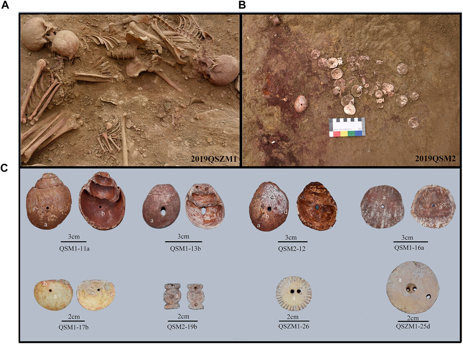
FIGURE 1. (A) Tomb 2019QSZM1 at Sazha Locus of Qulong Site; (B) Tombs 2019QSM2 at Sailaqinbopu Locus of Qulong Site; (C) Photos of samples in this study.
Although all the beads exhibit varying degrees of weathering, red substances remained on one or both sides of the beads. It is worth noting that about one-10th of the samples exhibit a relatively thick red layer, such as QSM1-11a, QSM1-13b, and QSM2-12 (see Figure 1C). At the same time, other shell artifacts only show red traces that could barely be detectable with the naked eye (e.g., Figure 1C, second row). As Figures 1A,B show, a red substance, unclearly paint coat or powder, was found covering the bodies of the tomb owners and the shell beads nearby. Thus, it could be reasonable that the red residue attached to the shell surface was the red substance intentionally splashed onto the deceased during the burial process, signifying an indigenous funeral practice. It may explain the situation of the shell beads with faint red traces (e.g., sample QSM1-17b, Figure 1C), but not all the objects. Consequently, if the composition of this thick layer differs from that of the weak residues that remained on other shell decorations, it is possible that these thick red layers were not left behind due to the burial practice. If the result is opposite, we cannot rule out the possibility that the thick layer was also formed during funeral rites. In addition, take QSM1-11a as an example. It shows different textures of the pigment layers on two sides: the inner surface (of the original conch) displays a thin layer of red paint that is homogenous and bright; the outside, on the contrary, is crusted by a much thicker and darker layer, although part of it has flaked away. The contrast in texture of the substances may suggest a discrepancy in their composition. If it is the case, the formation of the red layers on QSM1-11a could not all have resulted from the red pigment splashing behavior. In short, two questions arise: 1) Are the thick coatings on some of the shell beads comprised of the same material as the faint red pigment remnant on the remaining shell objects? 2) Are the compositions of the red layers on the inner and outer surfaces of sample QSM1-11a different?
2.2 Methods
To answer these questions, non-destructive methods of pXRF and Raman spectrum were used to analyze the chemical composition and phase composition of pigments on the inner and outer surfaces of eight shell beads. In addition, the powder scraped from the outer surface of sample QSM1-11a was detected by micro-invasive methods including XRD and FTIR.
2.2.1 Portable energy-dispersive X-ray fluorescence spectrometer (pXRF)
The pXRF test was performed on all eight shell beads, and the test points are marked in Figure 1C. The chemical compositions of the samples were analyzed via pXRF (OURSTEX 100FA) at the Shanghai Institute of Optics and Fine Mechanics, Chinese Academy of Sciences. The target material of this pXRF spectrometer is palladium (Pd). The excitation voltage of the X-ray tube is 40 or 15 kV, the current is 0.5 or 1.0 mA, the maximum power is 50 W, and the diameter of the X-ray focal spot is about 2.5 mm.
2.3.2 X-ray diffraction (XRD)
About 10 mg of pigment powder was scraped from the outer surface of sample QSM1-11a for XRD analysis. XRD analyses were performed on a Rigaku TTR-III X-ray diffractometer using Cu Kα irradiation (λ = 0.154056 nm) at the University of Science and Technology of China. A small amount of pigment powder was scraped from the sample for analysis. The 2θ range was 10–70. The crystal phase of the samples was determined by comparison with standard spectra in the Jade software.
2.3.3 Laser Raman spectroscopy (Raman)
Raman spectroscopic analysis of samples QSM2-12 and QSM1-16a was performed with a LabRAM HR Evolution (HORIBA JY) at the Instruments Center for Physical Science, USTC. The equipment’s parameters for this analysis were set up as follows: excitation light sources, 785 nm; the detection range, 100–800 cm−1; power, 17 mW; and common objective lens, ×50.
2.3.4 Fourier transform infra-red spectroscopy (FTIR)
The sample used for FTIR analysis was from the powder remaining after the XRD analysis. FTIR analysis was performed at the Instruments Center for Physical Science, USTC. KBr tablet method was used for the FTIR test. The model of the instrument is Nicolet 8700, with a spectral range of 4000-400 cm−1 and a resolution of 0.1 cm−1.
3 Results
3.1 Pigment composition
The red pigment of eight samples contains Fe, indicating that the coloration may be Fe2O3 (Table 1). To further confirm the composition of the pigment, samples QSM2-12 and QSM1-16a were analyzed by Raman spectroscopy, and the external surface of QSM1-11a was analyzed by XRD and FTIR. The Raman spectrum of QSM2-12 and QSM1-16a (Figure 2) shows bands at 222 cm−1, 289 cm−1, 406 cm−1, and 606 cm−1, which are entirely characteristic of hematite (Guglielmi et al., 2022). The infrared spectrum of QSM1-11a (Figure 3; Table 1) shows bands at 467 cm−1 and 529 cm−1 which may be assigned to hematite (Bikiaris, et al., 2000), while there are also obvious diffraction peaks of hematite in the XRD spectrum (Figure 4). Therefore, the chemical composition and phase analysis show that the red pigment used in the shell beads unearthed at the Qulong Site is hematite. In addition, XRD results show the presence of quartz, which may be derived from the soil on the surface of the sample.
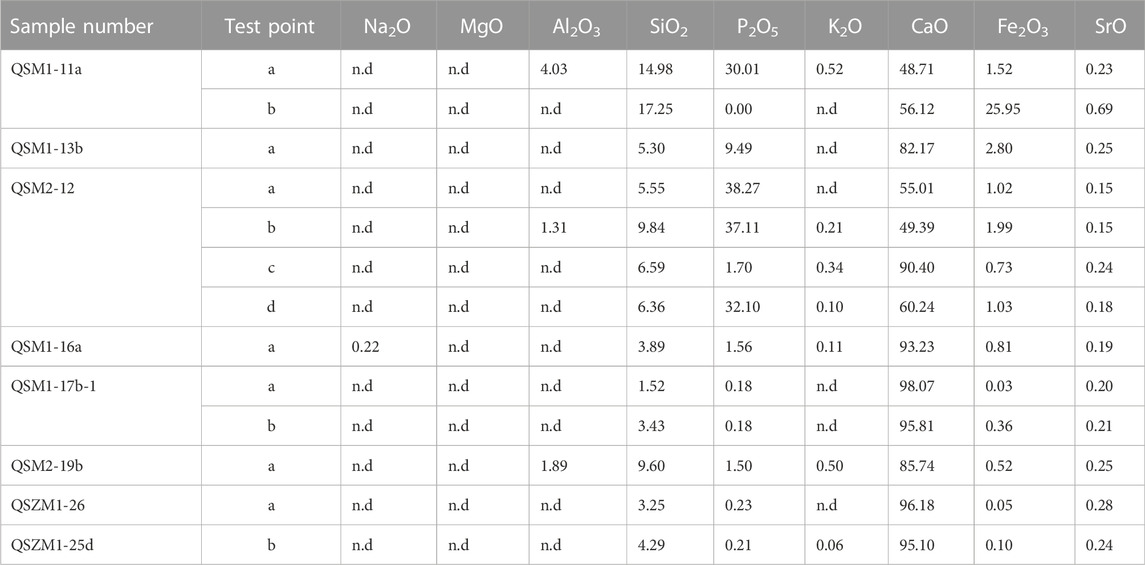
TABLE 1. The chemical composition of the shell beads from the Qulong Site (wt%).
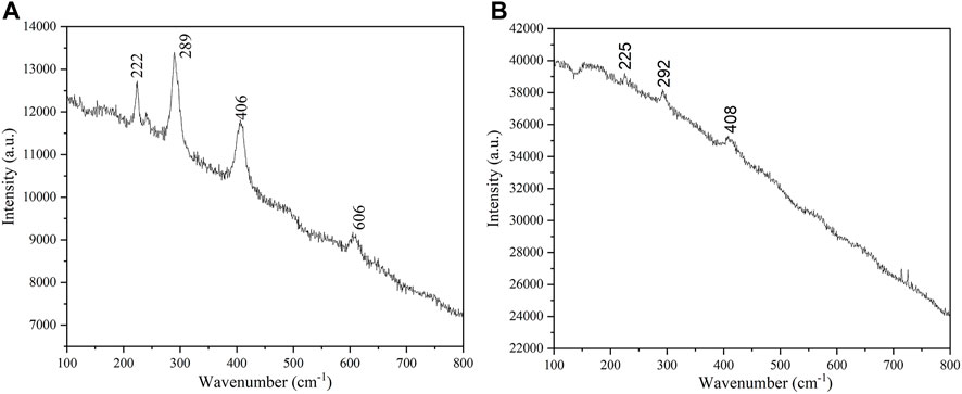
FIGURE 2. Raman results of sample QSM2-12 (A) and QSM1-16a (B).
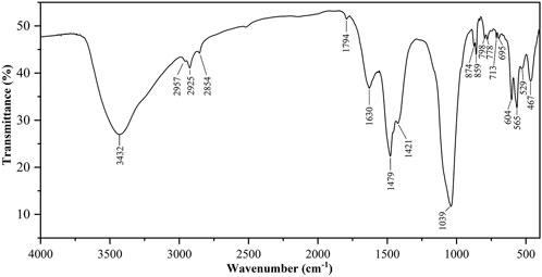
FIGURE 3. FTIR result of sample QSM1-11a.
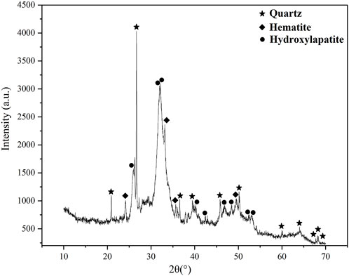
FIGURE 4. XRD result of sample QSM1-11a.
Interestingly, Ca and P were detected in the outer surface pigments of samples QSM1-11a, QSM2-12, and QSM1-13b. The shell beads matrix is CaCO3, which will have a certain impact on pXRF analysis. But theoretically, the influence of the matrix on the compositional analysis is relatively small, given the thick coating layer. Therefore, the possibility that a kind of substance rich in Ca and P, such as bone ash, was mixed into the pigment should be considered. The XRD result of samples QSM1-11a indicated that hydroxyapatite was indeed present in the red pigment. The FTIR spectra recorded on the same samples also show the peaks of hydroxyapatite. A strong peak at 1,039 cm−1 is due to the asymmetric ν3 stretching modes of (PO4)3-, the 562 and 598 cm−1 peaks are due to the asymmetric bending ν4 mode of (PO4)3- groups (Opris et al., 2022). Besides the phosphate groups peaks, we observed weaker peaks due to the carbonate groups as follows: the 875 cm−1 peak is due to the symmetric ν2 mode of (CO3)2- groups, and the 1,421 and 1,479 cm−1 are due to the asymmetric ν3 stretching modes of (CO3)2- groups (Opris et al., 2022). The results showed that the red pigment was indeed doped with bone powder. However, the inner surface of the sample QSM1-11a lacks P, suggesting that no bone meal was added during the production of the outer surface pigments.
To further understand the production process of the pigments, the SF value of the infrared spectrum were calculated to evaluate whether the bone meal was heated. The FTIR SF calculation for bioapatite is based on the degree of peak splitting in the PO43− antisymmetric bending mode (Andrew and John, 1996), which is an important index when discussing bone crystallinity after long-term depositing or burning. First, a local baseline was drawn between 750 and 495 cm−1. Then the heights of the υ3PO43- antisymmetric bending frequencies at approximately 565 cm−1 (A) and 604 cm−1 (B) above this local baseline were measured and divided by the height of the enclosed valley (C) (Weiner and Bar-Yosef, 1990; Todd and Mary, 2001). Finally, the SF value of the bone meal in the samples QSM1-11a was calculated to be 3.7 by the formula SF = (A+ B)/C. The SF index of modern bones is about 2.8–3.0, and that of archaeological samples is about 3.5–4.8 (Guo et al., 2017). SF values are relatively stable until 600°C, then strongly increase to reach values around 8–10 for samples heated from 700 to 900°C (Lebon et al., 2008). The SF of the bone meal in the QSM1-11a pigment is within the range of the SF value of the archaeological sample, indicating that it has not been heated at a high temperature above 600°C.
In general, the red pigments of the shell beads of the Qulong site are all hematite, and most of the samples are doped with bone powder that has not been heated at a high temperature (600°C). Unlike the outer surface, the pigment on the inner surface of sample QSM1-11a does not contain bone powder, which may indicate the different processes of pigment production.
3.2 Glue identification
Binding media is an important part of ancient painted cultural relics. In the process of making a color painting, mixing glue and mineral pigment can improve the force between pigment particles and strengthen the adhesion between the pigment and the painting matrix (Yang et al., 2011a). The study on the existence and types of binding media will help us to understand the production technology of the surface pigments of the shell beads at the Qulong site and the utilization of animal and plant resources by the ancients.
The binders in ancient painted cultural relics include proteins, lipids, polysaccharides, resins, etc. For QSM1-11a, the strong absorption bands in the 1,628–1,636 cm−1 were attributed to amide I (C=O stretching), a characteristic of proteinaceous binders (Pellegrini et al., 2016). Additionally, the asymmetrical N-H stretching vibration occurring near 3,300 cm−1, the methyl (CH3) and methylene (CH2) groups produce asymmetric stretching vibrations respectively at 2957 and 2925 cm−1 and small symmetric stretching bands at 2,854 cm−1 (CH2) (Invernizzi et al., 2018). Common ancient proteinaceous binders include animal glue (bone glue, skin glue, fish bladder glue), eggs (whole egg, egg yolk, egg white), and milk. Among them, the whole egg, egg yolk, skin glue, and milk are rich in lipids, and there are C=O stretching bands near 1745 cm−1 of their infrared spectra (Ma et al., 2020). There is no band near 1745 cm−1 in the infrared spectrum of sample QSM1-11a, which may indicate that the proteinaceous binders were not from the whole egg, egg yolk, skin glue, milk, and other lipids rich materials, but the degradation of lipids during the burial process increases the uncertainty of the deduction. Previous studies showed that during the evolution of organisms from the sea to the land, some amino acids in collagen, such as serine (Ser), were gradually converted into hydroxyproline (Hyp) to adapt to the new higher temperature living environment (Song et al., 2008). Therefore, the content of hydroxyproline (Hyp) in the collagen of terrestrial mammals is significantly higher than that of fish, while the content of serine (SER) is lower than that of fish. It is shown that there is an absorption peak generated by the stretching vibration of the C-O on the serine side chain in the 1,076 cm−1 of fish bladder gum, which is not significant in the infrared spectrum of terrestrial mammalian gum (Yang et al., 2011b). There is no peak around 1,076 cm−1 in the infrared spectrum of sample QSM1-11a, which may indicate that the proteinaceous binders are not fish bladder glue, but the effect of protein degradation cannot be excluded.
In addition, due to the presence of bone powder that has not been heated at a high temperature, it is still necessary to further explore whether the protein detected on the surface of the shell beads was attributed to bone powder or intentionally added glue. The amide-to-phosphate ratio (amide I/PO4) has been used to evaluate the preservation of collagen in bone (Trueman et al., 2004; Fredericks et al., 2012; Kontopoulos et al., 2020). LeBon et al. (2016) studied 42 bone samples from the Pleistocene to modern times. The results showed that the amide I/PO4 ratio had a good correlation with the nitrogen content, and thus established the relationship between the amide I/PO4 ratio and N wt% and Collagen wt%:
According to the guidance of Bouchard et al. (2019), the ratio of the area under the Amide I peak to the area under the phosphate ν3 peak was calculated, and the amide I/PO4 value of QSM1-11a was about 0.23. Thus, the nitrogen content is about 5.05% and the collagen content is about 27.71wt%. Depending on the species and type of bone, fresh bone contains 20–35% organic matrix, of which ∼90% is collagen, giving bone a %N of ∼3.5–4.5% (Brock et al., 2012; Currey 2012; Richter et al., 2022). Kontopoulos et al. (2020) tested 266 human and animal bone samples from 10,000 BC to 1850 AD, and found that the amide I/PO4 values were lower than 0.2 and the collagen content was lower than 25%. Meanwhile, the mean value of amide I/PO4 value in 42 bones was measured by LeBon et al. (2016) was 0.08, the highest value was 0.18, the mean value of N content was 2.29%, and the highest value was 4.25%. In the case of the degradation of ancient proteins and the reduction of the thermal stability of collagen by hematite (Pellegrini et al., 2016), proteinaceous binders should be added to the surface of shell beads at the Qulong Site, resulting in such high nitrogen content and collagen content, even higher than that of fresh bones.
Therefore, FTIR results showed that the outer pigment of sample QSM1-11a was doped with bone powder and then added with proteinaceous binders. There are no characteristic peaks of the whole egg, egg yolk, skin glue, milk, and other lipids-rich binders and fish bladder glue in the spectrum. However, the degradation limits the exploration of the specific ancient binder.
4 Discussion and conclusion
The scientific analyses show that the red pigment applied to chank shell beads included in this paper is red ochre, one of the most important pigments found at archaeological sites around the globe (Langley and O’Connor, 2018; Siddall, 2018). Nevertheless, different types of shell objects are treated differently. For example, phosphorus-rich substances were found in the pigments on the outer surfaces of samples QSM1-11a, QSM1-13b, and QSM2-12. XRD and FTIR results suggest that they were bone powder. However, phosphorus did not exist on the inner surfaces of these samples and both sides of the other analyzed shell beads, indicating the absence of bone powder. Moreover, proteinaceous binders were detected in the powder scraped from the outer paint of sample QSM1-11a, which are necessary for bonding thick pigments. Therefore, we believe that the shell beads with thick pigments on the outside were already coated with a red pigment mixed with ochre, bone powder and proteinaceous binders before burial. Although it is still challenging to determine the specific types of binders, anthropological evidence may provide a clue. Modern Tibetan herders often boil the milk dregs separated from the butter making them into a thick, dark brown paste, that is used as a binder to apply red paint on their faces. The glue can be immediately used to smear the face or stored for later use. Only a tiny amount of water must be added, and then it can be used after being heated to a thick paste (Melvyn and Cynnthia, 1990). The ethnographic record may shed light on future research.
For the other shell samples from Qulong, the situation is different. The photographs taken during fieldworks (Figures 1A, B) exhibit red pigments scattered around the shell beads and the human skeletons, suggesting a burial practice after the beads were placed in the tombs. Thus, for the shell objects as samples QSZM1-25d, QSM1-17b, QSM1-16a, and QSM2-19b, they might not have been painted before interment but stained red due to the ochre powder splashing activity in the funeral. In addition, samples QSM1-11a, QSM1-13b, and QSM2-12, showing similarities in shape and bearing a thick red layer on the outside, have a thin and brighter layer on the inner surface, apparent in sample QSM1-11a. Although the composition of the thin layer could not be distinguished from other weak red traces, its distinct texture and appearance may indicate that the three samples’ inner surfaces had also been painted red before the funeral, despite a pigment difference in composition from that of the outer surfaces. This may indicate an elaborate treatment of this type of shell ornaments. In brief, the diversity of material compositions of the red substances may indicate that the ancient people at Qulong treated a variety of shell bead types in different ways according to their distinct functions, or that the source of these shell objects had defined their forms and colors before being imported into Qulong.
Similar perforated chank shell beads, ranging in number from one to 17, were also sporadically recovered from a few sites in the Himalayan region, including Chuvthag (c. 2,400–2200 BP) (IACASS et al., 2015; Tong, 2021, 539-540), Gepa Serul (c. 3500–2200 BP) (CTSSU et al., 2001b) (The shell beads information from Gepa Serul as well as the archaeological background were provided by co-author Songmei Hu who participated the excavation at Gepa Serul in 2018.) and Piyang-Donggar (Gelingtang Locus, c. 2700–2200 BP) (CTSSU et al., 2001a) in western Tibet, Malari (c. 2400–2100 BP) (Deshpande-Mukherjee et al., 2015; Bhatt and Nautiyal, 1987–88), Lippa (c. 2600–2300 BP) (Nautiyal et al., 2014; Deshpande-Mukherjee et al., 2015) and Ropa (probably close to the period of Malari and Lippa) (Deshpande-Mukherjee et al., 2015) in northwestern Indian, and Chokhopani (c. 2800 BP) (Simons et al., 1994; Tiwari, 1984/85) in north Nepal (see locations in Figure 5). Most of these shells are disk-shaped and show faint or no traces of red pigment, except for two beads from Ropa in Himachal Pradesh, India (Figure 6A). As shown in Figure 6A, the photographed shell bead from Ropa is similar in shape and color to samples QSM1-11a, QSM1-13b and QSM2-12 from Qulong, demonstrating solid connections between these artifacts from the two sites. It is worth noting that besides these two red shell objects, the other 13 cowrie-shaped shell beads, which were stored together with the red ones in a large vessel when unearthed, are purely white (Figure 6B). Thus, the red beads should have been painted red before being buried with other white shell beads, just as the three samples from Qulong.
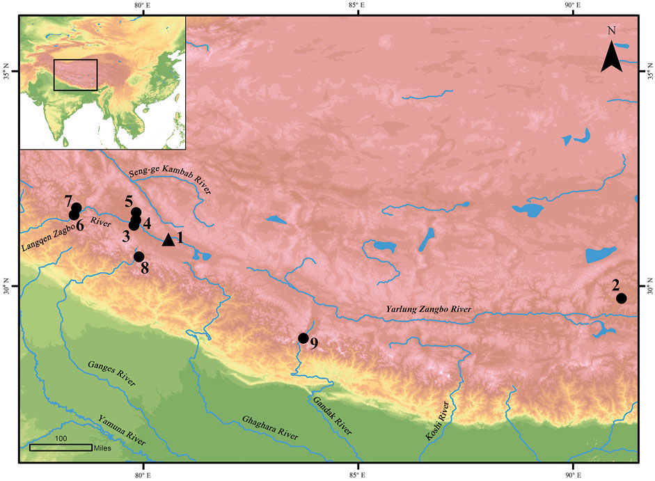
FIGURE 5. Location of the sites mentioned in the article. 1. Qulong; 2. Qugong; 3. Chuvthag; 4. Gepa Serul; 5. Gelingtang; 6. Lippa; 7. Ropa; 8. Malari; 9. Chokhopani.
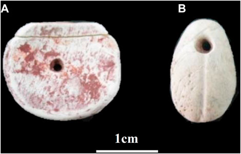
FIGURE 6. Chank shell beads from Ropa. (after Deshpande-Mukherjee et al., 2015, Figures 6, 7). (A) perforated disk-shaped bead; (B) cowrie-shaped shell bead.
Most of the archaeological data from the Qulong site has yet to be analyzed and published. Based on the limited information, only three gemstone beads of carnelian or agate and a handful of small bronze tools were recovered with the chank shell beads at Qulong, indicating a not very prominent social status of the tomb owners. Besides, although the red pigment splashing activity during the funeral ceremony might suggest a religious behavior, there is no evidence to support that the tomb owners were associated with indigenous shamanism. So far, we could assume that these shell beads were used as ornaments for the Qulong people, such as headdresses, necklaces, and buttons sewn onto clothing, in the same manner as in today’s traditional Tibetan dress. Moreover, the red-painted ones might have been the key design in a set of shell beads since the Tibetan people have been fond of red since ancient times. For example, as the Old Book of Tang (Jiutangshu) records, in the time of the Tubo King Songtsen Gampo’s reign (seventh century), there was a popular custom of painting faces red in Tibet (Liu, 1974; Li, 2006). Therefore, although it is difficult to determine whether the shell ornaments were manufactured and/or painted red locally, the affection for the color red by the Qulong people is perceptible.
The present study was designed to examine the material composition of the red substances adhering to eight chank shell beads recovered from the 2019 season excavation at Qulong in western Tibet. The results are as follows: 1) the coloring agent of all red pigments on the shell bead is iron oxide, i.e., red ocher; 2) bone powder that has not been heated to high temperatures (above 600°C) and proteinaceous binders were added to the paint on the outer surface of sample QSM1-11a, but the thin layer on its interior surface was without bone powder or organic binders; (3) neither bone powder nor proteinaceous binders were added to the red residues on samples other than QSM1-11a, QSM1-13b, and QSM2-12. The results of this study suggest the complexity and diversity of the red substances applied to shell beads from Qulong and clarify our understanding of human practices and local customs in the Tibetan plateau and the surrounding areas in prehistoric times. More research is required to determine the specific kind of protein glue mixed with ochre in the future.
Data availability statement
The original contributions presented in the study are included in the article/supplementary material, further inquiries can be directed to the corresponding authors.
Author contributions
JW, BZ, and AF conceived the study and designed the experiments; BZ and YZ performed the experiments; LX and SH provided the samples; all the authors discussed the data and contributed to writing the manuscript.
Funding
The research was supported by the National Natural Science Foundation of China (Grant No.41303080), the National Social Science Foundation of China (Grant No. 20BKG025), the Youth Innovation Promotion Association CAS (2018499), USTC Academic Leader Cultivation Program (2018), USTC Youth Innovation Fund (2021, WK2110000018), and the USTC Research Funds of the Double First-Class Initiative (YD2110002027).
Acknowledgments
We thank Qinghui Li and Song Liu (Shanghai Institute of Optics and Fine Mechanics, Chinese Academy of Sciences) for the pXRF test in this study, and Zhe Cui (School of Material Science and Engineering, Zhengzhou University) for the advice on FTIR. Their help formed the first step to this paper.
Conflict of interest
The authors declare that the research was conducted in the absence of any commercial or financial relationships that could be construed as a potential conflict of interest
The handling editor YH declared a past co-authorship with the author SH.
Publisher’s note
All claims expressed in this article are solely those of the authors and do not necessarily represent those of their affiliated organizations, or those of the publisher, the editors and the reviewers. Any product that may be evaluated in this article, or claim that may be made by its manufacturer, is not guaranteed or endorsed by the publisher.
References
Andrew, S., and John, P. (1996). Diagenesis of bones from eland’s bay cave. J. Archaeol. Sci. 23, 535–542. doi:10.1006/jasc.1996.0050
Bhatt, R. C., and Nautiyal, K. P. (1987). Trans himalayan burials vis-a-vis Malari: An assessment. J. Himal. Stud. Regional Dev., 95–101.
Bikiaris, D., Daniilia, S. X., Sotiropoulou, S., Katsimbiri, O., Pavlidou, E., Moutsatsou, A. P., et al. (2000). Ochre-differentiation through micro-Raman and micro-FTIR spectroscopies: Application on wall paintings at meteora and mount athos, Greece. Spectrochimica Acta Part A Mol. Biomol. Spectrosc. 56 (1), 3–18. doi:10.1016/s1386-1425(99)00134-1
Bouchard, G. P., Mentzer, S. M., Riel-Salvatore, J., Hodgkins, J., Miller, C. E., Negrino, F., et al. (2019). Portable FTIR for on-site screening of archaeological bone intended for ZooMS collagen fingerprint analysis. J. Archaeol. Sci. Rep. 26, 101862. doi:10.1016/j.jasrep.2019.05.027
Brock, F., Wood, R., Higham, T. F. G., Ditchfield, P., Bayliss, A., and Ramsey, C. B. (2012). Reliability of nitrogen content (%N) and carbon: Nitrogen atomic ratios (C:N) as indicators of collagen preservation suitable for radiocarbon dating. Radiocarbon 54 (3–4), 879–886. doi:10.1017/s0033822200047524
CTSSU (Center for Tibetan Studies of Sichuan University)DASU (Department of Archaeology, Sichuan University)BCRTAR (Bureau of Cultural Relics, Tibet Autonomous Region)BCRTNP (Bureau of Culture, Radio and Television, Ngari Prefecture) (2001b). A brief report on the survey of the Gepa Serul cemetery in Zanda county, Tibet. Archaeology (6), 39–44.
CTSSU (Center for Tibetan Studies of Sichuan University)DASU (Department of Archaeology, Sichuan University)BCRTAR (Bureau of Cultural Relics, Tibet Autonomous Region) (2001a). Brief report on the piyang-donggar ancient cemeteries, Zanda county, Tibet. Archaeology (6), 14–31.
Currey, J. D. (2012). The structure and mechanics of bone. J. Mater. Sci. 47 (1), 41–54. doi:10.1007/s10853-011-5914-9
Deshpande-Mukherjee, A., Bhatt, R. C., Saklani, P. M., Nautiyal, V., Chauhan, H., Bist, K., et al. (2015). A note on the Marine shell objects from the burial sites of Malari, Lippa and Ropa in the Trans-Himalayan region of India. Archaeo+Malacology Group Newsl. (25), 9–15.
Fredericks, J. D., Bennett, P., Williams, A., and Rogers, K. D. (2012). FTIR spectroscopy: A new diagnostic tool to aid dna analysis from heated bone. Forensic Sci. Int. Genet. 6 (3), 375–380. doi:10.1016/j.fsigen.2011.07.014
Gao, Z. (2011). On the ancient sources of ocher, vermilion, red lead and its application. J. Qinghai Natl. Univ. Soc. Sci. 37 (01), 102–109.
Guglielmi, V., Andreoli, M., Comite, V., Baroni, A., and Fermo, P. (2022). The combined use of SEM-EDX, Raman, ATR-FTIR and visible reflectance techniques for the characterisation of roman wall painting pigments from monte d'Oro area (rome): An insight into red, yellow and pink shades. Environ. Sci. Pollut. Res. 29 (20), 29419–29437. doi:10.1007/s11356-021-15085-w
Guo, Y., Xiang, C., Xia, Y., Xu, X., and Zhang, G. (2017). Preliminary study on the feasibility of hydroxyapatite stable isotope analysis of ancient human bone in South China: Take the Zhuangqiaofen sit, Zhejiang Province as an example. Quat. Sci. 37 (1), 143–154.
Huo, W. (1997). Discussion on ancient Zhang Zhuang and Zhang Zhung civilization. Tibet. Stud. (3), 44–54.
IACASS (Institute of Archaeology, Chinese Academy of Social Science)BCRTAR (Bureau of Cultural Relics, Tibet Autonomous Region) (1999). Qugong in Lhasa: Excavations of an ancient site and tombs. Beijing: Encyclopedia of China Publishing House.
IACASS (Institute of Archaeology, Chinese Academy of Social Science)TCRCI (Tibetan Cultural Relics Conservation Institute)CRBNP (Cultural Relics Bureau of Ngari Prefecture)CRBZC (Cultural Relics Bureau of Zanda County) (2015). The gurugyam and Chuvthag cemeteries in Ngari prefecture, Tibet. Archaeology (7), 29–50.
Invernizzi, C., Rovetta, T., Licchelli, M., and Malagodi, M. (2018). Mid and near-infrared reflection spectral database of natural organic materials in the cultural heritage field. Int. J. Anal. Chem. 2018, 1–16. doi:10.1155/2018/7823248
Kenoyer, J. M. (1991). Ornament styles of the indus valley tradition: Evidence from recent excavations at harappa, Pakistan. paleo. 17 (2), 79–98. doi:10.3406/paleo.1991.4553
Kontopoulos, I., Penkman, K., Mullin, V. E., Winkelbach, L., Unterlaender, M., Scheu, A., et al. (2020). Screening archaeological bone for palaeogenetic and palaeoproteomic studies. Plos One 15 (6), e0235146. doi:10.1371/journal.pone.0235146
Langley, M. C., and O’Connor, S. (2018). “Exploring red ochre use in Timor-Leste and surrounds: Headhunting, burials, and beads,” in The archaeology of portable art: Southeast asian, pacific, and Australian perspectives. Editors M. C. Langley, M. Litster, D. Wright, and S. K. May (London: Routledge), 25–36.
Lebon, M., Reiche, I., Froehlich, F., Bahain, J. J., and Falgueres, C. (2008). Characterization of archaeological burnt bones: Contribution of a new analytical protocol based on derivative FTIR spectroscopy and curve fitting of the ν 1 ν 3 PO4 domain. Anal. Bioanal. Chem. 392 (7–8), 1479–1488. doi:10.1007/s00216-008-2469-y
Lebon, M., Reiche, I., Gallet, X., Bellot-Gurlet, L., and Zazzo, A. (2016). Rapid quantification of bone collagen content by ATR-FTIR spectroscopy. Radiocarbon 58 (1), 131–145. doi:10.1017/rdc.2015.11
Li, Y. (2017). A preliminary Study of khyung lung Site in Ngari, Tibet. Master’s thesis. Northwest University.
Li, Y. (2006). Taim bilong sipak: Nasioi alcohol, 1962-1978. Ethnology 25, 21–39. doi:10.2307/3773719
Ma, Z., Wang, L., Yang, L., and Zhao, X. (2020). The influence of the soil aging exerting on the stability of proteinaceous binders in Chinese polychromy artworks. Microchem. J. 157, 104955. doi:10.1016/j.microc.2020.104955
Melvyn, G., and Cynnthia, B. (1990). Nomads of western Tibet. Berkeley Los Angeles: University of California Press.
Nautiyal, V., Bhatt, R. C., Saklani, P. M., Tripathy, V. M., Nautiyal, C. M., and Chauhan, H. (2014). Lippa and Kanam: Trans-Himalayan cist burial culture and pyrotechnology in Kinnaur, Himachal Pradesh, India. Antiq. Proj. Gallery 88 (339).
Opris, V., Velea, A., Secu, M., Rostas, A.-M., Buruiana, A.-T., Simion, C.-A., et al. (2022). Put variety in White”: Multi-analytical investigation of the white pigments inlaid on Early Chalcolithic pottery from Southern Romania. J. Archaeol. Sci. Rep. 42, 103402. doi:10.1016/j.jasrep.2022.103402
Pellegrini, D., Duce, C., Bonaduce, I., Biagi, S., Ghezzi, L., Colombini, M. P., et al. (2016). Fourier transform infrared spectroscopic study of rabbit glue/inorganic pigments mixtures in fresh and aged reference paint reconstructions. Microchem. J. 124, 31–35. doi:10.1016/j.microc.2015.07.018
Richter, K. K., Codlin, M. C., Seabrook, M., and Warinner, C. (2022). A primer for ZooMS applications in archaeology. Proc. Natl. Acad. Sci. U. S. A. 119 (20), e2109323119. doi:10.1073/pnas.2109323119
Siddall, R. (2018). Mineral pigments in archaeology: Their analysis and the range of available materials. Minerals 8, 201. doi:10.3390/min8050201
Simons, A., Schön, W., and Shrestha, S. S. (1994). Preliminary report on the 1992 campaign of the team of the Institute of prehistory, university of cologne. Anc. Nepal J. Dep. Archaeol. 136, 51–75.
Song, Q., Dong, X., and Yu, X. (2008). Comparison of components and contents of collagen amino acids in some mammal and fishes. Mod. Food Sci. Technol. 24 (12), 1239–1242.
Subba Rao, N. V. (2003). Indian seashells (Part 1) polyplacophora and gastropoda. Kolkata: Zoological Survey of India.
Tang, Q., Wang, J., Dong, S., Chen, J., Zhang, Y., Jiang, F., et al. (2018). Design of a near-infrared sky brightness monitor and field measurement at the Ngari Observatory, Tibet. J. Astron. Telesc. Instrum. Syst. 4 (4), 1. doi:10.1117/1.JATIS.4.4.046002
Tian, L. (2018). Study on red Pigments in ancient China. Master’s thesis. Shaanxi Normal University.
Tiwari, D. N. (1984/85). Cave burials from Western Nepal, mustang. Anc. Nepal J. Dep. Archaeol. 85, 1–12.
Todd, A. S., and Mary, C. S. (2001). Standardizing infra-red measures of bone mineral crystallinity: An experimental approach. J. Archaeol. Sci. 28 (6), 633–642. doi:10.1006/jasc.2000.0633
Tong, T. (2021). Archaeological study on the silk road of the qinhai-tibet plateau. Beijing: Wenwu Press.
Trueman, C. N. G., Behrensmeyer, A. K., Tuross, N., and Weiner, S. (2004). Mineralogical and compositional changes in bones exposed on soil surfaces in amboseli national park, Kenya: Diagenetic mechanisms and the role of sediment pore fluids. J. Archaeol. Sci. 31 (6), 721–739. doi:10.1016/j.jas.2003.11.003
Wang, R. (2014). “Applying red phenomenon on stone tools of Qugong Culture,” in Chinese Tibetan ArchaeologyCTSSU (center for Tibetan studies of sichuan university) and HCSSU (history and culture school of sichuan university) (Chengdu: Tiandi Press), 1909–1914.
Weiner, S., and Bar-Yosef, O. (1990). States of preservation of bones from prehistoric sites in the near east: A survey. J. Archaeol. Sci. 17 (2), 187–196. doi:10.1016/0305-4403(90)90058-d
Yang, L., Huang, J., Wang, L., Ma, T., Cao, X., and Li, X. (2011a). The study of binding media’s amino acids components and their FTIR characteristics used in ancient Chinese colored relics. Sci. Conservation Archaeol. 23 (01), 36–39.
Yang, L., Wang, L., Huang, J., Yu, S., and Li, Y. (2011b). The investigation of fish bladder glue, a kind of binder commonly used in painted relics, by using FTIR, micro-Raman spectroscopy, and amino acid analysis. J. Northwest Univ. Nat. Sci. Ed. 41 (1), 63–66.
Keywords: applying red, Tibetan plateau, prehistoric period, Qulong site, chank shell beads
Citation: Wang J, Zhang B, Xi L, Hu S, Zhang Y and Fan A (2023) Investigation of red substances applied to chank shell beads from prehistoric site of Qulong in Ngari Prefecture, Tibet, China. Front. Earth Sci. 10:1063851. doi: 10.3389/feart.2022.1063851
Received: 07 October 2022; Accepted: 05 December 2022;
Published: 26 January 2023.
Edited by:
Yaowu Hu, Fudan University, ChinaReviewed by:
Wugan Luo, University of Chinese Academy of Sciences, ChinaRong Wang, Fudan University, China
Copyright © 2023 Wang, Zhang, Xi, Hu, Zhang and Fan. This is an open-access article distributed under the terms of the Creative Commons Attribution License (CC BY). The use, distribution or reproduction in other forums is permitted, provided the original author(s) and the copyright owner(s) are credited and that the original publication in this journal is cited, in accordance with accepted academic practice. No use, distribution or reproduction is permitted which does not comply with these terms.
*Correspondence: Lin Xi, xilin1015@126.com; Anchuan Fan, anchuan@ustc.edu.cn
†These authors have contributed equally to this work and share first authorship