Characterization of Esophageal Microbiota in Patients With Esophagitis and Esophageal Squamous Cell Carcinoma
 Zongdan Jiang
 Jun Wang2†
  Shukui Wang
- 1Department of Gastroenterology, Nanjing First Hospital, Nanjing Medical University, Nanjing, China
- 2Department of Gastroenterology and Hepatology, Jinhu County People’s Hospital, Huaian, China
- 3Department of Endocrinology, Nanjing First Hospital, Nanjing Medical University, Nanjing, China
- 4General Clinical Research Center, Nanjing First Hospital, Nanjing Medical University, Nanjing, China
Microbial imbalances have been well elucidated in esophageal adenocarcinoma. However, few studies address the microbiota in esophageal squamous cell carcinoma (ESCC) and esophagitis (ES). We aimed to explore the association of esophageal microbiota with these patients. Esophageal tissues were obtained from healthy controls and ES and ESCC patients undergoing upper endoscopy. 16S rRNA gene sequencing was applied to analyze the microbiome. The α and β diversity differences were tested by Tukey test and partial least squares-discriminant analysis (PLS-DA), respectively. Linear discriminant analysis effect size (LEfSe) analysis was performed to assess taxonomic differences between groups. A total of 68 individuals were enrolled (control = 21, ES = 15, ESCC = 32). Microbial diversity was significantly different between the ESCC patients and healthy controls by Chao1 index, Shannon index, and PLS-DA. Firmicutes, Proteobacteria, Bacteroidetes, Actinobacteria, and Fusobacteria were the five dominant bacterial phyla among the three groups. Megamonas, Collinsella, Roseburia, and Ruminococcus_2 showed a significantly continuous decreasing trend from the control group to the ESCC group at the genus level. When compared with the control group, decreased Fusobacteria at phylum level and Faecalibacterium, Bacteroides, Curvibacter, and Blautia at genus level were detected. ESCC samples also displayed a striking reduction of Bacteroidetes, Faecalibacterium, Bacteroides, and Blautia in comparison with the ES patients. LEfSe analysis indicated a greater abundance of Streptococcus, Actinobacillus, Peptostreptococcus, Fusobacterium, and Prevotella in the ESCC group. Our study suggests a potential association between esophageal microbiome dysbiosis and ESCC and provides insights into potential screening markers for esophageal cancer.
Introduction
Esophageal cancer is a highly lethal malignancy with a rapidly increasing incidence globally. It has been ranked the fourth leading cause of cancer-related death, which brings a substantial public health burden (Feng et al., 2019). The main histological types of esophageal carcinoma are esophageal adenocarcinoma (EAC) and esophageal squamous cell carcinoma (ESCC), with the latter predominating in the Chinese population (Kelly, 2019). Although the treatment for ESCC has been notably improved, the prognosis of ESCC is still not satisfactory, with a 5-year overall survival rate of 30.3% (Baba et al., 2014; Torre et al., 2015). Thus, we still need to further advance our insights into ESCC and attempt to offer new therapeutic alternatives.
Esophagitis (ES), smoking, drinking, and heredity are the known risk factors related to ESCC (Kavin et al., 1996; Reichenbach et al., 2019). Recently, microorganisms have been considered to exert essential functions in the occurrence and progression of gastrointestinal diseases. There are at least 38 trillion microorganisms colonizing the human gastrointestinal tract, which associate with the immunological homeostasis. Increasing evidence suggests that an imbalance of certain species is involved in tumor onset and development via producing carcinogenic toxins, dampening the immunity, and damaging DNA structure. Previous studies reported that there was a significant decrease in bacterial counts and alterations in microbial communities in the gastroesophageal reflux disease (GERD) and Barrett’s esophagus groups (Blackett et al., 2013; Elliott et al., 2017) compared with the healthy group. Alterations of microbial diversity, including a lower level of Veillonella and Streptococcus and a higher level of Lactobacillus, Enterobacteriaceae, and Akkermansia, are associated with EAC (Lopetuso et al., 2020). However, the relationship between human esophageal microbiota and ESCC has not garnered sufficient scientific attention.
Hence, in the present study, we aimed to assess and compare the diversity and composition of the microbiota between ESCC, ES, and healthy tissues and complemented the clinical data in this area. This might further illustrate the role of microbiota in the pathogenesis of ESCC and bring light to the treatment of ESCC.
Materials and Methods
Study Participants
Samples were obtained from patients undergoing routine upper endoscopy for the screening of upper gastrointestinal cancer or clinical indications. Finally, we recruited 15 esophagitis patients (ES group) and 21 healthy volunteers (Control group) in Nanjing First Hospital from 2018 to 2019. A total of 32 ESCC patients (ESCC group) were enrolled from Jinhu People’s Hospital. Sampling from esophagectomy was done with a sterile scalpel blade (cutting down to submucosa) within 1 h of surgical resection. All samples were flash frozen in liquid nitrogen and stored at –80°C. The inclusion criteria were age older than 18 years and no contraindications for endoscopic examination. The ES and ESCC groups were composed of patients diagnosed as ES and ESCC, respectively, which were confirmed by both endoscopy and pathology. The control group was defined as individuals with normal esophagus confirmed by endoscopy and pathology and no digestive symptoms. To minimize the potential influence on the microbiota, all patients enrolled should not receive antibiotics, H2 receptor antagonists, proton pump inhibitors, and probiotics 1 month before sample collection. Patients who had received radiotherapy, chemotherapy, and/or prior surgery were excluded. The study protocol was approved by the institutional review board of Nanjing Medical University, and all experiments were performed in accordance with approved guidelines and regulations.
DNA Extraction
Total genome DNA from samples was extracted using the QIAamp DNA Mini Kit (QIAGEN, Valencia, CA, USA) combined with the bead-beating method. The DNA concentrations of each sample were adjusted to 50 ng/μl for subsequent 16S rDNA gene analysis. The bacterial DNA samples were stored at –80°C for sequencing.
PCR Amplification
16S rDNA genes of V3-V4 region were amplified using universal primers, namely, 338F (5′-ACTCCTACGGGAGGCAGCAG-3′) and 806R (5′ -GGACTACHVGGGTWTCTAAT-3′). All PCR reactions (including denaturation, annealing, and elongation) were carried out with Phusion® High-Fidelity PCR Master Mix (New England Biolabs). After electrophoresis of PCR products, samples with bright main strip between 400 and 450 bp were chosen for next mixing and purification with Qiagen Gel Extraction Kit (Qiagen, Germany).
Sequencing Processing and Analysis
The purified amplifications were paired-end sequenced (PE300) on an Illumina MiSeq platform (Illumina, San Diego, CA, USA). Barcodes and sequencing primers were trimmed before assembly. All raw reads were stored in NCBI Sequence Read Archive (SRA) database, and the accession number is PRJNA759579.
Statistical Analysis
The raw data were filtered with QIIME (V1.8.0), discarding the reads that were dereplicated or shorter than 150 bp. Filtered reads were clustered into operational taxonomic units (OTUs) assuming 97% similarity. Compared with the SILVA database (version 128), the species classification information of each OTU was obtained. For continuous variables, independent t-test, White’s nonparametric t-test, and Mann–Whitney U test were applied. For categorical variables between groups, Pearson chi-square or Fisher’s exact test was used, depending on assumption validity. QIIME software was used to evaluate the α diversity by calculating the Shannon index and Simpson index. To compare the differences of diversity among groups, β diversity was tested by partial least squares-discriminant analysis (PLS-DA). Linear discriminant analysis (LDA) effect size (LEfSe) was performed to find key microbes associated with different groups with the LDA threshold of 3. We used Phylogenetic Investigation of Communities by Reconstruction of Unobserved States (PICRUSt) analysis to predict Kyoto Encyclopedia of Genes and Genomes (KEGG) biochemical pathways. Statistical analysis was performed using the SPSS V19.0 (SPSS Inc., Chicago, IL, USA) and STAMP V2.1.3. GraphPad Prism V6.0 (San Diego, CA, USA) was used for preparation of graphs. A p value <0.05 was considered statistically significant.
Results
Baseline Characteristics of Participants
A total of 21 healthy volunteers, 15 ES patients, and 32 ESCC patients were enrolled in this study. Demographic characteristics of all included individuals were shown in Table 1. Older age was observed in ESCC group compared with the healthy controls. There was no significant difference in sex, alcohol intake, smoking, diabetic background, and family history of cancer among the groups.
Table 1 Clinical characteristics of enrolled patients and healthy controls.
Microbial Richness and Diversity Among the Three Groups
After sequencing and quality filtering, more than 3.2 million tags and a total of 2,134 OTUs were obtained with the dominant length of tags located among 400–440 bp (Figure 1A). To test the sequencing depth, we created the rarefaction curves and showed a reasonable amount of sampling (Supplementary Figure S1).
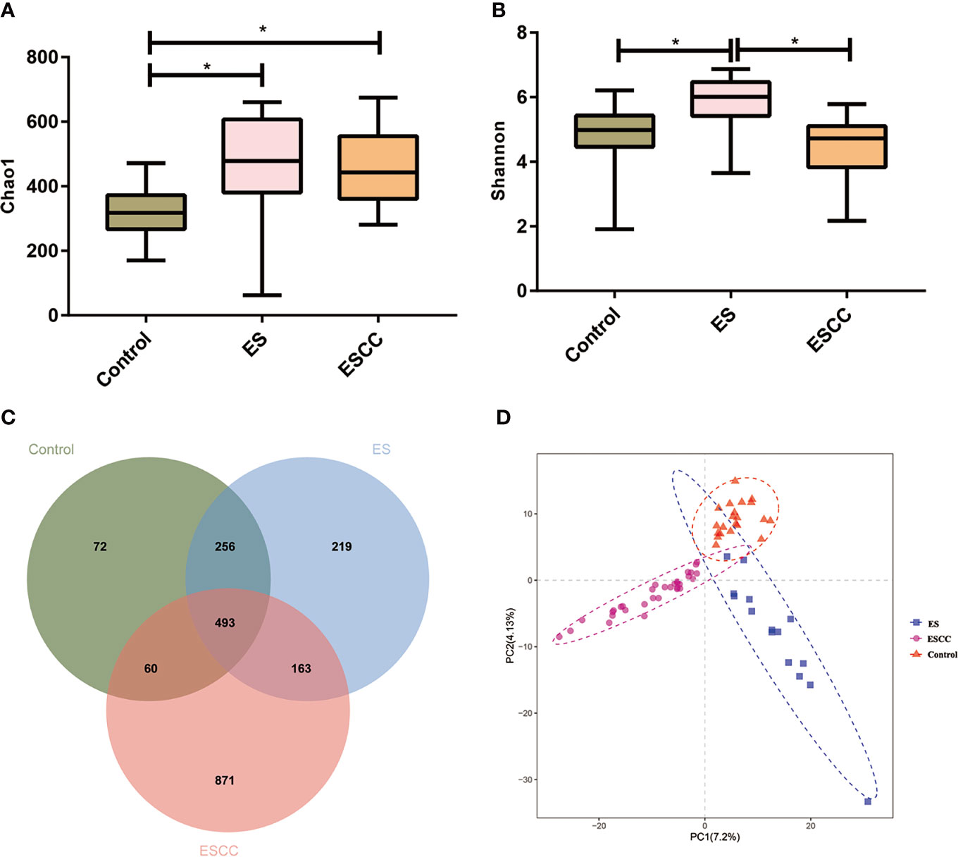
Figure 1 The microbial α diversity and β diversity analysis in different groups. (A) Chao 1 index was higher in the ESCC and ES group than in the control group. (B) The ES group had a significantly higher Shannon index, in comparison with the ESCC group and control group. Data were reported as minimum (min) to the maximum (max) with the line at median. (C) A Venn diagram displayed the overlaps among the groups. (D) PLS-DA revealed different microbial community structures in the three groups. *p < 0.05.
Each sample was reflected as a curve in the figure. The curve tends to be flat when the depth of sequencing increases, supporting the adequate volume of sequencing data.
The microbial α diversity and β diversity were applied to analyze the microbiota biodiversity and composition among the groups. We used Chao1 index and Shannon index to describe the community richness and diversity. A higher richness of microbiota was observed in the ESCC and ES groups than that in the control group according to the Chao1 index (ESCC vs. control, p = 0.0002, ES vs. control, p = 0.0012; Figure 1A). Compared with the control group, the Shannon index of the ESCC group showed a decreasing trend (p = 0.4171; Figure 1B), whereas the ES group owned a significantly higher Shannon index in comparison with the ESCC group (p < 0.0001) and the control group (p = 0.0022). Moreover, the Venn diagram indicated that 493 of the total 2,134 OTUs were shared among the three groups, with 72, 219, and 871 OTUs unique for the control, ES, and ESCC groups, respectively (Figure 1C). About β diversity, PLS-DA at the OTU level revealed a statistically significant clustering (Figure 1D), suggesting different microbial community structures.
The Changes of Esophageal Microbiota Composition Among the Three Groups
As shown in Figures 2A–C, each group showed a different bacterial composition at the phylum, family, class, and genus levels. We explored taxa distribution at the phylum, family, and genus levels to reveal distinctive characteristics of each group. Firmicutes, Proteobacteria, Bacteroidetes, Actinobacteria, and Fusobacteria were the five dominant bacterial phyla in the three groups.
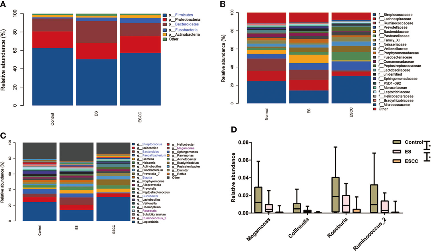
Figure 2 Comparison of relative abundance among each group Barplots of the relative abundance of the main bacterial taxa at (A) phylum, (B) family and (C) genus level for the control, ES and ESCC group. Significant taxa were highlighted in the blue font. (D) Mean relative abundance of continuous changing genera among the groups. Significant taxa were highlighted in the purple font. Data were reported as min to the max with the line at median. *p < 0.05.
The healthy esophageal microbiota was composed mainly of Firmicutes (62.5%), Proteobacteria (18.2%), Bacteroidetes (13.9%), Actinobacteria (2.6%), and Fusobacteria (1.3%), with another ~1.5% of unidentified bacteria. At the genus level, Streptococcus (24.3%) was the main contributor to the microbiota profile, followed by Faecalibacterium and Bacteroides (6.1% and 4.3%, respectively); other subdominant genera were Lactobacillus, Neisseria, Curvibacter, and Blautia, accounting for about 3% each (Figures 2A–C).
The ES group showed a significant decrease of Firmicutes (p = 0.0370) together with a statistically significant robust increase of Fusobacteria (p = 0.0280) and Bacteroidetes (p = 0.0060) with its corresponding genus Bacteroides (p = 0.0240) as compared to the control group (Figures 2A–C).
The ESCC samples also displayed a striking reduction in its microbial composition, such as in Fusobacteria (p = 0.0010) at phylum level and Faecalibacterium (p = 0.0010), Bacteroides (p = 0.0090), Curvibacter (p = 0.0010), and Blautia (p = 0.0040) in comparison with the control group at the genus level. We observed an increasing tendency of Streptococcus in the ESCC group. When compared with the ES group, fewer Bacteroidetes (p = 0.0010), Faecalibacterium (p = 0.0010), Bacteroides (p = 0.0010), and Blautia (p = 0.0040) with more Streptococcus (p = 0.0070) in ESCC tissues were identified (Figures 2A–C). In addition, Megamonas, Collinsella, Roseburia, and Ruminococcus_2 showed a significantly continuous decreasing trend from the control group to the ESCC group at the genus level (Figure 2D).
Characterized Microbial Taxa Associated With Esophageal Squamous Cell Carcinoma Patients
We used multi-level LEfSe analysis to explore potential important microbe biomarkers for the groups in all taxa, and among the three groups, abundance of 138 bacterial species was significantly different. Here, 41, 45, and 52 taxa were abundant in healthy volunteers, ES patients, and ESCC patients, respectively. Given the large number of different bacterial species, we focused on the taxa with LDA scores >4.0. As shown in Figure 3, at the genus level, increased Streptococcus (LDA score = 4.9115, p = 0.0021), Actinobacillus (LDA score = 4.5193, p < 0.0001), Peptostreptococcus (LDA score = 4.3049, p < 0.0001), Fusobacterium (LDA score = 4.2109, p = 0.0004), and Prevotella (LDA score = 4.0768, p = 0.0020) were identified as powerful markers in ESCC patients. Particularly, Streptococcus anginosus at the species level (LDA score = 4.0115, p < 0.0001) showed greater abundance in the ESCC group. Besides, we observed a high level of Roseburia (LDA score = 4.0412, p = 0.0001), Faecalibacterium (LDA score = 4.4607, p < 0.0001), and Curvibacter (LDA score = 4.0812, p < 0.0001) at the genus level and Alphaproteobacteria (LDA score = 4.2618, p = 0.0002) at the class level in the control group. Bacteroides (LDA score = 4.6561, p = 0.0002) and Blautia (LDA score = 4.0883, p < 0.0001) at the genus level were abundant in ES patients (Figure 3).
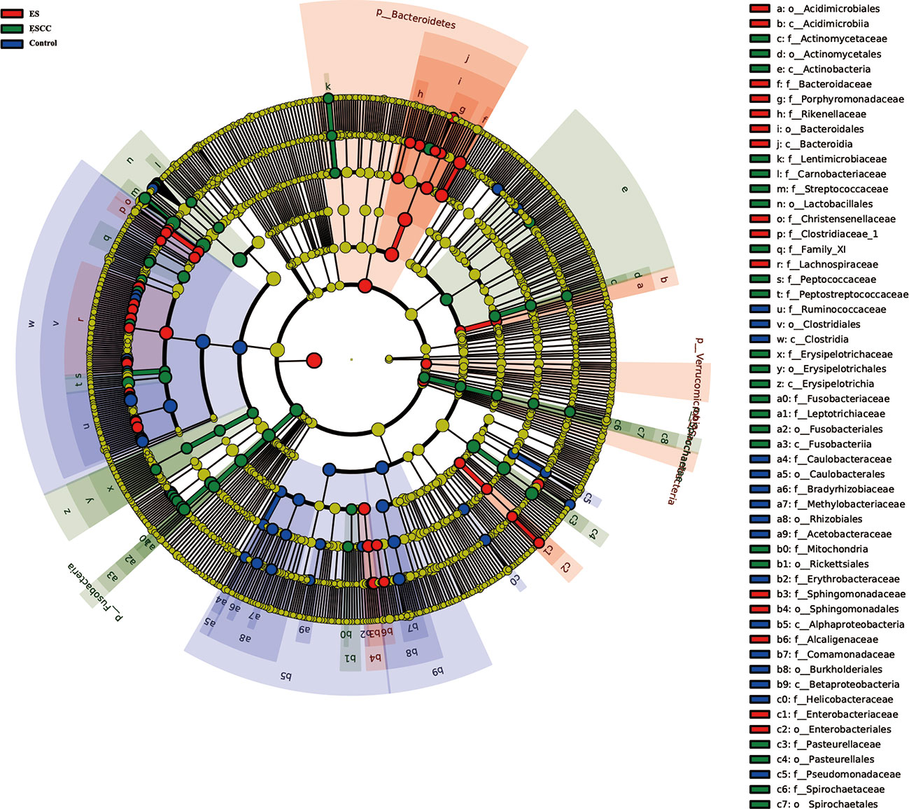
Figure 3 Linear discriminant analysis effect size (LEfSe) analysis showed the most abundant taxa from the phylum to the genus level among the control, esophagitis (ES), and esophageal squamous cell carcinoma (ESCC) groups.
Functional Analysis of Esophageal Microbiota Across The Groups
Finally, PICRUSt was conducted to predict the metagenomes and identify the KEGG pathways involved in each group.
Compared with the control group, patients with ESCC showed a significant upregulation of microbial genes involved in signaling molecules and interaction, excretory system, cellular community, cell growth and death, membrane transport, energy metabolism, metabolism of other amino acids, nucleotide metabolism, folding, sorting and degradation, translation, glycan biosynthesis and metabolism, replication and repair, and metabolism of cofactors and vitamins, while there was a reduction of genes related to lipid metabolism, xenobiotics biodegradation and metabolism, cell motility, amino acid metabolism, carbohydrate metabolism, transcription, and signal transduction (Figure 4A). On the other hand, microbiota of the ES group was characterized by a higher potential for excretory system, digestive system, folding, sorting and degradation, energy metabolism, glycan biosynthesis and metabolism, and metabolism of cofactors and vitamins while showing reduced xenobiotic biodegradation and metabolism, lipid metabolism, cell motility, membrane transport, and signal transduction (Figure 4B).
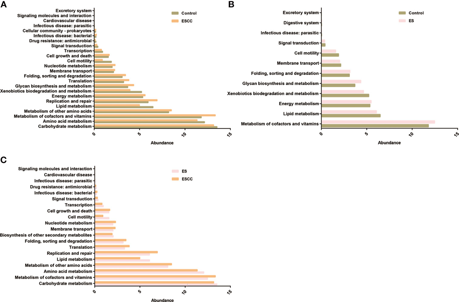
Figure 4 The function prediction of the three groups. Differential Kyoto Encyclopedia of Genes and Genomes (KEGG) pathways were analyzed using PICRUSt for the three groups. Significant differences between the control and esophageal squamous cell carcinoma (ESCC) groups (A), the control and esophagitis (ES) groups (B), and the ES and ESCC groups (C) were presented.
Moreover, when comparing the ES and ESCC tissues, ESCC-associated microbiota showed significantly increased signaling molecules and interaction, infectious disease, cell growth and death, membrane transport, nucleotide metabolism, folding, sorting and degradation, metabolism of other amino acids, translation, metabolism of cofactors and vitamins, and replication and repair. Conversely, it displayed a consistently decreased lipid metabolism, amino acid metabolism, cell motility, carbohydrate metabolism, transcription, biosynthesis of other secondary metabolites, and signal transduction pathways (Figure 4C).
Discussion
Increasing evidence has shown the crucial roles of bacterial imbalance in tumor development, including esophageal cancers (Garrett, 2015). In China, ESCC constitutes more than 90% of all esophageal cancers (Chen et al., 2021). Although imbalanced microbiome has been well elucidated in EAC, microbial alterations of ESCC were still inconclusive. In the present study, we profiled the structure of esophageal microbiota in the ES and ESCC patients and the matched controls through 16S rRNA gene sequencing and predicted the functional changes. We found a lower microbial diversity in the ESCC patients than in the healthy controls, which was supported by previous findings (Li M. et al., 2020), whereas other studies indicated a decreasing tendency or a higher diversity without significant difference (Shao et al., 2019; Li M. et al., 2020; Yang et al., 2021). Although studies conducted by Li D. et al. (2020) and Castaño-Rodríguez et al. (2017) suggested decreased richness of microbiota in the ESCC patients, our study observed a contrary trend. It could be partly explained by factors that could affect microbial structures such as geographic area or the organ studied. In accordance with previous research, the β diversity was statistically different between the ESCC and the control group (Shao et al., 2019; Li D. et al., 2020). The microbial dysbiosis of ESCC tissues was characterized by decreased Faecalibacterium, Bacteroides, Curvibacter, and Blautia and increased Fusobacteria.
Bacteroides is a predominant member of the gut microbiota, with Bacteroides fragilis as the most prevalent form, which was an opportunistic pathogen related to abdominal, soft tissue, and bloodstream infections (Gilbert et al., 2018). Later studies revealed that a subtype of Bacteroides could produce a heat-labile toxin named Bacteroides fragilis toxin. Secreted toxin promoted interleukin (IL)-18 production and cleared E-cadherin that led to profound inflammation and epithelial homeostasis (Valguarnera and Wardenburg, 2020). Indeed, higher levels of toxigenic B. fragilis strains have been reported in secretory diarrhea and various types of cancer including colorectal cancer and prostate cancer (Mármol et al., 2017; Sha et al., 2020). It seems to be inconsistent between our study and previous reports. Yang et al. (2021) have demonstrated significantly enriched Bacteroides in ESCC. This discrepancy might be explained by samples isolated from different sites and diverse diets of the individuals recruited. Meanwhile, recent findings also revealed the protective effect of B. fragilis in the development of colitis-related colorectal cancer. Thus, we proposed that the Bacteroides owned a bidirectional role in the oncogenesis, and a future functional and theoretical investigation is urgent to confirm its role in ESCC.
Specifically, a higher abundance of S. anginosus in the ESCC tissues was identified. S. anginosus, as an oral bacterium, was frequently found in the oral cavity, gastrointestinal tract, and genitourinary tract (Morita et al., 2003). It occupied up to 82% of patient-unique strains collected from hospitalized patients and was involved in purulent infections, including endocarditis (Siegman-Igra et al., 2012; Kawaguchi et al., 2014). In addition, the presence of S. anginosus has been reported in head and neck squamous cell carcinomas, gastric cancer, dysplasia of esophagus, and esophageal cancer tissues, which indicated the involvement of S. anginosus in the carcinogenic process (Sasaki et al., 1998; Tateda et al., 2000). Viable S. anginosus isolated from the esophageal cancer tissues could adhere to cultured epithelial cells and induce the mRNA expression of two CXC-chemokine genes, IL-8 and growth related oncogene (GRO). These results were supported by a higher content of inflammatory cytokines in esophageal cancer tissues (Narikiyo et al., 2004). Streptolysin S encoded by the sag gene cluster was supposed to be responsible for the cytotoxicity of S. anginosus (Asam et al., 2015). The involvement in sulfur metabolism might be the alternative strategy for S. anginosus in carcinogenesis (Coker et al., 2018). However, more clinical strains were urgent to better verify the pathogenic genes and cytotoxicity in future studies. Interestingly, increased abundance of Fusobacterium, another common elongated anaerobic Gram-negative bacterium of the oral cavity, was also observed in the ESCC tissues. Similar results have been confirmed in previous studies (Li D. et al., 2020; Yang et al., 2021). However, Li D. et al. (2020) found no significant difference of Fusobacterium between ESCC and healthy control. Fusobacterium caused periodontal disease and was related to the development of human cancers. Increased Fusobacterium nucleatum in esophageal cancer was a biomarker for predicting a poor clinical outcome (Yamamura et al., 2016). Experimental studies have shown that F. nucleatum could promote carcinogenesis by induction of chemokines and activation of β-catenin signaling pathway (Rubinstein et al., 2013; Yamamura et al., 2016). In our study, we failed to find the different abundance of Porphyromonas gingivalis among the three groups, which contributed to the development of ESCC via enhancing IL-6 secretion and promoting epithelial–mesenchymal transition (Chen et al., 2021). Because esophageal microbiome was partly shaped by the oral microbiome that linked the periodontal disease to ESCC (Norder et al., 2013), it suggested the possibility of protection against periodontal disease to prevent oncogenesis. This hypothesis was partly supported by the findings that numbers of lost teeth and lifestyle factors, including alcohol use and oral hygiene, were related to increased risk of ESCC (Chen et al., 2017).
Apart from compositional changes in bacterial taxa, we also predicted alterations in function across the groups. Metabolic reprogramming is a hallmark of cancer. Dysregulated metabolites including glucose, lipids, and amino acids have been reported in upper gastrointestinal cancers. For example, increased lactic acid, citrate, and glyceraldehyde were related to gastric cancer and esophageal cancer, although opposite changes were documented by other studies. In our study, we observed a reduction of carbohydrate and amino acid metabolism, which suggested a potential underlying mechanism of ESCC.
Nevertheless, two limitations about this study should be addressed. Firstly, the relatively small sample size of each group limited the generalizability of our findings, and larger studies will provide more credible results. Secondly, a cross-sectional study urged a follow-up prospective trial to fully demonstrate the role of microbiota in esophageal diseases.
Conclusion
Taken together, our data investigated the microbiota spectrum of ESCC patients and demonstrated a significant difference in the microbial diversity and richness between the ESCC patients and the healthy subjects. Our results provided a potential association of Streptococcus, Actinobacillus, Peptostreptococcus, Fusobacterium, and Prevotella with ESCC. Further studies are required to confirm our results and elucidate mechanisms of the causal relationship.
Data Availability Statement
The datasets presented in this study can be found in online repositories. The names of the repository/repositories and accession number(s) can be found below: https://www.ncbi.nlm.nih.gov/, PRJNA759579.
Ethics Statement
The studies involving human participants were reviewed and approved by Nanjing first hospital. The patients/participants provided their written informed consent to participate in this study.
Author Contributions
ZJ, ZZ, and SW conceived, organized, and supervised the project and proofread the article. ZJ and ZS collected and analyzed the data and drafted the article. JW supervised statistical analysis. All authors critically revised and approved the final version of the article.
Funding
This study was supported by the Nanjing Health Science and Technology Development Special Fund Project Plan (YKK20108) and the Science and Technology Development Project of Nanjing Medical University (NMUB2019134).
Conflict of Interest
The authors declare that the research was conducted in the absence of any commercial or financial relationships that could be construed as a potential conflict of interest.
Publisher’s Note
All claims expressed in this article are solely those of the authors and do not necessarily represent those of their affiliated organizations, or those of the publisher, the editors and the reviewers. Any product that may be evaluated in this article, or claim that may be made by its manufacturer, is not guaranteed or endorsed by the publisher.
Supplementary Material
The Supplementary Material for this article can be found online at: https://www.frontiersin.org/articles/10.3389/fcimb.2021.774330/full#supplementary-material
Supplementary Figure 1 | Quality Control and Basic Analysis
References
Asam, D., Mauerer, S., Spellerberg, B. (2015). Streptolysin S of Streptococcus Anginosus Exhibits Broad-Range Hemolytic Activity. Med. Microbiol. Immunol. 204, 227–237. doi: 10.1007/s00430-014-0363-0
Baba, Y., Watanabe, M., Yoshida, N., Kawanaka, K., Yamashita, Y., Baba, H. (2014). Radiofrequency Ablation for Pulmonary Metastases From Gastrointestinal Cancers. Ann. Thorac. Cardiovasc. Surg. Off. J. Assoc. Thorac. Cardiovasc. Surgeons Asia 20, 99–105. doi: 10.5761/atcs.ra.13-00343
Blackett, K. L., Siddhi, S. S., Cleary, S., Steed, H., Miller, M. H., Macfarlane, S., et al. (2013). Oesophageal Bacterial Biofilm Changes in Gastro-Oesophageal Reflux Disease, Barrett's and Oesophageal Carcinoma: Association or Causality? Aliment. Pharmacol. Ther. 37, 1084–1092. doi: 10.1111/apt.12317
Castaño-Rodríguez, N., Goh, K. L., Fock, K. M., Mitchell, H. M., Kaakoush, N. O. (2017). Dysbiosis of the Microbiome in Gastric Carcinogenesis. Sci. Rep. 7, 15957. doi: 10.1038/s41598-017-16289-2
Chen, M. F., Lu, M. S., Hsieh, C. C., Chen, W. C. (2021). Porphyromonas Gingivalis Promotes Tumor Progression in Esophageal Squamous Cell Carcinoma. Cell. Oncol. (Dordrecht) 44, 373–384. doi: 10.1007/s13402-020-00573-x
Chen, X., Yuan, Z., Lu, M., Zhang, Y., Jin, L., Ye, W. (2017). Poor Oral Health Is Associated With an Increased Risk of Esophageal Squamous Cell Carcinoma - A Population-Based Case-Control Study in China. Int. J. Cancer 140, 626–635. doi: 10.1002/ijc.30484
Coker, O. O., Dai, Z., Nie, Y., Zhao, G., Cao, L., Nakatsu, G., et al. (2018). Mucosal Microbiome Dysbiosis in Gastric Carcinogenesis. Gut 67, 1024–1032. doi: 10.1136/gutjnl-2017-314281
Elliott, D. R. F., Walker, A. W., O'Donovan, M., Parkhill, J., Fitzgerald, R. C. (2017). A Non-Endoscopic Device to Sample the Oesophageal Microbiota: A Case-Control Study. Lancet Gastroenterol. Hepatol. 2, 32–42. doi: 10.1016/S2468-1253(16)30086-3
Feng, R. M., Zong, Y. N., Cao, S. M., Xu, R. H. (2019). Current Cancer Situation in China: Good or Bad News From the 2018 Global Cancer Statistics? Cancer Commun. (London England) 39, 22. doi: 10.1186/s40880-019-0368-6
Garrett, W. S. (2015). Cancer and the Microbiota. Sci. (New York NY) 348, 80–86. doi: 10.1126/science.aaa4972
Gilbert, J. A., Blaser, M. J., Caporaso, J. G., Jansson, J. K., Lynch, S. V., Knight, R. (2018). Current Understanding of the Human Microbiome. Nat. Med. 24, 392–400. doi: 10.1038/nm.4517
Kavin, H., Yaremko, L., Valaitis, J., Chowdhury, L. (1996). Chronic Esophagitis Evolving to Verrucous Squamous Cell Carcinoma: Possible Role of Exogenous Chemical Carcinogens. Gastroenterology 110, 904–914. doi: 10.1053/gast.1996.v110.pm8608902
Kawaguchi, Y., Tabata, A., Nagamune, H., Ohkura, K. (2014). Molecular Analysis of Streptococcus Anginosus-Derived SagA Peptides. Anticancer Res. 34, 4627–4631. doi: 10.1016/j.urolonc.2014.05.010
Kelly, R. J. (2019). Emerging Multimodality Approaches to Treat Localized Esophageal Cancer. J. Natl. Compr. Cancer Netw. JNCCN 17, 1009–1014. doi: 10.6004/jnccn.2019.7337
Li, D., He, R., Hou, G., Ming, W., Fan, T., Chen, L., et al. (2020). Characterization of the Esophageal Microbiota and Prediction of the Metabolic Pathways Involved in Esophageal Cancer. Front. Cell. Infect. Microbiol. 10, 268. doi: 10.3389/fcimb.2020.00268
Li, M., Shao, D., Zhou, J., Gu, J., Qin, J., Chen, W., et al. (2020). Signatures Within Esophageal Microbiota With Progression of Esophageal Squamous Cell Carcinoma. Chin. J. Cancer Res. = Chung-kuo yen cheng yen chiu 32, 755–767. doi: 10.21147/j.issn.1000-9604.2020.06.09
Lopetuso, L. R., Severgnini, M., Pecere, S., Ponziani, F. R., Boskoski, I., Larghi, A., et al. (2020). Esophageal Microbiome Signature in Patients With Barrett's Esophagus and Esophageal Adenocarcinoma. PloS One 15, e0231789. doi: 10.1371/journal.pone.0231789
Mármol, I., Sánchez-de-Diego, C., Pradilla Dieste, A., Cerrada, E., Rodriguez Yoldi, M. J. (2017). Colorectal Carcinoma: A General Overview and Future Perspectives in Colorectal Cancer. Int. J. Mol. Sci. 18 (1), 197. doi: 10.3390/ijms18010197
Morita, E., Narikiyo, M., Yano, A., Nishimura, E., Igaki, H., Sasaki, H., et al. (2003). Different Frequencies of Streptococcus Anginosus Infection in Oral Cancer and Esophageal Cancer. Cancer Sci. 94, 492–496. doi: 10.1111/j.1349-7006.2003.tb01471.x
Narikiyo, M., Tanabe, C., Yamada, Y., Igaki, H., Tachimori, Y., Kato, H., et al. (2004). Frequent and Preferential Infection of Treponema Denticola, Streptococcus Mitis, and Streptococcus Anginosus in Esophageal Cancers. Cancer Sci. 95, 569–574. doi: 10.1111/j.1349-7006.2004.tb02488.x
Norder, G. E., Dahlén, G., Ruth, M., Ny, L., Quiding-Järbrink, M., Bergquist, H., et al. (2013). Bacterial Flora of the Human Oral Cavity, and the Upper and Lower Esophagus. Dis. Esophagus: Off. J. Int. Soc. Dis. Esophagus 26, 84–90. doi: 10.1111/j.1442-2050.2012.01328.x
Reichenbach, Z. W., Murray, M. G., Saxena, R., Farkas, D., Karassik, E. G., Klochkova, A., et al. (2019). Clinical and Translational Advances in Esophageal Squamous Cell Carcinoma. Adv. Cancer Res. 144, 95–135. doi: 10.1016/bs.acr.2019.05.004
Rubinstein, M. R., Wang, X., Liu, W., Hao, Y., Cai, G., Han, Y. W. (2013). Fusobacterium Nucleatum Promotes Colorectal Carcinogenesis by Modulating E-Cadherin/β-Catenin Signaling via Its FadA Adhesin. Cell Host Microbe 14, 195–206. doi: 10.1016/j.chom.2013.07.012
Sasaki, H., Ishizuka, T., Muto, M., Nezu, M., Nakanishi, Y., Inagaki, Y., et al. (1998). Presence of Streptococcus Anginosus DNA in Esophageal Cancer, Dysplasia of Esophagus, and Gastric Cancer. Cancer Res. 58, 2991–2995.
Sha, S., Ni, L., Stefil, M., Dixon, M., Mouraviev, V. (2020). The Human Gastrointestinal Microbiota and Prostate Cancer Development and Treatment. Invest. Clin. Urol. 61, S43–s50. doi: 10.4111/icu.2020.61.S1.S43
Shao, D., Vogtmann, E., Liu, A., Qin, J., Chen, W., Abnet, C. C., et al. (2019). Microbial Characterization of Esophageal Squamous Cell Carcinoma and Gastric Cardia Adenocarcinoma From a High-Risk Region of China. Cancer 125, 3993–4002. doi: 10.1002/cncr.32403
Siegman-Igra, Y., Azmon, Y., Schwartz, D. (2012). Milleri Group Streptococcus–a Stepchild in the Viridans Family. Eur. J. Clin. Microbiol. Infect. Dis. Off. Publ. Eur. Soc. Clin. Microbiol. 31, 2453–2459. doi: 10.1007/s10096-012-1589-7
Tateda, M., Shiga, K., Saijo, S., Sone, M., Hori, T., Yokoyama, J., et al. (2000). Streptococcus Anginosus in Head and Neck Squamous Cell Carcinoma: Implication in Carcinogenesis. Int. J. Mol. Med. 6, 699–703. doi: 10.3892/ijmm.6.6.699
Torre, L. A., Bray, F., Siegel, R. L., Ferlay, J., Lortet-Tieulent, J., Jemal, A. (2015). Global Cancer Statistic. CA: Cancer J. Clin. 65, 87–108. doi: 10.3322/caac.21262
Valguarnera, E., Wardenburg, J. B. (2020). Good Gone Bad: One Toxin Away From Disease for Bacteroides Fragilis. J. Mol. Biol. 432, 765–785. doi: 10.1016/j.jmb.2019.12.003
Yamamura, K., Baba, Y., Nakagawa, S., Mima, K., Miyake, K., Nakamura, K., et al. (2016). Human Microbiome Fusobacterium Nucleatum in Esophageal Cancer Tissue Is Associated With Prognosis. Clin. Cancer Res. an Off. J. Am. Assoc. Cancer Res. 22, 5574–5581. doi: 10.1158/1078-0432.CCR-16-1786
Keywords: esophageal microbiota, esophagitis patients, esophageal squamous cell carcinoma (ESCC), Streptococcus anginosus, Nanjing area
Citation: Jiang Z, Wang J, Shen Z, Zhang Z and Wang S (2021) Characterization of Esophageal Microbiota in Patients With Esophagitis and Esophageal Squamous Cell Carcinoma. Front. Cell. Infect. Microbiol. 11:774330. doi: 10.3389/fcimb.2021.774330
Received: 11 September 2021; Accepted: 19 October 2021;
Published: 11 November 2021.
Edited by:
Nar Singh Chauhan, Maharshi Dayanand University, IndiaReviewed by:
Valeria D’Argenio, University of Naples Federico II, ItalyGajender Aleti, University of California, San Diego, United States
Copyright © 2021 Jiang, Wang, Shen, Zhang and Wang. This is an open-access article distributed under the terms of the Creative Commons Attribution License (CC BY). The use, distribution or reproduction in other forums is permitted, provided the original author(s) and the copyright owner(s) are credited and that the original publication in this journal is cited, in accordance with accepted academic practice. No use, distribution or reproduction is permitted which does not comply with these terms.
*Correspondence: Zhenyu Zhang, ahzhangzhenyu@sina.com; Shukui Wang, sk_wang@njmu.edu.cn
†These authors have contributed equally to this work