-
- Academic Editor
-
-
-
Objective: The alterations of the functional network (FN) in
anti-N-methyl-Daspartate receptor (NMDAR) encephalitis have been recognized by
functional magnetic resonance imaging studies. However, few studies using the
electroencephalogram (EEG) have been performed to explore the possible FN changes
in anti-NMDAR encephalitis. In this study, the aim was to explore any FN changes
in patients with anti-NMDAR encephalitis. Methods: Twenty-nine
anti-NMDAR encephalitis patients and 29 age- and gender-matched healthy controls
(HC) were assessed using 19-channel EEG examination. For each participant, five
10-second epochs of resting state EEG with eyes closed were extracted. The
cortical source signals of 84 Brodmann areas were calculated using the exact low
resolution brain electromagnetic tomography (eLORETA) inverse solution by
LORETA-KEY. Phase Lag Index (PLI) matrices were then obtained and graph and
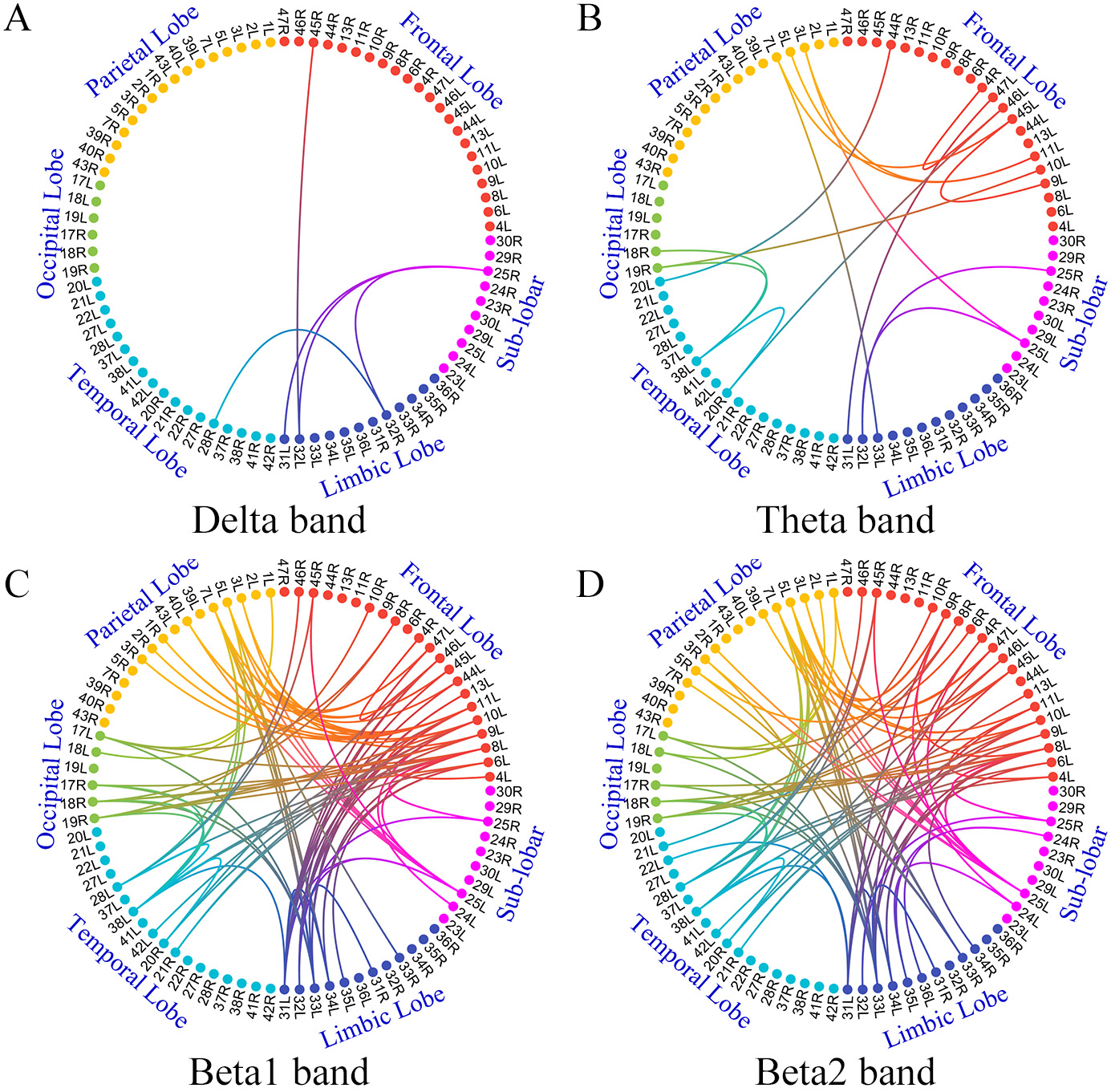
relative band power (RBP) analyses were performed. Results: Compared
with healthy controls, functional connectivity (FC) in the delta, theta, beta 1
and beta 2 bands significantly increased within the 84 cortical source signals of
anti-NMDAR encephalitis patients (p