Article Text

Statistics from Altmetric.com
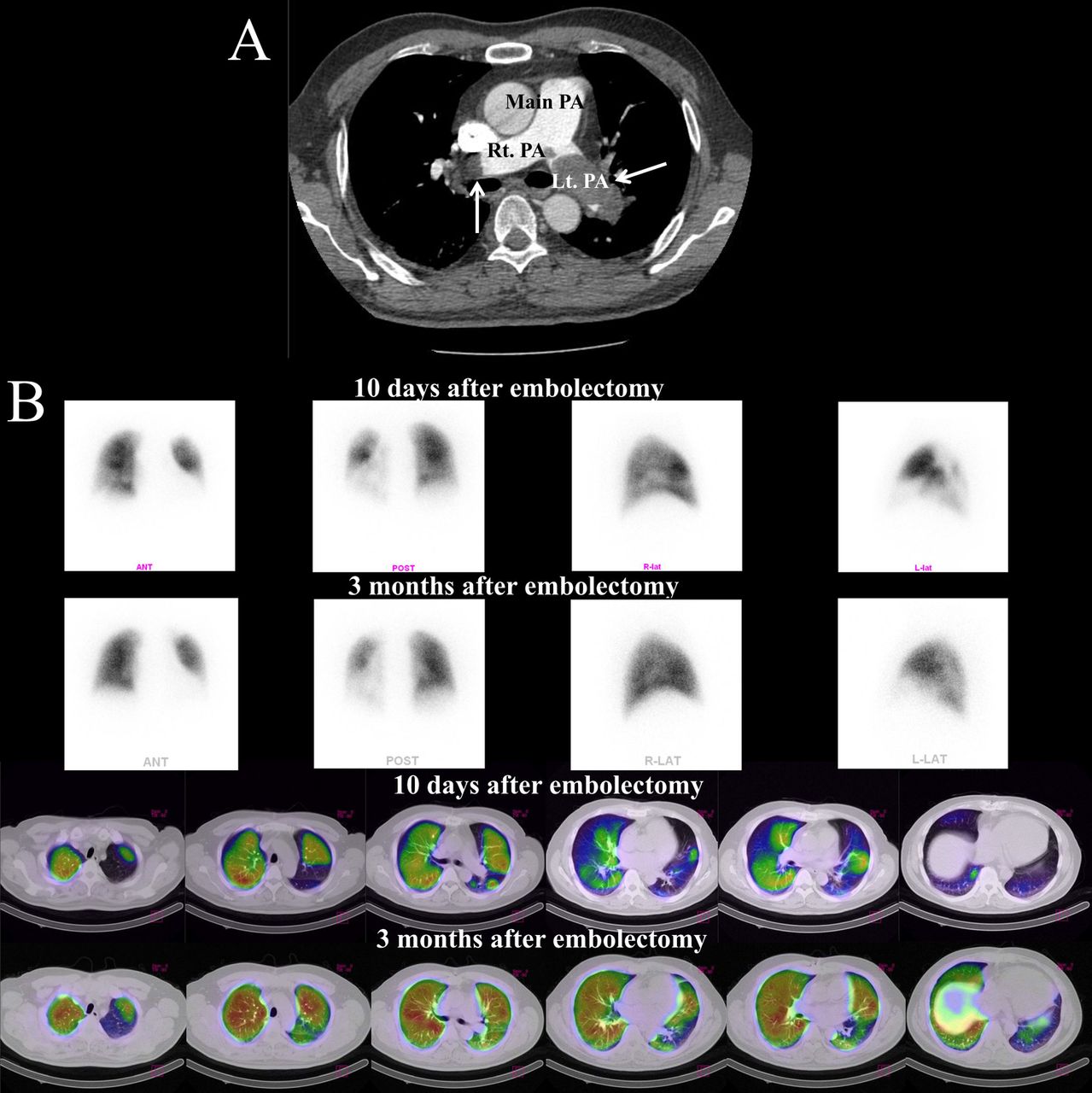
A 46-year-old man was referred to our department with dyspnoea associated with precordial T-wave inversions on a 12-lead ECG and an increased level of d-dimer. Enhanced CT of the chest revealed multiple thrombi in the pulmonary arteries and right side of the heart, with one occluding the left pulmonary artery and another almost occluding the right pulmonary artery (figure 1A, arrows). The patient immediately underwent surgical embolectomy and the thrombi were successfully extracted. Tc-99m macro aggregated albumin pulmonary perfusion scintigraphy 10 days after the embolectomy (acute phase) showed little perfusion to the lower lobe and lingular segment of the left lung or to the medial and lower lobes of the right lung (figure 1B). The patient had an uneventful postoperative course but showed shortness of breath on mild exertion at discharge. Follow-up pulmonary perfusion (3 months after the embolectomy; chronic phase) showed improvement in the perfusion of the entire lung (figure 1B). Single photon emission CT (SPECT)/CT was used to evaluate the changes between the acute and chronic phases. The results of SPECT/CT revealed the improvement more clearly than those of pulmonary perfusion alone (figure 1B).1 In addition, the percentage perfusion defect was determined and was found to improve from 54% to 17%, with associated symptom relief. The SPECT/CT results indicated that the stunned area cannot be evaluated during the acute phase. This case showed that SPECT/CT can contribute to the correct diagnosis of pulmonary thromboembolisms and monitoring of the treatment effect.
(A) Contrast-enhanced CT showing pulmonary emboli (white arrows) in the bilateral pulmonary artery. PA, pulmonary artery; Lt. left; Rt., right. (B) Both Tc-99m macro aggregated albumin pulmonary perfusion scintigraphy and single photon emission CT (SPECT)/CT perfusion show multiple perfusion defects in both lungs at baseline (10 days after the embolectomy) and significant improvement in perfusion at 3 months of follow-up. SPECT/CT reveals the improvement more clearly than perfusion scintigraphy only.
Footnotes
-
Contributors KM conceived and designed the study and approved the final version of the article, SK performed analysis and interpreted the data, and SN drafted the article and revised it for important intellectual content.
-
Competing interests None.
-
Ethics approval Nippon Medical School Ethics Committee.
-
Provenance and peer review Not commissioned; internally peer reviewed.