Article Text

Statistics from Altmetric.com
Description
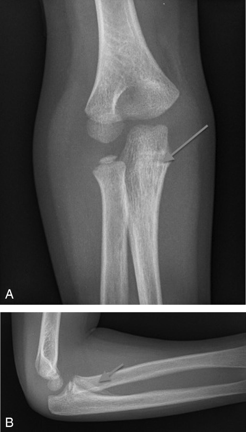
A 6-year-old boy, who fell while playing at school, presented to the fracture clinic with a suspected right distal radius fracture. On examination, the child was tender over the dorsal aspect of the distal radius and also had pain and stiffness around the elbow joint with a lack of the last 25° of elbow flexion. An acute haemarthrosis was also suspected at presentation. Plain anteroposterior (AP) and lateral radiographs of the elbow joint revealed an abnormal calcification superimposed on the anterior aspect of the coronoid process (figure 1A, B), which had not been visualised on plain radiographs in A&E.

(A) Anteroposterior plain radiograph of the right elbow joint showing a possible cortical breech of proximal ulna surface. (B) Lateral plain radiograph of the right elbow joint showing abnormal calcification on anterior aspect of proximal ulna.
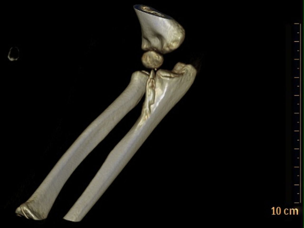
A CT scan, as suggested by the radiologists, revealed a 3 cm avulsion fracture of the anterior surface of the proximal ulna, which corresponded to the calcification seen on lateral plain radiographs (figure 1B) and was thought to be an avulsion fracture of the insertion of brachialis muscle (figure 2). The child was immobilised at the wrist and elbow for 3 weeks with a plaster cast and repeat radiographs were performed to monitor the migration of the avulsion fragment.
CT three-dimensional reconstruction of the right elbow showing a 3 cm avulsion fracture of the anterior surface of the proximal ulna.
The brachialis muscle insertion comprises two distinct tendons originating from the deep and superficial heads of the brachialis muscle inserting into the ulna tuberosity and coronoid process. Studies investigating elbow trauma using ultrasound have shown it to commonly be the deep head that is responsible for avulsion injuries caused by a sudden unexpected movement at the elbow joint.1
There have been cases of coronoid process fractures due to avulsion by the medial collateral ligaments, biceps tendon insertion and avulsion fracture in combination with other injuries, for example, olecranon fractures or elbow dislocations.2 ,3 In this case, a 6-year-old child sustaining a brachialis avulsion fracture of this extent following low-energy trauma has not been reported in the literature. Initial concerns for this child were slow resolution of stiffness and poor functional recovery at the elbow joint as reported in similar patients.2
Despite our concerns at the 3-month follow-up, the child had full range of movement in his right elbow with no tenderness and was undertaking normal activities at school; therefore, conservative management was continued and the child was followed up at six monthly intervals.
Learning points
-
This case emphasises the importance of careful clinical and radiological examination in treating paediatric elbow injuries.
-
Surgery should be avoided in these types of paediatric elbow injuries if they are asymptomatic and have no functional deficit.
-
Regular follow-ups on these types of injuries in children are necessary to monitor the risk of ectopic ossification.
Footnotes
-
Contributors SC wrote the case report, assisted by CAM. AH obtained the patient consent and RT supervised the case report.
-
Competing interests None.
-
Patient consent Obtained.
-
Provenance and peer review Not commissioned; externally peer reviewed.