
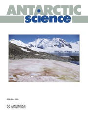


Introduction
The exposure of new islands in polar regions is a visually powerful symptom of a warming climate. These islands are appearing due to the retreat of ice caps and ice sheets, compounded by local sea-level fall caused by isostatic uplift of surrounding coastal areas and/or reduction in gravitational ‘pull’ on the ocean by the waning ice sheet (e.g. Ziaja & Ostafi Reference Ziaja and Ostafin2019, Hansen et al. Reference Hansen, Stevens and Shuman2022). In 2020, a hitherto uncharted small island, informally named Sif Island, was discovered during expedition NBP20-02 of RV/IB Nathaniel B. Palmer, part of the Thwaites Offshore Research (THOR) project (see https://thwaitesglacieroffshoreresearch.org/). Sif Island is located in Pine Island Bay of the eastern Amundsen Sea Embayment (latitude: -75°05'42'', longitude: -102°49'30''; Fig. 1). Subsequent analysis of satellite imagery revealed that Sif Island became isolated from the main West Antarctic Ice Sheet (WAIS) by a decade of retreat of slow-flowing ice between Thwaites Glacier and Pine Island Glacier (Fig. 2).

Figure 1. Location of Sif Island in the eastern Amundsen Sea Embayment relative to other rock exposures, which are mapped based on Cox et al. (Reference Cox and Smith Lyttle2019) and classified based on Simões Pereira et al. (Reference Simões Pereira, van de Flierdt, Hemming, Frederichs, Hammond and Brachfeld2020). The location of the main map within Antarctica is shown in the top-left insert (red box), and the position of Sif Island (corresponding to Fig. 2) is indicated in the main map (red box). The approximate position of Pine Island Rift is from Jordan et al. (Reference Jordan, Ferraccioli, Vaughan, Holt, Corr, Blankenship and Diehl2010) and the position of the strike-slip movement zone near Mount Murphy is from Spiegel et al. (Reference Spiegel, Lindow, Kamp, Meisel, Mukasa and Lisker2016), although the sense of the fault motion is indicated as uncertain due to disagreement with Müller et al. (Reference Müller, Gohl, Cande, Goncharov and Golynsky2007). The uncertain Thurston Island (TI) and Marie Byrd Land (MBL) crustal block boundaries are displayed following Zundel et al. (Reference Zundel, Spiegel, Mehling, Lisker, Hillenbrand, Monien and Klügel2019b). Hornblende and biotite 40Ar/39Ar age populations, ɛNd values and 87Sr/86Sr data for detritus shed from Pine Island and Thwaites glaciers are from Simões Pereira et al. (Reference Simões Pereira, van de Flierdt, Hemming, Frederichs, Hammond and Brachfeld2020). Offshore bathymetry is from Hogan et al. (Reference Hogan, Larter, Graham, Arthern, Kirkham and Totten Minzoni2020), with BedMachine Antarctica v.1 (Morlighem et al. Reference Morlighem, Rignot, Binder, Blankenship, Drews and Eagles2020) used where these data were absent. Ice-sheet extent data are from the Scientific Committee on Antarctic Research (SCAR) Antarctic Digital Database, accessed 2022 (Gerrish et al. Reference Gerrish, Fretwell and Cooper2022). On the ice, MODIS ice imagery is displayed (Haran et al. Reference Haran, Bohlander, Scambos, Painter and Fahnestock2014), and red shading indicates faster ice-flow velocities (Mouginot et al. Reference Mouginot, Rignot and Scheuchl2019), where Thwaites and Pine Island glaciers are located.

Figure 2. The exposure of Sif Island caused by a retreating ice front captured in LANDSAT imagery from 23 November 2008 (a. Landsat 7), 18 November 2012 (b. Landsat 7), 11 January 2016 (c. Landsat 8) and 26 January 2020 (d. Landsat 8). Landsat Operational Land Imager/Thermal Infrared Sensor (OLI/TIRS) imagery courtesy of the United States Geological Survey. The ice margin retreated by ~500 m in the 12 year period. Panel d. shows previous ice extents in a.–c. as magenta, yellow and cyan lines, respectively. The position of Sif Island is circled in green, with an example iceberg circled in teal in panel b. for comparison. The location of the map within Pine Island Bay is shown in Fig. 1.
The newly exposed granitic rock at Sif Island is uniquely situated to address unresolved tectonic and glaciological hypotheses that hinder progress towards a better understanding of the ice-bed interaction, and thus flow dynamics, of Thwaites and Pine Island glaciers. Together, these two glaciers drain approximately a third of the largely marine-based and inherently unstable WAIS into the Amundsen Sea Embayment (Rignot et al. Reference Rignot, Mouginot, Scheuchl, Van Den Broeke, Van Wessem and Morlighem2019), with an ice volume sufficient to raise sea level by ~1.2 m (Morlighem et al. Reference Morlighem, Rignot, Binder, Blankenship, Drews and Eagles2020). As these glaciers comprise one of the most sensitive parts of the Antarctic cryosphere, understanding them is critical for predicting the magnitude and rate of future global sea-level rise from WAIS loss (Scambos et al. Reference Scambos, Bell, Alley, Anandakrishnan, Bromwich and Brunt2017, Intergovernmental Panel on Climate Change Reference Pörtner, Roberts, Masson-Delmotte, Zhai, Tignor and Poloczanska2019).
Underlying geological features and boundaries influence the flow of overlying ice and fundamentally shape ice drainage patterns in the Amundsen Sea sector of the WAIS. Sif Island is the only rock exposure between Pine Island and Thwaites glaciers. Characterizing the petrology and geochemistry of Sif Islands granitic bedrock is therefore essential to refine the geological and tectonic framework of the Amundsen Sea Embayment and affirm the origins of large-scale features of the modern seabed extending beneath the ice. Additionally, measurements of cosmogenic nuclides can help determine the exposure history of the island and place the observed recent ice-sheet margin retreat in a long-term context.
Geological setting
The Amundsen Sea Embayment coincides with a lithospheric boundary between the crustal blocks of Thurston Island and Marie Byrd Land in West Antarctica (Fig. 1; e.g. Jordan et al. Reference Jordan, Riley and Siddoway2020). Deep bed topography and bedrock lineaments imply the localization of the WAIS outlet glaciers/ice streams along fault zones. For example, Pine Island Rift lies beneath Pine Island Glacier (Jordan et al. Reference Jordan, Ferraccioli, Vaughan, Holt, Corr, Blankenship and Diehl2010, Gohl Reference Gohl2012), and a wrench system bordering Mount Murphy lies on the western side of Thwaites Glacier (Spiegel et al. Reference Spiegel, Lindow, Kamp, Meisel, Mukasa and Lisker2016). Sif Island is located on the southern margin of Pine Island Bay (Fig. 1). Despite diverse geophysical data obtained from gravimetric, magnetic, bathymetric and seismic surveys of the region (e.g. Diehl et al. Reference Diehl, Holt, Blankenship, Young, Jordan and Ferraccioli2008, Jordan et al. Reference Jordan, Ferraccioli, Vaughan, Holt, Corr, Blankenship and Diehl2010, Kalberg et al. Reference Kalberg, Gohl, Eagles and Spiegel2015), consensus has not been reached regarding the specific position of the tectonic boundary between the Marie Byrd Land and Thurston Island crustal blocks (Fig. 1). Some workers portray the Thurston Island crustal block boundary along Pine Island Rift (e.g. Gohl et al. Reference Gohl, Teterin, Eagles, Netzeband, Grobys and Parsiegla2007, Dunham et al. Reference Dunham, O'Donnell, Stuart, Brisbourne, Rost and Jordan2020, Jordan et al. Reference Jordan, Riley and Siddoway2020), whereas others project it beneath Thwaites Glacier (e.g. Kalberg et al. Reference Kalberg, Gohl, Eagles and Spiegel2015, Spiegel et al. Reference Spiegel, Lindow, Kamp, Meisel, Mukasa and Lisker2016, Zundel et al. Reference Zundel, Spiegel, Mehling, Lisker, Hillenbrand, Monien and Klügel2019b). The kinematics of parallel structures within the boundary zone have been determined using diverse criteria and are also uncertain. Spiegel et al. (Reference Spiegel, Lindow, Kamp, Meisel, Mukasa and Lisker2016) interpreted sinistral transtension along the Mount Murphy wrench system based on bedrock thermochronology and structural geometries. In contrast, Müller et al. (Reference Müller, Gohl, Cande, Goncharov and Golynsky2007) determined a dextral sense for the Ferrigno Rift, on the eastern margin of the Thurston Island block, using plate motions during the Cenozoic opening of the West Antarctic Rift System.
A further debate resulting from a dearth of bedrock exposure is whether Cenozoic structures reactivate faults that formed during Mesozoic tectonism across West Antarctica (e.g. Kalberg et al. Reference Kalberg, Gohl, Eagles and Spiegel2015, Dziadek et al. Reference Dziadek, Ferraccioli and Gohl2021). In the Amundsen Sea Embayment and its WAIS drainage sector, the prominent crustal structures controlling the bathymetry/bed topography and lithospheric boundaries formed during the Cenozoic phase of deformation in this part of the West Antarctic Rift System (Müller et al. Reference Müller, Gohl, Cande, Goncharov and Golynsky2007, Dziadek et al. Reference Dziadek, Ferraccioli and Gohl2021), with Pine Island Rift having been active during the early Cenozoic (Jordan et al. Reference Jordan, Ferraccioli, Vaughan, Holt, Corr, Blankenship and Diehl2010, Gohl et al. Reference Gohl, Denk, Eagles and Wobbe2013, Spiegel et al. Reference Spiegel, Lindow, Kamp, Meisel, Mukasa and Lisker2016). However, it is unclear whether Pine Island Rift exploited a pre-existing structure or whether it formed across the crust of the Thurston Island block. Characterizing the age, origin and thermal history of the Sif Island granite will inform this debate, as well as helping constrain the location of the Marie Byrd Land-Thurston Island block boundaries.
Exposed rocks elsewhere around the Amundsen Sea Embayment show that both Marie Byrd Land and Thurston Island contain plutonic rocks formed in a convergent plate boundary setting since the mid-Palaeozoic (Pankhurst et al. Reference Pankhurst, Weaver, Bradshaw, Storey and Ireland1998, Riley et al. Reference Riley, Flowerdew, Pankhurst, Leat, Millar, Fanning and Whitehouse2017). The two tectonic provinces differ as Marie Byrd Land is underlain by Proterozoic lithosphere and contains exposures of Cretaceous migmatite, whereas Thurston Island contains Jurassic evolved granites and volcanics (e.g. Jordan et al. Reference Jordan, Riley and Siddoway2020). Neogene volcanic rocks are present in the Hudson Mountains, north of the Pine Island Glacier terminus, and as isolated volcanoes, such as Mount Murphy, to the west of Thwaites Glacier (Fig. 1). Distributed volcanic centres are larger and more numerous across the Marie Byrd Land province (Wilch et al. Reference Wilch, McIntosh, Panter, Smellie, Panter and Geyer2021), and Neogene volcanic rocks probably continue beneath the WAIS, inferred from magnetic anomalies, radar data and geochemical data (e.g. Behrendt Reference Behrendt2013, van Wyk de Vries et al. Reference van Wyk de Vries, Bingham and Hein2017, Simões Pereira et al. Reference Simões Pereira, van de Flierdt, Hemming, Frederichs, Hammond and Brachfeld2020). Cretaceous to Cenozoic sedimentary strata underlie parts of the drainage basins of both glaciers as well as the continental shelf (Smith et al. Reference Smith, Jordan, Ferraccioli and Bingham2013, Schroeder et al. Reference Schroeder, Blankenship, Young, Witus and Anderson2014, Muto et al. Reference Muto, Anandakrishnan, Alley, Horgan, Parizek and Koellner2019, Klages et al. Reference Klages, Salzmann, Bickert, Hillenbrand, Gohl and Kuhn2020, Simões Pereira et al. Reference Simões Pereira, van de Flierdt, Hemming, Frederichs, Hammond and Brachfeld2020).
Field observations
Sampling of Sif Island was conducted opportunistically on behalf of the THOR project during expedition NBP20-02 with RV/IB Nathaniel B. Palmer in early 2020. The island was named after the Norse goddess Sif, the wife of Thor, in association with the THOR project. THOR is part of the wider International Thwaites Glacier Collaboration (ITGC) programme, a joint US (National Science Foundation) and UK (Natural Environment Research Council) initiative to substantially improve decadal and longer-term projections of ice loss and sea-level rise originating from Thwaites Glacier (https://thwaitesglacier.org/).
Sif Island was visited and circumnavigated by small inflatable boat (zodiac) shortly after its discovery. The main island is ~350 m long and ~120 m wide (Fig. 2). At the time of the boat landing in February 2020, ~1–2 m of bare bedrock was exposed above the waterline around the island perimeter (Fig. 3), although the tidal state was unknown. Above this bedrock, ~40–50 m-thick snow and ice cover the entire island. The substantial thickness of the snow and ice cover upon the island and the history of ice-margin retreat documented in satellite imagery (Fig. 2) suggest that this cover is an ice-sheet remnant (i.e. dead ice) rather than the result of local snow accumulation alone.

Figure 3. Field photographs of Sif Island. a. The ~40–50 m-thick ice cover of the island (right), remaining after retreat of the West Antarctic Ice Sheet margin, which is visible in the background. Grounded icebergs are present between the modern ice-sheet margin and the island (left). b. Exposed rock on the promontory (i.e. sampling location), with the main island visible in the top-left of the image. Exposed rock is ~5 m × ~20 m. c. A typical section of the basal ice and rock exposure. Melt layers are visible in the ice, and the height of the exposed rock between the water line and ice cover is ~1–2 m. d. Promontory rising ~1 m above sea level, with the main island in the background.
Safe sampling was possible thanks to a small ~100 m2, ice-free rocky promontory that extends out ~50–60 m away from the main island (Fig. 3b,d). This rose to ~1 m above sea level at the time of landing. The blocky, jointed pink granite cropping out on the promontory is identical in appearance to the rock examined during circumnavigation by zodiac around the perimeter of the main island (Fig. 3). The granite is holocrystalline and has a phaneritic texture. In hand specimen, most visible mineral grains are K-feldpsar, plagioclase, quartz and biotite, typically up to a few millimetres in diameter and approximately equigranular (Fig. 4). Finer-grained regions with a similar mineralogy are also present (Fig. 4b). The bedrock composition and its surface are consistent with geophysical interpretations that hard, crystalline bedrock forms part of the subglacial substrate in this region (Schroeder et al. Reference Schroeder, Blankenship, Young, Witus and Anderson2014, Muto et al. Reference Muto, Anandakrishnan, Alley, Horgan, Parizek and Koellner2019).

Figure 4. Photographs of the Sif Island rock exposure. a. Typical exposed bedrock surface with GPS handset for scale. b. Contact between a coarse-grained granite forming the bulk of the outcrop and a finer-grained enclave. c. Hand specimen of a ‘pinker’ sample (e.g. SIA and SIC; SI = Sif Island). d. Hand specimen of sample SIB, collected just below the exposure shown in panel a. Each yellow and white section on the folding rule is 10 cm.
Exposed rock surfaces are smooth and lack glacial surface features such as striations (Fig. 4), consistent with low surface ice-flow velocities in the order of just 10 m year-1 along the nearby coast (Rignot et al. Reference Rignot, Mouginot, Morlighem, Seroussi and Scheuchl2014), implying little basal slip. Alternatively, if the promontory only recently emerged above sea level due to isostatic uplift in response to mass loss from the Pine Island Glacier and Thwaites Glacier catchments, the rock surface exposed at present may have previously lain just below the seawater surface, making it vulnerable to wave erosion. This could have removed any glacial surface features and erratics over centuries or millennia prior to uplift. The rock exposure remains low-lying, so it may still be subject to wave erosion at the present day.
Current bed topographies from inversion of ice flow velocities (Morlighem et al. Reference Morlighem, Rignot, Binder, Blankenship, Drews and Eagles2020) and gravity-derived bathymetric data (Millan et al. Reference Millan, Rignot, Bernier, Morlighem and Dutrieux2017) show the bed to be below sea level at the location of Sif Island. This highlights that these methods underestimate high-resolution topography, consistent with observed smoothing of data when comparing these datasets to higher-resolution multibeam swath bathymetry data from deglaciated former beds (Hogan et al. Reference Hogan, Larter, Graham, Arthern, Kirkham and Totten Minzoni2020, Graham et al. Reference Graham, Wåhlin, Hogan, Nitsche, Heywood and Totten2022). These smaller-scale topographic and bathymetric features are important as they can impact basal sliding beneath grounded ice, provide pinning points for ice shelves and also prevent/restrict delivery of warm deep water to the ice-sheet grounding zone and undersides of ice shelves (e.g. Hogan et al. Reference Hogan, Larter, Graham, Arthern, Kirkham and Totten Minzoni2020).
Analytical methods
Sampling
Samples from Sif Island were obtained from different parts of the accessible exposures, and three samples with designated identifiers SIA, SIB and SIC (SI = Sif Island) were analysed. Sample SIB is shown in Fig. 4d and samples SIA and SIC closely resembled the sample in Fig. 4c.
To determine the time of granite formation, all three rock samples were zircon U-Pb dated using laser ablation inductively coupled plasma mass spectrometry (LA-ICP-MS). Zircon fission track (ZFT) and apatite fission track (AFT) and zircon and apatite (U-Th)/He dating was performed on sample SIA to determine the cooling history of the granite and to gain indirect evidence of the timing of fault activity in the vicinity of Sif Island. Zircon Hf isotope analyses were also conducted. For bulk rock geochemical and radiogenic Nd and Sr isotope analyses, ~230 g of sample SIB was powdered, ensuring a representative bulk sample.
Neodymium and strontium isotopes on bulk rock samples
Analyses were performed on 50 mg of the sample SIB rock powder digested on a hotplate in concentrated HF, HClO4 and HNO3 for 3–5 days. The samples were processed alongside ~75 mg of United States Geological Survey (USGS) BCR-2 rock standard. The Nd was isolated from the sample matrix using a cation-exchange resin (AG50W-X8, 200–400 μm mesh) and HCl in increasing molarity, followed by a low-molarity HCl Ln-Spec resin procedure (50–100 μm mesh). The sample matrix from the cation-exchange step was dried, taken up in HNO3 and loaded onto Eichrom Sr Spec resin to wash down the matrix and elute the Sr (Simões Pereira et al. Reference Simões Pereira, van de Flierdt, Hemming, Hammond, Kuhn and Brachfeld2018).
Neodymium isotopes were measured in the MAGIC laboratories at Imperial College London on a Nu Instruments high-resolution multi-collector inductively coupled plasma mass spectrometer (HR MC-ICP-MS). To account for instrumental mass bias, isotope ratios were corrected using an exponential law and a 146Nd/144Nd ratio of 0.7219. Although negligible, interference of 144Sm on 144Nd was corrected for. Bracketing standards were used to correct measured 143Nd/144Nd ratios to the commonly used JNdi-1 ratio of 0.512115 (Tanaka et al. Reference Tanaka, Togashi, Kamioka, Amakawa, Kagami and Hamamoto2000). USGS BCR-2 rock standard processed alongside the samples yielded measured 143Nd/144Nd ratios with a mean of 0.512636 ± 0.000012 (2 SD, n = 4), which is within error of the published ratio of 0.512638 ± 0.000015 (Weis et al. Reference Weis, Kieffer, Maerschalk, Barling, De Jong and Williams2006). A full procedural blank for Nd was 41 pg. 143Nd/144Nd ratios are expressed using epsilon notation (ɛNd), which denotes the deviation of a measured ratio from the modern Chondritic Uniform Reservoir (0.512638) in parts per 10 000 (Jacobsen & Wasserburg Reference Jacobsen and Wasserburg1980).
Strontium isotopes were measured in the MAGIC laboratories at Imperial College London on a thermal ionization mass spectrometer (TIMS). Samples were loaded in 1 μl of 6 M HCl onto degassed tungsten filaments with 1 μl of TaCl5 activator. The measured 87Sr/86Sr ratios were corrected for instrumental mass bias using an exponential law and an 88Sr/86Sr ratio of 8.375. Interference of 87Rb was corrected for using an 87Rb/85Rb ratio of 0.386. Analyses of the NIST 987 standard reference material yielded a mean of 0.710256 ± 0.000009 (2 SD, n = 4), and samples were corrected to the published value of 0.710252 ± 0.000013 (Weis et al. Reference Weis, Kieffer, Maerschalk, Barling, De Jong and Williams2006). The accuracy of the results was confirmed using rock standard USGS BCR-2 processed alongside the sample, which yielded an 87Sr/86Sr ratio of 0.705000 ± 0.00008 (2 SE, n = 2), which is within error of the published ratio of 0.705013 ± 0.00010 (Weis et al. Reference Weis, Kieffer, Maerschalk, Barling, De Jong and Williams2006).
Zircon U-Pb dating
Apatite and zircon grains for all of the geochronological methods described below were concentrated using standard crushing, density separation and magnetic separation techniques at the London Geochronology Centre, University College London, UK, and by ZirChron LLC, Tucson, AZ, USA.
At the London Geochronology Centre, the entire separate was mounted in epoxy resin and 20 zircon grains were analysed from two different rock samples (SIB and SIC). A New Wave Research (NWR) 193 nm laser ablation system coupled to an Agilent 7900 ICP-MS was used. Plešovice zircon was used as a primary standard to correct for instrumental mass bias and depth-dependent inter-element fractionation (Sláma et al. Reference Sláma, Košler, Condon, Crowley, Gerdes and Hanchar2008), whereas GJ1 zircon was used as a secondary standard to verify the accuracy of the data (Jackson et al. Reference Jackson, Pearson, Griffin and Belousova2004). Uranium and thorium concentrations were estimated by comparison with NIST 612 glass (Pearce et al. Reference Pearce, Perkins, Westgate, Gorton, Jackson, Neal and Chenery1997). Data reduction of the time-resolved mass spectrometer data was performed using GLITTER 4.5 (Griffin Reference Griffin and Sylvestor2008). Although the number of discordant ages was high for sample SIB, the GJ1 ages measured alongside the samples, with a mean of 612 ± 8 Ma and a range from 604 to 619 Ma, indicate that the data are accurate (Table S1).
Zircon U-Pb dating at the Arizona LaserChron Center used the methods of Gehrels et al. (Reference Gehrels, Valencia and Ruiz2008) applied to 50 zircon grains selected from sample SIA. Instrumentation comprised a Photon Machines Analyte G2 excimer laser equipped with a HelEx ablation cell using a spot diameter of 20 μm, coupled to an Element2 HR-ICP-MS. Sri Lanka and FC-1 zircon crystals were used as the primary zircon standard reference materials, and R33 zircon crystals were used as a secondary standard. Data reduction was performed in MATLAB® using the Arizona LaserChron Center AgeCalcML software. Detailed methods are available at www.laserchron.org. Laser spot selection was conducted using colour cathodoluminescence (CL) images generated with a Hitachi 3400N SEM and a Gatan CL2 detector system (www.geoarizonasem.org). These CL images show typical euhedral igneous grains with internal growth zoning.
All final data analyses and visualizations were performed using IsoplotR (Vermeesch Reference Vermeesch2018). Discordant grains were filtered using the log ratio distance to the concordia composition (Vermeesch Reference Vermeesch2021). Instead of applying a fixed threshold to select between the use of the 206Pb/238U ratio or 207Pb/206Pb ratio to calculate the age, a single-grain concordia age was calculated. Final ages were calculated as concordia ages.
Zircon Hf isotopes
Methods used for Hf isotope analyses are based on those in Cecil et al. (Reference Cecil, Gehrels, Ducea and Patchett2011) and were conducted at the Arizona LaserChron Centre. Measurements were conducted using a Nu Instruments HR-ICP-MS connected to a Photon Machines Analyte G2 excimer laser. Solutions of 10 ppb of JMC475 and a Spex Hf solution, then 10 ppb solutions containing Spex Hf, Yb and Lu, were used to determine instrument settings. When all solutions yielded 176Hf/177Hf ratios of ~0.28216, instrument settings were optimized for laser ablation analyses, and seven different standard zircons (Mud Tank, 91500, Temora, R33, FC52, Plešovice and Sri Lanka) were analysed mounted in the same epoxy pucks. When precision and accuracy were ≤1 unit of ɛHf at the 2σ level, unknowns were analysed using identical acquisition parameters.
Laser ablation pits with a diameter of 40 μm were placed on top of the U-Pb analysis pits, with CL images used to ensure that the ablation pits did not overlap multiple age domains or inclusions (Fig. 5). Each acquisition consisted of one 40 s integration on backgrounds (on peaks with no laser firing) followed by 60 1 s integrations with the laser firing. Using a typical laser fluence of ~5 J/cm2 and a pulse rate of 7 Hz, the ablation rate is ~0.8 μm/s. Each standard was analysed at the beginning and end of the sample run.

Figure 5. Cathodoluminescence images and U-Pb dates of the 50 zircon grains analysed from sample SIA (SI = Sif Island). Red text indicates discordant ages, and the two Palaeozoic inherited cores are highlighted using green text. Also shown are the ɛHf values (blue text).
Isotope fractionation was accounted for using the method of Woodhead et al. (Reference Woodhead, Hergt, Shelley, Eggins and Kemp2004): βHf (mass fractionation factor in the exponential law) was determined from the measured 179Hf/177Hf; βYb was determined from the measured 173Yb/171Yb (except for very low Yb signals). βLu was assumed to be the same as βYb. Ytterbium and Lu interferences were corrected by measurement of 176Yb/171Yb and 176Lu/175Lu (respectively), as advocated by Woodhead et al. (Reference Woodhead, Hergt, Shelley, Eggins and Kemp2004). Stable isotope ratios used as reference were 179Hf/177Hf = 0.73250 (Patchett & Tatsumoto Reference Patchett and Tatsumoto1980), 173Yb/171Yb = 1.132338 (Vervoort et al. Reference Vervoort, Patchett, Söderlund and Baker2004), 176Yb/171Yb = 0.901691 (Vervoort et al. Reference Vervoort, Patchett, Söderlund and Baker2004, Amelin & Davis Reference Amelin and Davis2005) and 176Lu/175Lu = 0.02653 (Patchett Reference Patchett1983). All corrections were done line by line. For very low Yb signals, βHf was used for fractionation correction of Yb isotope ratios. The corrected 176Hf/177Hf values were filtered for outliers (2σ filter), and the average and standard error were calculated from the resulting ~58 integrations.
The cut-off for using βHf vs βYb was determined by monitoring the average offset of the standards from their known values, and the cut-off was set at the minimum offset. This was achieved at ~6 mV of 171Yb. The 176Hf/177Hf at the time of crystallization was calculated from measurement of present-day 176Hf/177Hf and 176Lu/177Hf, using the decay constant of 176Lu (λ = 1.867e-11) from Scherer et al. (Reference Scherer, Münker and Mezger2001) and Söderlund et al. (Reference Söderlund, Patchett, Vervoort and Isachsen2004). No capability is provided for calculating Hf-depleted mantle model ages because the 176Hf/177Hf and 176Lu/177Hf of the source material(s) from which the zircon crystallized are not known. However, a graphical estimate of the model age is made (Fig. S2).
(U-Th)/He dating
(U-Th)/He dating was conducted at the Arizona Radiogenic Helium Dating Laboratory (ARDHL), University of Arizona. This was performed for zircon and apatite grains; the former have a closure temperature range of ~150–200°C (Guenthner et al. Reference Guenthner, Reiners, Ketcham, Nasdala and Giester2013). Zircon and apatite grains were examined under a polarizing stereo-microscope and selected for (U-Th)/He on the basis of grain size (>60 μm diameter), morphology, clarity and lack of inclusions (Fig. S3). In apatite grains, zircon inclusions containing relatively high concentrations of uranium were common; care was taken to avoid these when picking grains. Final grains were imaged and their dimensions measured, and then they were loaded into Nb packets. To measure He, aliquots were heated with a diode laser to ~1300°C for 18–20 min for zircon and to ~900°C for 4 min for apatite. One or more gas re-extractions (lasing) for 20–21 min at higher temperatures were performed for zircon grains, and no gas re-extracts were done for apatite grains. Extracted He was spiked with 3He, purified using cryogenic and gettering methods and measured with a quadrupole mass spectrometer. A known amount of 4He was measured at every eighth analysis to monitor instrument drift.
Degassed apatite grains were retrieved, spiked with a 233U-229Th-147Sm-42Ca tracer, and dissolved in HNO3, and U, Th, Sm and Ca isotopes were analysed on an Element2 HR-ICP-MS. Degassed zircon grains were spiked with a 233U-229Th tracer, equilibrated and dissolved in HF in a Parr bomb. The U and Th isotopes of the zircon aliquots were measured on an Element2 HR-ICP-MS. Grain masses were used to calculate U, Th, Sm and He concentrations. For apatite grains, the mass was calculated from Ca measurements and stoichiometry following the protocols of Guenthner et al. (Reference Guenthner, Reiners and Chowdhury2016). For zircon grains, we report the dimensional mass calculated from grain length measurements and assumptions about morphology following the protocols of Hourigan et al. (Reference Hourigan, Reiners and Brandon2005). Durango apatite and Fish Canyon tuff zircon were used as standards to assess dissolution protocols and HR-ICP-MS analyses. Blank-corrected (U-Th-Sm)/He and (U-Th)/He ages were calculated with propagated analytical uncertainties from U, Th, Sm and He measurements. Alpha-ejection corrections were applied using grain measurements and assuming apatite and zircon are unzoned with respect to U, Th and Sm (Hourigan et al. Reference Hourigan, Reiners and Brandon2005, Ketcham et al. Reference Ketcham, Gautheron and Tassan-Got2011).
The zircon and apatite grains analysed here may have been subject to parentless He contamination from neighbouring U-rich grains or coatings (Murray et al. Reference Murray, Orme and Reiners2014). This probably biased ages to older than the true cooling age, as typically only extreme enrichment of U and Th in zircon rims can lead to ages being biased younger (Hourigan et al. Reference Hourigan, Reiners and Brandon2005), and we observed only minor U zoning when counting fission track densities during ZFT analysis.
Fission track dating
Fission track dating was performed at the Arizona Fission Track Laboratory, University of Arizona. Again, this method was applied to both zircon and apatite grains, with respective closure temperature ranges of ~205 ± 18°C (Bernet Reference Bernet2009) and ~100–120°C (Reiners & Brandon Reference Reiners and Brandon2006).
For AFT dating, 100 apatite grains were mounted in epoxy resin and polished with alumina powder, and spontaneous fission tracks were revealed by etching with 5.5 M HNO3 at 21°C (±1°C) for 20 (±0.5) s (Donelick et al. Reference Donelick, O'Sullivan and Ketcham2005). For ZFT dating, 100 zircon grains were mounted in PFA Teflon, diamond polished and etched in an oven at 220°C using a KOH-NaOH eutectic melt (Gleadow et al. Reference Gleadow, Hurford and Quaife1976) in a zirconium crucible for 3–9 h. The optimum fission track etch time is dependent on age and radiation damage and was monitored by repeated etching and observation at 3 h time intervals. Samples were analysed via the external detector method (Gleadow Reference Gleadow1981) using very low uranium, annealed muscovite mica detectors and irradiated at the Oregon State University Triga Reactor, Corvallis, OR, USA. The neutron fluence was monitored using European Institute for Reference Materials and Measurements (IRMM) uranium-dosed glasses IRMM 540R for apatite and IRMM 541 for zircon. After irradiation, induced fission tracks in the mica external detectors were revealed by etching with 48% HF at 20°C for 20 min. Spontaneous and induced fission track densities were counted using an Olympus BX61 microscope at 1250× magnification using an automated Kinetek Stage system. Apatite horizontal confined fission track lengths and D par (mean fission track etch pit diameter parallel to the crystallographic c-axis) values were measured using FTStage software, an attached drawing tube and a digitizing tablet (supplied by Trevor Dumitru of Stanford University) calibrated against a stage micrometer. The apatite uranium concentrations were generally low (average of ~7 ppm), with relatively few fission tracks, meaning only 57 horizontal confined track lengths could be measured. Central ages (Galbraith & Laslett Reference Galbraith and Laslett1993, Galbraith Reference Galbraith2005) were calculated using the International Union of Geological Sciences (IUGS) recommended zeta-calibration approach of Hurford & Green (Reference Hurford and Green1983). Apatite and zircon IRMM 540R and IRMM 541 weighted mean zeta-calibration factors of 343.1 ± 8.7 and 116.0 ± 0.3, respectively, were obtained by repeated calibration against internationally agreed age standards including Fish Canyon tuff and Durango apatite, as well as Fish Canyon tuff and Tardree rhyolite zircon, according to the recommendations of Hurford (Reference Hurford1990).
Inverse thermal history modelling
The fission track and (U-Th)/He dating methods described above constrain the timing and rate of cooling of the sample (Guenthner et al. Reference Guenthner, Reiners, Ketcham, Nasdala and Giester2013, Ault et al. Reference Ault, Gautheron and King2019). Such data can be better visualized and interpreted using inverse thermal history modelling; here, this was conducted using the software QTQt version 5.8.0 (Gallagher Reference Gallagher2012). For the ZFT data, the fission track annealing of Yamada et al. (Reference Yamada, Murakami and Tagami2007) was used. For AFT, the annealing model of Ketcham et al. (Reference Ketcham, Carter, Donelick, Barbarand and Hurford2007) for 5.5 M etchant was used, with D par as an additional kinetic parameter and initial track length calculated from the compositional (D par) information. The time-temperature ranges for the inverse model prior were set at 89 ± 89 Ma and 300 ± 300°C, with the maximum allowed heating/cooling rate set at 100°C/Ma. A present-day temperature of 5 ± 5°C is used, as we assume that, until recently, the sample was at the pressure melting point at the base of the ice or in contact with seawater. We also set one constraint box at 175 ± 3 Ma and 575 ± 25°C to allow any thermal history to begin at the granite crystallization age and minimum crystallization temperature. A total of 1 000 000 iterations were run: 500 000 burn-in and 500 000 post-burn-in, with a thinning parameter of 1. We also set QTQt to reject proposed models (time-temperature points) outside the initial prior ranges and to reject complex models that do not improve the data fit, with default values used for all other parameters.
The preferred thermal history predicted by QTQt is the so-called Expected Model, which is effectively the weighted mean time-temperature path, where the weighting is provided by the posterior probability of post-burn-in accepted models (Gallagher Reference Gallagher2012). QTQt produces several other model thermal histories, including a maximum likelihood model (the thermal history that best predicts the data, but that is often too complex), a maximum posterior model (the thermal history with maximum probability, but that is generally too simplistic) and a maximum mode model (a thermal history that represents the maximum probability distribution over time and temperature). The latter model tends to be overly complex, but its general trends often provide the most geologically reasonable thermal history, as well as a better fit to the observed data than the Expected Model. Given that QTQt will always produce a ‘most likely’ thermal history regardless of the quality of input data, we made sure to examine the quality of fit between the input data and model predictions made by QTQt (Vermeesch & Tian Reference Vermeesch and Tian2014). Our results show that both the Expected and Maximum Mode models predict the measured ZFT and AFT age and length data within 1σ uncertainty.
Major and trace element geochemistry
Approximately 50 mg of the powdered SIB sample was digested in a mixture of concentrated HNO3, HF and HClO4 in the MAGIC clean laboratories at Imperial College London. Following digestion, samples were diluted to exactly 1000 times the original sample weight in a 2% HNO3 matrix. Different sample aliquots were digested for major and trace element analyses and isotopic analyses. Concentrations of selected major and trace elements were analysed at the Open University using an Agilent 8800 ICP triple-quadrupole mass spectrometer (see methods in Simões Pereira et al. Reference Simões Pereira, van de Flierdt, Hemming, Hammond, Kuhn and Brachfeld2018), which includes an integrated collision reaction cell filled with either no gas, He or O2 to remove isobaric interference ions. Oxide formation (CeO+/Ce+) was < 1.03% in no gas mode, and doubly charged species (Ce2+/Ce+) were < 1.68%. In He mode, oxide formation was < 0.40%, and doubly charged species were < 1.00%. In no gas mode, machine sensitivity varied from 1 × 10⁵ cps/ppb (Li) to 7 × 10⁵ cps/ppb (Y), and in He mode it varied from 5 × 10⁴ cps/ppb (Li) to 1 × 10⁵ cps/ppb (Tl). Measurements were calibrated using USGS reference materials BIR-1, W-2, DNC-1, BHVO-2, AGV-1 and RGM-1 (digested at the Open University), and instrument drift was corrected for using BIR-1. Overall precision (assessed as repeated measurements of a separate digest of BHVO-2 at the Open University) was mainly better than 2% (or 5% for U and Th). The accuracy of the BHVO-2 reference material was predominantly better than 5% (or 10% for Al, Th and U). The BCR-2 standard was processed and measured alongside samples, with elemental concentrations agreeing mostly within 20% (or 37% for Rb).
Cosmogenic nuclides
Satellite imagery suggests that the promontory at Sif Island was ice covered or submerged below sea level until recently (Fig. 2), but the exposure history prior to that time is unknown. We therefore measured the cosmogenic nuclides 10Be (half-life 1.387 ± 0.012 Myr) and in situ 14C (half-life 5700 ± 30 years) in quartz from bedrock to investigate the exposure history of Sif Island prior to this most recent, historical deglaciation. Cosmogenic nuclide concentrations are primarily produced by spallation reactions between cosmic ray particles and target nuclei in the uppermost few metres of rock at the surface of the Earth. Cosmogenic nuclides are also produced at much lower rates by muons, both at the surface and up to tens to hundreds of metres depth (e.g. Balco Reference Balco2017). Concentrations are directly proportional to the time that a surface has been exposed to the cosmic ray flux. As such, cosmogenic nuclides have long been measured in surfaces proximal to Antarctic glaciers to study their past (e.g. Ackert et al. Reference Ackert, Barclay, Borns, Calkin, Kurz, Fastook and Steig1999).
We note that the bedrock sample used here for cosmogenic nuclide analysis was collected from the ice-free promontory of Sif Island rather than the main island that is currently covered by 40–50 m of ice and snow (Fig. 3). The 10Be concentration was measured in two aliquots and the in situ 14C concentration was measured in one aliquot of clean quartz.
Samples for cosmogenic nuclide analysis were processed at Tulane University and Imperial College London. All sample pre-treatment, quartz isolation, cosmogenic nuclide extraction and analysis procedures follow those of Balco et al. (Reference Balco, Brown, Nichols, Venturelli, Adams and Braddock2023), with the notable exception that froth flotation was not used to isolate any quartz. To summarize, samples were crushed and sieved to extract the 125–710 μm grain size fraction, before the magnetic fraction was removed. Quartz was isolated using repeat etching in HF/HNO3 on a shaker table (in 5% HF/HNO3) and an ultrasonic bath (in 1% HF/HNO3) following Nichols & Goehring (Reference Nichols and Goehring2019).
Isotope dilution chemistry and ion-exchange chromatography were used to extract beryllium from the two aliquots of quartz (with masses of 20 and 10 g) following methods similar to Corbett et al. (Reference Corbett, Bierman and Rood2016). 10Be/9Be isotope ratios were measured at the Centre for Accelerator Science, Australian Nuclear Science and Technology Organisation (ANSTO; Wilcken et al. Reference Wilcken, Codilean, Fülöp, Kotevski, Rood and Rood2022). Beryllium was extracted from quartz in this study at the same time as the samples presented in Balco et al. (Reference Balco, Brown, Nichols, Venturelli, Adams and Braddock2023) and Nichols et al. (Reference Nichols, Rood, Venturelli, Balco, Adams and Guillaume2023). In other words, the two aliquots of quartz processed for 10Be analysis in this present study were incorporated into two different batches of samples that were processed in the CosmIC Laboratory at Imperial College London, one of which was composed of samples in Balco et al. (Reference Balco, Brown, Nichols, Venturelli, Adams and Braddock2023) and the second of samples in Nichols et al. (Reference Nichols, Rood, Venturelli, Balco, Adams and Guillaume2023). In Balco et al. (Reference Balco, Brown, Nichols, Venturelli, Adams and Braddock2023) and Nichols et al. (Reference Nichols, Rood, Venturelli, Balco, Adams and Guillaume2023), 10Be concentrations were background corrected using the mean and standard deviation (30 287 ± 8456 atoms) of procedural blanks for the entire series of batches that were processed in succession over a period of a few months. Thus, we use the same background correction for the 10Be concentrations of the Sif Island quartz.
Carbon was extracted from 5 g of quartz for in situ 14C analysis using the Tulane University Carbon Extraction and Graphitization (TU-CEGS) system (Goehring et al. Reference Goehring, Wilson and Nichols2019). 14C/12C ratios were measured at the National Ocean Science Accelerator Mass Spectrometry (NOSAMS) facility, stable carbon isotope ratios were measured at the University of California, Davis Stable Isotope Facility and data reduction follows the recommendations of Hippe & Lifton (Reference Hippe and Lifton2014).
The in situ 14C blank correction procedure follows that of Balco et al. (Reference Balco, Brown, Nichols, Venturelli, Adams and Braddock2023). In short, we measured 49 procedural blanks during the same measurement period as the in situ 14C analysis in this present study. Because blanks displayed a positively skewed distribution, we correct in situ 14C concentrations by approximating the frequency of blanks using a lognormal distribution fitted to the observed distribution of all blanks.
Sample information and analytical data, including geographical information and measured values required to calculate cosmogenic nuclide concentrations, are found in Tables S2 & S3. The 10Be exposure age discussed below is calculated using version 3 of the online calculators formerly known as the CRONUS-Earth online calculators (Balco et al. Reference Balco, Stone, Lifton and Dunai2008), along with the Lifton-Sato-Dunai (LSDn) scaling method (Lifton et al. Reference Lifton, Sato and Dunai2014) and the ‘primary’ production rate calibration dataset (Borchers et al. Reference Borchers, Marrero, Balco, Caffee, Goehring and Lifton2016). The 10Be exposure age is an apparent exposure age because we assume no erosion and constant surface exposure.
Scanning electron microscope modal mineralogy
Modal mineralogy data were collected using a Tescan Tima scanning electron microscope (SEM) system at the Natural History Museum, London. This instrument is equipped with four EDAX Element 30 energy-dispersive X-ray spectroscopy (EDS) detectors and was operated at 25 kV accelerating voltage and 14 nA probe current. Mineralogy was determined by conducted EDS analysis at a 7 μm pixel resolution. A separate backscattered image was acquired at a 0.5 μm pixel resolution. Mineral classifications were created based on spectra acquired from this sample.
Results
SEM mineralogy
Mineral abundances inferred from the elemental analysis suggest the Sif Island rocks have a typical granite composition, dominated by quartz (~33%), K-feldspar (~29%) and plagioclase (~32%; Table S4). The K-feldspar is typically perthitic. When plotted on a quartz-alkali feldspar-plagioclase-feldspathoid (QAPF) diagram, the modal analysis confirms that the sample is a granite. The accessory minerals present are also typical for a granite. Although elemental data acquired from the SEM are only qualitative, they suggest percentages of SiO2 (~73%), Na2O (~4%) and K2O (~5%) in line with those expected for a granite.
Geochronology and low-temperature thermochronology
LA-ICP-MS zircon U-Pb dating of samples SIA, SIB and SIC gave weighted mean single-grain concordia ages of 173.3 ± 1.0, 177.6 ± 1.4 and 174.0 ± 1.0 Ma, respectively (Fig. 6 & Table S1). The Sif Island granite therefore formed at ~177–174 Ma. The CL images show discontinuities in zoning in some grains; for example, grain 20 may show some resorption suggestive of slow, complex magma crystallization (Fig. 5). Dark areas, typical of higher U concentrations, are also visible (Fig. 5). The cores of grains 15 and 36 are particularly dark, which would support these younger ages being caused by Pb loss due to high radiation damage. Furthermore, despite a different texture not being obvious in the CL images, two concordant cores from sample SIA appear to be inherited, with ages of 305.1 ± 8.9 and 380.5 ± 14.6 Ma (Fig. 5 & Table S1). This implies that partial melting of Palaeozoic granitoids contributed to the melt.

Figure 6. Concordia diagrams for concordant laser ablation inductively coupled plasma mass spectrometer (LA-ICP-MS) U-Pb-dated zircon grains. Data are from samples a. SIA, b. SIB and c. SIC (SI = Sif Island). Concordia ages are presented ±2σ. The combined equivalence and concordance mean of the squared weighted deviates (MSWD) is reported as well as the number of concordant grains (n).
Following emplacement, the thermochronological data permit evaluation of the timing and rate of cooling through zones of crustal closure temperature, T c (see temperature ranges referenced in the ‘Analytical methods’ section). After emplacement and crystallization of the Sif Island granite at ~177–174 Ma, a period of rapid cooling is recorded by our ZFT date of 165.5 ± 7.2 Ma, which records the time of cooling through the ZFT T c (~205 ± 18°C). This implies that the Sif Island granite cooled to < ~200°C within ~20 million years of formation by exhumation, thermal relaxation or some combination thereof.
Zircon (U-Th)/He (ZHe) and AFT dates provide closure temperature constraints at slightly cooler temperatures, with respective T c ranges of ~150–200°C and ~100–120°C. The Sif Island granite contained one zircon grain with a ZHe alpha-ejection-corrected age of 92.3 ± 1.3 Ma (Table S5) and has an AFT central age of 91.7 ± 5.7 Ma (Table I). The close agreement of these thermochronometers suggests rapid cooling at ~100–90 Ma through the ZHe and AFT T c ranges. Rapid cooling at ~100–90 Ma is supported by relatively long apatite-confined fission track lengths (mean 14.30 μm; Fig. 7c).
Table I. Sif Island granite apatite and zircon fission track data. ρs = spontaneous track density, ρi = induced track density and ρd = dosimeter track density. N s, N i and N d are the respective track counts. Very low age dispersion percentages indicate that all of the data for each method are close to identical. Pχ2 is the probability of obtaining a χ2 value for v degrees of freedom where v = no. of crystals – 1. SE = standard error.


Figure 7. Thermal history of the Sif Island granite. Panel a. shows the QTQt temperature paths. Following crystallization (~177–174 Ma), there was rapid cooling, followed by another period of rapid cooling at ~100–90 Ma. b. Comparison of the expected model age and measured age for the two thermochronometers applied. Panel c. shows the measured fission track length data as a histogram (blue), overlain by the modelled fission track data (grey and red). Note that although the zircon (U-Th)/He (ZHe) date for the grain with an age of 92.3 ± 1.3 Ma may be accurate, we do not include this date in the thermal history modelling. AFT = apatite fission track; LL = log likelihood; MTL = mean track length (in micrometres); ZFT = zircon fission track.
Except for the single 92.3 ± 1.3 Ma ZHe date, all other apatite (U-Th)/He (AHe) and ZHe dates for the Sif Island granite were older than the zircon U-Pb dates, which is impossible given the much warmer temperature sensitivity of the zircon U-Pb system. This ‘age inversion’ is likely to be a consequence of contamination from neighbouring U-rich grains or coatings ('bad neighbours') injecting parentless He into the apatite and zircon grains. This hypothesis is supported by an anti-correlation between dates of grains and effective uranium concentrations (Tables S5 & S6), as well as high concentrations of fission tracks at the edges of apatite grains (Fig. S1). We note that ‘bad neighbours’ will not impact the fission track data, as fission tracks are physical features in the grain and any tracks ‘injected’ from outside the grain will only be seen on the outer edge and can easily be avoided when counting (Fig. S1).
Despite the ‘bad neighbour’ issue, we judge that the analysed zircon grain with a ZHe date of 92.3 ± 1.3 Ma escaped this effect and that its (U-Th)/He-corrected age is reliable. This is because only extreme enrichment of U and Th in zircon rims, which was not observed when counting fission tracks, can lead to ages being biased younger than the true cooling age. Furthermore, since the ZHe T c falls between the ZFT and AFT T c ranges, a ZHe date (92.3 ± 1.3 Ma) slightly older than the AFT date (91.7 ± 5.7 Ma) from the same sample is geologically reasonable. To avoid basing interpretations on a single grain, we do, however, exclude this ZHe date from our inverse thermal history modelling.
Inverse thermal history modelling
The cooling history of the Sif Island granite can be visualized and investigated further using QTQt inverse thermal history modelling (Fig. 7). For the early post-crystallization history, the probability density distribution of acceptable time-temperature paths (shown by the colour map and maximum mode model path in Fig. 7) is constrained by the ZFT annealing model (see ‘Analytical methods’ section). The modelling shows that the Sif Island granite cooled relatively quickly following emplacement at 177–174 Ma to temperatures < ~280°C by ~160 Ma (Fig. 7). It then remained at temperatures of ~180–280°C until the onset of rapid cooling between ~100 and 90 Ma, during which time temperatures rapidly cooled to < ~50°C. However, inverse thermal history modelling is unable to reveal the cause of rapid cooling. It cannot, therefore, discern whether the rapid cooling at ~100–90 Ma was due to thermal relaxation following a period of elevated heat flow or to bedrock cooling caused by rapid exhumation.
The absence of meaningful AHe data makes it difficult to constrain the more recent, lowest-temperature evolution of the Sif Island granite after the rapid cooling in the mid-Cretaceous. However, the lack of any resetting or partial resetting of AFT ages or lengths since ~85 Ma limits total cooling of the current sea-level exposure level of the Sif Island granite to < ~50°C since this time.
Neodymium, strontium and hafnium isotope composition
The 143Nd/144Nd ratio grants insight into the initial magma composition and, in combination with Sm and Nd concentrations, to the probable mantle separation age of a rock (i.e. Nd model age). The mean modern ɛNd value for Sif Island is -3.9 and the calculated mean initial value is -2.3 (Fig. 8 & Table II). The 87Sr/86Sr ratio of the Sif Island granite was initially 0.7061 but is now unusually high (0.7252) compared to the ɛNd value due to a high Rb/Sr ratio (Fig. 8 & Tables II & IV). The mean Nd model age is 1209 Ma.

Figure 8. Initial Nd and Sr isotopic composition of the Sif Island granite (black cross) compared to the isotopic composition of other late Early Jurassic rocks (~190–170 Ma) calculated at T = 174 Ma. These include the Mapple Formation in eastern Graham Land (blue; isotopic data from Riley et al. Reference Riley, Leat, Pankhurst and Harris2001), with sensitive high-resolution ion microprobe (SHRIMP) ages of 171–168 Ma (Pankhurst et al. Reference Pankhurst, Riley, Fanning and Kelley2000); the Mount Poster Formation in southern Palmer Land (purple; isotopic data from Riley et al. Reference Riley, Leat, Pankhurst and Harris2001, Bastias et al. Reference Bastias, Spikings, Riley, Ulianov, Grunow and Chiaradia2021) with U-Pb zircon ages of ~189–167 Ma (Fanning & Laudon Reference Fanning and Laudon1997, Reference Fanning and Laudon1999, Bastias et al. Reference Bastias, Spikings, Riley, Ulianov, Grunow and Chiaradia2021); the Jones Mountain granites (black), with a Rb-Sr age of 198 ± 2 Ma (Pankhurst et al. Reference Pankhurst, Millar, Grunow and Storey1993); the Whitmore Mountain granite (grey), zircon U-Pb dated to 175 and 208 Ma (Craddock et al. Reference Craddock, Schmitz, Crowley, Larocque, Pankhurst and Juda2017); Palmer Land granitoids (yellow), Sr and zircon U-Pb dated to 183–181 Ma (Wareham et al. Reference Wareham, Millar and Vaughan1997, Millar et al. Reference Millar, Willan, Wareham and Boyce2001, Bastias et al. Reference Bastias, Spikings, Riley, Ulianov, Grunow and Chiaradia2021); and Graham Land granitoids (green), Sr dated to 181–175 Ma (Millar et al. Reference Millar, Willan, Wareham and Boyce2001). Also shown are recalculated Nd-depleted mantle model ages (TDM). These data show a much closer agreement between the Sif Island granite and the Mapple Formation and Palmer Land granitoids than other Early-Middle Jurassic rocks.
Table II. Bulk rock neodymium and strontium isotope data for sample SIB (SI = Sif Island), including a full procedural replicate from the powdered sample. Initial compositions (i) are calculated at 174 Ma. Epsilon values assume a modern 143Nd/144Nd of 0.512638 (Jacobsen & Wasserburg Reference Jacobsen and Wasserburg1980). The 147Sm/143Nd ratio is calculated from the rock's elemental composition (Table IV). The 2 standard error (SE) values represent internal measurement error. The 2 standard deviation (SD) values are the reproducibility of the 143Nd/144Nd ratio of the JNdi-1 standard during the run (0.512136 ± 15; n = 19). Neodymium model ages (TDM) are calculated relative to the modern depleted mantle using a single-stage evolution model with a 147Sm/143Nd ratio of 0.2136 and a 143Nd/144Nd ratio of 0.51315.

Zircons in the Sif Island granite have a mean ɛHfi value of -1.3 ± 2.9 (1 S.D.), evenly distributed between extremes of -6.0 and +3.7 (Fig. 9 & Tables III & S7). The zircon Hf model age is ~1100 Ma (Fig. S2). Both Nd and Hf model ages therefore suggest a contribution from an older, ‘Grenvillian'-age crustal source.

Figure 9. Initial Sif Island ɛHf data (red crosses) compared to literature data from Thurston Island (hollow black circles; Riley et al. Reference Riley, Flowerdew, Pankhurst, Leat, Millar, Fanning and Whitehouse2017, Nelson & Cottle Reference Nelson and Cottle2018), the Antarctic Peninsula (black squares; Flowerdew et al. Reference Flowerdew, Millar, Vaughan, Horstwood and Fanning2006, Bastias et al. Reference Bastias, Spikings, Ulianov, Riley, Burton-Johnson and Chiaradia2020, Reference Bastias, Spikings, Riley, Ulianov, Grunow and Chiaradia2021) and eastern Marie Byrd Land (grey circles; Nelson & Cottle Reference Nelson and Cottle2018). The Sif Island granite falls within the range of values from Antarctic Peninsula and Thurston Island samples, whereas different ɛHf(i) values and a lack of magmatism in the Early-Middle Jurassic are apparent in the data from Marie Byrd Land.
Table III. Zircon hafnium isotope data for sample SIA (SI = Sif Island). Grain numbers correspond to cathodoluminescence images in Fig. 5. The initial 176Hf/177Hf ratio was calculated from the measurement of present-day 176Hf/177Hf and 176Lu/177Hf ratios using the decay constant of 176Lu (λ = 1.867e-11) from Scherer et al. (Reference Scherer, Münker and Mezger2001) and Söderlund et al. (Reference Söderlund, Patchett, Vervoort and Isachsen2004). For the estimation of the Hf model age, see Fig. S2. The 1σ errors on the 176Hf/177Hf ratio are internal measurement 1 standard errors. Grain ages were determined using the U-Pb method (Table S3).

Major and trace element composition of the Sif Island granite
The elemental concentration data show that the Sif Island granite has a light rare Earth element (REE) enrichment (LaN/LuN = 7.10). The Sif Island granite also displays a strong negative Eu anomaly of 0.32 (Eu/Eu* = Eu/[Sm + Gd]1/2) when normalized to chondrites (Fig. 10). Compared to rocks from islands on the northern side of Pine Island Rift (grey in Fig. 10), the Sif Island granite is rich in Rb, Th, U, Pb, Sm, Gd, Tb, Dy, Ho, Er, Tm and Yb and poor in Sr, Ti, Mn and Fe (Table IV).

Figure 10. Rare Earth element composition of the Sif Island granite (this study, black) in comparison to rocks from eastern Marie Byrd Land (MBL; orange; Kipf et al. Reference Kipf, Mortimer, Werner, Gohl, Van Den Bogaard, Hauff and Hoernle2012), northern Pine Island Bay (grey; Kipf et al. Reference Kipf, Mortimer, Werner, Gohl, Van Den Bogaard, Hauff and Hoernle2012), Triassic–Jurassic volcanic and plutonic rocks from the Antarctic Peninsula (purple; Bastias et al. Reference Bastias, Spikings, Riley, Ulianov, Grunow and Chiaradia2021) and the Mapple Formation in eastern Graham Land (blue; Riley et al. Reference Riley, Leat, Pankhurst and Harris2001). The data have been normalized to chondrites (McDonough & Sun Reference McDonough and Sun1995). Means are shown as thick lines, with the range in samples shown as shaded regions.
Table IV. Selected major and trace element concentrations for the Sif Island granite sample SIB. The results from two procedural replicates and mean values are shown.

Cosmogenic nuclides
Two aliquots of the same quartz yielded 77 607 ± 3876 and 36 241 ± 3610 (1σ) total 10Be atoms, statistically significantly above the background of 30 282 ± 8454 total 10Be atoms (mean ± 1 SD of process blanks; Table S2). After background correction and error propagation in quadrature, the resulting 10Be concentrations in the two aliquots are 2363 ± 662 and 593 ± 939 atoms g-1 (1σ; Table S2); these two analyses are statistically indistinguishable within the uncertainties and therefore reproducible. The average concentration of the two aliquots, 1775 ± 541 10Be atoms g-1 (weighted mean ± 1 SE), is used as the best estimate of the 10Be concentration in the Sif Island granite quartz.
Additionally, we extracted in situ 14C (one analysis) from the same quartz, resulting in a concentration of 55 840 ± 1296 atoms g-1 (Table S3). This in situ 14C concentration is higher than is realistic and therefore cannot be used as an indicator of Holocene exposure of Sif Island. The high concentration is probably an indicator that our pre-treatment protocol (e.g. Nichols & Goehring Reference Nichols and Goehring2019) did not sufficiently remove meteoric 14C from this sample. We speculate that endolithic algae or marine organisms could have supplied meteoric 14C in the form of carbonate/bicarbonate to the sample because the Sif Island promontory was either underwater or under ice on the coast.
Discussion
Exposure history
Because of the complexities of the in situ 14C data, our interpretation of Sif Islands exposure history is limited to the 10Be results (Table S2). The measured average 10Be concentration could result from surface or subsurface nuclide production or, more likely, a combination of the two.
If the measured 10Be concentration was produced entirely during surface exposure and neglecting any erosion, the exposure would have had a duration of 337 ± 105 years (1σ external error). However, this ‘end-member’ exposure scenario alone cannot explain the measured 10Be concentration, as the Sif Island promontory was probably overlain by ice (or seawater) until more recently (Fig. 2). An alternative ‘end-member’ scenario assumes that the measured 10Be concentration was produced entirely due to subsurface nuclide production. In this scenario, if enough time has elapsed, production by muons at significant depth beneath ice or seawater can reach secular equilibrium between production and radioactive decay. The average 10Be concentration would be equivalent to muon production under ~350 m of ice (at either 0 or 400 m above sea level), or slightly less seawater, over millions of years (Balco et al. Reference Balco, Brown, Nichols, Venturelli, Adams and Braddock2023). In either of these scenarios, we assume no subglacial erosion, which is arguably an improbable assumption when considering timescales of millions of years and subglacial erosion processes during multiple glacial-interglacial cycles. However, erosion rates are likely to be relatively low on the island given the cold climate, limited basal slip and hard granitic bedrock (Fernandez et al. Reference Fernandez, Anderson, Wellner, Totten, Hallet and Smith2016).
The measured 10Be concentration of the Sif Island granite is unlikely to be the result of purely subaerial exposure over 337 ± 105 years or purely burial under ~350 m of ice for millions of years. However, with only one cosmogenic nuclide measurement, we cannot determine the combination of exposure and burial history that produced the measured concentration. Instead, we speculate that the measured 10Be concentration is a function of some combination of thicker than present ice cover during glacial periods, periodic exposure during past warm periods of the Pleistocene/Pliocene and/or subglacial erosion. Future work involving the measurement of multiple cosmogenic nuclides and/or from multiple depths in the subsurface (e.g. from a bedrock core) would be needed to address the equifinality of these initial results.
Thermal history and structural control of Pine Island Rift
More conclusive inferences can be made regarding the cooling history of the Sif Island granite. Following plutonic emplacement at ~177–174 Ma, the thermal history of the Sif Island granite broadly resembles that of pre-Jurassic basement rocks from Thurston Island (Zundel et al. Reference Zundel, Spiegel, Mehling, Lisker, Hillenbrand, Monien and Klügel2019b). These rocks record cooling in the latest Early Jurassic, followed by a period of relative thermal stability and a second episode of cooling in the Early Cretaceous. The timing of Jurassic emplacement and cooling at Sif Island does, however, slightly lag the Early Jurassic cooling recorded in the Thurston Island rocks, as the Sif Island granite was intruded at the peak of the cooling recorded on Thurston Island (~175 Ma).
At Sif Island, the Late Jurassic and Early Cretaceous period of relative thermal stability was followed by rapid cooling between ~100 and 90 Ma (Fig. 7). This is a near ubiquitous cooling episode recorded in rocks along the entire length of the palaeo-Pacific margin of West Antarctica (Richard et al. Reference Richard, Smith, Kimbrough, Fitzgerald, Luyendyk and McWilliams1994, Adams et al. Reference Adams, Seward and Weaver1995, Storey et al. Reference Storey, Brown, Carter, Doubleday, Hurford, Macdonald and Nell1996, Lisker & Olesch Reference Lisker, Olesch, Haute and Corte1998, Lindow et al. Reference Lindow, Kamp, Mukasa, Kleber, Lisker and Gohl2016, Spiegel et al. Reference Spiegel, Lindow, Kamp, Meisel, Mukasa and Lisker2016, Zundel et al. Reference Zundel, Spiegel, Lisker and Monien2019a,b). This cooling episode has been proposed to be broadly a consequence of a switch from convergence to extension, potentially due to an ocean ridge or the Hikurangi Plateau colliding with the subduction zone (Mukasa & Dalziel Reference Mukasa and Dalziel2000, Lindow et al. Reference Lindow, Kamp, Mukasa, Kleber, Lisker and Gohl2016, Spiegel et al. Reference Spiegel, Lindow, Kamp, Meisel, Mukasa and Lisker2016, Nelson & Cottle Reference Nelson and Cottle2018, Zundel et al. Reference Zundel, Spiegel, Mehling, Lisker, Hillenbrand, Monien and Klügel2019b, Jordan et al. Reference Jordan, Riley and Siddoway2020). From ~115 to 90 Ma, extension occurred in Marie Byrd Land (McFadden et al. Reference McFadden, Teyssier, Siddoway, Whitney and Fanning2010), central West Antarctica (Jordan et al. Reference Jordan, Riley and Siddoway2020), the Ross Sea sector (Siddoway et al. Reference Siddoway, Baldwin, Fitzgerald, Fanning and Luyendyk2004) and the Campbell Plateau and Zealandia (Tulloch et al. Reference Tulloch, Mortimer, Ireland, Waight, Maas and Palin2019), leading to extensive magmatism in Marie Byrd Land and the West Antarctic Rift System (e.g. Jordan et al. Reference Jordan, Riley and Siddoway2020). Cooling linked to this extension began in Marie Byrd Land at ~105–100 Ma (McFadden et al. Reference McFadden, Teyssier, Siddoway, Cosca and Fanning2015, Zundel et al. Reference Zundel, Spiegel, Lisker and Monien2019a) and had progressed towards Thurston Island by ~95 Ma (Zundel et al. Reference Zundel, Spiegel, Mehling, Lisker, Hillenbrand, Monien and Klügel2019b). The cooling has been attributed to gravitational orogenic collapse (Lindow et al. Reference Lindow, Kamp, Mukasa, Kleber, Lisker and Gohl2016) or tectonic denudation (Spiegel et al. Reference Spiegel, Lindow, Kamp, Meisel, Mukasa and Lisker2016).
The new data from Sif Island allow assessment of the role of Pine Island Rift during this regional switch from convergence to extension. By comparing cooling histories from Sif Island on the southern side of Pine Island Rift (Fig. 7) to published data from granitoids on the northern side (Lindow et al. Reference Lindow, Kamp, Mukasa, Kleber, Lisker and Gohl2016), our data show that there was offset along Pine Island Rift at ~100–90 Ma. This is because, prior to the rapid cooling at ~100–90 Ma, the Sif Island granite was at temperatures below the ZFT T c (Fig. 7), whereas rocks from the northern flank of Pine Island Rift were at higher temperatures above the sensitivity of the ZFT T c (Lindow et al. Reference Lindow, Kamp, Mukasa, Kleber, Lisker and Gohl2016). The southern flank of Pine Island Rift (i.e. Sif Island) was therefore at significantly lower temperatures than the northern flank at ~100 Ma. However, both sides of the rift cooled to similar temperatures of < ~50°C following the rapid cooling (i.e. after ~90 Ma), equivalent to < ~1 km depth assuming a geothermal gradient similar to the present day (Dziadek et al. Reference Dziadek, Ferraccioli and Gohl2021). The southern side of Pine Island Rift with Sif Island must therefore have been subject to less uplift, requiring vertical offset along the fault.
Our data thus indicate an early Late Cretaceous age for most of the vertical offset along Pine Island Rift. This represents the first robust geological evidence to support geophysical studies suggesting that Pine Island Glacier lies on a major tectonic structure that was active in the Cretaceous (i.e. Pine Island Rift; Jordan et al. Reference Jordan, Ferraccioli, Vaughan, Holt, Corr, Blankenship and Diehl2010, Gohl Reference Gohl2012, Gohl et al. Reference Gohl, Denk, Eagles and Wobbe2013), as previous data have only constrained the cooling history on one flank of Pine Island Rift (Lindow et al. Reference Lindow, Kamp, Mukasa, Kleber, Lisker and Gohl2016). We note that this does not preclude later Cenozoic activity along Pine Island Rift, although this must have been comparatively limited given the similar AFT ages on both sides.
We can also estimate the minimum magnitude of offset along Pine Island Rift at ~100–90 Ma. While the absolute temperature bounds of the ZFT partial annealing zone can vary dependent on radiation damage (e.g. Reiners & Brandon Reference Reiners and Brandon2006), field-based estimates of the temperature range suggest it spans at least 60–80°C (Bernet et al. Reference Bernet2009, Rahn et al. Reference Rahn, Wang and Dunkl2019). This would represent a physical vertical offset of at least 1 km assuming a high geothermal gradient of 60–65°C/km, as estimated for Pine Island Bay granitoids in the Early Cretaceous (Mehling Reference Mehling2015).
The observed cooling at ~100–90 Ma precedes the break-up of West Antarctica and Zealandia, which occurred at ~88–83 Ma along this sector of the former Pacific-Gondwana margin (Larter et al. Reference Larter, Cunningham, Barker, Gohl and Nitsche2002, Riefstahl et al. Reference Riefstahl, Gohl, Davy, Hoernle, Mortimer and Timm2020). Along the Antarctic Peninsula, there were significant changes to convergence rates at ~95 Ma (Twinn et al. Reference Twinn, Riley, Fox and Carter2022), with subduction probably oblique to the plate boundary (McCarron & Larter Reference McCarron and Larter1998, Larter et al. Reference Larter, Cunningham, Barker, Gohl and Nitsche2002). The extension along Pine Island Rift may be linked to plate motion resulting from oblique dextral subduction along the Antarctic Peninsula at this time (McCarron & Larter Reference McCarron and Larter1998).
Comparisons to surrounding crustal blocks
The new data from the Sif Island granite can assign this crust to a tectonic province, reducing uncertainty regarding the position of the boundaries between West Antarctic crustal blocks in the Amundsen Sea sector. The granite's age provides the first clue. Evidence for Jurassic magmatism dating to ~190–170 Ma is notably lacking along the coast of Marie Byrd Land (Riley et al. Reference Riley, Flowerdew, Pankhurst, Leat, Millar, Fanning and Whitehouse2017; Fig. 9) and is rare in detritus shed from Thwaites Glacier (Fig. 1; Simões Pereira et al. Reference Simões Pereira, van de Flierdt, Hemming, Frederichs, Hammond and Brachfeld2020). On the other hand, rocks of Early-Middle Jurassic age (~180–170 Ma) are present in the Antarctic Peninsula and the Thurston Island crustal blocks (e.g. Pankhurst et al. Reference Pankhurst, Riley, Fanning and Kelley2000, Millar et al. Reference Millar, Willan, Wareham and Boyce2001, Riley et al. Reference Riley, Flowerdew, Pankhurst, Leat, Millar, Fanning and Whitehouse2017, Bastias et al. Reference Bastias, Spikings, Riley, Ulianov, Grunow and Chiaradia2021). In the Pine Island Bay area, Jurassic magmatism is recorded in granitoids of the Brownson Islands and Edwards Islands (Fig. 1) that have zircon U-Pb ages clustering around ~165 and ~194 Ma (Mukasa & Daziel Reference Mukasa and Dalziel2000). Further north and east, the Antarctic Peninsula contains widespread outcrops of Jurassic silicic igneous rocks, although they are predominantly extrusive. The ~177–174 Ma Sif Island granite formed during a pause in Antarctic Peninsula silicic magmatism (Pankhurst et al. Reference Pankhurst, Riley, Fanning and Kelley2000, Bastias et al. Reference Bastias, Spikings, Riley, Ulianov, Grunow and Chiaradia2021) or perhaps at the very start of a subsequent magmatic phase at ~173–160 Ma (labelled V2 by Pankhurst et al. Reference Pankhurst, Riley, Fanning and Kelley2000), which nearly overlaps with the youngest U-Pb zircon age of the Sif Island granite (173.3 Ma). These Antarctic Peninsula V2 rocks may have formed in a continental arc setting (Bastias et al. Reference Bastias, Spikings, Riley, Ulianov, Grunow and Chiaradia2021). The Jurassic emplacement age of the Sif Island granite therefore supports it belonging to the Thurston Island/Antarctic Peninsula crustal block.
The isotopic data of the Sif Island granite corroborate this hypothesis, as they display similarities to Antarctic Peninsula rocks. Comparing the initial 87Sr/86Sr ratio of 0.7061 and ɛNd value of -2.3 of Sif Island to rocks of similar Early-Middle Jurassic age reveals similarity to granitoids in north-western Palmer Land (Millar et al. Reference Millar, Willan, Wareham and Boyce2001, Bastias et al. Reference Bastias, Spikings, Riley, Ulianov, Grunow and Chiaradia2021) and other rocks on the Antarctic Peninsula (Wever et al. Reference Wever, Millar and Pankhurst1994, Scarrow et al. Reference Scarrow, Pankhurst, Leat and Vaughan1996, Riley et al. Reference Riley, Leat, Pankhurst and Harris2001) (Fig. 8 & Table S8). These rock types also have a similar Nd mantle separation age to that of Sif Island (~1200–1100 Ma). Furthermore, zircon hafnium isotope data corroborate a match with Antarctic Peninsula and Thurston Island tectonic development, matching the range of values observed here through the Jurassic (Fig. 9 & Table S9; Flowerdew et al. Reference Flowerdew, Millar, Vaughan, Horstwood and Fanning2006, Riley et al. Reference Riley, Flowerdew, Pankhurst, Leat, Millar, Fanning and Whitehouse2017, Nelson & Cottle Reference Nelson and Cottle2018, Bastias et al. Reference Bastias, Spikings, Ulianov, Riley, Burton-Johnson and Chiaradia2020, Reference Bastias, Spikings, Riley, Ulianov, Grunow and Chiaradia2021), which has been linked to the extensional tectonic setting (Nelson & Cottle Reference Nelson and Cottle2018).
The Mapple Formation of the Antarctic Peninsula consists of felsic extrusive rocks formed during the V2 phase, which are suggested to have formed through partial melting of andesitic Grenville-aged lower crustal rocks (Pankhurst & Rapela Reference Pankhurst and Rapela1995, Riley et al. Reference Riley, Leat, Pankhurst and Harris2001, Bastias et al. Reference Bastias, Spikings, Riley, Ulianov, Grunow and Chiaradia2021). The Sif Island granite may have crystallized from a shared parent magma, hypothesized to have an isotopically uniform 87Sr/86Sri ratio of ~0.707 and ɛNd(i) value of ~-3 (Riley et al. Reference Riley, Leat, Pankhurst and Harris2001). Similarity with such a magma is supported by the bulk geochemical data, revealing a similar light REE enrichment (LaN/LuN) for Sif Island (7.10) as in samples from the Mapple Formation (mean of 6.80; Riley et al. Reference Riley, Leat, Pankhurst and Harris2001) and other Antarctic Peninsula igneous rocks (mean of 7.14; Bastias et al. Reference Bastias, Spikings, Riley, Ulianov, Grunow and Chiaradia2021). The Sif Island granite also has a negative Eu*/Eu anomaly, which is consistent with these rock groups (Fig. 10). Jurassic igneous rocks along the Antarctic Peninsula contain evidence for a subduction-derived component (Bastias et al. Reference Bastias, Spikings, Riley, Ulianov, Grunow and Chiaradia2021). The Sif Island granite has similar isotopic and elemental compositions and may have been emplaced in a continental margin arc, contemporaneous with and bordering the Chon Aike silicic large igneous province.
The emplacement age and isotopic composition of the Sif Island granite show similarities with rocks in the Thurston Island/Antarctic Peninsula crustal blocks, suggesting that it belongs to this province (Fig. 11). This places the boundary between the Thurston Island and Marie Byrd Land crustal blocks either beneath or along the eastern shear margin (e.g. MacGregor et al. Reference MacGregor, Catania, Markowski and Andrews2012) of Thwaites Glacier, similar to what has previously been proposed by Spiegel et al. (Reference Spiegel, Lindow, Kamp, Meisel, Mukasa and Lisker2016) and Zundel et al. (Reference Zundel, Spiegel, Mehling, Lisker, Hillenbrand, Monien and Klügel2019b; Fig. 1). In contrast, placing this boundary along Pine Island Rift (e.g. Gohl et al. Reference Gohl, Teterin, Eagles, Netzeband, Grobys and Parsiegla2007, Jordan et al. Reference Jordan, Riley and Siddoway2020, Riley et al. Reference Riley, Millar, Carter, Flowerdew, Burton-Johnson and Bastias2023) would incorrectly include Sif Island in the Marie Byrd Land crustal block. Sif Island represents the south-western-most extent of exposed coastal rocks of Early-Middle Jurassic age, which probably extend beneath Pine Island Glacier (Simões Pereira et al. Reference Simões Pereira, van de Flierdt, Hemming, Frederichs, Hammond and Brachfeld2020), as well as the south-western-most exposure of Jurassic arc-related rocks.

Figure 11. The configuration of the crustal blocks at tectonic boundaries at ~90 Ma, when data from Sif Island (yellow star) indicate offset along Pine Island Rift. Convergence along the Antarctic Peninsula is shown in pink and spreading centres are shown in red, with the locations of margins based on Eagles et al. (Reference Eagles, Gohl and Larter2004) and Zundel et al. (Reference Zundel, Spiegel, Mehling, Lisker, Hillenbrand, Monien and Klügel2019b). We have added the thick red line, highlighting divergence along Pine Island Rift. Crustal blocks are marked by different colours. The striped, green area around Sif Island indicates the area between the crustal block boundaries of Jordan et al. (Reference Jordan, Riley and Siddoway2020) and those of Spiegel et al. (Reference Spiegel, Lindow, Kamp, Meisel, Mukasa and Lisker2016); our data suggest that this area belongs to the Thurston Island block. CI = Chatham Islands.
Conclusions
Ice-front retreat in an area of slow-flowing ice between Thwaites and Pine Island glaciers has revealed the new Sif Island. Sampling of the recently exposed rock from a promontory extending from the island has allowed characterization of its geology and thermal history, as well as providing constraint on its exposure history. Sif Island contains the only subaerial bedrock outcrop between Thwaites and Pine Island glaciers, enabling new inferences to be made regarding the geological and tectonic development of the bedrock underlying the ‘weak underbelly’ of the WAIS. The geochemical composition and age of the granite constituting Sif Island reveal similarities with Jurassic rocks in the Antarctic Peninsula and Thurston Island crustal blocks. This sets the south-western-most limit of intrusive Jurassic arc-related rocks in West Antarctica, placing the boundary between the Thurston Island and Marie Byrd Land crustal blocks under Thwaites Glacier or at its eastern shear margin.
In addition, the new data reveal that Sif Island has a different thermal history from islands on the northern side of Pine Island Bay, providing the first clear geological evidence that Pine Island Rift was active in the Late Cretaceous. Relative vertical motion along this fault probably exceeded 1 km. Since this time, the Sif Island granite has remained within ~1–2 km of the modern land surface. Cosmogenic nuclide data are consistent with Sif Island either having been buried by an average of ~350 m of ice for millions of years or exposed at the surface for only a few hundred years during the Pleistocene or Holocene. However, other scenarios between these ‘end-members’ are more plausible.
Future outlook
As the ice extent and grounding zones around Antarctica are likely to continue to retreat over the coming decades and centuries, more islands previously covered by ice will be exposed. This effect will be compounded by isostatic rebound of the lithosphere due to ice-sheet mass loss; the rate of uplift in the Amundsen Sea sector is currently estimated to be in the region of a few millimetres per year (Whitehouse et al. Reference Whitehouse, Gomez, King and Wiens2019) and locally can reach up to 41 mm/year (Barletta et al. Reference Barletta, Bevis, Smith, Wilson, Brown and Bordoni2018). This uplift could lead to significant increases in the area of exposed rock over centennial and millennial timescales, compounded by local sea-level fall due to reduced gravitational pull upon ocean water by the waning ice sheet. In Pine Island Bay, the bed underlying the slow-flowing sector between Thwaites and Pine Island glaciers is predicted to have other areas above or close to sea level (Morlighem et al. Reference Morlighem, Rignot, Binder, Blankenship, Drews and Eagles2020), which may be revealed as the ice front retreats inland. Whilst providing new opportunities to study the geological history of Antarctica, the retreat of ice grounded on such topographic pinning points will have major implications for grounding-zone stability and thus global sea-level rise.
Acknowledgements
We thank the NBP20-02 science party, support staff and crew of the RV/IB Nathaniel B. Palmer, who made it possible to circumnavigate and sample Sif Island, and particularly First Officer Peter Kaple for spotting the island.
This work is from the THOR project, a component of the International Thwaites Glacier Collaboration (ITGC). The cruise was made possible by support from the Natural Environment Research Council (NERC; grant NE/S006664/1 to Claus-Dieter Hillenbrand) and National Science Foundation (NSF; grant 1738942 to Julia Wellner). Ryan Venturelli (grant 2317097), Christine Siddoway and Stuart Thomson (grants 1917176 and 1917009) were supported by the NSF and Keir Nichols and Dylan H. Rood by the NERC (grant NE/S006753/1). Jim W. Marschalek and Tina van de Flierdt acknowledge funding by the additional SSCP NERC DTP and NERC grants NE/R018219/1 and NE/W000172/1. We thank Liam Holder (Imperial College), Barry Coles (Imperial College), Katharina Kreissig (Imperial College) and Mark Evans (BAS) for technical support. We also thank Mark Pecha and the Arizona LaserChron Center staff for help with the zircon U-Pb and Hf isotope analysis; Nicky Giesler (Arizona LaserChron Center) for acquiring the CL imagery; and Peter Reiners, Uttam Chowdhury and the staff at the Arizona Radiogenic Helium Dating Laboratory for assistance with the (U-Th)/He analyses. We are also very grateful to Will Brownscombe at the Natural History Museum (London) for conducting the SEM analyses and calculating the model mineralogy. We also thank Klaus Wilcken and the staff of the Centre for Accelerator Science (CAS) at ANSTO for support during cosmogenic isotope analyses. Discussion with Robert Larter (BAS) helped shape discussion of the tectonic implications of the data. Logistics were provided by NSF-US Antarctic Program and NERC-British Antarctic Survey. ITGC Contribution No. ITGC-100.
We thank Joaquin Bastias and an anonymous reviewer for their constructive feedback on the manuscript.
Author contributions
J.W. Marschalek measured the Nd and Sr isotope compositions and collated and interpreted all of the data with guidance from T. van de Flierdt and C.-D. Hillenbrand. S.N. Thomson and C. Siddoway collected and helped interpret the thermochronological (fission track and (U-Th)/He) data, as well as U-Pb zircon data and zircon Hf isotope data. S.N. Thomson and J.W. Marschalek performed the inverse thermal history modelling. P. Vermeesch, A. Carter, J.W. Marschalek, S.N. Thomson and C. Siddoway measured the U-Pb zircon data. K. Nichols, R.A. Venturelli and D.H. Rood provided and interpreted the cosmogenic nuclide surface exposure data. Major and trace element data were measured by S.J. Hammond. C.-D. Hillenbrand and J. Wellner assisted with collection of the rock samples and circumnavigation of the island during cruise NBP20-02. All authors contributed to writing the text.
Competing interests
The authors declare none.
Supplemental material
A supplemental table will be found at https://doi.org/10.1017/S0954102023000287.


 Open access