Review

The Multifaceted Role of Curcumin in Advanced Nanocurcumin Form in the Treatment and Management of Chronic Disorders

1
Amity Institute of Pharmacy, Amity University, Noida 201303, India
2
PRISAL Foundation (Pharmaceutical Royal International Society), Bhopa l462026, India
3
Practice of Medicine Department, Government Homeopathy College, Bhopa l462016, India
4
Department of Pharmacy, Faculty of Allied Health Sciences, Daffodil International University, Dhaka 1207, Bangladesh
5
Department of Botany, Abdul Wali Khan University Mardan, Mardan 23200, Pakistan
6
Integro Pharma Ltd., Dhaka 1206, Bangladesh
7
Department of Pharmacy, Southeast University, Banani, Dhaka 1213, Bangladesh
8
Department of Global Medical Science, Graduate School, Yonsei University, Wonju 26426, Korea
9
Department of Vegetable and Herbal Crops, University of Life Sciences in Lublin, 50A Doświadczalna Street, 20-280 Lublin, Poland
10
Department of Biology, Faculty of Sciences, University of Hafr Al Batin, Hafr Al Batin 39524, Saudi Arabia
11
Biology Department, College of Science, Jouf University, Sakaka P.O. Box 2014, Saudi Arabia
12
Zoology Department, Faculty of Science, Cairo University, Giza 12613, Egypt
13
Department of Biological Sciences, Faculty of Science, King Abdulaziz University, Jeddah 21589, Saudi Arabia
14
Department of Pharmacology and Toxicology, Faculty of Pharmacy, King Abdulaziz University, Jeddah 21589, Saudi Arabia
15
Department of Landscape Architecture, University of Life Science in Lublin, 28 Gleboka Street, 20-612 Lublin, Poland
16
Pharmacy Program, Department of Pharmaceutical Sciences, Batterjee Medical College, Jeddah 21442, Saudi Arabia
17
Pharmacology Department, Faculty of Veterinary Medicine, Suez Canal University, Ismailia 41522, Egypt
*
Authors to whom correspondence should be addressed.
Molecules 2021, 26(23), 7109; https://doi.org/10.3390/molecules26237109
Submission received: 30 September 2021 / Revised: 8 November 2021 / Accepted: 15 November 2021 / Published: 24 November 2021
(This article belongs to the Special Issue Recent Progress in Health Benefits from Curcumin)

Abstract

:
Curcumin is the primary polyphenol in turmeric’s curcuminoid class. It has a wide range of therapeutic applications, such as anti-inflammatory, antioxidant, antidiabetic, hepatoprotective, antibacterial, and anticancer effects against various cancers, but has poor solubility and low bioavailability. Objective: To improve curcumin’s bioavailability, plasma concentration, and cellular permeability processes. The nanocurcumin approach over curcumin has been proven appropriate for encapsulating or loading curcumin (nanocurcumin) to increase its therapeutic potential. Conclusion: Though incorporating curcumin into nanocurcumin form may be a viable method for overcoming its intrinsic limitations, and there are reasonable concerns regarding its toxicological safety once it enters biological pathways. This review article mainly highlights the therapeutic benefits of nanocurcumin over curcumin.

Graphical Abstract

1. Introduction

Curcumin is a bioactive compound and is the active component of Curcuma longa (C. longa), which is turmeric, a member of the ginger family. It is used as a spice, culinary coloring, and a component in ancient herbalism. Curcumin, a polyphenol, has been demonstrated to target various signaling molecules while also displaying cellular activity, contributing to its multiple health advantages. It also serves as an antioxidant, anti-inflammatory, and anticancer agent. Curcumin has been proven to have antioxidant, anti-inflammatory and anticancer effects and the ability to enhance cognitive skills and manage obesity and diabetes [1]. In Asian countries, C. longa has long been used as a prescription or supplement to treat diabetes, coronary disease, obesity, neurodegenerative disease, inflammatory bowel disease, allergy or asthma, and psoriasis [2]. C. longa is grown in tropical and subtropical climates. India is the world’s largest producer of turmeric, which has long been used as a home cure for various diseases [3]. Even though the extraction and isolation of curcumin from turmeric powder was first published in 1815, newer and more advanced extraction methods are still reported two centuries later [4]. The most frequent method for separating curcumin from turmeric has been solvent extraction followed by column chromatography, and numerous polar and nonpolar organic solvents have been utilized, including hexane, ethyl acetate, acetone, methanol, and others. For extracting curcumin, ethanol was determined to be the most preferred solvent among the organic solvents used. Although chlorinated solvents extract curcumin from turmeric quite effectively, they are not widely used in the food business because of their unacceptability. Soxhlet extraction, ultrasonic extraction, microwave extraction, zone refining, and dipping procedures have been tried, with the most being popular Soxhlet, ultrasonic, and microwave extractions [5]. Curcumin is insoluble in water; thus, it was isolated using an organic solvent. Ref. [6] developed a method for separating curcumin from turmeric powder. The authors magnetically stirred the ground turmeric in dichloromethane and heated it at reflux for one hour. The filtrate was concentrated in a hot-water bath maintained at 50 °C after being suction-filtered. Suction filtering was used to capture the reddish-yellow oil residue after it was triturated with hexane. The existence of all three components was confirmed by thin-layer chromatography (TLC) analysis (3% methanol and 97% dichloromethane) [7]. Curcumin was extracted from turmeric powder using a solvent that was a combination of ethanol and acetone. Turmeric comprises carbohydrates (96.4%), moisture (13.1%), protein (6.3%), fat (5.1%), and minerals (3.5%), according to chemical analysis. Its extracts produce curcuminoids, including curcumin (77%), demethoxycurcumin (DMC 17%), and bisdemethoxycurcumin (BDMC 3%). Curcuminoids, particularly curcumin, are used as medicines and supplements [8]. Curcumin is a symmetric molecule also known as diferuloylmethane. The IUPAC name of curcumin is 1,7-bis(4-hydroxy-3-methoxyphenyl)-1,6-heptadiene-3,5-dione, with chemical formula C21H20O6 and molecular weight of 368.38. Its structure has three chemical entities: two aromatic ring systems containing o-methoxy phenolic groups connected by a seven-carbon linker consisting of an α, β-unsaturated β-diketone moiety [9]. The source and chemical structure of curcumin are given in Figure 1.

2. Review Methodology

For this updated review, valuable data were collected from PubMed, Science Direct, Scopus, Web of Science, and Elsevier on molecular mechanism studies and pharmacological studies of natural curcumin and its nanocurcumin form. The following MeSH terms were used to search for curcumin: “chemistry”, “pharmacology”, “drug therapy”, “humans”, “clinical trials”, “diabetes”, “anti-inflammatory”, “neuroprotective”, “antibacterial”, “antifungal”, and “antiviral.” Inclusion criteria were preclinical pharmacological studies that highlighted molecular mechanisms of action and signaling pathways and experimental in vitro and in vivo pharmacological studies.

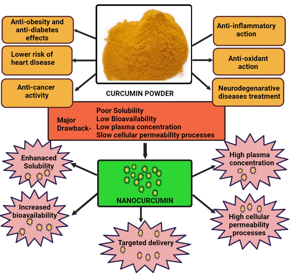
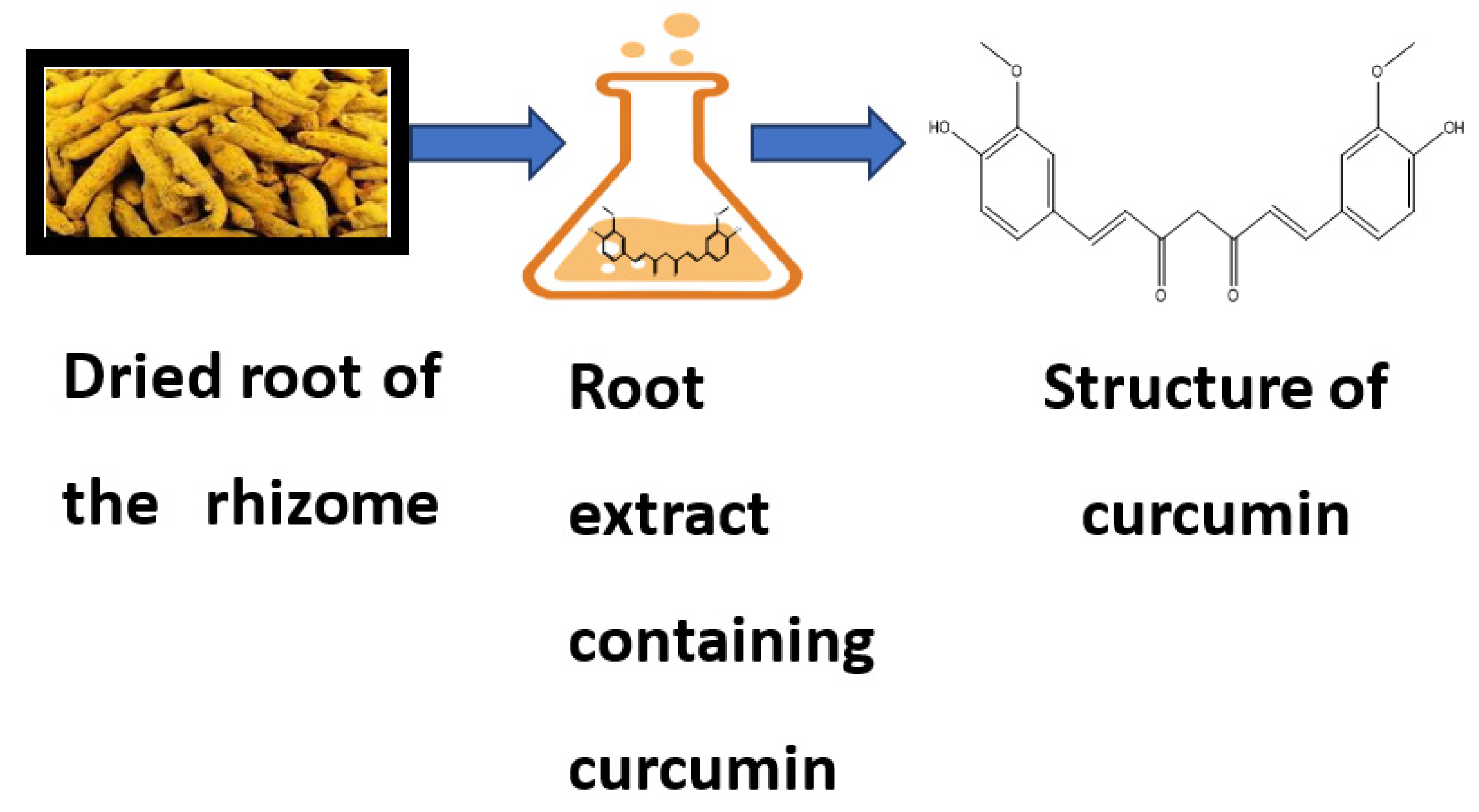
3. Bioavailability of Curcumin

Despite numerous health benefits, curcumin’s limited bioavailability is a fundamental criticism [10]. Low absorption, fast metabolism, chemical instability, and rapid systemic clearance have been suggested as possible causes [11]. According to various animal studies, most oral curcumin is eliminated in the feces (90%). Several strategies have been tried to boost the bioavailability of curcumin to solve this problem. Piperine, liposomal curcumin, curcumin nanoparticles, phospholipid complexes, and structural analogs of curcumin such as turmeric oil are a few examples of adjuvants [12]. Increased blood concentrations have been observed as a result of such efforts. However, human clinical trials comparing the therapeutic potencies and pharmacodynamic responses of these more bioavailable variants to those of conventional curcumin have yet to be undertaken extensively. Furthermore, the serum concentrations required to achieve a particular clinical or biological effect have yet to be determined. Piperine is an alkaloid found mainly in Piper nigrum, and it is this component provides black pepper its piquancy. Piperine has been shown to increase curcumin bioavailability [12]. Piperine suppresses the liver enzyme UDP-glucuronyl transferase, reducing the amount of glucuronidation of curcumin. Additionally, curcumin is accessible for ingestion as a result of this mechanism [13]. An oral dosage of 2 g/kg curcumin with 20 mg/kg piperine was delivered concurrently to animals and mice in an in vivo investigation [14]. Curcumin’s relative bioavailability was enhanced 1.54-fold in rats and 20-fold in healthy volunteers. Even though the increase in curcumin bioavailability was higher in humans than in rats, the quantity of curcumin ingested was more remarkable in rats than in humans. In another clinical experiment, healthy human volunteers were given 2 g of curcumin and 5 mg of piperine daily. The research found that administering curcumin plus piperine together boosted absorption by 200% then consuming curcumin alone [15]. Zeng et al. [16] looked at the effect of piperinepre administration on curcumin oral bioavailability and in this investigation, rats were given 20 mg/kg piperine first and subsequently 200 mg/kg curcumin at frequencies of 0.5 to 8 h after piperine treatment. Compared to those that received pure curcumin, the rats that received piperine before the curcumin exhibited a statistically huge increase in curcumin oral bioavailability, notably at 6 h following ingestion of piperine, with AUC0-t increasing 97-fold [17]. Thus, based on the aforementioned studies, it has been found that delivering natural substances such as piperine, quercetin, resveratrol, and silibinin in combination with curcumin resulted in improved curcumin absorption. This is a potentially cost-effective way to increase curcumin’s oral bioavailability and further explore it in developing novel drug delivery systems [18].
Nanocarrier-based delivery of curcumins one of the best approaches to improve curcumin’s solubility and bioavailability while also protecting it from hydrolysis-induced inactivation. Some nanocarriers emphasized long-term retention and circulation in the body, while others focused on intracellular release mechanisms and cellular delivery. Curcumin is solubilized in these environments by becoming entrapped in hydrophobic pockets, primarily through hydrophobic interactions. Curcumin’s fluorescence is boosted when solubilized in any of these systems, making it simple to measure its binding effectiveness. Because of their biocompatibility, these systems may be successfully studied for anticancer activity in cancer cells and in vivo systems, with considerable increases in anticancer activity due to enhanced curcumin bioavailability reported. Curcumin liposomal formulations have been proven to be the most effective for increasing curcumin bioavailability in cells [19], and products based on liposomal formulations are being commercialized. Nanocurcumin’s effectiveness is due to the size, surface area, charge, and hydrophobic nature of the particles, which make it superior to native curcumin [20] and regarded as an acceptable target for usage as a drug compared to standard curcumin. This feature is especially crucial in the fight against infectious illnesses caused by intracellular infections [21]. According to Ma et al. [22], nanocurcumin increased in vivo bioavailability and tissue distribution, with a 60-fold increase in biological half-life compared to native curcumin treatment in rat models.

4. Therapeutic Applications of Nanocurcumin over Curcumin

Nanocurcumin has the potential to prevent and treat a wide range of human diseases. Successful therapeutic applications of nanocurcumin are highlighted in the following section.

4.1. Antioxidant Effects

The antioxidant property of curcumin is due to its structure, which interconnects two methoxylated phenols with two unsaturated carbonyl groups grouped in stable enol form [23]. The phenolic O-H and C-H groups in curcumin are responsible for its bioactivity. Curcumin also plays a role in lipid peroxidation inhibition by oxidizing a polyunsaturated fatty acid called linoleate, which is oxidized to generate a fatty acid radical. Curcumin also participates in the intermolecular Diels–Alder reaction by breaking the chain at the 3’ site and neutralizing lipid radicals [24]. Besides lipid peroxidation inhibition, curcumin has been shown to have free radical scavenging activities in in vitro and in vivo models employing rat peritoneal macrophages. Curcumin actively scavenges numerous reactive oxygen species (ROS) produced by macrophages, such as hydrogen peroxide, nitrite radicals, and superoxide anions [25].
Nanocurcumin therapy is a promising method in modulating inflammatory cytokines, particularly IL-1 and IL-6 mRNA expression and cytokine production in COVID-19 patients, resulting in a better clinical presentation and over all recovery [26]. Significant levels of nitric oxide (NO) were created by iNOS, which then reacted with superoxide radicals in the surrounding oxidative environment to make peroxynitrite, a poisonous compound that was proven to be fatal to cells. Curcumin has been shown to inhibit the activity of iNOS and reduce ROS levels in cells. Additional research on microglial cells has demonstrated that this antioxidant spice can protect brain cells from oxidative damage by lowering NO production and diminishing neuroinflammation associated with chronic neurodegenerative diseases such as AD [27]. Alg-NP-Cur was analyzed and evaluated in a PD model in drosophila. It exhibited good antioxidant activity by decreasing lipid peroxidation in the PD drosophila brain after a diet supplemented with the nanocarrier for 24 days [28]. Curcumin nanocrystals were used to protect Wistar rats from cardiovascular damage by lowering lipid peroxidation and enriching the properties of antioxidants [29]. Using an aluminum phosphide (AIP) toxicity-induced rat model, Ranjbar et al. [30] studied the effects of curcumin and nanocurcumin on the oxidant and antioxidant system in the liver mitochondria and found that nanocurcumin enhanced oxidative stress variables and protected the liver by scavenging free radicals and stabilizing the oxidative state.

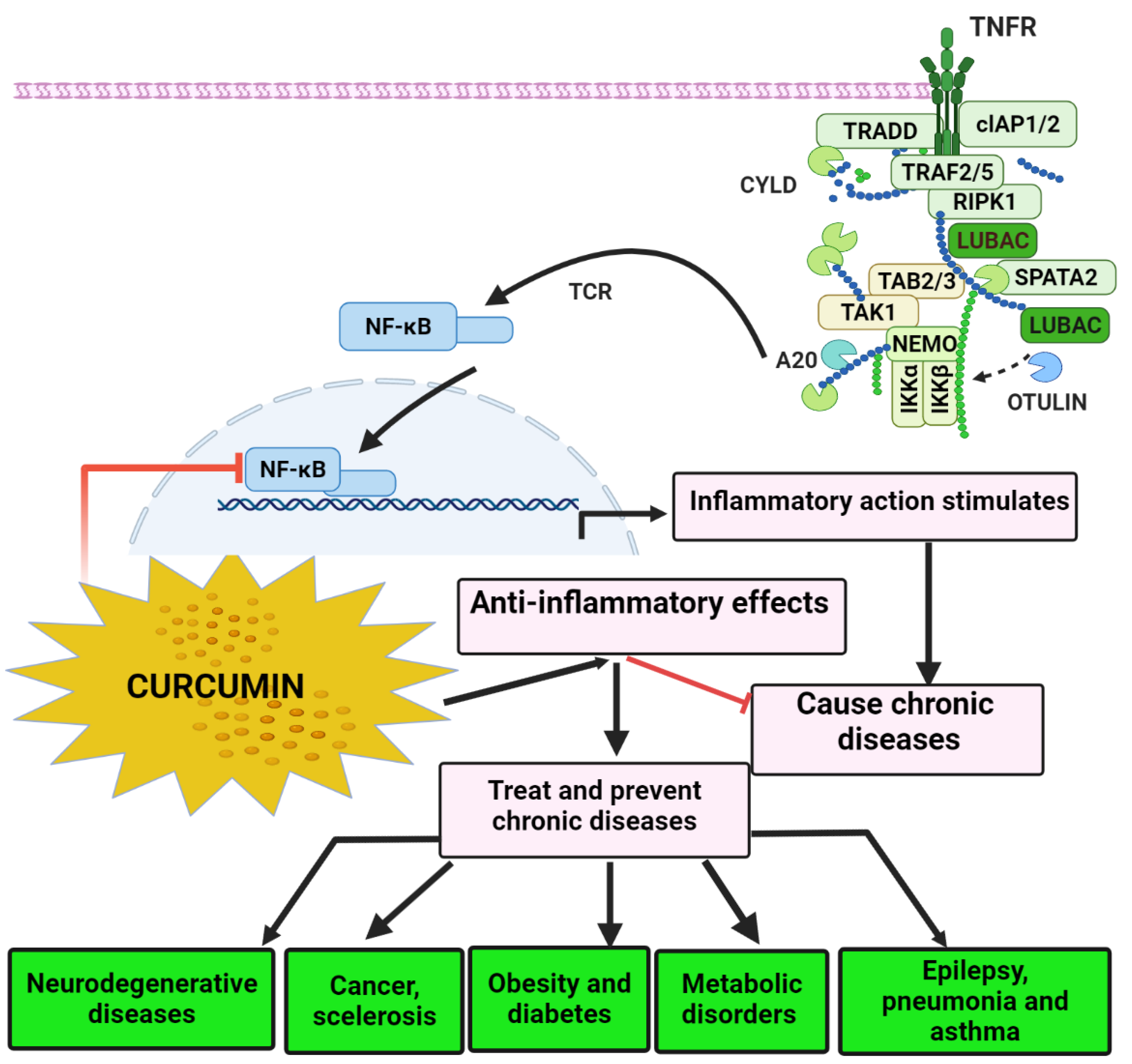
4.2. Anti-Inflammatory Effects

Curcumin aids in the battle against invaders and aids in the healing of the rift zone. While acute, short-term inflammation is helpful, it may become a problem if it becomes persistent and destroys cells. Excessive inflammatory activation for long periods of time can result in mitochondrial dysfunction. Traumatic brain injuries (TBI), spinal cord injuries (SCI), and hemorrhagic/ischemic stroke all cause substantial changes in mitochondrial dynamics, notably increased membrane permeabilization, oxidative phosphorylation, and the accumulation of mitochondrial ROS [31,32]. Excessive glial activation has been linked to the same outcomes as long-term inflammation. Advanced mitochondrial dysfunction has also been demonstrated to increase inflammatory processes, leading in neuronal injury and poor neurological consequences [33,34]. Studies have identified that the gene CDGSH iron sulfur domain 2(CISD2) protects against inflammatory reactions and apoptosis caused by mitochondrial dysfunction. The presence of CISD2 in the outer membrane of mitochondria has also been linked to the preservation of mitochondrial integrity. CISD2 deficiency resulted in mitochondrial dysfunction and cell death, according to reports on CISD2 knockout mice [35,36]. The attachment of BCL2 to BECN1, which governs cellular autophagy/apoptosis, has been found to be aided by CISD2. Anti-inflammatory and/or antiapoptotic therapeutics based on CISD2 are extremely likely to be developed to combat the consequences of aging, neurodegenerative disease, and CNS trauma [37].
A variety of radical oxygen/nitrogen species may also trigger a signal transduction pathway that boosts proinflammatory neural activity. Inflammation has been linked to the onset of a variety of chronic illnesses and disorders, as illustrated in Figure 2: Alzheimer’s disease (AD), Parkinson’s disease (PD), multiple sclerosis, epilepsy, brain damage, colorectal cancer, metabolic disorders, carcinoma, allergies, asthmatic, pneumonia, diarrhea, osteoarthritis, renal ischemic, lupus, diabetes, overweight status, anxiety, tiredness, and acquired immunodeficiency syndrome (AIDS) [38].
The stimulation of a transcriptional nuclear factor kappa B (NF-κB) regulates the impact of tumor necrosis factor (TNF-), a valuable inflammatory marker in most illnesses. Even though TNF- is the most potent NF-κB activator, TNF- production is low. NF-κB is also known to regulate this gene. TNF- is not the only cytokine that can be found in most cancers. Gram-negative bacteria; numerous disease-causing viruses; air toxins; biological, bodily, structural, and mental anxiety; high glucose; fatty acids; UV exposure; tobacco smoke; and other illness factors are also known to activate NF-κB [39]. As a result, medicines that inhibit NF-κB and NF-B-regulated gene products may be effective against various illnesses. Turmeric has been found to inhibit NF-κB activation, which is induced by several inflammation stimuli. Turmeric has also been demonstrated to reduce inflammation via different methods outside the scope of this study, indicating that it may be used as an anti-inflammation drug [40].
Wang et al. [41] produced curcuminsolid lipid nanoparticles (SLNPs) and improved efficacy in an allergic rat model of ovalbumin-induced asthma. According to Milano et al. [42], nanocurcuminis efficient against the Ehrlich ascites carcinoma (EAC) cell lines OE33 and OE19. It makes EAC cells more vulnerable to T cell-induced cytotoxicity and reduces T cell proinflammatory signals. Nanocurcumin improves oral bioavailability and, therefore, efficacy in preventing streptozotocin (STZ)-induced diabetes in rats, at least in part, by suppressing inflammation and pancreatic beta-cell death as compared to the native form. According to another investigation, loss of NF- activation led to downregulation of COX-2 and iNOS expression, blocking the inflammatory response and carcinogenesis [43]. W.S. Sarawi et al. [44] examined the impact of curcumin and nanocurcumin on copper sulphate (CuSO4)-induced brain oxidative stress, inflammation, and apoptosis in rats, and found that Akt/GSK-Akt/GSK-3β may be involved. Rats were given 100 mg/kg CuSO4 and were also given curcumin and nanocurcumin for 7 days. Cu-administered rats had significantly higher levels of MDA, NF-κB p65, TNF-, and IL-6 in the brain, as well as lower levels of GSH, SOD, and catalase. Cu produced DNA fragmentation in rats’ brains, elevated BAX, caspase-3, and p53, and decreased BCL-2, MDA, and NF-B p65. Nanocurcumin and curcumin, on the other hand, lowered proinflammatory cytokines while also decreasing proapoptotic genes, increasing BCL-2, and improving antioxidants and DNA integrity. In the brains of Cu-administered rats, both nanocurcumin and curcumin enhanced AKT Ser473 and GSK-3β Ser9phosphorylation. Finally, nanocurcumin and curcumin protected against Cu neurotoxicity by reducing oxidative damage, inflammation, and apoptosis while also increasing AKT/GSK-3AKT/GSK-3βsignaling. Nanocurcumin had a stronger neuroprotective impact than curcumin. Sinjari et al. [45] also investigated the anti-inflammatory action of curcumin liposomal formulations (CUR-LIP) and found it to be potent.

4.3. Antibacterial Effects

The dramatic rise in microbe resistance to multiple medications has necessitate daquest for novel and potentially effective antibacterial agents with minimal or lower human cytotoxic effects that can aid in treating a wide range of microbial diseases [46]. Curcumin antibacterial effectiveness against Gram-positive bacteria (Staphylococcus aureus) and Gram-negative bacteria (Escherichia coli) was studied by Tyagi et al. [47]. Curcumin was shown to have good antibacterial activity against all tested microorganisms [48]. Curcumin’s increased antibacterial action in the form of sunlight radiation mainly owed to photoexcitation, which caused the production of ROS that inhibited bacteria load [48].
Bhawana et al. [49] utilized the wet milling process to create water-soluble nanocurcuminin the 2–40 nm range of sizes and tested its antibacterial properties. S. aureus, B. subtilis, E. coli, P. aeruginosa, Penicillium notatum, and Aspergillus niger were shown to be resistant to nanocurcumin. Bhawana et al. also found that gram-positive bacteria had more potent antibacterial activity than Gram-negative bacteria. Similarly, employing surface-charged sodium dodecyl sulfate, Tween 20, and cetrimonium bromide, No et al. [50] developed nanocurcumin via an acoustoplastic deformation method. The investigators used L. monocytogenes, a gastrointestinal parasite, to evaluate the nanocurcumin they created. Shariati et al. [51] recently synthesized N-CUR and studied it against multidrug-resistant P. aeruginosa. The researchers observed that 128 g/mL of soluble curcuminin dimethyl sulfoxide (DMSO) inhibited bacterial growth, but 256 g/mL was necessary for soluble curcumin in DMSO to suppress it. At 64 g/mL, the nanocurcumin inhibited biofilm development and destroyed 58% of the biofilm. Based on these findings, the researchers hypothesized that nanocurcumin might lower P. aeruginosa pathogenicity and biofilm formation. Polymeric liposomes, micelles, and nanoparticles have all been described as intriguing biological systems. Nanocurcumin is quite desirable and effective for the treatment of different illnesses because of its excellent loading capacity and diminutive size. Curcumin therapeutic efficacy is increased when combined with nanomaterials, according to several kinds of research [52]. The antibacterial activity of silver-decorated polymeric micelles coupled with curcumin was investigated by Huang et al. [53].

4.4. Hepatoprotective Effects

Curcumin nanoliposomes were developed to lower the particle size while simultaneously increasing hydrophilicity [54]. The hydrodynamic layer around the nanoparticles was smaller as a consequence, resulting in a faster rate of surface-specific dissolving. It was said that liver damage, including necrosis and steatosis, might lead to a short event of the hepatotoxic substance carbon tetrachloride (CCl4). When the liver was exposed to untreated curcumin or curcumin nanoliposomes, the blood activities of alanine transaminase (ALT), aspartate aminotransferase (AST), and alkaline phosphatase (ALP) were significantly decreased. It was also discovered that the study group given curcumin nanoliposomes had less ALT, AST, and ALP serum activity than the study group provided untreated curcumin. This demonstrated that curcumin nanoliposomes might function as the most effective medication for treating any liver injury and might have a successful hepatoprotective impact [54]. Nanocurcumin was utilized for about the first time in liver tissues to manage the deleterious effects of CuSO4, since wat is thought that nanocurcumin’s compact size might be handy for associating with outer and inner biomolecules effectively [55]. Maghsoumi et al. [56] conducted research primarily to assess the preventive and rejuvenating properties of curcumin nanomicelles on prolonged liver damage produced by alcohol in mice. The findings indicated that nanocurcumin had a substantial effect on preventing the liver from CCl4-induced damage, and it was also found that nanocurcumin’s hepatoprotective activity was based on a specific dosage of 2 mL/kg of body weight [56]. A further study [57] found that in obese individuals with nonalcoholic fatty liver disease, nanocurcumin increased high-density lipoprotein (HDL) levels and lowered fatty liver and liver transaminase levels, demonstrating that nanocurcumin had a hepatoprotective effect.

4.5. Lower Risk of Heart Disease

Cardiac issues are the leading cause of mortality worldwide. Researchers have studied it for years and have learned a great deal about why it occurs. This chronic problem is unexpectedly complex, and various factors play a role in its development. Curcumin’s primary advantage in cardiovascular disease (CVD) is that it improves the performance of the endothelial, which lines blood vessels [58]. Curcumin has been linked to improved heart health in many studies [58]. Curcumin promotes cardiac repair and ameliorates cardiac dysfunction following myocardial infarction [59].
Although curcumin has been proven to positively impact on CVD and its risk factors, however it has a limited effectiveness due to its poor aqueous solubility. After oral dosing, this is ascribed to absorption, increased bioavailability, and systemic clearance. As a result, its therapeutic effects are substantially reduced [60]. Even though curcumin and nanocurcumin have the same crystal structure, nanocurcumin can be as helpful as curcumin in reducing CVD risk factors. Earlier in vitro and in vivo investigations, for particular, have shown that micro may be better than native curcumin in terms of certain testing products [61].

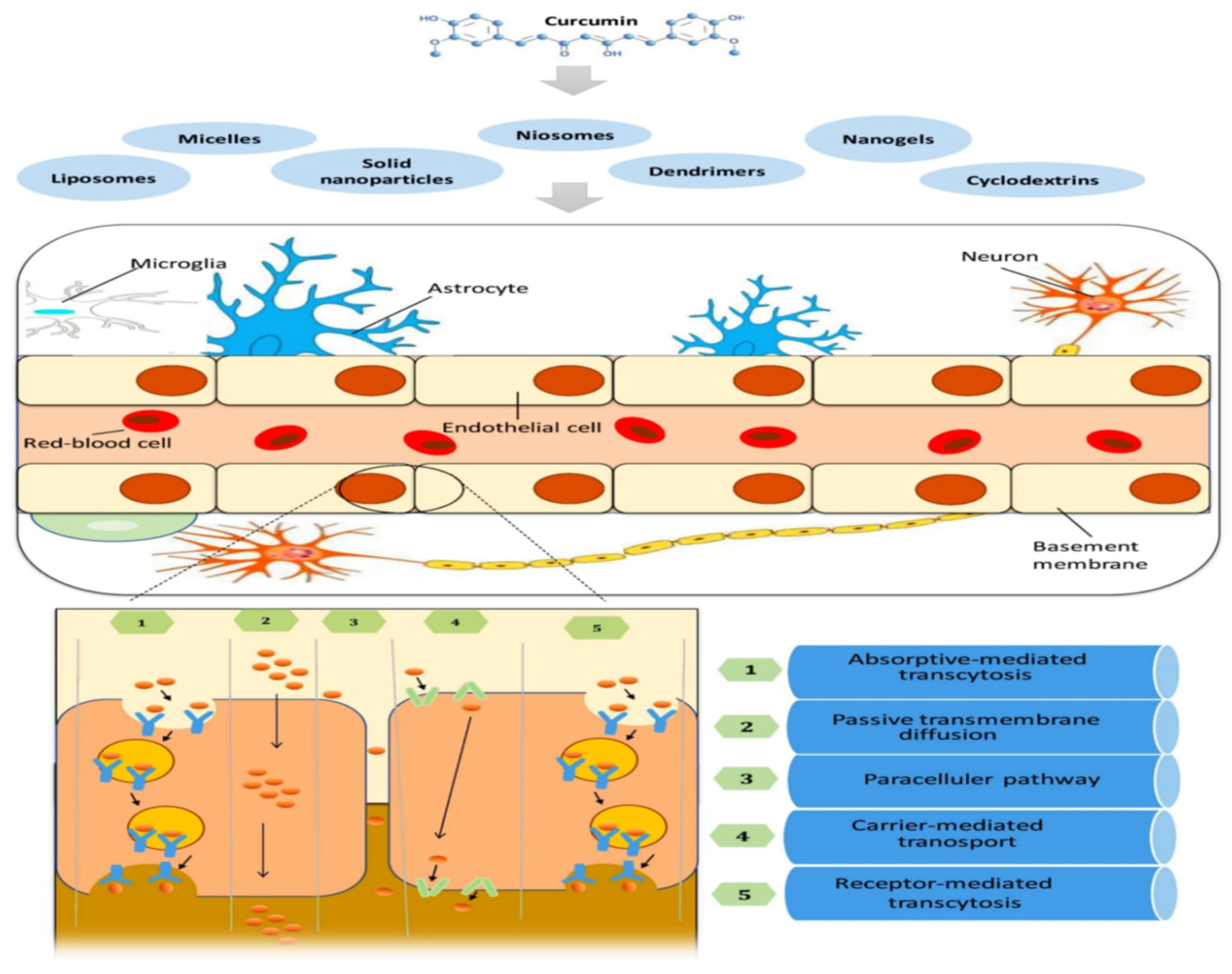
4.6. Nanoencapsulated Curcumin’s Potential in Crossing the Blood–Brain Barrier (BBB)

4.6.1. Boost Brain-Derived Neurotrophic Factor

Nerve cells can establish social contacts, and they may increase and grow in quantity in specific regions of the brain. Brain-derived neurotrophic factor (BDNF) is one of the most significant factors in this process [62]. It is a gene that is engaged in the manufacture of an enzyme that helps neurons live longer. The BDNF protein is located in regions of the brain that control feeding, drinks, and fat mass, and it plays a role in cognitive function [63]. Low levels of BDNF protein have been related to various common brain diseases, including stress and AD. Curcumin has been shown in animal experiments to boost BDNF levels in the brain [64]. It may be possible to postpone or even reverse many brain illnesses and age-related declines in brain function [65].
When primary tumors or metastases develop in unhealthy circumstances, the BBB is physically and functionally impaired beyond 1–2 mm in diameter, during this stage it becomes the blood–brain tumor barrier (BBTB). The deficiency of fenestration, the tight connections between endothelial cells (ECs), and the lower incidence of pinocytosis all restrict molecular exchange across the BBB, limiting transcellular transit [66]. Curcumin’s limited penetration through the BBB and its chemical–physical properties provide significant constraints for using these novel systems in brain diseases. The BBB between the blood and the central nervous system (CNS) has the physiological function of maintaining ionic homeostasis and chemical exchange, thereby shielding the CNS from infections and circulating toxins and sustaining the brain microenvironment [67]. The structure, function, and permeability of the BBB changed in brain cancers such as glioblastoma (GBM). Even though various methods for crossing the BBB have been developed, significant issues still make brain drug delivery problematic. The use of customized nanocarriers to preserve drugs while allowing them to pass across the BBB appears to be a successful approach [68].
The first stage of endocytosis determines the ultimate passage of nanoparticles into endothelial cells via transcytosis. The endothelial cells’ outer membrane influences the nanoparticles’ entrance; in fact, positively charged nanoparticles employ the adsorbing transcytosis route more readily than neutral or negatively charged nanoparticles, which have more negligible protein adsorption and thus longer circulation durations. Because of improper tight junctions, nanoparticles can penetrate the BBB via receptor-mediated transcytosis (RMT), adsorptive-mediated transcytosis (AMT), and extended permeability and retention (EPR) [68].
Endothelial cells (ECs), pericytes, and astrocytes occupy the BBB, and their end feet cover the basal lamina of the brain capillaries. Small soluble substances and endogenous macromolecules pass through the BBB via various transport pathways classified into diffusion and transcytosis (Figure 3). Hydrophilic and lipophilic compounds diffuse across cells via tight junctions or migrate through cells by dissolving in the plasma membrane [68]. Several aspects must be evaluated for proper drug transport across the BBB utilizing nanofabricated devices, including size, charge, biocompatibility, blood circulation stability, and controlled release capacity. Nanoparticles have been shown to penetrate the BBB efficiently [69]. Nanoparticles with diameters of 50 to 100 nm are most effective in animal models of CNS disorders. The specificity of BBB nanosystems, typically established by functionalizing them with particular proteins, is also critical [70].

4.6.2. Potential Role in the Treatment of Neurodegenerative Disease

Despite the limited number of in vivo, in vitro, and clinical investigations, data on the bioactive function of various nanoencapsulated curcumins in protection against and control of many CNS-related diseases have suggested nanocurcumin as a new approach to neurodegenerative diseases, including AD and PD [68]. Meng et al. [71] explored the effectiveness of a novel nanostructured low-density lipoprotein transporter that was tailored with curcumin-loaded lactoferrin for a targeted brain release that might potentially decrease the progression of AD. This nanocurcumin formulation, with a size of 100 nm, penetrated the BBB and released curcumin efficiently. The nanostructure had a protective impact on neuronal injury, according to histopathological evaluation. Furthermore, plasma levels of malondialdehyde (MDA), a key biomarker of lipid peroxidation, were lower in the nanocurcumin-treated groups, indicating BBB bridging and efficacy in decreasing oxidative damage [72]. It is well known that oxidative stress is linked to NO generation and mitochondrial dysfunction in the pathophysiology of PD [73]. Curcumin-loaded solid lipid nanoparticles (SLCN) were efficiently developed to have a particle size of about 86 nm and show no toxicity in endothelial brain cells. The effectiveness of SLCN against lipopolysaccharide-induced neuroinflammation in BV-2 microglial cells was also investigated. In a dose-dependent way, the SLCN inhibited NO generation more effectively than conventional curcumin [74].

Potential Role in AD

Curcumin has been proven to penetrate the venous barrier, so there may be some excellent signs on the way. Inflammatory and lipid peroxidation are known to have a role in AD. An accumulation of protein tangles known as an amyloid plaque is also a major characteristic of AD. Curcumin has been shown in studies to aid in the removal of these plaques. Curcumin’s ability to delay or perhaps reverse the development of AD in humans is unclear at this time, and more research is needed on this topic [75].The inclusion of curcumin into cultured neural stem cells has been shown to reduce brain risk of certain types of β-amyloid-induced neurotoxicity. When β-amyloid is used to create animal models of AD, curcumin guards against damage [76].
Curcumin prevents the agglomeration of β-amyloid into folded layers and reduces the production of β-amyloid from isozyme. Turmeric also has a preventive impact on mouse models against overproducing β-amyloid, according to studies. In these animal studies, brain activity was also enhanced. Curcumin clinical studies in AD have not proven particularly encouraging. This is likely due to turmeric’s low biocompatibility in humans; therefore, researchers have been working on ways to enhance delivery methods or produce analogues that replicate the neuroprotective benefits while being readily absorbed by the brain [77]. Curcumin’s lack of effectiveness in people with AD may possibly be due to treatment for too little time or beginning therapy too late in the illness’s progression, when significant neuronal death has already happened and cannot be reversed. Curcumin may be helpful in preventing the formation or advancement of AD. Many in vitro and in vivo investigations have examined how turmeric interacts with β-amyloid. Curcumin’s dose-dependent preventive activity against β-amyloid-induced damage in established neural stem cells has been widely studied. Several factors have been described to explain this preventive role. Inhibiting NF-κB with curcumin prevented β-amyloid-induced cell death in both human monocytes. Curcumin also inhibited NF-κB-induced suppression of peroxiredoxin-6 in rat hippocampus cells, reducing hypoxia-induced cell death [77].
Curcumin was found to reduce β-amyloid-induced affectation of the cytokines tumor necrosis factor-α and interleukin-1, as well as stimulation of mitochondrial protein kinase and phosphatases of intracellularly message kinase-1/2, in a human acute monocytic leukemia cell line (Sigma-Aldrich) [78]. The adding of turmeric to rat prefrontal cortical neurons reduced the β-amyloid-mediated rise in caspase-3 and triggered the defensive pathway involving Akt. Curcumin preserved cell viability in rat cortical cells following exposure to β-amyloid and reduced oxidative stress indicators and reactive oxygen species levels. Curcumin also seemed to decrease β-amyloid toxicity in APPs transfected SY5Y cells by reducing GSK-3 activity and boosting the protective Wnt/β-catenin pathway. As a result, curcumin seems to work on many levels to reduce tissue damage induced by inflammatory, peroxidation, or β-amyloid exposure [78].
Curcumin may also influence the formation and storage of β-amyloid, a protein that has long been believed to be a cause of neurotoxicity in AD [79].
Curcumin may be beneficial by lowering maintenance levels, according to other studies. Turmeric reduced the amount of amyloid precursor protein (APP) and prevented the formation of β-amyloid 1–42 in tissue culture. Curcumin did not seem to alter APP degradation by β-site amyloid precursor protein cleaving enzyme 1 (BACE-1), an enzyme implicated in the formation of –β-amyloid. Neither BACE-1 protein nor messenger planning to open acid levels was changed; thus, the action must involve an APP signal transduction process. Curcumin did not affect the amounts of mature APP in a later study, although it did reduce the levels of immature and total APP. The authors of [80] hypothesized that by disrupting the route that leads to the formation of both β-amyloid 1–40 and β-amyloid 1–42, inhibiting the APP proliferative phase may explain the participative management of both β-amyloid 1–40 and β-amyloid 1–42 [80]. Curcumin seems to influence the action of β-secretase via lowering the production of closed air, the enzyme’s active subunit. This may be due to glycogen synthase kinase-3 (GSK-3) suppression, which hydrolyzes presenilin-1 to activate β-secretase. GSK-3 activity may hydrolyze tau, enabling it to form Hrithik filaments in order to raise β-amyloid levels.
Curcumin’s capacity to bind iron is another method by which it may reduce the development of β-amyloid. Increases in iron may promote the creation of β-amyloid. Curcumin reduces iron-induced toxicity in basic cultures, and it has been suggested that turmeric’s therapeutic benefits in nova animal models of AD may be attributable in part to its iron-related actions. Curcumin may reduce β-amyloid formation and depositing via a variety of pathways, including changes in secretase activity and amyloid precursor protein (APP) maturity, decreasing GSK-3 activity and tau synthesis, and sorption of iron. Table 1 summarizes these activities [81].
Curcumin nanoformulation by altering the surface of the PLGA polymer and encapsulation of selenium nanoparticles decreased the Aβ load and inflammation in brain samples of AD mice and treated the memory loss of the model mice. Moreover, histopathological images of animal tissues displayed no deceptive abnormalities [82]. Singh et al. [83] developed curcumin-encapsulated Pluronic F127 nanoparticles (FCur-NPs) and compared the blood–brain barrier penetration of free curcumin (free CUR) and FCur-NPs. In the brains of mice, FCur-NPs had a 6.5-fold higher fluoresce intensity than free CUR. In vitro comparisons with Congo red then demonstrated that encapsulated CUR retains its capacity to bind to A plaques. When compared to free CUR, FCur-NPs showed antioxidant and antiapoptotic activities. In vitro and in vivo data implied that FCur-NPs might be useful as a theragnostic agent for AD.

Potential in the Treatment of GBM

Curcumin has been used as an adjuvant to GBM therapies in a number of studies. Various experimental models of GBM treated with different curcumin nanoencapsulations (methoxy polyethylene glycol polycaprolactone, mPEG-PCL; solid lipid nanoparticles, SLCP; curcumin-loaded lipid-core nanocapsules, C-LNCs) showed similar results, such as efficient endocytosis, increased apoptosis, and arrest of the G2/M cell cycle [84]. Curcumin encapsulated in nano micelles inhibited U373 cell growth by modulating the NF-κB pathways, resulting in early G2/M cell cycle arrest, increased sub-G1 cell cycle arrest, and cell damage induction [80]. A further new dendrosomal curcumin (DNC) prohibited U87MG cell proliferation in a time- and dose-dependent manner, increasing the number of apoptotic cells when used in combination with p53 overexpression [85]. In another study, curcumin-loaded zein nanoparticles (CUR-ZpD-G23 NPs) successfully crossed the BBB and administered curcumin to glioblastoma cells. When compared to free curcumin, the NPs increased cellular absorption of curcumin by C6 glioma cells and demonstrated great penetration into 3D tumor spheroids. BBB crossing and tumor spheroid penetration were increased by G23-functionalized NPs. Furthermore, in liquid and soft agar models of C6 glioma cell development, the NPs significantly reduced proliferation and migration, as well as causing cell death [86]. Finally, curcumin nanoencapsulation in polysaccharide matrices (diameters 210–240 nm) based on hyaluronic acid (HA) and curcumin–lactoferrin conjugated (Lf-Cur-PSNPs) showed excellent BBB penetration. Lf-Cur-PSNPs were occupied predominantly by brain capillary endothelial cells and remained constant and more efficient in targeting C6 glioma cells after crossing the BBB [87].

4.7. Anticancer Effects

Curcumin is efficacious against cancer by inhibiting all three phases of tumorigenesis: start, development, and advancement [88]. Curcumin is a chemosensitizer and radio-sensitizer for tumor cells, and a chemoprotector and radioprotector for body cells, in addition to its chemopreventive ability with low discomfort and susceptibility [89]. Curcumin is a high-epigenetic-regulation-potential anticancer drug that acts with many cancer targets and processes. It can prevent tumorigenesis through various mechanisms, including tumor apoptosis, inhibition of proliferation, antiangiogenesis, and stimulation of mitotic catastrophe, differentiation and atrophy, suppression of cytokines and migration, and genome control [90].

4.7.1. Activation of Tumor Apoptosis

In many cancer forms, curcumin therapy may trigger apoptosis (the process of programmed cell death; opposition to apoptosis can encourage tumor formation) via internal stress responses and external ligands. Cytotoxicity is initiated intrinsically in response to cellular cues such as stress and DNA damage. Turmeric was found to have two purposes: it upregulates proapoptotic B-cell lymphoma 2 (Bcl-2) family enzymes (Bim, Bax, Bak, Puma, and Noxa) and downregulates the antiapoptotic proteins X-linked inhibitor of apoptosis protein (XIAP), Bcl-2, and B-cell lymphoma-extra-large (Bcl-xL) [91]. The NF-κB regulated discovered genes Bcl-2, Bcl-XL, cIAP, survivin, and tumor necrosis factor receptor (TNFR) associated factors 1(TRAF1) and 2 (TRAF2), as well ascurcumin, were also shown to operate mainly via inhibiting the apoptotic pathway [92]. Autophagy can be triggered at the binding site via the intrinsic route, which fuels the growth of cellular membranes transmitters (Fas, TRAIL). Unlike the inner pathway, which is linked to endorphins, the extrinsic pathway is linked to death receptors. This route leads to the formation of the Fas, flavin adenine dinucleotide (FAD), and caspase-8 and -10 death-inducing signaling complexes. The activation of cell damage by apoptosis after combination therapy lends credence to curcumin’s potential effectiveness as cancer care. Another immune cell enzyme, p53, may be activated by curcumin and mediates apoptosis death in basal pituitary tumor lines in a dose- and time-dependent manner [93]. The mitogen-activated protein kinase (MAPK) family is a large set of cellular proteins known to bind p53’s trans activation domain (c-Jun N-terminal kinases (JNK) 1-3, ERK1/2, and p38 MAPK). On the other hand, curcumin has been shown to promote apoptosis by phosphorylating and activating JNK [94].

4.7.2. Antiangiogenesis

Curcumin stimulates neurogenesis (the process of supplying food and energy to a tumor while also removing toxins) by controlling endothelial features, which include a few other metabolic enzymes, e.g., basic fibroblast growth factor (bFGF), epidermal growth factor (EGF), granulocyte colony-stimulating factor, interleukin 8(IL-8), platelet-derived growth factor(PDGF), transforming growth factor–tumor necrosis factor (TGF– TNF), vascular endothelial growth factor (VEGF),and many small particles (e.g., adenosine, prostaglandin E). VEGF and bFGF seem to be the most significant of these chemicals in maintaining tumor development [95]
Attacking VEGF or its cellular receptor is a common strategy for anti-VEGF treatment. Antigens are the most frequent neurotransmitter medicines, while receptor tyrosine kinase agents are the most common direct amplifiers of VEGF activity. Curcumin inhibited VEGF and angiopoietin 1 and 2 gene production in EAT cells, VEGF and angiopoietin one oxidative stress in NIH3T3 cells, and KDR gene expression in HUVEC cells in a time-dependent manner [96]. Curcumin shown to inhibit VEGF production in blood clotting via mechanisms such as TGF release, COX-2 hypertrophy, hydrogen peroxide discharge, conventional and aberrant EGFR, Src signaling, and abnormal NF-B signaling [97]. In mice, curcumin and its derivatives inhibited primary b-FGF-mediated ocular neovascularization significantly. Curcumin has also been shown to decrease VEGF and b-FGF biomass production in estrogen receptor-negative MDA-MB-231 tumor tissues [98].
Vasculature, epithelial cell movement, and tube construction are all mediated by matrix metalloproteinases (MMPs). Gelatinase A and gelatinase B are metalloproteinases that activate transcription factors to develop new microvascular. Curcumin has been found to inhibit 72kDa MMP production and transposition. Phytochemicals suppress the production of gelatinase B, which is activated by the FGF-2-regulated gene transcription activator protein 1(AP-1) [99]. CD13/aminopeptidase N (APN) is a zinc-dependent vesicle protease involved in tumor invasion and morphogenesis. Curcumin linked to APN and reduced its function irreversibly, according to Shim and colleagues [100]. Liposomal curcumin reduced tumor size and decreased CD31 production, as well as VEGF and IL-8 inflexion, suggesting that curcumin prevented tumor angiogenesis and lowered pancreatic cancer development in mouse xenograft models [101]. Curcumin also had the capacity to incite apoptosis, inhibit cancer cell proliferation, and decrease cell cycle development, making it a potential therapy against human lung, breast, prostate, pancreatic, and melanoma malignancies [102]. Some researchers have claimed that further clinical trials on curcumin are unnecessary because of its fragile, reactive, and non-bioavailable nature [103]. Curcumin has been found to suppress cancer cells from growing. Curcumin inhibits the function of matrix metalloproteinases, which govern the process, preventing cancer cells from attacking normal tissue. Curcumin suppresses the production of genes including cyclin D1, c-myc, bcl-2, and Bcl-xL, which are implicated in tumor growth, proliferation, and apoptosis. NF-κB inhibition, for example, is crucial in carcinogenesis and proliferation [104]. Basniwal et al. [105] investigated the anticancer effects of curcumin nanoparticles in cancer cell lines from the lungs (A549) and skin (A431). In aqueous circumstances, curcumin nanoparticles were shown to have a significantly greater effect on cancer cells than inherent curcumin. One of the most common histological subtypes of breast cancer with a metastatic characteristic is triple-negative breast cancer (TNBC). Dendrosomal nanocurcumin and exogenous p53 have had anticancer effects on TNBC cells when used together [106].

4.8. Antidiabetic Effects

Diabetes and heart disease model mice (db mice) received dietary curcumin (0.2 g/kg, 6 weeks) that lowered HOMA-IR and HbA1c levels and the activity of the hepatic gluconeogenic enzyme. Curcumin raises the blood levels of adiposity, an adipocytokine that improves insulin resistance [107]. A few findings on curcumin’s antiobesity and antidiabetes benefits are included here. Ina study in which 14 healthy individuals were given 6 g turmeric powder along with glucose and observed no impact of oral curcumin consumption (capsules) on glucose tolerance. However, they discovered an increase in blood insulin. Another scholar performed a randomized quintuple placebo-controlled study in 240 people with the post as an example. For nine months, a group of patients was administered a curcumin capsule containing turmeric extract (75–85% curcuminoids) at a dose of 1.5 g per day. Insulin secretion improved considerably, and blood triglyceride levels increased, compared to the control group [108]. This same study team conducted a 6month experiment with 213 people with diabetes at risk of angiogenesis, in which one group of subjects received identical pills as in the prior trial. They had lower HOMA-IR, reduced central body fat, and more significant plasma adiponectin64 than the placebo group. An oral intake of 300 mg/day glucosamine composition (curcumin 36.06%, dimethoxycurcumin 18.85%, bisdemethoxycurcumin 42.58%) for 12 weeks resulted in lower HbA1c and HOMA-IR levels compared to the placebo group in a randomized, double-blind placebo clinical study consisting of a total of 100 people with diabetes with fatness. Several good reviews [109,110] may aid in gaining a better knowledge of the antidiabetes and antiobesity benefits. In rats and mice, foliar spray enriched diets substantially reduced body fat accumulation and improved hyperglycemia. Instead of turmeric, large dosages of ferulic acid were given in each instance. According to human research examining different polyphenol metabolites, ferulic acid secreted in urine is not substantially linked with type 2 diabetes risks. There have been no reports on vanillin, dicyclopentadiene, or curcumin glucuronic acid, curcumin’s major precursor [111]. Nanocurcumin has been shown to be efficacious against diabetes in rats caused by STZ in research. Nanocurcumin mitigated oxidative stress and lowered inflammation and apoptosis in pancreatic β-cells [112]. Curcumin promoted glucose uptake in cells by increasing GLUT4 translocation and enhancing insulin levels in body cells, according to Mohiti et al. [113].

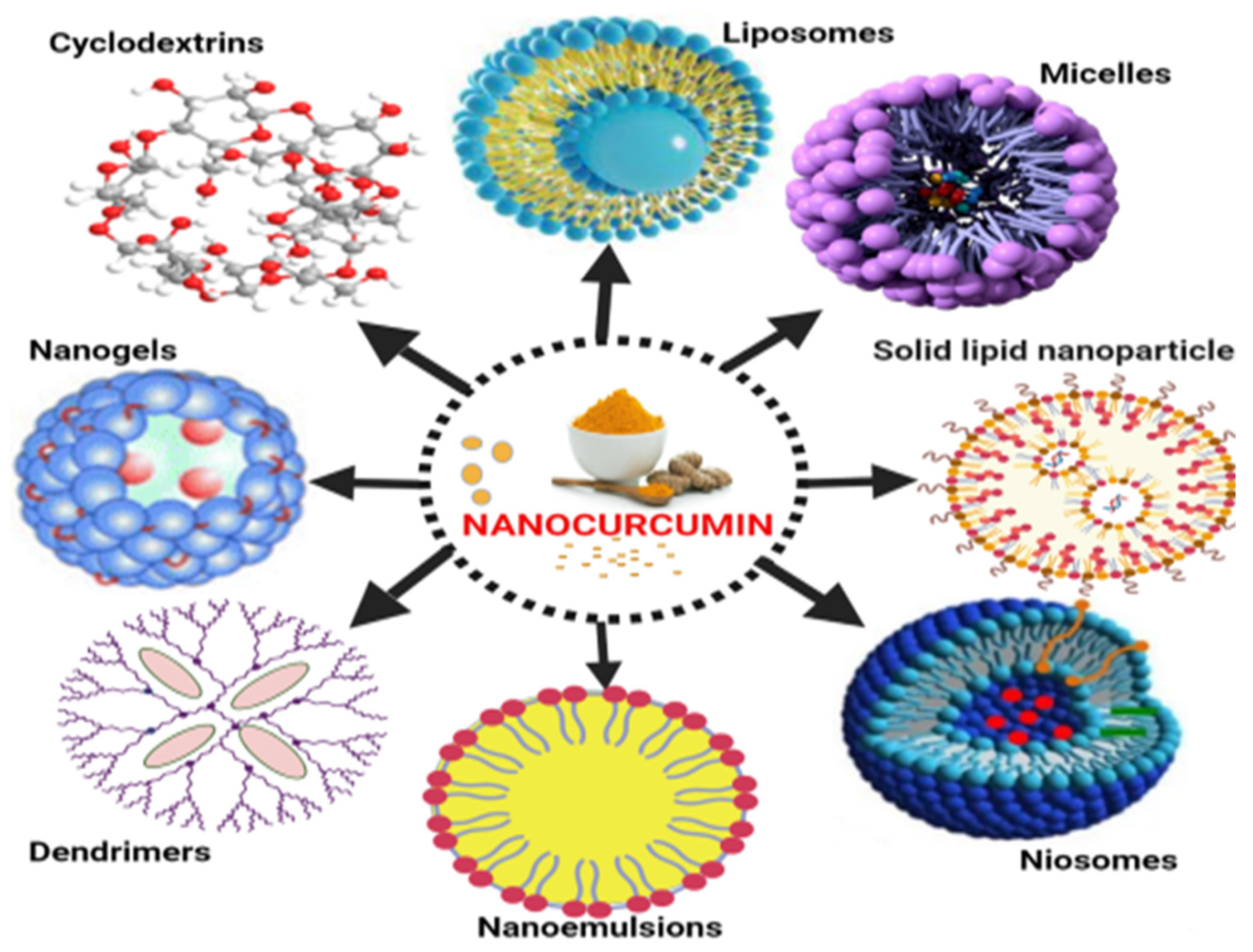
5. Various Nano Drug Delivery Systems for Curcumin
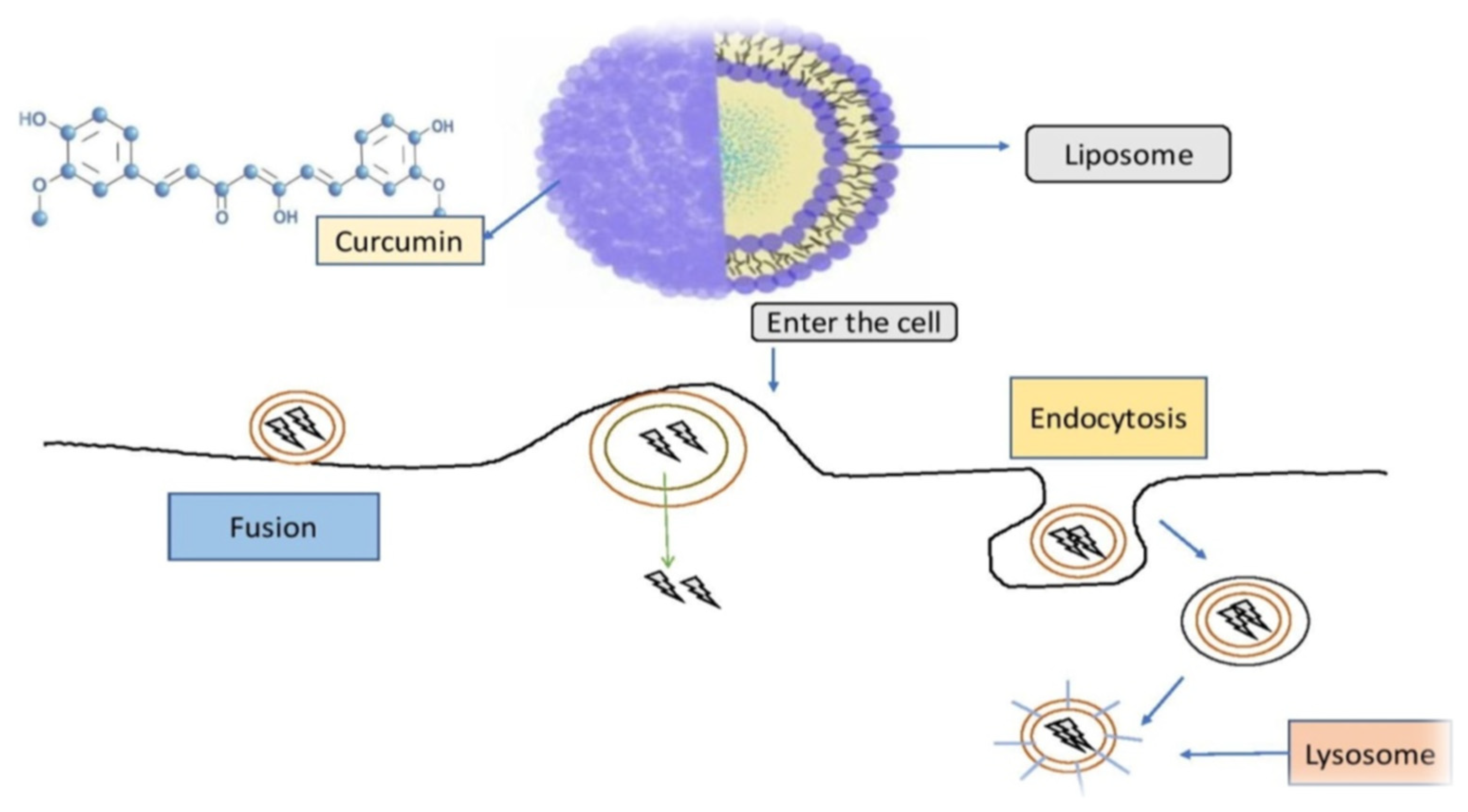
5.1. Liposomes

Natural phospholipids can be used to produce liposomes, which are globular synthetic vesicles. They are inner enclosed colloidal materials made up of lipid bilayers with an exterior lipid bilayer around a core aquatic region [114,115]. Liposomes range in size from 25 to 205 nm in diameter. Liposomes are supposed to be utilized as drug transporters and immunological assistance agents and are encapsulated either in the aquatic part of the lipid bilayers or at the bilayer surface. They can be used to encapsulate drugs with a wide range of solubilities or lipophilicities [116,117]. Furthermore, they can carry drugs into cells by fusion or endocytosis, and almost any chemical, regardless of solubility, can be encapsulated in liposomes (Figure 2). To enhance curcumin solubility, Rahman et al. [118] produced β-cyclodextrincurcumin binding interactions that incorporated both natural curcumin and the complexes independently into liposomes. Entire curcumin-containing preparations were efficient at suppressing cell growth in vitro cell study [119]. Shi et al. [120] used an enzyme-linked immunosorbent assay (ELISA) technique to evaluate curcumin’s therapeutic benefits on lung fibrosis in mice using a water-soluble liposomal curcumin that was found to successfully reduce radiation pneumonitis and lung fibrosis and sensitize LL/2 cells to irradiation. Some research has indicated that liposome-encapsulated medication is predicted to be delivered without accelerated deterioration and results in fewer adverse consequences and exhibit greater evidence of resilience in the receiver. Matabudul et al. [121] investigated whether various intervals of Lipocurc intravenous infusions affected metabolism and tissue delivery of curcumin, and whether reacting necropsied beagle dog tissues with phosphoric acid before evaluating curcumin and its active ingredient (tetrahydrocurcumin) could regulate the substances and allow effective assessments. Figure 4 illustrated various nano drug delivery system for curcumin delivery.
Curcumin-loaded nanoparticles have been used to assess liposomal curcumin’s potential against various cancers [122,123,124]. Liposomal curcumin demonstrated significant anticancer potential against osteosarcoma and breast cancer cell lines in vitro and in vivo via the caspase cascade, which resulted in apoptotic cell death. The xenograft osteosarcoma model in vivo was used to demonstrate the efficacy of liposomal curcumin nanoparticles. Curcumin was incorporated into a liposomal delivery system for intravenous administration by Li et al. [125]. These authors also used human pancreatic cancer cells in vitro and in vivo to demonstrate the effects of liposome-encapsulated curcumin on proliferation, apoptosis, signaling, and angiogenesis [126,127]. Curcumin encapsulated in liposomes inhibited tumor angiogenesis in vivo and suppressed pancreatic carcinoma growth in murine xenograft models [128]. It also inhibited proliferation, caused apoptosis, and inhibited the NF-κB pathway in human pancreatic cells in vitro, as well as having anticancer and antiangiogenesis actions in vivo [129,130]. Curcumin partitioning into liposomes containing dimyristoyl phosphatidyl choline (DMPC) and cholesterol suppressed cellular proliferation in human prostate cancer cell lines (LNCaP and C4B2) by 70–80% without affecting viability. When compared to curcumin, oral doses of liposome-encapsulated curcumin resulted in high bioavailability and quicker and better absorption of curcumin in rats [131]. Curcumin is incorporated into liposomes and then reaches cells are demonstrated in Figure 5.

5.2. Micelles

A micelle is a spherical vesicle made up of amphiphilic surfactant molecules that spontaneously assemble in water. Micelles are commonly utilized to deliver drugs that are not very water soluble, such as curcumin [133]. Curcumin can be added pre- or post-micelle production to solubilize it within the hydrophobic center of micelles. Micelles are colloidal dispersions that are highly thermally stable and include particulates. Because the particles are so tiny, they do not scatter light waves substantially and thus tend to be optically transparent. Micelle viscosities are determined by surfactant concentration and micelle structure. Micelles often seem to be fluids with low viscosities at low concentrations, but semisolids with greater viscosities or gel-like textures at higher concentrations. Since these variables impact the size, shape, interactions, and dynamics of the colloidal structures produced, the rheological characteristics of micellar systems are highly influenced by the surfactant type and ambient circumstances [134]. Micelles improve the bioavailability of hydrophobic substances (such as curcumin) by enhancing their bioaccessibility in gastrointestinal fluids and potentially boosting epithelial cell penetration [135]. Curcumin encapsulated polymeric micelles (CUR-M) were produced using a one-step solid dispersion method, and the efficacy of CUR-M was tested in a breast tumor model, by Liu et al. [136]. CUR-M exhibited better activity than pure curcumin at suppressing the generation of breast cancers and spontaneous pulmonary metastases of the lungs. Curcumin-poly (ethylene glycol) methyl ether (MPEG-PCL) micelles with solid dispersion improved curcumin’s antiangiogenesis and antitumor effects.
Curcumin micelles may be beneficial in the treatment of lung cancer, according to the findings in [137]. Chang et al. [138] investigated the cell uptake, intracellular localization, and cytotoxicity of different sizes of curcumin encapsulated micelles on human colon cancer cells in vitro. Their findings showed that smaller curcumin-loaded micelles had a greater possibility for inducing cytotoxicity in human colon cancer cells than larger micelles. As a result, drug loading, micelle size, and uptake/release kinetics are all critical factors to consider when it comes to nanoparticle drug delivery [139].
Another study by W. Ma et al. [140] on co-assembly of doxorubicin and curcumin targeted micelles for synergistic delivery by ultrasonication. The micelles demonstrated improved antitumor efficacy, improved tumor suppression and diminution, and improved cytotoxic effects. A study by Yang et al. [141] on a pH multistage responsive micellar system with charge-switch and PEG layer detachment for codelivery of paclitaxel and curcumin to synergistically eliminate breast cancer stem cells using the dialysis method found tumor growth inhibition with no substantial recurrence.

5.3. Nanoemulsions

Nanoemulsions (NEs) are biphasic dispersions, either oil-in-water (o/w) or water-in-oil (w/o), in which particles from one phase are dispersed in another and stabilised by an emulsifier that lowers the interfacial tension between the two immiscible liquids [142]. Regardless of the lack of an existing arrangement on the maximum limit, NEs can have a mean droplet diameter of 20 to 200 nm [143]. NEs can be made with a variety of emulsifiers, including biopolymers (such as proteins and polysaccharides), phospholipids, surfactants (such as Tween 80), and oils (such as fats and essential oils) [144,145,146]. Because of their prospective capacity to encapsulate bioactive chemicals for a variety of applications in the dietary, pharmaceutical, and medical fields, NEs have garnered much attention [147]. Among different nanoformulations, NEs are best suited for lipophilic drugs such as curcumin to improve their solubility and bioavailability [148]. Furthermore, NEs are more resistant to droplet agglomeration and gravitational dispersion than traditional emulsions [149]. As a result, numerous experiments have been conducted to demonstrate the feasibility of utilizing NEs to develop curcumin delivery systems [149]. Thermodynamically stable and transparent NEs are required. Despite this, because NEs are nonequilibrium systems, they take a great deal of energy to develop. Curcumin can therefore be enclosed in NEs with either high- or low-energy techniques. Low-energy techniques depend on the nature (i.e., molecular structure and solubility) of the molecules present in the solution to produce oil droplets by controlling interfacial alteration at the border between two immiscible phases [149].
A few in vitro and in vivo studies have been carried out to evaluate the therapeutic properties (e.g., anticarcinogenicity), biological effectiveness, and patient compliance of NEs containing curcumin. Curcumin enclosed in NEs also had better anticancer impact than free curcumin, according to research. For example, Nikolic et al. [150] studied the pharmacological safety and compliance profile of low-energy NEs stabilized by polysorbate 80 and soybean lecithin as curcumin delivery methods. Curcumin-NEshada PDI of 0.156 to 0.175, with an average size diameter of 150 nm. Curcumin-NEs were found to have considerable a cytotoxic effect against the HeLa cell line (IC50 = 22.89 g/mL) and human melanoma (Fem-x cell line, IC50 = 37.87 g/mL) in cytotoxicity assays. Free curcumin, on the contrary, had IC50 values of 7.77 and 20.64 g/mL, respectively, for HeLa and Fem-x cell lines. The scientists also noted that encapsulation improved curcumin-NEs’ safety profile when compared to free curcumin, with IC50 values of 67.72 and 26.97 g/mL for curcumin-NE and free curcumin, respectively, against a normal lung fibroblast (MRC-5) cell line.
The curcumin-NEs had an average, PDI, and zeta potential of 195 to 217 nm, 0.200, and −30 to −36 mV, respectively. In the Northeast, the efficiency of encapsulation (% EE) and drug loading (% DL) of curcumin were 95 and 2.1%, respectively. Human gastric adenocarcinoma (AGS), colorectal adenocarcinoma (HT29-ATCC), cells generated from HT29-ATCC with enhanced metastatic potential (HT29-US), human mammary gland adenocarcinoma (MDA-MB-231), and murine melanoma cell viability was evaluated after treatment with CUR-NE (B16F10). The IC50 values for AGS, MDA-MB-231, HT29-US, and HT29-ATCC cells were 24, 26.2, 75.7, and 84.6 M, respectively, indicating that this nanoformulation inhibited cancer cell growth [151].

5.4. Solid Lipid Nanoparticles

Solid lipid nanoparticles (SLNs) constitute a unique possible colloidal carrier system and comprise physiologically tolerable lipid components. At room temperature, their form is solid. SLNs are a popular way to improve the oral bioavailability of drugs that are not very water soluble [152]. Curcumin was incorporated into SLNs by Kakkar et al. [153] to enhance its oral bioavailability. A microemulsification method was used to generate curcumin-SLNs with a mean particle size of 134.6 nm and a maximum amount of drugs <92%. After oral delivery of curcumin-SLNs, in vivo pharmacokinetics was measured in rat plasma via a validated LC–MS/MS technique. When compared to curcumin-solid lipids, the results showed a substantial improvement in bioavailability times following delivery of curcumin-SLNs. Data have shown that improved and consistent bioavailability can aid in determining a drug’s therapeutic impact. In addition, Kakkar et al. [154] integrated curcumin with SLNs to enhance bioavailability. Plasma and brain cryosections were then examined under a fluorescent/confocal microscope for fluorescence. The biodistribution of 99m Tc-labeled curcumin-SLNs and curcumin-solid lipids in mice following oral and intravenous treatment was also studied. Yadav et al. [155] introduced a new formulation strategy for treating investigational colitis in rats using a colon-specific delivery system. Palmitic acid, stearic acid, and soya lecithin were used to make solid lipid microparticles (SLMPs) containing curcumin with an optimum proportion of poloxamer 188. The colonic delivery mechanism of the SLMP preparation of curcumin was then researched for its antiangiogenic and anti-inflammatory properties by employing chick embryo and rat colitis studies. In the chorioallantoic membrane test, solid lipid microparticles of curcumin were revealed to constitute a strong angioinhibitory drug. When compared to dextran sulfate solution-treated control rats, animals fed with curcumin and its SLMP complex gained weight quicker. When compared to free curcumin and controlled animals, the increase in total colon length was revealed to be substantially higher in solid lipid microparticle-treated rats. Furthermore, rats treated with curcumin-SLMPs had fewer mast cells in their colon mucosa. Colonic distribution of curcumin-SLMPs considerably reduced the severity of colitis produced by dextran sulfate solution administration [155].
In one study on SLNs loaded with curcumin and doxorubicin prepared by a cold dilution microemulsion method, it was found that curcumin-loaded SLNs were more effective than free curcumin at increasing doxorubicin efficacy in resistant TNBC cells. The curcumin-loaded SLNs decreased P-gp activity and expression and decreased P-gp transcription by reducing intracellular ROS and NF-κB activity. They also restored doxorubicin sensitivity by downregulating the ROS/NF-κB/Pgp axis in resistant TNBC cells cocultured with macrophages [156].
Another study showed that curcumin-SLNs prepared using an emulsification evaporation–low temperature solidification method resulted in drug loading and encapsulation efficienciesof23.38% and 72.47% and greater cytotoxicity against SKBR3 cells. In an in vitro cellular uptake study, curcumin-SLNs were found to have a good absorption efficiency by SKBR3 cells. Curcumin-SLNs also produced higher apoptosis in SKBR3 cells and decreased the manifestation of cyclin D1 and CDK4.These data suggest that curcumin-SLNs might be a promising chemotherapeutic formulation for breast cancer therapy [156].

5.5. Niosomes

Niosomes are tiny lamellar structures containing cholesterol and a nonionic surfactant of the alkyl or dialkyl polyglycerol ether family [157]. Because of hydrophilic nature niosomes can serve as a vessel for drug substances with a large variety of solubilities. They have amphiphilic, lipophilic, and amphiphilic moieties in their composition, and they act similarly to liposomes in vivo, making them a viable substitute to liposomal drug carriers. These properties are dependent on the bilayer’s constituent and the method of production [157]. The type of surfactant, the nature of the encapsulated medication, the storage temperature, the detergents, and the usage of bilayer lipids can all impact the stability of niosomes [158]. Niosomes are also expected to have a variety of therapeutic uses, including anticancer, and it can enhance drug therapeutic parameters by limiting drugs’ effect on target cells. They also use a new drug delivery system to enhance the oral bioavailability of improperly absorbed drugs, such as curcumin, and boost drug penetration via the skin [159]. Curcuminoid niosomes were generated with a variety of nonionic surfactants to improve skin penetration of curcuminoids in a report by Rungphanichkul et al. [160]. Entrapment potency and in vitro curcuminoid penetration through snakeskin were used to assess the results. When compared to a vehicle solution of curcuminoids, niosomes significantly improved curcuminoid penetration. Curcuminoids could be successfully produced as niosomes, according to the data, and such preparations offered superior characteristics for transdermal drug delivery [161].

5.6. Dendrimers

Dendrimers are polymers with a monodisperse, three-dimensional structure with nanoscale dimensions that are highly branching, synthetic, and radially symmetric [162]. Dendrimers are made up of two parts: (i) a central initiator core made up of a molecule or an atom with at least two similar chemical functions, and (ii) an inner structure made up of branches emanating from the core and constructed by repeat units with at least one branch junction [163]. As a consequence, a radial series of concentric layers known as “generations” is formed, which forms an exterior structure composed of terminal functional groups present on dendrimer surfaces [164]. Dendrimers can encapsulate both hydrophilic and hydrophobic bioactive substances and have been utilized to increase the bioavailability and water solubility of hydrophobic substances [164]. Dendrimers’ solubility may be easily increased by adjusting core, branch, and surface functions or changing the surface with hydrophilic moieties [165].
The immobilisation of gold nanoparticles on folate-conjugated dendritic mesoporous silica-coated reduced graphene oxide nanosheets provided a novel nanoplatform for curcumin distribution that was pH-controlled and targeted [166]. According to Wang et al. [167], incorporating curcumin with a PAMAM dendrimer enhanced curcumin’s water solubility 200-fold over free curcumin. In addition, the PAMAM dendrimer resulted in prolonged curcumin release, increased anticancer efficacy against A549 cell lines, and reduced intracellular ROS production [168].
Dendrimer nanoparticle articulated curcumin was formulated as a therapeutic target for glioblastoma in mice. The anti-inflammatory properties of D-Cys-Cur and D-Cys transfection of GL261 cells were similar. In mice, treatment with both complexes resulted in a similar increase in lifespan. There was no difference in the size of mouse tumors between treatment groups [169]. Accordingly, NP-encapsulated curcumin was found effective in decreasing the growth of medulloblastoma cell lines (i.e., DAOY and D283Med) and GBM neurosphere lines (i.e., HSR-GBM1 and JHH-GBM14), particularly affecting the CD133+ stem-like population [170]. Babaei et al. [171] developed curcumin-loaded dendrosomes and evaluated them in vitro and in a dose- and time-dependent manner. The dendrosomes had greater bioavailability and inhibitory effects against WEHI-164 and A431 cancer cell lines. Henceforth curcumin induced apoptosis.
In another study, curcumin dendrosomes were prepared by Tahmasebi et al. [172] as a nontoxic nanocarrier with chemical and physical stability and a spherical form. The dendrosomes had an inhibitory impact on U87MG glioblastoma cancer cell lines but had no effect on nonneoplastic cells. In another study, new dendritic silica/titania mesoporous nanoparticles (DSTNs) loaded with curcumin were synthesized and coated with polyethylenimine–folic acid groups (PEI–FA). FA groups on the surfaces of DSTNs increased cancer cell absorption through receptor-mediated endocytosis. The quantity of curcumin released from DSTNs could be regulated by tweaking the US radiation time, according to the release profiles of the CUR–PEI–FA–DSTN system. MTT cytotoxicity assays of free curcumin, free PEI–FA–DSTN nanocarrier, and CURL–PEI−FA–DSTNs against A549 (human lung cancer cell lines) and HeLa (human cervical carcinoma cell lines) revealed that CUR–PEI–FA–DSTNs were more harmful than curcumin and PEI–FA–DSTNs alone. The novel system, CUR–PEI–FA–DSTNs, were deemed a powerful drug delivery system for improving the efficacy of curcumin’s anticancer activity in combination chemosonodynamic treatment [173].

5.7. Nanogels

Curcumin’s biological activity can be amplified using nanogels. A nanogel is a nanoparticle (10 to 100 nm) made up of a hydrogel created by controlled physical or chemical cross-linking of polymers. Nanogel’s cross-linked structure provides a stable platform for drug storage and release. It is a method for preparing and delivering active medicines to cells in order to maintain activity, improve stability, and prevent drug immunogenicity [174]. Reeves et al. [175] developed and tested a colloidal nanogel carrier method for encapsulating curcuminin order to improve its solubility and cytotoxicity. When compared to curcumin alone, this curcumin–nanogel combination was responsible for destroying tumor cells. Dandekar et al. [176] combined hydroxypropyl methylcellulose and polyvinyl pyrrolidone to make curcumin-loaded hydrogel nanoparticles, which they evaluated for antimalarial potential in mice. It was demonstrated that curcumin-loaded hydrogel nanoparticles had a significant effect [177,178].
Curcumin nanoemulgel showed improved transdermal penetration against squamous cell carcinoma in research. The nanoemulgel produced substantially more drug release with substantially less toxicity [179]. The in vitro cytotoxic effect of curcumin and nanocurcumin on the human breast cancer cell line (MDAMB231) was investigated [180]. The process of self-assembly was used to make meristic acid chitosan (MAchitosan) nanogels. The nanogels were loaded with curcumin. Curcumin-loaded nanogels were shown to be at least twice as powerful as free curcumin, perhaps because of increased absorption [180]. Self-assembled nanogels made from hydrophobically modified dextrin were efficient curcumin nanocarriers, according to a study on the stability and loading effectiveness of curcumin loaded nanogels. This study showed that the preparation was more stable than a preparation in phosphate-buffered saline, as determined by dynamic light scattering and fluorescence tests. The therapeutic efficacy of curcumin-loaded nanogels in HeLa cell cultures was also examined in the paper. The nanogels were just as efficient as free CUR at inhibiting cancer cell proliferation [181].

5.8. Cyclodextrins

Cyclodextrins are a type of cyclic oligosaccharide made up of sugar molecules linked together [182]. To make them from starch, an enzyme switch is utilized. Cyclodextrins are glucose cyclic oligomers that can be combined with nanoparticles and fragments of larger complexes to produce water-soluble inclusion complexes. Cyclodextrins are also employed in agriculture and environmental research, drug delivery systems as well as synthetic industry [183].
Tønnesen et al. [184] used the UV/VIS scanned spectrophotometer and HPLC methods to produce cyclodextrin–curcumin complexes to enhance the water solubility and hydrolytic stability of curcumin. The complex improved curcumin’s hydrolytic stability and increased its photodecomposition rate in organic solvents when compared to the untreated curcumin. As a consequence, the stability and degradation of curcumin were impacted by the cavity size and charge of cyclodextrin side chains [185]. Curcumin compounds were shown to be more resistant to hydrolytic decomposition in cyclodextrin solutions than pure curcumin in previous findings on their hydrolyzing and thermo chemical durability, solubility, and phase distribution [186]. Another study used cyclodextrin-based nanosponges to improve the solubility of curcumin. When compared to untreated curcumin and aγ-cyclodextrin complex, the loaded nanosponges had higher solubilization efficiency. Curcumin’s interactions with nanosponges were verified by characterization of the curcumin–nanosponge complex. Curcumin drug release in vitro was also regulated over a long length of time, and the combination was nonhemolytic [187]. Below mentioned Table 2 has characterization properties of various nanoparticle- conjugated curcumin for the treatment of various diseases.

6. Clinical Studies and Patent Reviews

Nanocurcumin has been shown to be helpful in malignancy, multiple sclerosis, amyotrophic lateral sclerosis, ankylosing spondylitis, renal failure, and metabolic syndrome patients in many clinical studies. In Table 3, various nanocurcumins in clinical studies are mentioned, and Table 4 illustrated various nanocurcumin formulations in patent reviews.

7. Conclusions and Future Perspectives

Curcumin has generated much attention throughout the years because of its potential therapeutic applications. Based on thorough research, nanoencapsulation methods improved the pharmacokinetic characteristics of the curcumin formulation and provided more excellent therapeutic benefits. According to the information covered in the different units of this study, several curcumin nanoformulations have been produced and utilized to treat various diseases in humans, and curcumin nanoformulation has made remarkable development over the last decades. Curcumin, apart from targeting afflicted cells, interferes with healthy cells and tissues; hence, tissue specificity is an area that must be investigated. Other main challenges that must be addressed include storage stability and reducing production costs. However, using curcumin-loaded NPs in combination with the main therapeutic agent allows for a lower dose of the main therapeutic agent, enhancing therapeutic potential while lowering systemic toxicity.
Furthermore, while functionalized nanoparticles provide effective drug targeting, their nanosize structure and huge surface area may cause particle aggregation and low drug loading [230,231]. The toxicity of nanoparticles is affected by their state of aggregation and mechanical properties, which are dependent on their production and purifying methodologies. Thus, more research is needed to develop curcumin-loaded NPs with lesser toxicity. Concerns about the toxicity of NP-based delivery methods include neuroinflammation, excitotoxicity, and allergic reactions [232]. These can be addressed via a complete investigation of the chemicals used in nanocurcumin encapsulation, ensuring minimum cytotoxicity and increased biocompatibility. Furthermore, the majority of studies to date have focused on nanocurcumin’s in vitro effects. A series of conclusive in vivo tests in various disease experimental models are required to provide a more definite platform for promoting nanocurcumin up to the level of clinical trials.

Author Contributions

P.T. (Priti Tagde) & M.H.R.; conceptualization, writing—original draft preparation, A.N. and M.H.R.; investigation, M.S.; review and editing, P.T. (Pooja Tagde), S.T., I.S.A., M.H.R., M.O.G., H.R.H.M., M.M.A., M.Z.N., N.K. and F.I.; writing—review and editing, Z.D.H.; N.K.; visualization, M.M.A.-D.; supervision. All authors have read and agreed to the published version of the manuscript.

Funding

This research received no external funding.

Institutional Review Board Statement

Not Applicable.

Informed Consent Statement

Not Applicable.

Data Availability Statement

Not Applicable.

Conflicts of Interest

The authors declare that they have no conflicts of interest.

Abbreviations

TLCThin-layer chromatography
ADAlzheimer’s disease
PDParkinson’s disease
TNF-Tumor necrosis factor
NF-ĸBNuclear factor kappa B
ROSReactive oxygen species
iNOSNitric oxide synthase
NONitric oxide
BDNFBrain-derived neurotrophic factor
CDGSH iron sulfur domain 2CISD2
BPBlood pressure
CVDCardiovascular disease
APPAmyloid precursor protein
BACE-1β-site amyloid precursor protein cleaving enzyme 1
GSK-3Glycogen synthase kinase-3
BBBBlood–brain barrier
BBTBBlood–brain tumor barrier
EcsEndothelial cells
CNSCentral nervous system
GBMGlioblastoma
RMTReceptor-mediated transcytosis
AMTAdsorptive-mediated transcytosis
EPRExtended permeability and retention
MDAMalondialdehyde
Alg-Np-CurCurcumin–alginate nanoparticles
mPEG-PCLMethoxy polyethylene glycol polycaprolactone
SLCPSolid lipid nanoparticles
C-LNCsCurcumin-loaded lipid-core nanocapsules
DNCDendrosomal curcumin
HAHyaluronic acid
Lf-Cur-PSNPsCurcumin–lactoferrin conjugated
EACEhrlich ascites carcinoma
STZStreptozotocin
CurLIPCurcumin liposomal formulations
AIPAluminum phosphide
TNBCTriple-negative breast cancer
DSPNDiabetic sensorimotor polyneuropathy
DMSODimethyl sulfoxide
ALTAlanine transaminase
ASTAspartate aminotransferase
ALPAlkaline phosphatase
HDLHigh-density lipoprotein
ELISAEnzyme-linked immunosorbent assays
Cur-MCurcumin encapsulated polymeric micelles
SLMPsSolid lipid microparticles
PAMAMsPolyamidoamines
CYP3A4Cytochrome P4503A4
HEK 293Human embryonic kidney
hERGHuman ether-related gene
DSTNsDendritic silica/titania mesoporous nanoparticles
PEI–FAPolyethylenimine–folic acid groups

References

  1. Soleimani, V.; Sahebkar, A.; Hosseinzadeh, H. Turmeric (Curcuma longa) and its major constituent (curcumin) as nontoxic and safe substances: Review. Phytother. Res. 2018, 32, 985–995. [Google Scholar] [CrossRef] [PubMed]
  2. Tanvir, E.M.; Hossen, S.; Hossain, F.; Afroz, R.; Gan, S.H.; Khalil, I.; Karim, N. Antioxidant Properties of Popular Turmeric (Curcuma longa)Varieties from Bangladesh. J. Food Qual. 2017, 2017, 8471785. [Google Scholar] [CrossRef] [Green Version]
  3. Agarwal, S.; Mishra, R.; Gupta, A.K.; Gupta, A. Turmeric: Isolation and synthesis of important biological molecules. In Synthesis of Medicinal Agents; Plants Elsevier: Amsterdam, The Netherlands, 2018; pp. 105–125. [Google Scholar]
  4. Kannigadu, C.; N’Da, D. Recent Advances in the Synthesis and Development of Curcumin, its Combinations, Formulations and Curcumin-like Compounds as Anti-infective Agents. Curr. Med. Chem. 2021, 28, 5463–5497. [Google Scholar] [CrossRef] [PubMed]
  5. Jiang, T.; Ghosh, R.; Charcosset, C. Extraction, purification and applications of curcumin from plant materials-A comprehensive review. Trends Food Sci. Technol. 2021, 112, 419–430. [Google Scholar] [CrossRef]
  6. Sahne, F.; Mohammadi, M.; Najafpour, G.D.; Moghadamnia, A.A. Enzyme-assisted ionic liquid extraction of bioactive compound from turmeric (Curcuma longa L.): Isolation, purification and analysis of curcumin. Ind. Crop. Prod. 2017, 95, 686–694. [Google Scholar] [CrossRef]
  7. Santana, Á.L.; Meireles, M.A.A. Thin-layer chromatography profiles of non-commercial turmeric (Curcuma longa L.) products obtained via partial hydrothermal hydrolysis. Food Public Health 2016, 6, 15–25. [Google Scholar]
  8. Wangchuk, K.; Manochai, B.; Chulaka, P.; Wongchaochant, S.; Chintakovid, W.; Pumprasert, J. Monitoring of active constituents of turmeric (Curcuma longa L.) rhizome stored under supplemented white LED-light with different light intensities. Int. Forum Hortic. Prod. Qual. 2019, 131–138. [Google Scholar] [CrossRef]
  9. Nelson, K.M.; Dahlin, J.L.; Bisson, J.; Graham, J.; Pauli, G.F.; Walters, M.A. The essential medicinal chemistry of curcumin: Miniperspective. J. Med. Chem. 2017, 60, 1620–1637. [Google Scholar] [CrossRef]
  10. Sabet, S.; Rashidinejad, A.; Melton, L.D.; McGillivray, D.J. Recent advances to improve curcumin oral bioavailability. Trends Food Sci. Technol. 2021, 110, 253–266. [Google Scholar] [CrossRef]
  11. Scazzocchio, B.; Minghetti, L.; D’Archivio, M. Interaction between gut microbiota and curcumin: A new key of under-standing for the health effects of curcumin. Nutrients 2020, 12, 2499. [Google Scholar] [CrossRef]
  12. Bolat, Z.B.; Islek, Z.; Demir, B.N.; Yilmaz, E.N.; Sahin, F.; Ucisik, M.H. Curcumin-and piperine-loaded emulsomes as combinational treatment approach enhance the anticancer activity of curcumin on HCT116 colorectal cancer model. Front. Bioeng. Biotechnol. 2020, 8, 50. [Google Scholar] [CrossRef] [PubMed] [Green Version]
  13. Liu, Z.; Smart, J.D.; Pannala, A.S. Recent developments in formulation design for improving oral bioavailability of curcumin: A review. J. Drug Deliv. Sci. Technol. 2020, 60, 102082. [Google Scholar] [CrossRef]
  14. Huang, M.T.; Lou, Y.R.; Ma, W. Curcumin as a therapeutic agent: The evidence from in vitro, animal and human studies. Cancer Res. 1988, 48, 5941–5946. [Google Scholar] [PubMed]
  15. Atal, C.K.; Dubey, R.K.; Singh, J. Biochemical basis of enhanced drug bioavailability by piperine: Evidence that piperine is a potent inhibitor of drug metabolism. J. Pharmacol. Exp. Ther. 1985, 232, 258–262. [Google Scholar]
  16. Zeng, X.; Cai, D.; Zeng, Q.; Chen, Z.; Zhong, G.; Zhuo, J.; Gan, H.; Huang, X.; Zhao, Z.; Yao, N.; et al. Selective reduction in the expression of UGTs and SULTs, a novel mechanism by which piperine enhances the bioavailability of curcumin in rat. Biopharm. Drug Dispos. 2017, 38, 3–19. [Google Scholar] [CrossRef] [Green Version]
  17. Volak, L.P.; Ghirmai, S.; Cashman, J.R.; Court, M.H. Curcuminoids Inhibit Multiple Human Cytochromes P450, UDP-Glucuronosyltransferase, and Sulfotransferase Enzymes, whereas Piperine Is a Relatively Selective CYP3A4 Inhibitor. Drug Metab. Dispos. 2008, 36, 1594–1605. [Google Scholar] [CrossRef] [Green Version]
  18. Pandey, M.; Choudhury, H.; Gunasegaran, T.A.P.; Nathan, S.S.; Shadab; Gorain, B.; Tripathy, M.; Hussain, Z. Hyaluronic acid-modified betamethasone encapsulated polymeric nanoparticles: Fabrication, characterisation, in vitro release kinetics, and dermal targeting. Drug Deliv. Transl. Res. 2019, 9, 520–533. [Google Scholar] [CrossRef]
  19. Biswas, A.K.; Islam, M.R.; Choudhury, Z.S.; Mostafa, A.; Kadir, M.F. Nanotechnology based approaches in cancer therapeutics. Adv. Nat. Sci. Nanosci. Nanotechnol. 2014, 5, 043001. [Google Scholar] [CrossRef]
  20. Flora, G.; Gupta, D.; Tiwari, A. Nanocurcumin: A Promising Therapeutic Advancement over Native Curcumin. Crit. Rev. Ther. Drug Carr. Syst. 2013, 30, 331–368. [Google Scholar] [CrossRef]
  21. Zou, P.; Zhang, J.; Xia, Y.; Kanchana, K.; Guo, G.; Chen, W.; Huang, Y.; Wang, Z.; Yang, S.; Liang, G. ROS generation mediates the anti-cancer effects of WZ35 via activating JNK and ER stress apoptotic pathways in gastric cancer. Oncotarget 2015, 6, 5860. [Google Scholar] [CrossRef] [Green Version]
  22. Ma, Z.; Shayeganpour, A.; Brocks, D.R.; Lavasanifar, A.; Samuel, J. High-performance liquid chromatography analysis of curcu-min in rat plasma: Application to pharmacokinetics of polymeric micellar formulation of curcumin. Biomed. Chromatogr. 2007, 21, 546–552. [Google Scholar] [CrossRef] [PubMed]
  23. Khalil, I.; Yehye, W.A.; Etxeberria, A.E.; Alhadi, A.A.; Dezfooli, S.M.; Julkapli, N.B.M.; Basirun, W.J.; Seyfoddin, A. Nanoantioxidants: Recent Trends in Antioxidant Delivery Applications. Antioxidants 2019, 9, 24. [Google Scholar] [CrossRef] [PubMed] [Green Version]
  24. Enumo, A., Jr.; Pereira, C.I.D.; Parize, A.L. Temperature evaluation of curcumin keto–enolic kinetics and its interaction with two pluronic copolymers. J. Phys. Chem. B 2019, 123, 5641–5650. [Google Scholar] [CrossRef] [PubMed]
  25. Tagde, P.; Khan, F.; Gandhare, B. In Vitro Antioxidant Activity of Ipoema Biloba. Int. J. Phytopharm. 2012, 1, 50–54. [Google Scholar] [CrossRef]
  26. Mandal, M.; Jaiswal, P.; Mishra, A. Role of curcumin and its nanoformulations in neurotherapeutics: A comprehensive review. J. Biochem. Mol. Toxicol. 2020, 34, e22478. [Google Scholar] [CrossRef]
  27. Tsuda, T. Curcumin as a functional food-derived factor: Degradation products, metabolites, bioactivity, and future perspectives. Food Funct. 2018, 9, 705–714. [Google Scholar] [CrossRef] [PubMed]
  28. Siddique, Y.H.; Khan, W.; Singh, B.R.; Naqvi, A.H. Synthesis of Alginate-Curcumin Nanocomposite and Its Protective Role in Transgenic Drosophila Model of Parkinson’s Disease. ISRN Pharmacol. 2013, 2013, 794582. [Google Scholar] [CrossRef] [PubMed] [Green Version]
  29. Rajasekar, A. Facile synthesis of curcumin nanocrystals and validation of its antioxidant activity against circulatory toxici-ty in Wistar rats. J. Nanosci. Nanotechnol. 2015, 15, 4119–4125. [Google Scholar]
  30. Ranjbar, A.; Gholami, L.; Ghasemi, H.; Kheiripour, N.E. Effects of nano-curcumin and curcumin on the oxidant and antioxidant system of the liver mitochondria in aluminum phosphide-induced experimental toxicity. Nanomed. J. 2019, 7, 58–64. [Google Scholar]
  31. Nakamura, Y.; Park, J.-H.; Hayakawa, K. Therapeutic use of extracellular mitochondria in CNS injury and disease. Exp. Neurol. 2020, 324, 113114. [Google Scholar] [CrossRef]
  32. Sharma, A.; Khan, H.; Singh, T.G.; Grewal, A.K.; Najda, A.; Kawecka-Radomska, M.; Kamel, M.; Altyar, A.E.; Del-Daim, M.M. Pharmacological Modulation of Ubiquitin-Proteasome Pathways in Oncogenic Signaling. Int. J. Mol. Sci. 2021, 22, 11971. [Google Scholar] [CrossRef] [PubMed]
  33. Thomas, J.; Thomas, C.J.; Radcliffe, J.; Itsiopoulos, C. Omega-3 Fatty Acids in Early Prevention of Inflammatory Neurodegenerative Disease: A Focus on Alzheimer’s Disease. BioMed Res. Int. 2015, 2015, 172801. [Google Scholar] [CrossRef] [PubMed] [Green Version]
  34. Li, M.; Luo, X.; Long, X.; Jiang, P.; Jiang, Q.; Guo, H.; Chen, Z. Potential role of mitochondria in synoviocytes. Clin. Rheumatol. 2021, 40, 447–457. [Google Scholar] [CrossRef] [PubMed]
  35. Lin, C.-C.; Chiang, T.-H.; Chen, W.-J.; Sun, Y.-Y.; Lee, Y.-H.; Lin, M.-S. CISD2 serves a novel role as a suppressor of nitric oxide signalling and curcumin increases CISD2 expression in spinal cord injuries. Injury 2015, 46, 2341–2350. [Google Scholar] [CrossRef]
  36. Liao, H.-Y.; Liao, B.; Zhang, H.-H. CISD2 plays a role in age-related diseases and cancer. Biomed. Pharmacother. 2021, 138, 111472. [Google Scholar] [CrossRef]
  37. Xu, H.D.; Qin, Z.H. Beclin 1, Bcl-2 and autophagy. Autophagy Biol. Dis. 2019, 109–126. [Google Scholar]
  38. Mollazadeh, H.; Cicero, A.F.G.; Blesso, C.N.; Pirro, M.; Majeed, M.; Sahebkar, A. Immune modulation by curcumin: The role of interleukin-10. Crit. Rev. Food Sci. Nutr. 2019, 59, 89–101. [Google Scholar] [CrossRef] [PubMed]
  39. Zhang, X.; Wang, J.; Shao, H.; Zhu, W. Function of tumor necrosis factor alpha before and after mutation in gastric cancer. Saudi J. Biol. Sci. 2017, 24, 1920–1924. [Google Scholar] [CrossRef]
  40. Li, Y. Copper homeostasis: Emerging target for cancer treatment. IUBMB Life 2020, 72, 1900–1908. [Google Scholar] [CrossRef]
  41. Wang, W.; Zhu, R.; Xie, Q.; Li, A.; Xiao, Y.; Li, K.; Liu, H.; Cui, D.; Chen, Y.; Wang, S. Enhanced bioavailability and efficiency of cur-cumin for the treatment of asthma by its formulation in solid lipid nanoparticles. Int. J. Nanomed. 2012, 7, 3667. [Google Scholar] [CrossRef] [Green Version]
  42. Khan, M.; Lin, J.; Liao, G.; Tian, Y.; Liang, Y.; Li, R.; Liu, M.; Yuan, Y. ALK Inhibitors in the Treatment of ALK Positive NSCLC. Front. Oncol. 2019, 8, 557. [Google Scholar] [CrossRef] [PubMed] [Green Version]
  43. Karthikeyan, A.; Senthil, N.; Min, T. Nanocurcumin: A Promising Candidate for Therapeutic Applications. Front. Pharmacol. 2020, 11, 487. [Google Scholar] [CrossRef]
  44. Sarawi, W.S.; Alhusaini, A.M.; Fadda, L.M.; Alomar, H.A.; Albaker, A.B.; Aljrboa, A.S.; Alotaibi, A.M.; Hasan, I.H.; Mahmoud, A.M. Curcumin and Nano-Curcumin Mitigate Copper Neurotoxicity by Modulating Oxidative Stress, Inflammation, and Akt/GSK-3β Signaling. Molecules 2021, 26, 5591. [Google Scholar] [CrossRef]
  45. Sinjari, B.; Pizzicannella, J.; D’Aurora, M.; Zappacosta, R.; Gatta, V.; Fontana, A.; Trubiani, O.; Diomede, F. Curcumin/Liposome Nanotechnology as Delivery Platform for Anti-inflammatory Activities via NFkB/ERK/pERK Pathway in Human Dental Pulp Treated With 2-HydroxyEthyl MethAcrylate (HEMA). Front. Physiol. 2019, 10, 633. [Google Scholar] [CrossRef]
  46. Betts, J.W.; Wareham, D.W. In vitro activity of curcumin in combination with epigallocatechin gallate (EGCG) versus multi-drug-resistant Acinetobacter baumannii. BMC Microbiol. 2014, 14, 172. [Google Scholar] [CrossRef] [PubMed] [Green Version]
  47. Tyagi, P.; Singh, M.; Kumari, H.; Kumari, A.; Mukhopadhyay, K. Bactericidal Activity of Curcumin I Is Associated with Damaging of Bacterial Membrane. PLoS ONE 2015, 10, e0121313. [Google Scholar] [CrossRef] [Green Version]
  48. Qian, T.; Wang, M.; Wang, J.; Zhu, R.; He, X.; Sun, X.; Sun, D.; Wang, Q.; Wang, S. Transient spectra study on photo-dynamics of curcumin. Spectrochim. Acta Part A Mol. Biomol. Spectrosc. 2016, 166, 38–43. [Google Scholar] [CrossRef]
  49. Bhawana; Basniwal, R.K.; Buttar, H.S.; Jain, V.K.; Jain, N. Curcumin Nanoparticles: Preparation, Characterization, and Antimicrobial Study. J. Agric. Food Chem. 2011, 59, 2056–2061. [Google Scholar] [CrossRef]
  50. No, D.S.; AlGburi, A.; Huynh, P.; Moret, A.; Ringard, M.; Comito, N.; Drider, D.; Takhistov, P.; Chikindas, M.L. Antimicrobial efficacy of curcumin nanoparticles against Listeria monocytogenes is mediated by surface charge. J. Food Saf. 2017, 37, e12353. [Google Scholar] [CrossRef]
  51. Shariati, A.; Asadian, E.; Fallah, F.; Azimi, T.; Hashemi, A.; Sharahi, J.Y.; Moghadam, M.T. Evaluation of Nano-curcumin effects on expression levels of virulence genes and biofilm production of multidrug-resistant Pseudomonas aeruginosa isolated from burn wound infection in Tehran, Iran. Infect. Drug Resist. 2019, 12, 2223–2235. [Google Scholar] [CrossRef] [Green Version]
  52. Maghsoudi, A.; Yazdian, F.; Shahmoradi, S.; Ghaderi, L.; Hemati, M.; Amoabediny, G. Curcumin-loaded polysaccharide nano-particles: Optimization and anticariogenic activity against Streptococcus mutans. Mater. Sci. Eng. C 2017, 75, 1259–1267. [Google Scholar] [CrossRef] [Green Version]
  53. Huang, F.; Gao, Y.; Zhang, Y.; Cheng, T.; Ou, H.; Yang, L.; Liu, J.; Shi, L.; Liu, J. Silver-Decorated Polymeric Micelles Combined with Curcumin for Enhanced Antibacterial Activity. ACS Appl. Mater. Interfaces 2017, 9, 16880–16889. [Google Scholar] [CrossRef] [PubMed]
  54. Li, J.; Niu, R.; Dong, L.; Gao, L.; Zhang, J.; Zheng, Y.; Shi, M.; Liu, Z.; Li, K. Nanoencapsulation of Curcumin and Its Protective Effects against CCl4-Induced Hepatotoxicity in Mice. J. Nanomater. 2019, 2019, 7140132. [Google Scholar] [CrossRef]
  55. Alhusaini, A.; Hasan, I.; AlDowsari, N.; Alsaadan, N. Prophylactic Administration of Nanocurcumin Abates the Incidence of Liver Toxicity Induced by an Overdose of Copper Sulfate: Role of CYP4502E1, NF-κB and Bax Expressions. Dose-Response 2018, 16. [Google Scholar] [CrossRef] [PubMed] [Green Version]
  56. Maghsoumi, F.; Bidgoli, S.A. Hepatoprotective Effects of Curcumin Nanomicells in Alcohol-induced Liver Injury: Comparison with Curcumin and Silymarin in Mice Model. J. Med. Plants 2020, 4, 64–77. [Google Scholar] [CrossRef]
  57. Ng, P.Q.; Ling, L.S.; Chellian, J.; Madheswaran, T.; Panneerselvam, J.; Kunnath, A.P.; Gupta, G.; Satija, S.; Mehta, M.; Hansbro, P.M.; et al. Applications of nanocarriers as drug delivery vehicles for active phytoconstituents. Curr. Pharm. Des. 2020, 26, 4580–4590. [Google Scholar] [CrossRef]
  58. Oliver, J.M.; Stoner, L.; Rowlands, D.S.; Caldwell, A.R.; Sanders, E.; Kreutzer, A.; Mitchell, J.B.; Purpura, M.; Jäger, R. Novel form of curcumin improves endothelial function in young, healthy individuals: A double-blind placebo controlled study. J. Nutr. Metab. 2016, 2016. [Google Scholar] [CrossRef] [Green Version]
  59. Wang, N.P.; Wang, Z.F.; Tootle, S.; Philip, T.; Zhao, Z.Q. Curcumin promotes cardiac repair and ameliorates cardiac dysfunction following myocardial infarction. Br. J. Pharmacol. 2012, 167, 1550–1562. [Google Scholar] [CrossRef] [Green Version]
  60. Banez, M.J.; Geluz, M.I.; Chandra, A.; Hamdan, T.; Biswas, O.S.; Bryan, N.S.; Von Schwarz, E.R. A systemic review on the antioxidant and anti-inflammatory effects of resveratrol, curcumin, and dietary nitric oxide supplementation on human cardiovascular health. Nutr. Res. 2020, 78, 11–26. [Google Scholar] [CrossRef]
  61. Qin, S.; Huang, L.; Gong, J.; Shen, S.; Huang, J.; Ren, H.; Hu, H. Efficacy and safety of turmeric and curcumin in lowering blood lipid levels in patients with cardiovascular risk factors: A meta-analysis of randomized controlled trials. Nutr. J. 2017, 16, 68. [Google Scholar] [CrossRef]
  62. Sarraf, P.; Parohan, M.; Javanbakht, M.H.; Ranji-Burachaloo, S.; Djalali, M. Short-term curcumin supplementation enhances serum brain-derived neurotrophic factor in adult men and women: A systematic review and dose-response meta-analysis of randomized controlled trials. Nutr. Res. 2019, 69, 31279955. [Google Scholar] [CrossRef] [PubMed]
  63. Tiekou, L.H.; Fitzsimons, O.; Mursaleen, L.; Renshaw, D.; Begum, G.; Zariwala, M.G. Co-administration of iron and a bioavailable curcumin supplement increases serum BDNF levels in healthy adults. Antioxidants 2020, 9, 645. [Google Scholar] [CrossRef]
  64. Xiong, J.; Zhang, Z.; Sun, R.; Yan, M.A.; Liu, Q. Curcumin combined with electroacupuncture promotes the expression of brain-derived neurotrophic factor and nerve growth factor after cerebral infarction. Chin. J. Phys. Med. Rehabil. 2017, 39, 170–174. [Google Scholar]
  65. Nardi, J. Increasing Brain Derived Neurotrophic Factor with Traumatic Brain Injuries. Nutr. Perspect. J. Counc. Nutr. 2020, 43. [Google Scholar]
  66. Yavarpour-Bali, H.; Ghasemi-Kasman, M.; Pirzadeh, M. Curcumin-loaded nanoparticles: A novel therapeutic strategy in treatment of central nervous system disorders. Int. J. Nanomed. 2019, 14, 4449–4460. [Google Scholar] [CrossRef] [Green Version]
  67. Obermeier, B.; Daneman, R.; Ransohoff, R.M. Development, maintenance and disruption of the blood-brain barrier. Nat. Med. 2013, 19, 1584–1596. [Google Scholar] [CrossRef] [PubMed]
  68. Panzarini, E.; Mariano, S.; Tacconi, S.; Carata, E.; Tata, A.M.; Dini, L. Novel Therapeutic Delivery of Nanocurcumin in Central Nervous System Related Disorders. Nanomater 2020, 11, 2. [Google Scholar] [CrossRef] [PubMed]
  69. Mulvihill, J.J.; Cunnane, E.M.; Ross, A.M.; Duskey, J.T.; Tosi, G.; Grabrucker, A.M. Drug delivery across the blood–brain barrier: Re-cent advances in the use of nanocarriers. Nanomedicine 2020, 15, 205–214. [Google Scholar] [CrossRef]
  70. Bhaskar, S.; Tian, F.; Stoeger, T.; Kreyling, W.; de la Fuente, J.M.; Grazú, V.; Borm, P.; Estrada, G.; Ntziachristos, V.; Razansky, D. Multifunctional Nanocarriers for diagnostics, drug delivery and targeted treatment across blood-brain barrier: Perspectives on tracking and neuroimaging. Part. Fibre Toxicol. 2010, 7, 3. [Google Scholar] [CrossRef] [Green Version]
  71. Meng, F.; Asghar, S.; Gao, S.; Su, Z.; Song, J.; Huo, M.; Meng, W.; Ping, Q.; Xiao, Y. A novel LDL-mimic nanocarrier for the targeted delivery of curcumin into the brain to treat Alzheimer’s disease. Colloids Surf. B Biointerfaces 2015, 134, 88–97. [Google Scholar] [CrossRef]
  72. Ahmed, T.; Enam, S.A.; Gilani, A.H. Curcuminoids enhance memory in an amyloid-infused rat model of Alzheimer’s disease. Neuroscience 2010, 169, 1296–1306. [Google Scholar] [CrossRef] [PubMed]
  73. Pardridge, W.M. Drug and gene targeting to the brain with molecular trojan horses. Nat. Rev. Drug Discov. 2002, 1, 131–139. [Google Scholar] [CrossRef] [PubMed]
  74. Ganesan, P.; Kim, B.; Ramalingam, P.; Karthivashan, G.; Revuri, V.; Park, S.; Choi, D.K. Antineuroinflammatory activities and neurotoxicological assessment of curcumin loaded solid lipid nanoparticles on LPS-stimulated BV-2 microglia cell models. Molecules 2019, 24, 1170. [Google Scholar] [CrossRef] [Green Version]
  75. Shabbir, U.; Rubab, M.; Tyagi, A.; Oh, D.-H. Curcumin and Its Derivatives as Theranostic Agents in Alzheimer’s Disease: The Implication of Nanotechnology. Int. J. Mol. Sci. 2021, 22, 196. [Google Scholar] [CrossRef] [PubMed]
  76. Chainoglou, E.; Hadjipavlou-Litina, D. Curcumin in Health and Diseases: Alzheimer’s Disease and Curcumin Analogues, Derivatives, and Hybrids. Int. J. Mol. Sci. 2020, 21, 1975. [Google Scholar] [CrossRef] [Green Version]
  77. de la Monte, M.S. Brain insulin resistance and deficiency as therapeutic targets in Alzheimer’s disease. Curr. Alzheimer Res. 2012, 9, 35–66. [Google Scholar] [CrossRef] [PubMed]
  78. Zanforlin, E.; Zagotto, G.; Ribaudo, G. An overview of new possible treatments of Alzheimer’s disease, based on natural products and semi-synthetic compounds. Curr. Med. Chem. 2017, 24, 3749–3773. [Google Scholar] [CrossRef]
  79. Serafini, M.M.; Catanzaro, M.; Rosini, M.; Racchi, M.; Lanni, C. Curcumin in Alzheimer’s disease: Can we think to new strategies and perspectives for this molecule? Pharmacol. Res. 2017, 124, 146–155. [Google Scholar] [CrossRef]
  80. Farooqi, A.A.; Desai, N.N.; Qureshi, M.Z.; Nogueira-Librelotto, D.R.; Gasparri, M.L.; Bishayee, A.; Nabavi, S.M.; Curti, V.; Daglia, M. Exosome biogenesis, bioactivities and functions as new delivery systems of natural compounds. Biotechnol. Adv. 2018, 36, 328–334. [Google Scholar] [CrossRef]
  81. Sambon, M.; Wins, P.; Bettendorff, L. Neuroprotective Effects of Thiamine and Precursors with Higher Bioavailability: Focus on Benfotiamine and Dibenzoylthiamine. Int. J. Mol. Sci. 2021, 22, 5418. [Google Scholar] [CrossRef]
  82. Yallapu, M.M.; Nagesh, P.K.B.; Jaggi, M.; Chauhan, S.C. Therapeutic Applications of Curcumin Nanoformulations. AAPSJ 2015, 17, 1341–1356. [Google Scholar] [CrossRef] [Green Version]
  83. Singh, A.; Mahajan, S.D.; Kutscher, H.L.; Kim, S.; Prasad, P.N. Curcumin-Pluronic Nanoparticles: A Theranostic Nanoformulation for Alzheimer’s Disease. Crit. Rev. Biomed. Eng. 2020, 48, 153–168. [Google Scholar] [CrossRef] [PubMed]
  84. Tagde, P.; Tagde, P.; Tagde, S.; Bhattacharya, T.; Garg, V.; Akter, R.; Rahman, H.; Najda, A.; Albadrani, G.M.; Sayed, A.A.; et al. Natural bioactive molecules: An alternative approach to the treatment and control of glioblastoma multiforme. Biomed. Pharmacother. 2021, 141, 111928. [Google Scholar] [CrossRef] [PubMed]
  85. Zhang, H.; van Os, W.L.; Tian, X.; Zu, G.; Ribovski, L.; Bron, R.; Bussmann, J.; Kros, A.; Liu, Y.; Zuhorn, I.S. Development of curcu-min-loaded zein nanoparticles for transport across the blood–brain barrier and inhibition of glioblastoma cell growth. Biomater. Sci. 2021, 9, 7092–7103. [Google Scholar] [CrossRef]
  86. Hesari, A.; Rezaei, M.; Rezaei, M.; Dashtiahangar, M.; Fathi, M.; Rad, J.G.; Momeni, F.; Avan, A.; Ghasemi, F. Effect of curcumin on glioblastoma cells. J. Cell. Physiol. 2019, 234, 10281–10288. [Google Scholar] [CrossRef] [PubMed]
  87. Sadeghizadeh, M.; Mirgani, M.T.; Isacchi, B.; Marra, F.; Bilia, A.R.; Mowla, S.J.; Najafi, F.; Babaei, E. Dendrosomal curcumin nanoformulation downregulates pluripotency genes via miR-145 activation in U87MG glioblastoma cells. Int. J. Nanomed. 2014, 9, 403–417. [Google Scholar] [CrossRef] [Green Version]
  88. Xu, Y.; Asghar, S.; Yang, L.; Li, H.; Wang, Z.; Ping, Q.; Xiao, Y. Lactoferrin-coated polysaccharide nanoparticles based on chitosan hydrochloride/hyaluronic acid/PEG for treating brain glioma. Carbohydr. Polym. 2017, 157, 419–428. [Google Scholar] [CrossRef]
  89. Willenbacher, E.; Khan, S.Z.; Mujica, S.C.A.; Trapani, D.; Hussain, S.; Wolf, D.; Willenbacher, W.; Spizzo, G.; Seeber, A. Curcumin: New Insights into an Ancient Ingredient against Cancer. Int. J. Mol. Sci. 2019, 20, 1808. [Google Scholar] [CrossRef] [Green Version]
  90. Hesari, A.; Azizian, M.; Sheikhi, A.; Nesaei, A.; Sanaei, S.; Mahinparvar, N.; Derakhshani, M.; Hedayt, P.; Ghasemi, F.; Mirzaei, H. Chemopreventive and therapeutic potential of curcumin in esophageal cancer: Current and future status. Int. J. Cancer 2019, 144, 1215–1226. [Google Scholar] [CrossRef]
  91. Elbialy, N.S.; Aboushoushah, S.F.; Sofi, B.F.; Noorwali, A. Multifunctional curcumin-loaded mesoporous silica nanoparticles for cancer chemoprevention and therapy. Microporous Mesoporous Mater. 2020, 291, 109540. [Google Scholar] [CrossRef]
  92. Termini, D.; Hartogh, D.J.D.; Jaglanian, A.; Tsiani, E. Curcumin against Prostate Cancer: Current Evidence. Biomolecules 2020, 10, 1536. [Google Scholar] [CrossRef]
  93. Guesmi, F.; Prasad, S.; Tyagi, A.K.; Landoulsi, A. Antinflammatory and anticancer effects of terpenes from oily fractions of Teucruim alopecurus, blocker of IκBα kinase, through downregulation of NF-κB activation, potentiation of apoptosis and suppression of NF-κB-regulated gene expression. Biomed. Pharmacother. 2017, 95, 1876–1885. [Google Scholar] [CrossRef]
  94. McGlorthan, L.; Paucarmayta, A.; Casablanca, Y.; Maxwell, G.L.; Syed, V. Progesterone induces apoptosis by activation of caspase-8 and calcitriol via activation of caspase-9 pathways in ovarian and endometrial cancer cells in vitro. Apoptosis 2021, 26, 184–194. [Google Scholar] [CrossRef]
  95. Jayakumar, T.; Hou, S.M.; Chang, C.C.; Fong, T.H.; Hsia, C.W.; Chen, Y.J.; Huang, W.C.; Saravanabhavan, P.; Manubolu, M.; Sheu, J.R.; et al. Columbianadin Dampens In Vitro Inflammatory Actions and Inhibits Liver Injury via Inhibition of NF-κB/MAPKs: Impacts on OH° Radicals and HO-1 Expression. Antioxidants 2021, 10, 553. [Google Scholar] [CrossRef] [PubMed]
  96. Radomska-Leśniewska, D.; Białoszewska, A.; Kamiński, P. Angiogenic Properties of NK Cells in Cancer and Other Angiogenesis-Dependent Diseases. Cells 2021, 10, 1621. [Google Scholar] [CrossRef] [PubMed]
  97. Tagde, P.; Kulkarni, G.T.; Mishra, D.K.; Kesharwani, P. Recent advances in folic acid engineered nanocarriers for treatment of breast cancer. J. Drug Deliv. Sci. Technol. 2020, 56, 101613. [Google Scholar] [CrossRef]
  98. Zahariah, S.; Winarsih, S.; Baktiyani, S.C.; Rahardjo, B.; Kalsum, U. The Effect of Turmeric Decoctum to the Angiogenic Molecules Expression on Chicken Embryo. J. Trop. Life Sci. 2017, 7, 61–65. [Google Scholar]
  99. Barui, A.K.; Kotcherlakota, R.; Bollu, V.S.; Nethi, S.K.; Patra, C.R. Biomedical and drug delivery applications of functionalized inorganic nanomaterials. Biopolym. Based Compos. 2017, 2017, 325–379. [Google Scholar] [CrossRef]
  100. Mogharrabi, M.; Rahimi, H.R.; Hasanzadeh, S.; Dastani, M.; Kazemi-Oskuee, R.; Akhlaghi, S.; Soukhtanloo, M. The effects of nanomicelle of curcumin on the matrix metalloproteinase (MMP-2, 9) activity and expression in patients with coronary artery disease (CAD): A randomized controlled clinical trial. ARYA Atheroscler. 2020, 16, 136–145. [Google Scholar]
  101. Amin, S.A.; Adhikari, N.; Jha, T. Design of Aminopeptidase N Inhibitors as Anti-cancer Agents. J. Med. Chem. 2018, 61, 6468–6490. [Google Scholar] [CrossRef]
  102. Tomeh, M.A.; Hadianamrei, R.; Zhao, X. A Review of Curcumin and Its Derivatives as Anticancer Agents. Int. J. Mol. Sci. 2019, 20, 1033. [Google Scholar] [CrossRef] [PubMed] [Green Version]
  103. Shishodia, S.; Sethi, G.; Aggarwal, B.B. Curcumin: Getting Back to the Roots. Ann. N. Y. Acad. Sci. 2005, 1056, 206–217. [Google Scholar] [CrossRef] [PubMed] [Green Version]
  104. Tan, B.L.; Norhaizan, M.E. Curcumin Combination Chemotherapy: The Implication and Efficacy in Cancer. Molecules 2019, 24, 2527. [Google Scholar] [CrossRef] [PubMed] [Green Version]
  105. Basniwal, R.K.; Khosla, R.; Jain, N. Improving the Anticancer Activity of Curcumin Using Nanocurcumin Dispersion in Water. Nutr. Cancer 2014, 66, 1015–1022. [Google Scholar] [CrossRef]
  106. Baghi, N.; Bakhshinejad, B.; Keshavarz, R.; Babashah, S.; Sadeghizadeh, M. Dendrosomal nanocurcumin and exogenous p53 can act synergistically to elicit anticancer effects on breast cancer cells. Gene 2018, 670, 55–62. [Google Scholar] [CrossRef]
  107. Noce, A.; Di Lauro, M.; Di Daniele, F.; Zaitseva, A.P.; Marrone, G.; Borboni, P.; Di Daniele, N. Natural Bioactive Compounds Useful in Clinical Management of Metabolic Syndrome. Nutrients 2021, 13, 630. [Google Scholar] [CrossRef]
  108. Saad, B.; Zaid, H.; Shanak, S.; Kadan, S. Anti-diabetes and Anti-obesity Medicinal Plants and Phytochemicals. Anti-Diabetes Anti-Obes. Med. Plants Phytochem. 2017, 59–93. [Google Scholar] [CrossRef]
  109. Kumar, G.; Mittal, S.; Sak, K.; Tuli, H.S. Molecular mechanisms underlying chemopreventive potential of curcumin: Current challenges and future perspectives. Life Sci. 2016, 148, 313–328. [Google Scholar] [CrossRef]
  110. Kunnumakkara, A.B.; Bordoloi, D.; Padmavathi, G.; Monisha, J.; Roy, N.K.; Prasad, S.; Aggarwal, B.B. Curcumin, the golden nutraceutical: Multitargeting for multiple chronic diseases. Br. J. Pharmacol. 2016, 174, 1325–1348. [Google Scholar] [CrossRef] [Green Version]
  111. Wojcik, M.; Krawczyk, M.; Wojcik, P.; Cypryk, K.; Wozniak, L.A. Molecular Mechanisms Underlying Curcumin-Mediated Therapeutic Effects in Type 2 Diabetes and Cancer. Oxidative Med. Cell. Longev. 2018, 2018, 9698258. [Google Scholar] [CrossRef] [Green Version]
  112. Rahimi, H.R.; Nedaeinia, R.; Shamloo, A.S.; Nikdoust, S.; Oskuee, R.K. Novel delivery system for natural products: Nano-curcumin formulations. Avicenna J. Phytomed. 2016, 6, 383, PMID: 27516979; PMCID: PMC4967834. [Google Scholar]
  113. Mohiti-Ardekani, J.; Asadi, S.; Ardakani, A.M.; Rahimifard, M.; Baeeri, M.; Momtaz, S. Curcumin increases insulin sensitivity in C2C12 muscle cells via AKT and AMPK signaling pathways. Cogent Food Agric. 2019, 5. [Google Scholar] [CrossRef]
  114. Faraji, A.H.; Wipf, P. Nanoparticles in cellular drug delivery. Bioorg. Med. Chem. 2009, 17, 2950–2962. [Google Scholar] [CrossRef] [PubMed]
  115. Rahman, M.A.; Shin, D.M. CCR 20th Anniversary Commentary: Prospects and Challenges of Therapeutic Nanoparticles in Cancer. Clin. Cancer Res. 2015, 21, 4499–4501. [Google Scholar] [CrossRef] [Green Version]
  116. Aqil, F.; Munagala, R.; Jeyabalan, J.; Vadhanam, M.V. Bioavailability of phytochemicals and its enhancement by drug delivery systems. Cancer Lett. 2013, 334, 133–141. [Google Scholar] [CrossRef] [Green Version]
  117. Witika, B.; Makoni, P.; Matafwali, S.; Mweetwa, L.; Shandele, G.; Walker, R. Enhancement of Biological and Pharmacological Properties of an Encapsulated Polyphenol: Curcumin. Molecules 2021, 26, 4244. [Google Scholar] [CrossRef]
  118. Rahman, S.; Cao, S.; Steadman, K.J.; Wei, M.; Parekh, H.S. Native and β-cyclodextrin-enclosed curcumin: Entrapment within liposomes and their in vitro cytotoxicity in lung and colon cancer. Drug Deliv. 2012, 19, 346–353. [Google Scholar] [CrossRef]
  119. Fischer, M.; Zimmerman, A.; Zhang, E.; Kolis, J.; Dickey, A.; Burdette, M.K.; Weick, J.P. Biodistribution and inflam-matory response to intracranial delivery of scintillating nanoparticles. bioRxiv 2019, 609354. [Google Scholar]
  120. Gao, X.X.; Shi, H.-S.; Li, D.; Zhang, Q.-W.; Wang, Y.-S.; Zheng, Y.; Cai, L.-L.; Zhong, R.-M.; Rui, A.; Li, Z.-Y.; et al. A systemic administration of liposomal curcumin inhibits radiation pneumonitis and sensitizes lung carcinoma to radiation. Int. J. Nanomed. 2012, 7, 2601–2611. [Google Scholar] [CrossRef] [Green Version]
  121. Matabudul, D.; Pucaj, K.; Bolger, G.; Vcelar, B.; Majeed, M.; Helson, L. Tissue distribution of (Lipocurc™) liposomal cur-cumin and tetrahydrocurcumin following two-and eight-hour infusions in beagle dogs. Anticancer. Res. 2012, 32, 4359–4364. [Google Scholar]
  122. Dhule, S.S.; Penfornis, P.; Frazier, T.; Walker, R.; Feldman, J.; Tan, G.; Pochampally, R. Curcumin-loaded γ-cyclodextrin liposomal nanoparticles as delivery vehicles for osteosarcoma. Nanomed. Nanotechnol. Biol. Med. 2012, 8, 440–451. [Google Scholar] [CrossRef] [PubMed] [Green Version]
  123. Pandey, A. Cyclodextrin-based nanoparticles for pharmaceutical applications: A review. Environ. Chem. Lett. 2021, 1–14. [Google Scholar] [CrossRef]
  124. De Silva, L.; Goh, B.H.; Lee, L.H.; Chuah, L.H. Curcumin-loaded nanoparticles and their potential as anticancer agents in breast cancer. Nat. Bioact. Compd. 2019, 147–178. [Google Scholar]
  125. Li, L.; Braiteh, F.S.; Kurzrock, R. Liposome-encapsulated curcumin: In vitro and in vivo effects on proliferation, apoptosis, signaling, and angiogenesis. Cancer Interdiscip. Int. J. Am. Cancer Soc. 2005, 104, 1322–1331. [Google Scholar] [CrossRef]
  126. Kong, Z.L.; Kuo, H.P.; Johnson, A.; Wu, L.C.; Chang, K.L.B. Curcumin-loaded mesoporous silica nanoparticles mark-edly enhanced cytotoxicity in hepatocellular carcinoma cells. Int. J. Mol. Sci. 2019, 20, 2918. [Google Scholar] [CrossRef] [Green Version]
  127. Kabir, M.; Rahman, M.; Akter, R.; Behl, T.; Kaushik, D.; Mittal, V.; Abdel-Daim, M.M. Potential role of curcumin and its nanoformulations to treat various types of cancers. Biomolecules 2021, 11, 392. [Google Scholar] [CrossRef] [PubMed]
  128. Hosseini-Zare, M.S.; Sarhadi, M.; Zarei, M.; Thilagavathi, R.; Selvam, C. Synergistic effects of curcumin and its analogs with other bioactive compounds: A comprehensive review. Eur. J. Med. Chem. 2021, 210, 113072. [Google Scholar] [CrossRef]
  129. Wang, D.; Veena, M.S.; Stevenson, K.; Tang, C.; Ho, B.; Suh, J.D.; Wang, M.B. Liposome-encapsulated curcumin sup-presses growth of head and neck squamous cell carcinoma in vitro and in xenografts through the inhibition of nuclear factor κB by an AKT-independent pathway. Clin. Cancer Res. 2008, 14, 6228–6236. [Google Scholar] [CrossRef] [Green Version]
  130. Feng, T.; Wei, Y.; Lee, R.J.; Zhao, L. Liposomal curcumin and its application in cancer. Int. J. Nanomed. 2017, 12, 6027. [Google Scholar] [CrossRef] [Green Version]
  131. Deljoo, S.; Rabiee, N.; Rabiee, M. Curcumin-hybrid nanoparticles in drug delivery system. Asian J. Nanosci. Mater. 2019, 2, 66–91. [Google Scholar]
  132. Ghalandarlaki, N.; Alizadeh, A.M.; Ashkani-Esfahani, S. Nanotechnology-applied curcumin for different diseases ther-apy. BioMed Res. Int. 2014, 2014, 394264. [Google Scholar] [CrossRef] [Green Version]
  133. Karthika, C.; Hari, B.; Mano, V.; Radhakrishnan, A.; Janani, S.K.; Akter, R.; Kaushik, D.; Rahman, M.H. Curcumin as a great contributor for the treatment and mitigation of colorectal cancer. Exp. Gerontol. 2021, 152, 111438. [Google Scholar] [CrossRef]
  134. Zheng, B.; McClements, D.J. Formulation of more efficacious curcumin delivery systems using colloid science: En-hanced solubility, stability, and bioavailability. Molecules 2020, 25, 2791. [Google Scholar] [CrossRef] [PubMed]
  135. Wang, X.; Gao, Y. Effects of length and unsaturation of the alkyl chain on the hydrophobic binding of curcumin with Tween micelles. Food Chem. 2018, 246, 242–248. [Google Scholar] [CrossRef] [PubMed]
  136. Liu, L.; Sun, L.; Wu, Q.; Guo, W.; Li, L.; Chen, Y.; Li, Y.; Gong, C.; Qian, Z.; Wei, Y. Curcumin loaded polymeric micelles inhibit breast tumor growth and spontaneous pulmonary metastasis. Int. J. Pharm. 2013, 443, 175–182. [Google Scholar] [CrossRef]
  137. Na, Q.; Xiyou, D.; Ji, J.; Zhai, G. A review of stimuli-responsive polymeric micelles for tumor-targeted delivery of cur-cumin. Drug Dev. Ind. Pharm. 2021, 47, 839–856. [Google Scholar]
  138. Chang, T.; Trench, D.; Putnam, J.; Stenzel, M.H.; Lord, M.S. Curcumin-Loading-Dependent Stability of PEGMEMA-Based Micelles Affects Endocytosis and Exocytosis in Colon Carcinoma Cells. Mol. Pharm. 2016, 13, 924–932. [Google Scholar] [CrossRef]
  139. Vickers, N.J. Animal communication: When i’m calling you, will you answer too? Curr. Biol. 2017, 27, R713–R715. [Google Scholar] [CrossRef]
  140. Ma, W.; Guo, Q.; Li, Y.; Wang, X.; Wang, J.; Tu, P. Co-assembly of doxorubicin and curcumin targeted micelles for synergistic delivery and improving anti-tumor efficacy. Eur. J. Pharm. Biopharm. 2017, 112, 209–223. [Google Scholar] [CrossRef]
  141. Yang, Z.; Sun, N.; Cheng, R.; Zhao, C.; Liu, Z.; Li, X.; Liu, J.; Tian, Z. pH multistage responsive micellar system with charge-switch and PEG layer detachment for co-delivery of paclitaxel and curcumin to synergistically eliminate breast cancer stem cells. Biomaterials 2017, 147, 53–67. [Google Scholar] [CrossRef]
  142. Jiang, T.; Liao, W.; Charcosset, C. Recent advances in encapsulation of curcumin in nanoemulsions: A review of encapsu-lation technologies, bioaccessibility and applications. Food Res. Int. 2020, 132, 109035. [Google Scholar] [CrossRef]
  143. Choi, S.J.; McClements, D.J. Nanoemulsions as delivery systems for lipophilic nutraceuticals: Strategies for improving their formulation, stability, functionality and bioavailability. Food Sci. Biotechnol. 2020, 29, 149–168. [Google Scholar] [CrossRef]
  144. Pinheiro, A.C.; Coimbra, M.A.; Vicente, A.A. In vitro behaviour of curcumin nanoemulsions stabilized by biopolymer emulsifiers—Effect of interfacial composition. Food Hydrocoll. 2016, 52, 460–467. [Google Scholar] [CrossRef] [Green Version]
  145. Salvia-Trujillo, L.; Soliva-Fortuny, R.; Rojas-Graü, M.A.; McClements, D.J.; Martín-Belloso, O. Edible Nanoemulsions as Carriers of Active Ingredients: A Review. Annu. Rev. Food Sci. Technol. 2017, 8, 439–466. [Google Scholar] [CrossRef] [PubMed]
  146. Ahmad, N.; Ahmad, R.; Al-Qudaihi, A.; Alaseel, S.E.; Fita, I.Z.; Khalid, M.S.; Pottoo, F.H. Preparation of a novel cur-cumin nanoemulsion by ultrasonication and its comparative effects in wound healing and the treatment of inflammation. RSC Adv. 2019, 9, 20192–20206. [Google Scholar] [CrossRef] [Green Version]
  147. Beloqui, A.; Memvanga, P.B.; Coco, R.; Reimondez-Troitino, S.; Alhouayek, M.; Muccioli, G.G.; Préat, V. A compara-tive study of curcumin-loaded lipid-based nanocarriers in the treatment of inflammatory bowel disease. Colloids Surf. B Biointerfaces 2016, 143, 327–335. [Google Scholar] [CrossRef] [PubMed]
  148. Rafiee, Z.; Nejatian, M.; Daeihamed, M.; Jafari, S.M. Application of different nanocarriers for encapsulation of curcumin. Crit. Rev. Food Sci. Nutr. 2019, 59, 3468–3497. [Google Scholar] [CrossRef]
  149. Sari, T.P.; Mann, B.; Kumar, R.; Singh, R.R.B.; Sharma, R.; Bhardwaj, M.; Athira, S. Preparation and characterization of nanoemulsion encapsulating curcumin. Food Hydrocoll. 2015, 43, 540–546. [Google Scholar] [CrossRef]
  150. Nikolić, I.; Mitsou, E.; Damjanović, A.; Papadimitriou, V.; Antić-Stanković, J.; Stanojević, B.; Savić, S. Curcumin-loaded low-energy nanoemulsions: Linking EPR spectroscopy-analysed microstructure and antioxidant potential with in vitro evaluated biological activity. J. Mol. Liq. 2020, 301. [Google Scholar] [CrossRef]
  151. Guerrero, S.; Inostroza-Riquelme, M.; Contreras-Orellana, P.; Diaz-Garcia, V.; Lara, P.; Vivanco-Palma, A.; Oyarzun-Ampuero, F. Curcumin-loaded nanoemulsion: A new safe and effective formulation to prevent tumor reinci-dence and metastasis. Nanoscale 2018, 10, 22612–22622. [Google Scholar] [CrossRef]
  152. Ekambaram, P.; Sathali, A.A.H. Formulation and Evaluation of Solid Lipid Nanoparticles of Ramipril. J. Young-Pharm. 2011, 3, 216–220. [Google Scholar] [CrossRef] [PubMed] [Green Version]
  153. Kakkar, V.; Singh, S.; Singla, D.; Kaur, I.P. Exploring solid lipid nanoparticles to enhance the oral bioavailability of curcumin. Mol. Nutr. Food Res. 2011, 55, 495–503. [Google Scholar] [CrossRef]
  154. Kakkar, V.; Muppu, S.K.; Chopra, K.; Kaur, I.P. Curcumin loaded solid lipid nanoparticles: An efficient formulation ap-proach for cerebral ischemic reperfusion injury in rats. Eur. J. Pharm. Biopharm. 2013, 85, 339–345. [Google Scholar] [CrossRef]
  155. Yadav, V.R.; Suresh, S.; Devi, K.; Yadav, S. Novel formulation of solid lipid microparticles of curcumin for an-ti-angiogenic and anti-inflammatory activity for optimization of therapy of inflammatory bowel disease. J. Pharm. Pharmacol. 2009, 61, 311–321. [Google Scholar] [CrossRef] [PubMed]
  156. Wang, W.; Chen, T.; Xu, H.; Ren, B.; Cheng, X.; Qi, R.; Liu, H.; Wang, Y.; Yan, L.; Chen, S.; et al. Curcumin-Loaded Solid Lipid Nanoparticles Enhanced Anticancer Efficiency in Breast Cancer. Molecules 2018, 23, 1578. [Google Scholar] [CrossRef] [Green Version]
  157. Fathy Abd-Ellatef, G.-E.; Gazzano, E.; Chirio, D.; Hamed, A.R.; Belisario, D.C.; Zuddas, C.; Peira, E.; Rolando, B.; Kopecka, J.; Assem Said Marie, M.J.P. Curcumin-loaded solid lipid nanoparticles bypass P-glycoprotein mediated doxorubicin resistance in triple negative breast cancer cells. Pharmaceutics 2020, 12, 96. [Google Scholar] [CrossRef] [PubMed] [Green Version]
  158. Kazi, K.M.; Mandal, A.S.; Biswas, N.; Guha, A.; Chatterjee, S.; Behera, M.; Kuotsu, K. Niosome: A future of targeted drug delivery systems. J. Adv. Pharm. Technol. Res. 2010, 1, 374. [Google Scholar]
  159. Jain, S.; Singh, P.; Mishra, V.; Vyas, S.P. Mannosylatedniosomes as adjuvant–carrier system for oral genetic immuniza-tion against Hepatitis B. Immunol. Lett. 2005, 101, 41–49. [Google Scholar] [CrossRef]
  160. Rungphanichkul, N.; Nimmannit, U.; Muangsiri, W.; Rojsitthisak, P. Preparation of curcuminoid niosomes for enhance-ment of skin permeation. Die Pharm. Int. J. Pharm. Sci. 2011, 66, 570–575. [Google Scholar]
  161. Davletshina, R.; Ivanov, A.; Shamagsumova, R.; Evtugyn, V.; Evtugyn, G. Electrochemical Biosensor Based on Polyelec-trolyte Complexes with Dendrimer for the Determination of Reversible Inhibitors of Acetylcholinesterase. Anal. Lett. 2021, 54, 1709–1728. [Google Scholar] [CrossRef]
  162. Abbasi, E.; Aval, S.F.; Akbarzadeh, A.; Milani, M.; Nasrabadi, H.T.; Joo, S.W.; Pashaei-Asl, R. Dendrimers: Synthesis, applications, and properties. Nanoscale Res. Lett. 2014, 9, 247. [Google Scholar] [CrossRef] [PubMed] [Green Version]
  163. Scott, R.W.; Wilson, O.M.; Crooks, R.M. Characterization, and applications of dendrimer-encapsulated nanoparticles. J. Phys. Chem. B 2005, 109, 692–704. [Google Scholar] [CrossRef] [PubMed]
  164. Kesharwani, P.; Jain, K.; Jain, N.K. Dendrimer as nanocarrier for drug delivery. Prog. Polym. Sci. 2014, 39, 268–307. [Google Scholar] [CrossRef]
  165. Yang, H. Targeted nanosystems: Advances in targeted dendrimers for cancer therapy. Nanomed. Nanotechnol. Biol. Med. 2016, 12, 309–316. [Google Scholar] [CrossRef] [PubMed] [Green Version]
  166. Malekmohammadi, S.; Hadadzadeh, H.; Farrokhpour, H.; Amirghofran, Z. Immobilization of gold nanoparticles on folate-conjugated dendritic mesoporous silica-coated reduced graphene oxide nanosheets: A new nanoplatform for curcumin pH-controlled and targeted delivery. Soft Matter 2018, 14, 2400–2410. [Google Scholar] [CrossRef]
  167. Wang, L.; Xu, X.; Zhang, Y.; Zhang, Y.; Zhu, Y.; Shi, J.; Sun, Y.; Huang, Q. Encapsulation of curcumin within poly(amidoamine) dendrimers for delivery to cancer cells. J. Mater. Sci. Mater. Electron. 2013, 24, 2137–2144. [Google Scholar] [CrossRef]
  168. Qin, W.; Yang, K.; Tang, H.; Tan, L.; Xie, Q.; Ma, M.; Yao, S. Improved GFP gene transfection mediated by polyami-doamine dendrimer-functionalized multi-walled carbon nanotubes with high biocompatibility. Colloids Surf. B Biointerfaces 2011, 84, 206–213. [Google Scholar] [CrossRef]
  169. Akhtar, N.; Mohammed, S.A.; Singh, V.; Abdellatif, A.A.; Mohammad, H.A.; Ahad, A.; Yusuf, M.; Khadri, H.; Naz, M.; Khan, O.J.P.P.A. Liposome-based drug delivery of various anticancer agents of synthetic and natural product origin: A patent over-view. Pharm. Pat. Anal. 2020, 9, 87–116. [Google Scholar] [CrossRef]
  170. Guo, G.; Fu, S.; Zhou, L.; Liang, H.; Fan, M.; Luo, F.; Qian, Z.; Wei, Y. Preparation of curcumin loaded poly(ε-caprolactone)-poly(ethylene glycol)-poly(ε-caprolactone) nanofibers and their in vitro antitumor activity against Glioma 9L cells. Nanoscale 2011, 3, 3825–3832. [Google Scholar] [CrossRef]
  171. Babaei, E.; Sadeghizadeh, M.; Hassan, Z.M.; Feizi, M.A.H.; Najafi, F.; Hashemi, S.M. Dendrosomal curcumin signifi-cantly suppresses cancer cell proliferation in vitro and in vivo. Int. Immunopharmacol. 2012, 12, 226–234. [Google Scholar] [CrossRef]
  172. Tahmasebi, B.M.; Erfani, M.V.; Babaei, E.; Najafi, F.; Zamani, M.; Shariati, M.; Sadeghizadeh, M. Dendrosomal Nano-curcumin, the Novel Formulation to Improve the Anticancer Properties of Curcumin. Prog. Biol. Sci. 2015, 5, 143–158. [Google Scholar]
  173. Malekmohammadi, S.; Hadadzadeh, H.; Rezakhani, S.; Amirghofran, Z. Design and synthesis of gatekeeper coated dendritic silica/titania mesoporous nanoparticles with sustained and controlled drug release properties for targeted syn-ergetic chemo-sonodynamic therapy. ACS Biomater. Sci. Eng. 2019, 5, 4405–4415. [Google Scholar] [CrossRef]
  174. Wang, S.; Ha, Y.; Huang, X.; Chin, B.; Sim, W.; Chen, R. A New Strategy for Intestinal Drug Delivery via pH-Responsive and Membrane-Active Nanogels. ACS Appl. Mater. Interfaces 2018, 10, 36622–36627. [Google Scholar] [CrossRef] [PubMed]
  175. Reeves, A.; Vinogradov, S.V.; Morrissey, P.; Chernin, M.; Ahmed, M.M. Curcumin-encapsulating nanogels as an effective anticancer formulation for intracellular uptake. Mol. Cell. Pharmacol. 2015, 7, 25. [Google Scholar] [PubMed]
  176. Dandekar, P.P.; Jain, R.; Patil, S.; Dhumal, R.; Tiwari, D.; Sharma, S.; Patravale, V. Curcumin-loaded hydrogel nano-particles: Application in anti-malarial therapy and toxicological evaluation. J. Pharm. Sci. 2010, 99, 4992–5010. [Google Scholar] [CrossRef]
  177. Zhang, Y.; Rauf Khan, A.; Fu, M.; Zhai, Y.; Ji, J.; Bobrovskaya, L.; Zhai, G. Advances in curcumin-loaded nanopreparations: Improving bioavailability and overcoming inherent drawbacks. J. Drug Target. 2019, 27, 917–931. [Google Scholar] [CrossRef]
  178. Kesharwani, P.; Jain, A.; Srivastava, A.K.; Keshari, M.K. Systematic development and characterization of curcumin-loaded nanogel for topical application. Drug Dev. Ind. Pharm. 2020, 46, 1443–1457. [Google Scholar] [CrossRef] [PubMed]
  179. Ganesh, G.N.K.; Singh, M.K.; Datri, S.; Karri, V.V.S.R. Design and Development of Curcumin Nanogel for Squamous Cell Carcinoma. J. Pharm. Sci. Res. 2019, 11, 1683. [Google Scholar]
  180. Khosropanah, M.H.; Dinarvand, A.; Nezhadhosseini, A.; Haghighi, A.; Hashemi, S.; Nirouzad, F.; Dehghani, H. Anal-ysis of the antiproliferative effects of curcumin and nanocurcumin in MDA-MB231 as a breast cancer cell line. Iran. J. Pharm. Res. IJPR 2016, 15, 231. [Google Scholar]
  181. Chopra, H.; Dey, P.S.; Das, D.; Bhattacharya, T.; Shah, M.; Mubin, S.; Alamri, B.M. Curcumin Nanoparticles as Promising Therapeutic Agents for Drug Targets. Molecules 2021, 26, 4998. [Google Scholar] [CrossRef]
  182. Menuel, S.; Joly, J.P.; Courcot, B.; Elysée, J.; Ghermani, N.E.; Marsura, A. Synthesis and inclusion ability of a bis-β-cyclodextrin pseudo-cryptand towards Busulfan anticancer agent. Tetrahedron 2007, 63, 1706–1714. [Google Scholar] [CrossRef]
  183. Davis, M.E.; Brewster, M.E. Cyclodextrin-based pharmaceutics: Past, present and future. Nat. Rev. Drug Discov. 2004, 3, 1023–1035. [Google Scholar] [CrossRef] [PubMed]
  184. Tønnesen, H.H.; Másson, M.; Loftsson, T. Studies of curcumin and curcuminoids. XXVII. Cyclodextrin complexation: Solubility, chemical and photochemical stability. Int. J. Pharm. 2002, 244, 127–135. [Google Scholar] [CrossRef]
  185. Tomren, M.A.; Masson, M.; Loftsson, T.; Tønnesen, H.H. Studies on curcumin and curcuminoids: XXXI. Symmetric and asymmetric curcuminoids: Stability, activity and complexation with cyclodextrin. Int. J. Pharm. 2007, 338, 27–34. [Google Scholar] [CrossRef]
  186. Darandale, S.S.; Vavia, P.R. Cyclodextrin-based nanosponges of curcumin: Formulation and physicochemical character-ization. J. Incl. Phenom. Macrocycl. Chem. 2013, 75, 315–322. [Google Scholar] [CrossRef]
  187. Sesarman, A.; Tefas, L.; Sylvester, B.; Licarete, E.; Rauca, V.; Luput, L.; Porfire, A. Anti-angiogenic and an-ti-inflammatory effects of long-circulating liposomes co-encapsulating curcumin and doxorubicin on C26 murine colon cancer cells. Pharmacol. Rep. 2018, 70, 331–339. [Google Scholar] [CrossRef]
  188. Chen, Y.; Du, Q.; Guo, Q.; Huang, J.; Liu, L.; Shen, X.; Peng, J. AW/O emulsion mediated film dispersion method for cur-cumin encapsulated pH-sensitive liposomes in the colon tumor treatment. Drug Dev. Ind. Pharm. 2019, 45, 282–291. [Google Scholar] [CrossRef]
  189. Huo, X.; Zhang, Y.; Jin, X.; Li, Y.; Zhang, L. A novel synthesis of selenium nanoparticles encapsulated PLGA nanospheres with curcumin molecules for the inhibition of amyloid β aggregation in Alzheimer’s disease. J. Photochem. Photobiol. B Biol. 2019, 190, 98–102. [Google Scholar] [CrossRef]
  190. Reddy, A.S.; Lakshmi, B.A.; Kim, S.; Kim, J. Synthesis and characterization of acetyl curcumin-loaded core/shell lipo-some nanoparticles via an electrospray process for drug delivery, and theranostic applications. Eur. J. Pharma-Ceutics Biopharm. 2019, 142, 518–530. [Google Scholar] [CrossRef]
  191. Raveendran, R.; Bhuvaneshwar, G.S.; Sharma, C.P. In vitro cytotoxicity and cellular uptake of curcumin-loaded Pluron-ic/Polycaprolactone micelles in colorectal adenocarcinoma cells. J. Biomater. Appl. 2013, 27, 811–827. [Google Scholar] [CrossRef]
  192. Yu, H.; Li, J.; Shi, K.; Huang, Q. Structure of modified ε-polylysine micelles and their application in improving cellular antioxidant activity of curcuminoids. Food Funct. 2011, 2, 373–380. [Google Scholar] [CrossRef] [Green Version]
  193. Podaralla, S.; Averineni, R.; Alqahtani, M.; Perumal, O. Synthesis of novel biodegradable methoxy poly (ethylene gly-col)–zein micelles for effective delivery of curcumin. Mol. Pharm. 2012, 9, 2778–2786. [Google Scholar] [CrossRef]
  194. Song, Z.; Feng, R.; Sun, M.; Guo, C.; Gao, Y.; Li, L.; Zhai, G. Curcumin-loaded PLGA-PEG-PLGA triblock copolymeric micelles: Preparation, pharmacokinetics and distribution in vivo. J. Colloid Interface Sci. 2011, 354, 116–123. [Google Scholar] [CrossRef]
  195. Maran, A.; Yaszemski, M.J.; Kohut, A.; Voronov, A. Curcumin and osteosarcoma: Can invertible polymeric micelles help? Materials 2016, 9, 520. [Google Scholar] [CrossRef] [PubMed]
  196. Javadi, S.; Rostamizadeh, K.; Hejazi, J.; Parsa, M.; Fathi, M. Curcumin mediated down-regulation of αVβ3 integrin and up-regulation of pyruvate dehydrogenase kinase 4 (PDK4) in Erlotinib resistant SW480 colon cancer cells. Phytother. Res. 2018, 32, 355–364. [Google Scholar] [CrossRef] [PubMed] [Green Version]
  197. Chen, S.; Li, Q.; Li, H.; Yang, L.; Yi, J.-Z.; Xie, M.; Zhang, L.-M. Long-circulating zein-polysulfobetaine conjugate-based nanocarriers for enhancing the stability and pharmacokinetics of curcumin. Mater. Sci. Eng. C 2020, 109, 110636. [Google Scholar] [CrossRef] [PubMed]
  198. Lazăr, L.F.; Olteanu, E.D.; Iuga, R.; Burz, C.; Achim, M.; Clichici, S.; Tefas, L.R.; Nenu, I.; Tudor, D.; Baldea, I.; et al. Solid lipid nanoparticles: Vital characteristics and prospective applications in cancer treatment. Crit. Rev. Ther. Drug Carr. Syst. 2019, 36, 537–581. [Google Scholar] [CrossRef] [PubMed]
  199. Sun, M.; Su, X.; Ding, B.; He, X.; Liu, X.; Yu, A.; Lou, H.; Zhai, G. Advances in nanotechnology-based delivery systems for curcumin. Nanomedicine 2012, 7, 1085–1100. [Google Scholar] [CrossRef]
  200. Bhatt, H.; Rompicharla, S.V.; Komanduri, N.; Aashma, S.; Paradkar, S.; Ghosh, B.; Biswas, S. Development of curcu-min-loaded solid lipid nanoparticles utilizing glyceryl monostearate as single lipid using QbD approach: Characterization and evaluation of anticancer activity against human breast cancer cell line. Curr. Drug Deliv. 2018, 15, 1271–1283. [Google Scholar] [CrossRef]
  201. Li, Z.; Shi, M.; Li, N.; Xu, R. Application of functional biocompatible nanomaterials to improve curcumin bioavailability. Front. Chem. 2020, 8, 929. [Google Scholar] [CrossRef]
  202. Liu, J.; Chen, S.; Lv, L.; Song, L.; Guo, S.; Huang, S. Recent progress in studying curcumin and its nano-preparations for cancer therapy. Curr. Pharm. Des. 2013, 19, 1974–1993. [Google Scholar] [CrossRef]
  203. Debnath, S.; Saloum, D.; Dolai, S.; Sun, C.; Averick, S.; Raja, K.; Fata, J.E. Dendrimer-Curcumin Conjugate: A Water Soluble and Effective Cytotoxic Agent Against Breast Cancer Cell Lines. Anti-Cancer Agents Med. Chem. 2013, 13, 1531–1539. [Google Scholar] [CrossRef] [PubMed]
  204. Chauhan, S.C.; Yallapu, M.M.; Ebeling, M.C.; Chauhan, N.; Jaggi, M. Interaction of curcumin nanoformulations with human plasma proteins and erythrocytes. Int. J. Nanomed. 2011, 6, 2779–2790. [Google Scholar] [CrossRef] [PubMed] [Green Version]
  205. Cao, J.; Zhang, H.; Wang, Y.; Yang, J.; Jiang, F. Investigation on the interaction behavior between curcumin and PAMAM dendrimer by spectral and docking studies. Spectrochim. Acta Part A Mol. Biomol. Spectrosc. 2013, 108, 251–255. [Google Scholar] [CrossRef] [PubMed]
  206. Mollazade, M.; Zarghami, N.; Nasiri, M.; Nejati, K.; Rahmati, M.; Pourhasan, M. Polyamidoamine (PAMAM) encapsulat-ed curcumin inhibits telomerase activity in breast cancer cell line. Clin. Biochem. 2011, 13, S217. [Google Scholar] [CrossRef]
  207. Falconieri, M.C.; Adamo, M.; Monasterolo, C.; Bergonzi, M.C.; Coronnello, M.; Bilia, A.R. New dendrimer-based na-noparticles enhance curcumin solubility. Planta Med. 2017, 83, 420–425. [Google Scholar] [PubMed]
  208. Elmi, T.; Ardestani, M.S.; Hajialiani, F.; Motevalian, M.; Mohamadi, M.; Sadeghi, S.; Tabatabaie, F. Novel chloroquine loaded curcumin based anionic linear globular dendrimer G2: A metabolomics study on Plasmodium falciparum in vitro using 1H NMR spectroscopy. Parasitology 2020, 147, 747–759. [Google Scholar]
  209. Mangalathillam, S.; Rejinold, N.S.; Nair, A.; Lakshmanan, V.K.; Nair, S.V.; Jayakumar, R. Curcumin loaded chitin nanogels for skin cancer treatment via the transdermal route. Nanoscale 2012, 4, 239–250. [Google Scholar] [CrossRef]
  210. Wei, X.; Senanayake, T.H.; Warren, G.; Vinogradov, S.V. Hyaluronic acid-based nanogel–drug conjugates with en-hanced anticancer activity designed for the targeting of CD44-positive and drug-resistant tumors. Bioconj. Chem. 2013, 24, 658–668. [Google Scholar] [CrossRef] [Green Version]
  211. Amanlou, N.; Parsa, M.; Rostamizadeh, K.; Sadighian, S.; Moghaddam, F. Enhanced cytotoxic activity of curcumin on cancer cell lines by incorporating into gold/chitosan nanogels. Mater. Chem. Phys. 2019, 226, 151–157. [Google Scholar] [CrossRef]
  212. Wang, Z.; Zhang, R.X.; Zhang, C.; Dai, C.; Ju, X.; He, R. Fabrication of Stable and Self-Assembling Rapeseed Protein Nanogel for Hydrophobic Curcumin Delivery. J. Agric. Food Chem. 2019, 67, 887–894. [Google Scholar] [CrossRef]
  213. Priya, P.; Raj, R.M.; Vasanthakumar, V.; Raj, V. Curcumin-loaded layer-by-layer folic acid and casein coated carbox-ymethyl cellulose/casein nanogels for treatment of skin cancer. Arab. J. Chem. 2020, 13, 694–708. [Google Scholar] [CrossRef]
  214. Nabih Maria, D.; Mishra, S.R.; Wang, L.; Helmy Abd-Elgawad, A.E.; Abd-Elazeem Soliman, O.; Salah El-Dahan, M.; Jablonski, M. Water-soluble complex of curcumin with cyclodextrins: Enhanced physical properties for ocular drug delivery. Curr. Drug Deliv. 2017, 14, 875–886. [Google Scholar]
  215. Guo, S. Encapsulation of curcumin into β-cyclodextrins inclusion: A review. In E3S Web of Conferences; EDP Sciences: Les Ulis, France, 2019; Volume 131, p. 1100. [Google Scholar]
  216. Parohan, M.; Sarraf, P.; Javanbakht, M.H.; Foroushani, A.R.; Ranji-Burachaloo, S.; Djalali, M. The synergistic effects of nano-curcumin and coenzyme Q10 supplementation in migraine prophylaxis: A randomized, placebo-controlled, double-blind trial. Nutr. Neurosci. 2021, 24, 317–326. [Google Scholar] [CrossRef] [PubMed]
  217. Honarvar, N.M.; Soveid, N.; Abdolahi, M.; Djalali, M.; Hatami, M.; Karzar, N.H. Anti-Neuroinflammatory Properties of n-3 Fatty Acids and Nano-Curcumin on Migraine Patients from Cellular to Clinical Insight: A Randomized, Double-Blind and Pla-cebo-Controlled Trial. Endocr. Metab. Immune Disord. Drug Targets 2021, 21, 365–373. [Google Scholar]
  218. Karimi, A.; Mahmoodpoor, A.; Kooshki, F.; Niazkar, H.R.; Shoorei, H.; Tarighat-Esfanjani, A. Effects of nanocurcumin on in-flammatory factors and clinical outcomes in critically ill patients with sepsis: A pilot randomized clinical trial. Eur. J. Integr. Med. 2020, 36, 101122. [Google Scholar] [CrossRef]
  219. Talebi, S.; Safarian, M.; Jaafari, M.R.; Sayedi, S.J.; Abbasi, Z.; Ranjbar, G.; Kianifar, H.R. The effects of nano-curcumin as a nutritional strategy on clinical and inflammatory factors in children with cystic fibrosis: The study protocol for a randomized con-trolled trial. Trials 2021, 22, 292. [Google Scholar] [CrossRef]
  220. Cheragh-Birjandi, S.; Moghbeli, M.; Haghighi, F.; Safdari, M.R.; Baghernezhad, M.; Akhavan, A.; Ganji, R. Impact of resistance ex-ercises and nano-curcumin on synovial levels of collagenase and nitric oxide in women with knee osteoarthritis. Transl. Med. Commun. 2020, 5, 3. [Google Scholar] [CrossRef]
  221. Bakhshi, M.; Gholami, S.; Mahboubi, A.; Jaafari, M.R.; Namdari, M. Combination Therapy with 1% Nanocurcumin Gel and 0.1% Triamcinolone Acetonide Mouth Rinse for Oral Lichen Planus: A Randomized Double-Blind Placebo Controlled Clinical Trial. Dermatol. Res. Pract. 2020, 2020, 4298193. [Google Scholar] [CrossRef]
  222. Dolati, S.; Babaloo, Z.; Ayromlou, H.; Ahmadi, M.; Rikhtegar, R.; Rostamzadeh, D.; Roshangar, L.; Nouri, M.; Mehdizadeh, A.; Younesi, V.; et al. Nanocurcumin improves regulatory T-cell frequency and function in patients with multiple sclero-sis. J. Neuroimmunol. 2019, 327, 15–21. [Google Scholar] [CrossRef]
  223. Singh, P.K.; Prabhune, A.A.; Ogale, S.B. Curcumin-Sophorolipid Complex. U.S. Patent 9,931,309, 3 April 2018. [Google Scholar]
  224. Sezgin, V.C.; Bayraktar, O. Development of Curcumin and Piperine Loaded Double-Layered Biopolymer Based Nano Delivery Systems by Using Electrospray/Coating Method. U.S. Patent 10,398,650, 3 September 2019. [Google Scholar]
  225. Ranjan, A.P.; Mukerjee, A.; Vishwanatha, J.K. Solid in Oil/Water Emulsion-Diffusion-Evaporation Formulation for Preparing Curcumin-Loaded PLGA Nanoparticles. U.S. Patent 12/766,068, 18 November 2010. [Google Scholar]
  226. Haas, H.; Fattler, U. Liposomal Formulations of Lipophilic Compounds. U.S. Patent 10,413,511, 17 September 2019. [Google Scholar]
  227. Ranjan, A.P.; Mukerjee, A.; Vishwanatha, J.K.; Helson, L. Curcuminer, a Liposomal-PLGA Sustained Release Nanocurcumin for Minimizing QT Prolongation for Cancer Therapy. U.S. Patent 9,138,411, 22 September 2015. [Google Scholar]
  228. Kurzrock, R.; Li, L.; Mehta, K.; Aggarwal, B.B. Liposomal Curcumin for Treatment of Cancer. U.S. Patent 9,283,185, 15 March 2016. [Google Scholar]
  229. Di Martino, R.M.; Luppi, B.; Bisi, A.; Gobbi, S.; Rampa, A.; Abruzzo, A.; Belluti, F. Recent progress on curcumin-based therapeutics: A patent review (2012–2016). Part I: Curcumin. Expert Opin. Ther. Patents 2017, 27, 579–590. [Google Scholar] [CrossRef] [PubMed]
  230. Gupta, S.; Kesarla, R.; Chotai, N.; Misra, A.; Omri, A. Systematic Approach for the Formulation and Optimization of Solid Lipid Nanoparticles of Efavirenz by High Pressure Homogenization Using Design of Experiments for Brain Targeting and Enhanced Bioavailability. BioMed Res. Int. 2017, 2017, 5984014. [Google Scholar] [CrossRef] [PubMed]
  231. Wechsler, M.E.; Vela Ramirez, J.E.; Peppas, N.A. 110th anniversary: Nanoparticle mediated drug delivery for the treatment of Alzheimer’s disease: Crossing the blood–brain barrier. Ind. Eng. Chem. Res. 2019, 58, 15079–15087. [Google Scholar] [CrossRef] [PubMed]
  232. Su, C.; Liu, Y.; Li, R.; Wu, W.; Fawcett, J.P.; Gu, J. Absorption, distribution, metabolism and excretion of the biomaterials used in Nanocarrier drug delivery systems. Adv. Drug Deliv. Rev. 2019, 143, 97–114. [Google Scholar] [CrossRef] [PubMed]
Figure 1. Source and chemical structure of curcumin.
Molecules 26 07109 g001
Figure 2. Inflammatory action leading to chronic diseases, and mechanism of action of curcumin against inflammatory response.
Molecules 26 07109 g002
Figure 3. The major types of curcumin nanocarriers, the structure of the blood–brain barrier (BBB), and the BBB crossing mechanism.
Molecules 26 07109 g003
Figure 4. Various nano drug delivery systems for curcumin.
Molecules 26 07109 g004
Figure 5. Curcumin is incorporated into liposomes and then reaches cells in this diagram. Curcumin is enclosed within the liposomal vessel and chemically attached to the liposome, preventing it from being destroyed on its approach to the target. Phospholipids, which are found in biological membranes and may deliver curcumin to cells via fusion and endocytosis, are frequently used in liposome membranes [132].
Molecules 26 07109 g005
Table 1. The effects of curcumin on mechanisms involved in the degeneration in AD.
Mechanisms Involved in Degeneration in ADEffects of Curcumin
β-amyloid
  • Increased production
  • Decrease in β-amyloid
  • β-sheet formation
  • Inhibition of sheet formation
  • Neurotoxicity
  • Decrease in neuronal toxicity
  • NF-κB activation
  • Decrease in NF-κB activation
  • ERK1/2
  • Decrease in ERK-1/2 expression
  • γ-secretase activity
  • Inhibition of γ-secretase
Oxidative stress
  • IL-1β
  • Decrease in IL-1β
  • GSK-3β
  • Decrease in GSK-3β
  • Caspase-3
  • Prevention ofβ-amyloid-induced damage
  • Akt
  • Activate neuroprotective pathway
Abbreviations: NF-κB, nuclear factor kappa B; ERK1/2, extracellular signal regulated protein kinase; IL-1β, interleukin 1 beta; GSK-3β, glycogen synthase kinase-3beta; Akt, protein kinase B.
Table 2. Characterization of nanoparticle-conjugated curcumin for the treatment of various diseases.
TypesFormSize (nm)Study ModelsModelsOutcomesReferences
LiposomeGlobular25–205Breast cancer,
lung cancer,
renal ischemia, and
malaria
In vivo, in vitroAntitumor and antiangiogenesis effects were improved; antimelanoma, anti-inflammatory, and antimalarial activities were demonstrated[188,189,190]
MicelleSpherical10–100Lung, colorectal, and breast cancerIn vivo, in vitroImproved antioxidative and anticancer properties; enhanced solubility and bioavailability; longer circulation period and enhanced fluorescence impact[191,192,193,194,195,196,197]
Solid lipid nanoparticlesSpherical50–1000Ischemia of the brain, colitis, allergies, and breast cancerIn vivo, in vitroImproved anti-inflammatory properties; enhanced blood circulation; enhanced brain delivery[198,199,200,201,202]
NiosomeLamellar190–1140Cancer cellsIn vivo, in vitroImproved fluorescence intensity; anticancer properties[158]
DendrimerGlobular polymer15–150Breast cancer and colon cancerIn vivo, in vitroEnhanced antitumor and antiproliferative effects; improved stability[203,204,205,206,207,208]
NanogelNetwork of cross-linked polymers10–200Melanoma and
breast, pancreatic, colorectal, andskin cancer
In vitroImproved fluorescence effects; improved bioavailability; increased anticancer activity; more regulated release; extended half-life; improved melanoma therapy[209,210,211,212,213]
CyclodextrinCyclic150–500Cancers of the bowel, breast, lung, pancreas, and prostateIn vivo, in vitroIncreased solubility; stronger antiproliferation effects; improved anticancer and anti-inflammatory properties;
Improved bioavailability
[214,215]
Table 3. Nanocurcumin in clinical trial studies.
S. No.Clinical Studies IdentifierStudy TitleInterventionsPhase, Recruitment StatusPlace Intended for StudyReferences
1IRCT2017080135444N1The synergistic effects of nanocurcumin and coenzyme Q10 supplementation in migraine prophylaxis: a randomized, placebo-controlled, double-blind trialMigraine-related impairmentPhase 2 and 3, completedTehran University of Medical Sciences, Tehran, Iran[216]
2NCT02532023Effects of nanocurcumin on inflammatory factors and clinical outcomes in critically ill patients with sepsis: A pilot randomized clinical trialPatients with sepsis who are severely sickPhase 4, completedTabriz University of Medical Sciences, Tabriz, Iran[217]
3IRCT20200705048018N1The effects of nanocurcumin as a nutritional strategy on clinical and inflammatory factors in children with cystic fibrosis: the study protocol for a randomized controlled trialCystic fibrosisPhase 1, RecruitingAkbar Children’s Hospital, Mashhad, Iran.[218]
4IRCT20161208031300N1Impact of resistance exercises and nanocurcumin on synovial levels of collagenase and nitric oxide in women with knee osteoarthritisOsteoarthritisPhase 3, completedImam Ali Hospital, Bojnourd, Iran[219]
5IRCT20190523043678N1Combination Therapy with 1% Nanocurcumin Gel and 0.1% Triamcinolone Acetonide Mouth Rinse for Oral Lichen Planus: A Randomized Double-Blind Placebo Controlled Clinical TrialOral lichen planusPhase 3, completedShahid Beheshti University of Medical Sciences, Iran[220]
6NCT03150966The immunomodulatory effects of oral nanocurcumin in multiple sclerosis patientsMultiple sclerosisPhase 2, completedTabriz University of Medical Sciences, Iran[221]
7NCT01201694Study on surface controlled water soluble curcuminCancerPhase 1, completedUT MD Anderson Cancer Center Houston, Texas, United States[222]
Table 4. Nanocurcumin in patent reviews.
S. No.Patent No.Study TitleInterventionsReferences
1US 9, 931, 309 B2Complex curcumine-sophorolipidsNanoencapsulated in acidic sophorolipids to enhance curcumin’s bioavailability and solubility to boost its pharmacological response, including cancer[223]
2US20180028447Development of curcumin and piperine loaded double-layered biopolymer based nano delivery systems by using electrospray/coating methodCurcumin was contained in the core network, which was made up of zein protein, and piperinewas encased in the outermost casing, which was chitosan. Although the precise method emphasizing the molecular mechanism of piperine for curcumin improvement was not defined, it was demonstrated that decreasing the efficiency of cytochrome P4503A4 (CYP3A4), which plays a role in curcumin metabolism, enhanced the residence duration of curcumin.[224]
3US20100290982A1Solid in oil/water emulsion-diffusion-evaporation formulation for preparing curcumin-loaded PLGA nanoparticlesFindings were obtained by producing the solid in oil/water emulsion diffusion evaporation composition for generating curcumin-loaded PLGA nanoparticles.[225]
4US10413511B2Liposomal formulations of lipophilic compoundsRevealed unique preparations for curing refractory and resistant pancreatic malignancies with a paclitaxel in a cationic liposomal form; gemcitabine, a ribonucleotide reductase inhibitor that prevents DNA synthesis in cancerous cells; and other anticancer drugs[226]
5US9138411B2Curcumin-ER, a liposomal-PLGA sustained release nanocurcumin for minimizing QT prolongation for cancer therapyThe bioactive substance curcumin and curcumin–PLGA analogues were utilized in the liposome, which consisted of a polymeric core with ground lipidic components. Human embryonic kidney (HEK 293) cell lines treated with the human ether-related gene (hERG) were used to test it. The whole-cell patch-clamp present review and approval approach was used to examine the in vitro consequences of the curcumin liposomal preparation of potassium-selective IKr currents produced in normoxia in stably transfected HEK 293 cells.[227]
6US9283185B2Liposomal curcumin for treatment of cancerIn human patients, curcumin analogues and curcumin enclosed as liposomal preparations were revealed to treat pancreatic cancer, breast cancer, and melanoma.[228]
7WO 2013132457Nanocrystalline solid dispersion compositions and process of preparation thereofA curcumin–stearic acid mixture was produced and nebulized by spray-drying to create a dry powder to produce nanocrystalline solid dispersion. Oral dosing of spray-dried curcumin and curcumin–stearic acid nanocrystalline solid dispersion in rats resulted in a 15-fold increase in curcumin oral bioavailability with nanocrystalline solid dispersion compared to the control.[229]
Publisher’s Note: MDPI stays neutral with regard to jurisdictional claims in published maps and institutional affiliations.

