
Green Synthesis and Applications of ZnO and TiO2 Nanostructures


1
Department of Physics, School of Engineering, São Paulo State University (UNESP), Guaratinguetá, Sao Paulo 12516-410, Brazil
2
São Carlos Institute of Physics, University of São Paulo, 369, São Carlos, Sao Paulo 13560-970, Brazil
*
Authors to whom correspondence should be addressed.
Molecules 2021, 26(8), 2236; https://doi.org/10.3390/molecules26082236
Submission received: 12 March 2021
/
Revised: 30 March 2021
/
Accepted: 9 April 2021
/
Published: 13 April 2021
(This article belongs to the Special Issue Inorganic Nanochemistry)
Abstract
:Over the last two decades, oxide nanostructures have been continuously evaluated and used in many technological applications. The advancement of the controlled synthesis approach to design desired morphology is a fundamental key to the discipline of material science and nanotechnology. These nanostructures can be prepared via different physical and chemical methods; however, a green and ecofriendly synthesis approach is a promising way to produce these nanostructures with desired properties with less risk of hazardous chemicals. In this regard, ZnO and TiO2 nanostructures are prominent candidates for various applications. Moreover, they are more efficient, non-toxic, and cost-effective. This review mainly focuses on the recent state-of-the-art advancements in the green synthesis approach for ZnO and TiO2 nanostructures and their applications. The first section summarizes the green synthesis approach to synthesize ZnO and TiO2 nanostructures via different routes such as solvothermal, hydrothermal, co-precipitation, and sol-gel using biological systems that are based on the principles of green chemistry. The second section demonstrates the application of ZnO and TiO2 nanostructures. The review also discusses the problems and future perspectives of green synthesis methods and the related issues posed and overlooked by the scientific community on the green approach to nanostructure oxides.
1. Introduction
Green technologies have gained enormous attention over the last decade. Natural resources are being depleted daily, and the green approach appears to be a prominent solution without destroying natural resources. This technology deals with the fabrication of nanomaterials and their applications in the medical, sensor, optoelectronics, energy, food industries, etc. [1]. There are many physical and chemical methods of preparing metal nanoparticles (NPs) and metal oxide nanostructures such as sputtering, lithography, and electrospinning. However, they are quite expensive, and involvement with toxic chemicals results in health risks. In this regard, the green synthesis approach does not require any harmful chemicals, high-pressure reactors, or high temperatures. Most importantly, it results in degradable waste with less risk of contamination at the end [2,3]. Over the past few years, researchers have shown interest in green chemistry to synthesize NPs using environmentally benign agents such as plants, fruits, flowers, algae, yeasts, bacteria, fungi. Additionally, extensive research has been carried out using plant extracts for the synthesis of NPs, and it was observed that compared to other means, plants are more suitable for the production of NPs, even at the pilot scale [4,5,6,7,8,9].
Nanostructured semiconductor metal oxides are a class of materials that play an important role in the development of most electronic devices such as solar cells, transistors, diodes, and sensors [10,11,12,13,14,15,16,17,18,19,20,21,22,23,24,25,26]. Among various metal oxides, ZnO and TiO2 NPs are of much interest in the scientific community due to the fact of their unique electronic, chemical, and physical properties, their high surface-to-volume ratio, and availability of more surface atoms for an immediate chemical reaction [11,14,27,28]. Zinc oxide and titanium dioxide are n-type wide bandgap semiconductors (Eg = 3.37 and 3.6 eV, at 300 K, respectively) [9,29]. These compounds have drawn the interest of many researchers due to the wide range of their application. They are highly acceptable for commercialization due to the fact of their shape, size, conductivity, etc. Meanwhile, depending on their morphology, these materials can be prepared via various top-down and bottom-up methods. However, the green synthesis approach has motivated researchers to achieve the desired properties, size, and shape because of their facile one-step approach and environmentally friendly protocol. Nevertheless, some factors are always kept in mind while performing green synthesis, such as pH, reaction temperature and time, stability, risk assessment, and regulatory challenges [30,31]. Figure 1 shows the schematic illustration of green synthesis approaches and applications that we explore in the following section, along with the progress in the scientific articles on green synthesis to synthesize ZnO and TiO2 nanostructures in recent years.
2. Green Synthesis Methods of TiO2 and ZnO Nanostructures
This review summarizes the most widely used green synthesis methods in the fabrication of TiO2 and ZnO nanostructures for technological applications such as photocatalytic, supercapacitor, solar cell, and gas sensors. Our main objective was to shed light on the scope of solvothermal, hydrothermal, co-precipitation, and sol-gel methods and their advantages, drawbacks, and research advancements.
2.1. Sol-Gel Synthesis
Sol-gel is a widely used method of synthesizing ceramic oxide nanostructures from solution by transforming liquid precursors to “sol” and ultimately to a network structure called “gel” in wet chemical phases [32]. The composition of the sol is usually achieved by hydrolysis and condensation of metal alkoxide precursors. Still, a sol may generally be called a colloidal suspension involving a more comprehensive range of systems. There are also several different ways to form a gel, which Flory described in 1974. He divided gel into four groups: lamellar gel, ordered gel, disordered particulate gels, and physically aggregated polymers [33]. Later in 1996, Kakihana demonstrated more classifications of different gels [34]. Table 1 outlines the five major categories of gel types used in sol-gel chemistry. Since there are many articles on the sol-gel synthesis approach used to fabricate various nanostructures, our goal was to show the green approach using sol-gel chemistry.
Recently, researchers have demonstrated a high interest in the development of metal oxide NPs through a greener approach because they are eco-friendly, less toxic, and generate less hazardous waste. In this context, nanostructured oxide semiconductors have generated considerable interest due to the fact of their fundamental importance in addressing some of the main issues in fundamental physics and their possible applications as advanced materials. These nanostructures have been prepared by a variety of different fabrication methods. However, the reproducible and scalable synthesis of these nanostructures is the major difficulty for technological applications. Furthermore, many of these methods require expensive equipment and have little control over the scale, shape, and composition of NPs. In this way, the green approach of sol-gel processes can overcome some of the major limitations.
2.1.1. Green Sol-Gel Synthesis Approach for ZnO Nanostructures
Tu Uyen Doan Thi et al. [36] utilized the green sol-gel method to produce ZnO NPs by orange fruit peel extracts and studied the impact of pH and temperature on the morphology and antibacterial activities. Figure 2a shows a schematic of the ZnO NPs’ synthesis in which zinc nitrate and orange extract powder were mixed, followed by annealing at 400 °C for one hour. Figure 2b shows the reaction mechanism between the orange peel extract and zinc precursor, where the orange peel extract acts as a ligand agent. They analyzed the morphology and antibacterial properties by annealing the ZnO NPs at different temperatures ranging from 300–900 °C. Figure 2c shows the X-ray diffraction pattern (XRD) of the ZnO NPs annealed at a different temperature; it can be seen that compared to high-temperature samples, the crystal structure and diffraction peaks were poor at a lower temperature. By increasing the annealing temperature, the crystalline size increased, and NPs became reoriented and reduced the number of defects in grain boundaries. Figure 2d shows the Fourier transform infrared spectra (FTIR) of ZnO NPs under different annealing temperatures; a vibration bonding Zn–O and orange peel extract at 450 and 1640 cm−1 were observed, respectively.
Moreover, at low annealing temperatures, residual organic extract vibrations in the NPs were present, which gradually disappeared at higher annealing temperatures. Thermal stability and weight loss of the ZnO samples were evaluated by thermogravimetry Analysis (TGA). Significant weight loss was observed at a lower annealing temperature due to the loss of moisture and organic substances. In contrast, no weight loss was observed at higher annealing temperatures (Figure 2e).
Similarly, Sasirekha et al. [37] fabricated the ZnO/C nanocomposite via a cost-effective sol-gel method via a green approach in which they used sucrose as a capping agent. They studied the structural and electrochemical behavior of the prepared ZnO/C. Moreover, they performed electrochemical measurements which revealed a maximum specific capacitance of 820 F g−1 with a current density of 1 A g−1. Moreover, when they performed charge–discharge up to 400 cycles, a power retention of 92% was observed. In another work by Silva and co-workers [38], it was reported for the first time the facile green sol-gel synthesis of ZnO NPs using whey as a chelating agent, characterizing the samples using different analytical techniques.
Sahoo and co-workers [39] reported ZnO NPs using acacia concinna fruit extract as a surfactant. They used the acacia concinna powder to prepare the Zinc precursor solution, followed by a mixing and calcination process to achieve pure ZnO NPs. Figure 3a shows the XRD pattern of pure ZnO NPs, which reveals the hexagonal wurtzite phase. The ZnO sample’s sharp peak indicates the crystalline nature of the materials, and the Debye–Scherer equation measured the crystalline size (26 nm). Structural analysis of the ZnO sample was confirmed by scanning electron microscopy (SEM), and it can be seen that all the NPs were agglomerated (Figure 3b,c). The UV-Vis spectra of the ZnO NPs were analyzed at 498 nm at different time intervals, and it was found that there was a steady decrease in the intensity of the Congo red (CR) dye over time (Figure 3d). The FTIR analysis was carried out for the ZnO NPs, CR dye, and CR-loaded ZnO NPs (Figure 3e). The peaks observed at 1386 cm−1 and 1681 cm−1 indicate the C=O group’s asymmetric stretching vibration. In addition, the peak at 1057 cm−1 was due to the occurrence of m (C–N), and the peaks at 1235 and 1178 cm−1 were due to the aromatic C–N stretching and absorption of CR. Even after loading the CR with ZnO NPs, there were no changes in peak positions and bands. Figure 3f displays the transmission electron microscopy (TEM) images of ZnO NPs. It appears that the particles were irregular with an average particle size of approximately 30 to 50 nm. Similarly, the research group of Guy Van Assche [40] reported the cost-effective green synthetic route “sol-gel injection” to incorporate ZnO NPs onto porous silica matrix. They also confirmed that by enclosing ZnO NPs in the silica matrix, there is a way to monitor the size of the particles, the size distribution, and the NPs’ ability to aggregate and open up a new possibility to explore the application for catalysis and optical detection.
Majid Darroudi et al. [41] studied the temperature effect of zinc oxide NPs prepared using gum tragacanth (GT)—a green, economical, and readily available polysaccharide component. They mixed zinc nitrate as a zinc source and GT in the water and fixed it at 80 °C in an oil bath. The final product was washed, cleaned, and calcined at different temperatures (400–700 °C). Figure 4a displays the powder XRD of the ZnO NPs at different calcination temperatures. It can be seen that all the diffraction peaks with miller indices were indexed to a pure ZnO wurtzite structure with a crystalline size below 50 nm. The UV-Vis spectra of the ZnO NPs showed a 3.3 eV bandgap, and a sharp absorption peak at 370 nm was observed, which can be assigned to the absorption of the intrinsic bandgap because of the transition from the valence band to the conduction band (Figure 4b). The TEM images (Figure 4c,d) indicate the narrow size distribution of the ZnO NPs in GT media with a mean size of approximately 33 nm confirmed with Image J software. These NPs are expected to find potential applications in various fields such as cosmetics, paints and coatings, sensors, and medicines.
Similarly, Araujo et al. [42] reported a novel approach to preparing ZnO NPs using karaya gum—a polysaccharide extracted from low-cost Sterculia species. The prepared ZnO NPs were characterized by analytical techniques and their photocatalytic performance was studied. Moreover, some biosynthesis approaches for the synthesis of ZnO nanostructures were reported using plant extracts, fruit, and leaves (Table 2).
2.1.2. Green Sol-Gel Synthesis Approach for TiO2 Nanostructures
Several synthetic approaches to the preparation of TiO2 nanostructures have been developed to date. The sol-gel method’s green approach is widely used because of the eco-friendly and cost-effective concept for obtaining TiO2 nanostructures. Li Yang et al. [89] reported the sol-gel route to synthesize TiO2 microtubes using Platanus acerifolia seed fibers (SFs). Figure 5a,b show SEM images of the natural seed fibers that display the hollow tubular structures with a diameter of 25–30 μm and a wall thickness of 3–5 μm. Figure 5c,d show the titanium sol-impregnated fibers (TIFs), and Figure 5e shows the titanium fiber hybrid (TFH). The TiO2 microtubes were obtained with further calcination of the TFH at 500 °C for two hours with an average diameter of 24 μm and a wall thickness of 2 μm (Figure 5f–h). With the high magnification of the image with the TiO2 microtubes, it can be seen that the wall of the microtubes consisted of two layers, where NPs covered the outer wall with a rough surface, and the inner wall was very smooth and compact (Figure 5i).
Furthermore, Figure 5j shows the enlarged view of the TiO2 microtubes’ inner wall that was composed of a dense structure of TiO2 nanospheres of a diameter of 25 ± 5 nm. We can further understand the double soaking sol-gel route of the synthesized TiO2 microtubes based on the morphological changes. Figure 5k shows a schematic of the fabrication process of the TiO2 microtubes, and the first soaking step was to hydrolyze natural seed fibers with titanium sol to generate the metal alkoxide layer on the fibers’ surface. Once the Titanium sol was completely hydrolyzed on the deionized water, they formed the rough external wall that was the second soaking step. The final product was obtained with the calcination step to obtained anatase TiO2 microtubes. The XRD pattern of the TiO2 microcubes and the microtubes’ internal wall (Figure 5l) were analyzed and confirmed that all the peaks were indexed to the pure anatase TiO2 phase. There was a slight shift towards a lower degree in the XRD pattern of the internal wall (TiO2 microcubes) because of fine TiO2 NPs, which we had seen in the SEM image. Figure 5m shows the FTIR spectra of the natural SFs and TiO2 microtubes, TIF, and TFH prepared from natural SFs. The major characteristics peaks of C=O, C=C, and C–OH at 2937, 1735, and 1033 cm−1 from cellulose, hemicelluloses, and lignin, while the enlarged view of flexural vibration of O–H at 1630 cm−1 was attributed due to the existence of the moisture in the Platanus acerifolia seed fibers. Figure 5n shows the surface area obtained by the Brunauer–Emmett–Teller (BET) method and pore distribution of the TiO2 microtubes. A mesoporous size distribution was observed with a surface area of 128.2 m2/g with a pore diameter of 3.553 nm, which is higher than that reported for the commercial P25 TiO2. The UV-Vis spectra of synthesized TiO2 microtubes and commercial P25 TiO2 were analyzed (Figure 5o). The sharp absorption edge for TiO2 microtubes and commercial P25 TiO2 microtubes were observed at 400 and 390 nm. The bandgaps were measured and were 2.81 eV and 3.17 eV for TiO2 microtubes and commercial P25 TiO2, respectively; this difference was observed due to the carbon in the densely packed TiO2 NPs. This research demonstrated the use of a novel double soaking sol-gel route to synthesize TiO2 microtubes that have two advantages: they are environmentally friendly and green and they have excellent properties.
In another work, Muhammad Atif Irshad and co-workers reported a novel sol-gel approach using plant extracts (Trianthema portulacastrum (T2) and Chenopodium quinoa (T3)) to synthesize TiO2 NPs. They also compared this green method with a chemical process to analyze the antifungal activity and observed that TiO2 prepared via the green approach showed a better antifungal response against wheat rust. The various green sources have been used for TiO2–NPs synthesis via the sol-gel route, as reported in Table 3, which gives a broader view of the sol-gel method’s green approach.
Despite the beneficial aspects of TiO2 NPs, a few minor drawbacks limit the practicality of its application. The bandgap of anatase TiO2 is 3.23 eV, which can hamper the photocatalyst’s efficiency due to the fast recombination rate of the generated charge carriers, combined with a slow transfer rate of electrons to oxygen. Therefore, doping or modification of TiO2 NPs with noble metals, such as gold, silver, platinum, and palladium, is an alternative way to lower the bandgap and to promote and enhance visible light absorption. Hariharan et al. [90] reported the fabrication of Pd@TiO2 NPs using Aloe vera gel, which acts as a capping and reducing agent during fabrication. In another work, Rostami-Vartooni et al. [91] reported a novel sol-gel approach to fabricating Ag–TiO2 nanocomposites using C. acinaciformis leaf and flower extracts to achieve the desired photocatalytic properties. They confirmed that silver-doped-TiO2 nanocomposites showed faster photocatalytic degradation than pure TiO2 NPs.
2.2. Co-Precipitation Method
Metal oxides can be synthesized using the co-precipitation method through a two-step process: first, the precipitation of metal hydroxides and, second, a heat treatment to crystallize the oxide. In the homogeneous liquid phase, the nucleation and growth kinetics of the particles are determined by the controlled release of anions and cations in the solution, and the shape and size distribution can be adjusted by controlling parameters such as pH and concentration of reagents/ions [99]. The co-precipitation method has some advantages over other chemical routes such as low cost, low energy and time consumption, and the possibility of large-scale production [100,101]. In addition, green synthesis can be easily adapted to this method including TiO2 and ZnO nanostructures.
Mainly, green co-precipitation synthesis involves the use of plant extracts (leaf, root, fruit, bark). The biocomponents present in the extract can act as a stabilizing, reducing, capping, or chelating agent, changing both the morphology and the properties of the nanostructures, improving their performance in applications. Other strategies for obtaining nanostructures through green routes include the use of less aggressive solvents, fewer reagents, less energy consumption, the use of renewable feedstocks, reduced derivatives, and production of self-degrading products [102]. In general, the green syntheses of TiO2/ZnO nanostructures produced by the plant extract-mediated co-precipitation method follows an approximately similar route: First, a precursor solution is mixed with a solution of the plant extract under agitation. Then, changes in the temperature and/or pH of the mixture (addition of NaOH) initiates the precipitation process and hydroxide formation. The third step consists of the separation (simple or by centrifugation) of the precipitate, washing with deionized water and/or ethanol accompanied or not by a drying process at low temperatures. The last step consists of thermal treatment (calcination) at higher temperatures to crystallize the oxide. The final product can also be ground in a crystal mortar pestle. Figure 6 depicts a schematic diagram containing all these steps for a generic green synthesis using the co-precipitation method.
2.2.1. Green Co-Precipitation Synthesis Approach for TiO2 Nanostructures
In general, reports of the green syntheses of TiO2 nanostructures using the co-precipitation method in the literature are still modest but have shown notable growth in recent years due to the method’s ease of use and timesaving qualities. Next, we report on some works in this area. In recent years, Rawat and collaborators have produced different TiO2 nanostructures using a green co-precipitation method. They synthesized spherical TiO2 NPs (20–30 nm) using Phyllanthus emblica (Amla) leaf extract and TTIP as a titanium source [103]. In a typical process, the TiO2 precursor and the leaf extract (1:1 ratio by volume) were mixed and stirred at room temperature for 20 min until the color of the solution changed from transparent to whitish brown. Then, ammonia was added drop by drop to the solution, initiating the formation of the precipitate. Anatase NPs were obtained after filtering the solution and rinsing the precipitate with alcohol, calcining in a muffle furnace (400 °C, 3 h), and grinding in a crystal mortar pestle. This same method was previously used to obtain anatase nanocubes using Tinospora cordifolia leaf extract [104]. In this, it was observed that the control of the morphology provided by the extract’s biomolecules proved to be fundamental in the performance of the nanostructures for degradation of the neutral red (NR) dye: cubic TiO2 was more efficient for dye photodegradation than non-cubic NPs, reaching a percentage of 93.4% against 65.3% after 120 min under UV illumination.
Many other works have reported using plant extract mediating the synthesis of anatase TiO2 NPs using the co-precipitation method [105,106,107,108]. However, as with traditional syntheses, green syntheses of the rutile phase are scarcer. One of the disadvantages of the co-precipitation method is the rapid nucleation and growth of the nanostructures; the accelerated growth can result in a strong agglomeration of the final product. However, many green syntheses have shown that using plant extracts can improve the dispersion of the synthesized NPs using this method. Goutam et al. [95], for example, synthesized TiO2 NPs using Jatropha curcas L. leaf extract and showed that the low formation of NP agglomerates was related to the performance of phytochemicals present in the extract as capping agents. A similar effect was observed by Subhapriya and Gomathipriya [109] and by Kaur et al. [94] in the green synthesis of polydisperse TiO2 NPs from Trigonella foenum leaf extract and Lagenaria siceraria leaf extract, respectively.
In this scenario, we highlight the green co-precipitation synthesis in which no plant extract was used, as reported by Muniandy et al. [110], for the production of mesoporous anatase NPs using TTIP as a titanium source, water as a solvent, and starch as a template. According to the authors, starch plays a key role in the formation of mesoporous structures: the nucleation and initial crystal growth occur when the precursor diffuses and forms complexes with amylose molecules close to the interspaces between the swollen starch microspheres. With the calcination process, these starch granule templates are removed, giving rise to mesoporous structures. The morphology of the synthesized structures in this work was analyzed by Field Emission scanning Electron Microscopy (FESEM) and High-Resolution Transmission Electron Microscopy (HRTEM) and can be seen in Figure 7. The influence of the amount of Ti precursor and the pH of the solution on the morphology and photocatalytic activity of the material was also evaluated in this work.
2.2.2. Green Co-Precipitation Synthesis Approach for ZnO Nanostructures
As for the production of ZnO nanostructures, there is a more abundant number of papers using the co-precipitation method, especially when this process is mediated by plant extract. This is largely due to the work of Singh et al. [56], who reported a simple and efficient method for green synthesis of ZnO NPs using this method. In the mentioned work, zinc oxide NPs were produced using latex from Calotropis procera as an alternative to chemical syntheses. For the synthesis, 0.02 M of aqueous zinc acetate dihydrate solution was mixed with 50 mL of distilled water with vigorous stirring. After 10 min, 0.25, 0.5, and 1.0 mL of latex were added, one part at a time, to the acetate solution. Then, NaOH (2.0 M) was added to the solution until it reached pH 12, then the obtained mixture was placed on a magnetic stirrer for 2 h. The precipitate was collected, rinsed repeatedly with distilled water and ethanol to remove impurities, and left to dry overnight in a vacuum at 60 °C. The final product was spherical NPs (5–40 nm) and granular nanosized with little agglomeration; it remained stable and without visible changes even one month after synthesis. These characteristics were attributed to the effective role of latex as a stabilizing and reducing agent. From this pioneering work, many other green syntheses mediated by plant extracts have been reported for the production of zinc oxide NPs using the co-precipitation method. Table 4 presents works in the literature reporting the use of this type of green synthesis to produce ZnO nanostructures and the functionality of each of the biocomponents used in its preparation.
Regarding the syntheses that do not use plant extract for the synthesis of ZnO nanostructures, we can highlight the work of Akir et al. [125] in which structures with different morphologies were produced from three different protocols for the addition of basic solution in the zinc aqueous solution: spherical NPs, nanosheets, and hexagonal prismatic NPs, indicating that the speed of addition of the basic solution to the zinc precursor is a key factor for the morphology of the structures. Charoenthai and Yomma [126] synthesized ZnO NPs with a hexagonal wurtzite structure using a process similar to that adopted by Akir et al. [125]. In this case, the authors showed that the use of water as a solvent, in comparison with ethanol, methanol, propanol, and butanol solvents, results in the formation of smaller NPs with greater surface area and greater pore volume that caused an increased photodegradation for methylene blue (MB) and methyl orange (MO) dyes.
2.3. Hydrothermal Method
Nanoparticle synthesis is widely studied today, and many processing methods have been developed to produce homogeneous structures with high crystalline quality. Among all methods used to synthesize nanostructures, hydrothermal synthesis has been considered one of the most promising methods in this regard. In this method, the stability provided by using a closed system, where temperature and pressure are controlled, allows greater control over the size, nucleation, and degree of crystallinity of the NPs [127,128]. Thus, the hydrothermal method has been used to synthesize several nanostructures, including TiO2 and ZnO nanostructures (mono-dispersed and highly homogeneous NPs, nano-hybrid materials, and nanocomposites), as part of the framework of green synthesis [99]. In general, green hydrothermal syntheses are synthesized by non-toxic solvents and non-corrosive solutions in their process, which minimizes damage to the environment and reduces the consumption of raw materials. There are still few reports on green synthesized TiO2 and ZnO nanostructures compared to other methods such as co-precipitation and sol-gel. However, promising results have been presented in the literature and are summarized as follows.
2.3.1. Green Hydrothermal Synthesis for ZnO Nanostructures
Recently, Chang et al. [129] used a green hydrothermal route for the synthesis of versatile nanostructured zinc oxide particles (nZnOs) from zinc acetate precursors (Figure 8a). The morphological characterization showed that the NPs were shaped like nanorods (Figure 8b), nanoplatelets (Figure 8c), and multibranched flower-like particles (Figure 8d) for growth times of 6, 12, and 24 h, respectively. Figure 8e shows the nZnO nanostructure used in antimicrobial activity testing. The multibranched flower-type ZnO presented more remarkable, reliable, and stable antifungal activity than the other nZnOs, probably because it has a larger surface area.
Guo et al. [130] showed that the reaction temperature and time in the hydrothermal method were also fundamental to defining the crystalline phase of the material and the morphology. Changing the autoclave treatments at 100–120 °C for 6 h (or 170 °C for 0 h) to treatment at 170 °C for 3–6 h, the obtained product changed from cubic ZnO2 nanocrystals to hexagonal ZnO nanorods. The UV-Vis absorption spectra showed the synthesized ZnO2 nanocrystals had optical bandgaps around 4.1 eV, and the ZnO nanorods presented at 3.3 eV, which makes both structures good candidates for applications in photocatalysis and optoelectronic devices with a short wavelength. In another work, Lam et al. [77] proposed a green hydrothermal approach for the large-scale synthesis of ZnO nanotubes (NTs) using powder ZnO and aqueous H2O2 solution (volume 30%) as starting materials and performing an autoclave treatment at 130 °C for one day. The ZnO NTs with an average diameter of 10 nm and a wall thickness of 3–5 nm (rolling the nanosheet layer) were applied as photocatalysts for degradation of endocrine chemical disruptor methylparaben under UV irradiation. The degradation of methylparaben has been associated with the unique tubular structure and the large surface area of the NTs of ZnO that give rise to increased separation of electrons and holes and the formation of a large number of reactive radicals in the photocatalytic process. Liu et al. [131] also used the same method with an aqueous solution of H2O2 as a solvent for the production of hollow ZnO NTs and nanospheres (treatment in an autoclave at 120 °C for 6 h). An interesting study was also presented by Patrinoiu et al. [111] regarding control over ZnO nanostructures’ morphology; different nanostructures were produced by the variation in concentrations of zinc acetate precursor and starch reagent in the hydrothermal synthesis (autoclave treatment at 180 °C for 24 h). The authors claimed that the key parameter for the morphological alterations was the gelation capacity of amylose released by the starch. The study also showed that all the ZnO nanostructures exhibited antibacterial activity and antibiofilm potential.
2.3.2. Green Hydrothermal Synthesis for TiO2 Nanostructures
Wang et al. [132] successfully synthesized TiO2 NPs with different morphologies (i.e., nanorods, nanospheres, and microspheres) and crystalline phases by variating oxalic acid (OA) and TTIP precursor concentrations in a surfactant-free green hydrothermal approach. At first, OA was dissolved into H2O and magnetically stirred until a transparent solution was obtained. The TTIP was dropped until a colorless solution was reached. The solution was hydrothermally treated at 180 °C for 12 h. Deposits were collected by vacuum filtration, washed in deionized water and anhydrous ethanol, and calcinated at 80 °C for 12 h in a vacuum box. To reach different TiO2 phases (i.e., anatase and rutile), several TTIP/OA molar ratios were used, from 2:1 to 1:1, 1:3, 1:6, and 1:9 with the same initial route. Their work also showed that the microspheres with mixed anatase and rutile phases presented better photocatalytic performance.
Similarly, Spada et al. [133] used annealing temperature variations of 400–1000 °C (4 h) to control the crystalline phase and the particle size of TiO2 NPs. The XRD data show that at temperatures above 600 °C, the anatase phase started a transition to the rutile phase, and at temperatures above 1000 °C pure rutile was found. The size of the crystals increased proportionally with the increase in temperature, ranging from 17 to 57 nm; on the other hand, the bandgap decreased from 3.21 to 2.93 eV. Degeneration tests of the rhodamine B (RhB) dye showed that the NPs obtained at 600 °C, with small fractions of the rutile phase, presented improved photocatalytic performance. Green hydrothermal synthesis mediated by plant extracts were also used for the production of TiO2 NPs. Recently, Hariharan et al. [134] synthesized TiO2 NPs using Aloe vera gel and deionized water as starting materials (autoclave treatment at 180 °C for 4 h). Sample characterization showed TiO2 anatase NPs with good crystalline quality and sizes ranging from 6 to 13 nm. In addition, the use of plant extract resulted in a better performance of NPs for picric acid photodegradation than NPs synthesized by the chemical hydrothermal route. Subsequently, Hariharan et al. [135] used the same method for producing Ag-doped TiO2 NPs. The Ag–TiO2 NPs were found to have an improved activity for photodegradation of picric acid under visible light and also showed anticancer activity, decreasing the growth of cancer cells and increasing the reactive oxygen species.
2.4. Solvothermal Method
Solvothermal synthesis is a widely used process to synthesize several technological materials such as ferrite [136,137,138], Sn: In2O3 [139,140], CeO2 [141,142], ZnO [143,144,145,146,147], Metal Organic Frameworks (MOFs) [148,149,150,151,152], TiO2 [153,154,155,156,157,158]. In this approach, an organic solvent (non-aqueous) is used as reactional media in which a solute is dissolved and, subsequently, crystallized under high-temperature and high-pressure conditions. These conditions are achieved by confining the solution to a special sealed system such as an autoclave. There are several advantages associated with using the solvothermal approach instead of other inorganic synthesis, but by far the most attractive one is the simplicity of the approach. Other significant advantages are the control over shape, size distribution, and crystalline phases. Despite the tremendous success of the solvothermal approach for synthesizing TiO2 and ZnO nanostructures [143,144,145,146,147,153,154,155,156,157,158], it is still an approach that uses highly corrosive and toxic chemical precursors for human beings. For this reason, it is essential to seek greener approaches to synthesize these metal oxide nanostructures, since they are of high technological interest. In this context, our goal was to present the most recent bibliography on the green solvothermal approach to synthesize TiO2 and ZnO NPs.
2.4.1. Green Solvothermal Synthesis Approach for ZnO Nanostructures
In this section, the green solvothermal approach is presented in a slightly broader concept by considering papers that use plant extracts and natural templates and references in which authors tried to simplify their methodologies, reducing the quantity of precursors, or substituting hazards solvents or not using surfactants. In general, there are still few reports on the green synthesis of ZnO and TiO2 nanostructures in which the solvothermal approach was employed. Despite this fact, Zhang et al. [159] successfully synthesized Er–Al co-doped ZnO NPs by using a one-pot and surfactant-free, non-toxic solvothermal approach. The authors characterized the samples’ structural features and photocatalytic activity by degrading methyl orange (MO) in water under visible light irradiation. The XRD, energy-dispersive X-ray spectroscopy (EDX), and X-ray photoelectron spectroscopy (XPS) measurements revealed that Er and Al ions doped the ZnO’s pristine structure. The UV-Vis measurements revealed that the co-doping caused a widening in the ZnO bandgap from 3.14 eV (i.e., pristine structure) to 2.95 (Er–Al co-doped sample) and, consequently, an increase in the visible light absorption of ZnO. The authors also showed that Er–Al co-doped ZnO presented an enhanced photocatalytic activity under visible light illumination with 98.9% MO degradation efficiency. Šutka et al. [160] showed a straightforward and ethanol-based solvothermal synthesis of plasmonic Al-doped ZnO (AZO) NPs using Al and Zn salts as starting materials.
The control of ZnO samples was synthesized by mixing zinc acetate dehydrate (Zn(CH3CO2)2∙2H2O) in ethanol, resulting in a 0.1 M solution be mixed with a second solution prepared with NaOH in ethanol. Previously to their mixing, both solutions were vigorously stirred and heated to 80 °C. After this, the two solutions were mixed and left under stirring at 80 °C for 10 h. The final mixture was then transferred into a 50 mL Teflon-lined, stainless-steel autoclave, sealed, and heated at 150 °C for 24 h. The resulting material was filtered and washed with methanol and dried in air at 60 °C for 5 h. Doped samples were synthesized by substituting an amount of Zn(CH3CO2)2∙2H2O in ethanol by Al(NO3)3∙9H2O. Structural characterization showed that the Al atoms effectively substituted Zn in the zincite structure. Morphological features of the undoped and doped ZnO samples were studied by SEM and TEM measurements as presented in Figure 9.
Figure 9a,c show that the ZnO samples have a nanowire morphology with diameters ranging from 20 to 70 nm, lengths ranging from 0.2 μm to 1 μm, and aspect ratios up to 50. The increase of the Al dopant in the ZnO structure (Figure 9b,d) causes the aspect ratio to decrease, leading the final products to be NPs and nanorods (lengths below 50 nm and diameters around 10 nm). The authors showed via diffuse reflectance measurements the presence of localized surface plasmon absorption in the NIR region for all doped samples. They demonstrated the doped samples could be used for technological applications such as a hyper-realistic piezoresistive sensor based on a composite material formed by mixing Zn0.925Al0.075O nanocrystals into polydimethylsiloxane. In another work, Liu and co-workers [161] reported the synthesis of an octahedral ZnO/ZnFe2O4 heterostructure through a surfactant-free solvothermal method followed by thermal treatment. At the first step, the precursor to the ZnO/ZnFe2O4 final product was synthesized using the solvothermal method in which ZnCl2 and FeCl3·6H2O were dissolved in ethylene glycol (EG), and to this mixture, it was added CH3COONa. This mixture was then stirred for 30 min and then sealed into a Teflon-lined, stainless-steel autoclave (200 °C for 12 h). The final octahedral ZnO/ZnFe2O4 was achieved after the precursor was annealed in air at 500 °C. The authors showed the water treatment promising character of octahedral ZnO/ZnFe2O4 samples because of their excellent adsorption capacity of malachite green (MG) and selectivity in mixtures of dyes such as in MG/MO and MG/RhB. Mahlaule-Glory et al. [162] synthesized ZnO NPs using an eco-friendly approach to the traditional solvothermal method in which Sutherlandia frutescens extract was used as a reducing and capping agent for the synthesis of ZnO NPs. The plant composite formed by the Sutherlandia frutescens and ZnO NPs showed bactericidal effects against Gram-positive and -negative strains and antiproliferative effects against the A549 human alveolar lung cancer cell line. Although the authors claim that they used the solvothermal approach to synthesize the ZnO nanostructures, the parameters used in the synthesis were not described in the text.
2.4.2. Green Solvothermal Synthesis Approach for TiO2 Nanostructures
Several authors reported the use of titanium alkoxide as the metal precursor of solvothermal green synthesis of titanium oxide nano- and microstructures [163,164,165,166]. Wang and co-workers [164] reported a successful, single-step, green synthesis of the monoclinic metastable phase of TiO2 known as TiO2 (B). The samples’ structural characterizations confirmed the crystalline phase, and their composition was mainly formed by titanium and oxide atoms. The HRTEM measurements revealed that the as-synthesized TiO2 (B) comprised tiny crystallites and nanoporous structures (Figure 10a,b). In Figure 10c,d, the fast Fourier transition (FFT) image revealed the crystalline character of the sample, and the inverse fast Fourier transition (IFFT) image showed the (0 0 1) plane of TiO2 (B), respectively. Wang et al. [164] also found that green synthesized TiO2 (B) was highly efficient and stable for the decomposition of MO dye in agreement with previously reported studies in which non-green synthesized TiO2 (B) was found to be a highly efficient phase for the degradation of this dye.
Additionally, TiO2 core–shell microspheres were synthesized by a template-free and hydrofluoric acid-free solvothermal synthesis starting from TTIP, isopropyl alcohol, and organic amine [166]. The photocatalytic activity was also studied under visible irradiation and UV-Vis irradiation. Structural characterization data showed that the TiO2 core–shell was mainly composed of NPs aggregates covered by perpendicular assembled nanosheets with high-energy {116} facets exposed. The XPS measurements revealed that in situ doping with nitrogen at the interstitial sites of TiO2 shells occurred and induced local states above the valence band edge, leading to the narrowing of the bandgap and resulting in a visible light response of the material. Zhao et al. [167] reported the synthesis of spinous hollow pure anatase TiO2 and ZrO2-doped TiO2 microspheres using a solvothermal green approach in which sunflower pollen acted as bio templates. Shortly, the methodology used for the TiO2 pure phase was based on the dispersion of sunflower pollen template in absolute ethanol and then the addition of titanium butoxide under continuous magnetic stirring. After 2 h, water was added, and the mixture was submitted to the solvothermal conditions. A calcination process was also conducted after the solvothermal synthesis to ensure the samples’ crystalline and stoichiometry. Doped samples were synthesized in a very similar way by mixing titanium butoxide and zirconium n-butoxide instead of titanium butoxide.
In Figure 11a, the sunflower pollen morphology was close to a sphere covered with spines at the surface. Without calcination, ZrO2-doped TiO2 spinous hollow microspheres presented a very similar morphology as depicted in Figure 11b. The micrographs obtained for pure TiO2 and doped samples synthesized with 4.6%, 8.8%, 12.6%, and 18.2% molar ratio of zirconium n-butoxide in mixed esters can be seen in Figure 11c–f, respectively. These results indicated that all samples retain the spherical shape of the pollen templated besides the different amounts of ZrO2 introduced in the synthesis. Calcined samples presented smaller diameters due to the removal of the pollen templates (Figure 11g), and the hollow structure is presented in Figure 11h by observation of a broken microsphere in the micrograph. The authors claim that the samples’ hollow features probably be originated from the release of CO2 during the carbonization process of organic matter in pollen. Figure 11i depicts the samples synthesized without templates. Zhou and collaborators [168] reported the synthesis of anatase TiO2 mesocrystals using a green solvothermal method based on a halide precursor TiCl3. The authors showed that the as-synthesized samples were mainly composed of anatase mesocrystals with the Wulff construction in which the facet exposed was {101}.
3. Applications of TiO2 and ZnO Nanostructures
3.1. Gas Sensor Applications
The living standards of the human race in the 20th century grew rapidly due to the industrial revolution. Industrialization demands specific gas detection and monitoring for the benefit of society [16,18,169,170,171,172,173]. These include hydrocarbons (for the exploration of oil fields), oxygen (for breathable atmospheres and combustion processes, e.g., in boilers and internal combustion engines), and other various gaseous chemicals (for medical applications, manufacturing of different chemicals, etc.). However, extensive industrialization has a negative aspect: the emission of polluting gases into the environment poses a risk to public health. Therefore, gas sensors need to measure pollution in the atmosphere to take adequate control measures [13,17,21,174].
Due to the fact of their unique optical, electrical, and chemical properties, semiconductor metal oxide (SMO) nanomaterials, such as SnO2, ZnO, and TiO2, have created high expectations as sensitive layers. Moreover, SMO-based resistive gas sensors have been extensively used because of their low cost, compact size, and easy production. The SMO’s sensing properties depend on their morphology and the type and concentration of defects generated during the synthesis and doping. As discussed earlier, there are many techniques to fabricate ZnO and TiO2 nanomaterials to control their morphology. Moreover, recent studies show that various nanostructures, such as nanowires, nanocubes, and nanobelts, have enhanced sensor response to toxic gases because of the high surface-to-volume ratio. However, selectivity and high operation temperature hamper the use of these sensors for commercial purposes. With green synthesis, we can achieve the same morphology as other techniques, since they are eco-friendly, energy efficient, and take less time to process than different approaches. However, reproducibility and mass production of NPs via the green approach still requires more research.
The ZnO NPs prepared from Aloe vera plant extract [26] were tested for gas sensing properties and compared with the chemical method. Those NPs were observed to show maximum sensitivity towards 1000 ppm of liquified petroleum gas (LPG) at 250 °C, and it was concluded that both techniques showed similar responses towards LPG; however, green synthesized nanomaterials are recommended due to the fact of their facile approach. The ZnO nanocubes were synthesized using alginate, a water-soluble polysaccharide and a desirable candidate for aqueous processing; it showed room temperature selective sensing towards ammonia gas [175]. Besides their advantages, one of the most challenging issues for SMO-based gas sensors is to achieve selectivity towards target analytes under controlled humidity. Previous works to improve the specificity of metal oxides include: (i) incorporating suitable additives, (ii) temperature control, and (iii) using appropriate filters. A recent study showed that selectivity and sensitivity could be further enhanced by doping some oxides/2D materials, decorating the surface with noble metals, and using UV-light illumination [13,174,176,177,178,179,180]. In summary, nanomaterials prepared via a green approach could help to achieve high sensitivity; however, selectivity and operation temperature are still a challenge with metal oxide, and there are still many experiments and discoveries required to overcome this issue.
3.2. Photocatalysis Applications
Photocatalysis is usually defined as the process in which a photoinduced reaction is accelerated by a catalyst material. When irradiated with photons, the catalyst produces electron–hole pairs that can interact with other molecules in the reaction medium, giving rise to reactive oxidative species (ROS) that can degrade toxic components in less dangerous species. Traditionally, semiconductor materials have been used as a catalyst due to the fact of their unique electronic structure, the bandgap between the full valence band and the empty conduction band is low enough to allow these materials to be sensitizers for light-induced redox processes. In this category, ZnO and TiO2 oxides also stand out for their low cost, chemical inertness, thermal stability, and low hazard. [181]. In the particular case of nanostructures produced by green syntheses, the main photocatalytic application of TiO2 and ZnO oxides is in the treatment of wastewater, with the decolorization of water contaminated by dyes being the primary target. Outstanding works that report the use of these nanostructures for the degradation of dyes are listed in Table 5.
Notably, most of the photodegradation processes in Table 5 are associated with RhB, MB, and MG; this is because they are the primary dyes found in effluents [206]. In green syntheses, in which ZnO and TiO2 nanostructures are produced by means of plant extract, the degradation of harmful agents can be aided by the oxidizing potential of biocomponents such as quinones, phenols, and flavonoids present in the extracts; reduction potential will depend on the plant species, the type of dye analyzed and the temperature [207]. As highlighted in Table 5, NPs obtained with the aid of plant extracts presented excellent catalytic performances for dye degradation (synthesis marked with an asterisk) comparable to that of NPs obtained by other types of green syntheses and also chemical syntheses. Despite the excellent performances, the phenomenon of reduction and the role of biocomponents in photodegradation have been little discussed and need further investigation. Some studies have suggested that these biocomponents can help to generate more hydroxyl radicals on the semiconductor surface, resulting in an increase in photocatalytic activity [185,188].
More recently, in addition to the usual applications in dye photodegradation, TiO2 NPs synthesized by green approaches have also been used to remove Cr6+ ions and chemical oxygen demand of real tannery effluents (efficiency of 82.26% and 76.48% under solar illumination) [95], the photoreduction of Cr6+ ions (79.6% under UV irradiation) [208], removal of Pb from explosive industrial wastewater (82.53% removal after a 12 h treatment with UV light) [108], removal of total organic carbon and total nitrogen in refinery wastewater (efficiency of 32% and 67% under UV-C lighting, respectively) [209], and also organic compounds such as picric acid (100% under visible light at 120 min) [134], ornidazole antibiotic (67% efficiency under UV illumination after 100 min) [210], and ciprofloxacin (CIP) antibiotic (CIP removal of 90% after 60 min under UV-Vis illumination) [211]. The photodegradation of phenol (80% and 100% under irradiation with visible and UV light, respectively) [212], anthracene (96% efficiency a 4 h treatment with UV illumination) [213], photocatalytic generation of H2 (360 µmol/g under UV-Vis irradiation) [214], and photocatalytic treatment against Enterococcus faecalis bacteria (99.2% efficiency under visible illumination) [118] was also achieved by using ZnO nanostructures produced by green synthesis. In general, the photocatalysis process can be affected by factors such as surface defects that can both reduce the bandgap, decreasing the energy needed to produce photoexcited carriers [198,212,215], act in the prevention of the recombination of carriers [110], or increase the production of •OH radicals [84,203]; texture effects [53]; morphology, by an increase of surface area and/or active sites; and, in this case of ZnO, to its absorption capacity in a wide range of the solar spectrum [193]. The improvement in these aspects constitutes the best way to tailor the use of nanostructures produced by green routes in photocatalytic processes, and efforts should be made in this regard. Although the biggest challenge in the area is to show a superior performance of the ZnO and TiO2 catalysts for applications other than the well-established dye photodegradation and to elucidate the role of biocomponents (derived from plant extracts used in most green synthesis) in the photocatalytic process using them in their interest.
3.3. Supercapacitor Application
Over the last decades, many researches have focused on developing high-performance supercapacitors with greater storage capacity, faster loading, high-temperature resistance, and low cost. Metal oxides have been widely used as electrode materials in supercapacitors because of their high specific capacitance and low resistance, which allow the construction of high-energy devices. Currently, there has been an increase in the interest in producing oxide nanostructures for the development of new supercapacitors by using green approaches in which the environmental impacts can be reduced without impairing the good properties of the electrodes [216,217]. Recently, Reddy et al. [217] synthesized high surface area TiO2 NPs from the Ocimum tenuiflorum (OT) extracts, and Calotropis gigantea (CG) plants for the development of electrodes. The synthesized NPs showed a high specific capacitance of 105 F·g−1 for the OT and 224 F·g−1 for the CG compared with conventional TiO2-based electrodes, which indicates that green synthesized TiO2 NPs are efficient for electrochemical energy storage devices. Dhanemozhi and co-authors [218] reported ZnO NPs synthesis using the Camellia sinensis plant extracts (i.e., green tea) and evaluated their capacitance features as potential candidates to the development of supercapacitor applications. The ZnO NPs demonstrated excellent CV characteristics and good electrochemical stability, indicating that the as-prepared material can be used for supercapacitors applications. Similar results were achieved by Anand et al. [219] and Lee et al. [220] on ZnO nanostructures synthesized with the aid of Prunus dulcis and Chlorella vulgaris plant extracts, respectively.
As in traditional synthesis, strategies such as functionalization with metallic NPs and the formation of composites have also been used to improve the capacitance of NPs produced by green approaches. Aravinda et al. [221] showed that ZnO decorated carbon NTs nanocomposite electrodes presented a significant increase in specific capacitance and good stored energy density than pure NTs electrodes. Rajangam et al. [222] observed a similar behavior from Ag decorated TiO2 NPs synthesized using rose petals. Despite the reported significant advance in this area, many challenges need to be overcome to produce high-quality devices such as durability, electrode stability, high-capacity retention, good cycling stability. Although the use of oxide nanostructures itself has been proving that such challenges can be overcome, improvements are still needed in their synthesis approaches to develop active electrodes in which imbalance at redox sites, degradation, rise in internal resistance, and increase in equivalent series resistance can be minimized. In this scenario, the green synthesis approach can be the solution to these issues with the great advantage of low cost since the precursors are abundant in nature [223,224].
3.4. Solar Cell Application
Solar cells are an important way to produce clean and renewable energy, given their abundance and continuous availability. Over the last decades, research in this area has expanded due to the high global energy demand and the effects caused to the environment and climate by using fossil fuels. The ZnO and TiO2 nanostructures synthesized by green approaches have been used in the manufacture of third-generation solar cells (comprising emerging technologies not yet available on the market), especially of Dye-sensitized Solar Cells (DSSCs), a subclass of thin-film solar cells that has shown to be a promising alternative to silicon solar cells in view of the low cost, efficiency and easy manufacturing. In this device, the process of converting sunlight into electricity is based on the sensitization of a wide bandgap semiconductor used as photoanode material [102,225].
The TiO2 semiconductor is commonly used in DSSC photoanodes due to the fact of its properties such as its small particle size, high surface area, highly active anatase phase, high bandgap energy, low density, and high electron mobility [102,226], in addition to its non-toxicity, easy availability, and low cost. However, the ZnO semiconductor has also gained attention as a photoanode material. Characteristics, such as a wide bandgap, the high exciton binding energy (60 meV), strong luminescence, high thermal conductivity, and greater electron mobility than TiO2, have driven these studies [225,227]. Despite the advantages of combining the use of the TiO2 and ZnO semiconductors produced by green synthesis with the manufacture of solar cells, studies in this area are still scarce, although promising. Deng et al. [201] recently found a very expressive result when manufacturing a DSSC photoanode depositing mesoporous spheres of rutile TiO2 produced by green hydrothermal synthesis on FTO substrate coated with a dense layer of TiO2 anatase NPs and sensitizing them with the dye N719. Due to the greater light capture and the high specific surface area provided by the mesoporous spheres, the device achieved a conversion efficiency of 8.43%, 18% higher than that found for the reference DSSC (single layer of NPs). Ullattil and Periyat et al. [228] reached a slightly lower performance using anatase mesoporous NPs synthesized by a green microwave method as photoanode material in a DSSC (6.58% conversion efficiency). However, in general, the conversion efficiency achieved in DSSCs has been more modest with values changing from 2.79% to 4.33% for DSSC with photoanodes based on anatase NPs biosynthesized [229,230,231], a 3.8% efficiency for a DSSC based on mixed-phase anatase and rutile TiO2 nanorods synthesized from Phellinus linteus mushroom extract [232], an efficiency between 0.63–2.10% for DSSCs with ZnO NPs-based photoanodes [114,233,234,235].
Strategies to improve the performance of the DSSCs photoanodes have evolved doping of titania with Zn2+ ions [236] (efficiency increases from 4.4% to 4.8%), morphology changes [227], use of quantum dots instead of dye as sensibilization agent [237], use of a polymer-based electrolyte instead of the traditional liquid electrolyte in DSSCs (5.2% efficiency, TiO2 NPs) [226], and quasi-solid DSSCs (5.50–6.46% efficiency, ZnO nanosheets, and building blocks) and formation of composites between ZnO and natural graphite (3.12% efficiency) [238], biosynthesized ZnO, commercial TiO2 and graphene oxide (4.61–6.18% efficiency) [239]. In addition to DSSCs, other types of solar cells have also been developed using TiO2 and ZnO nanostructures synthesized by green routes, the records include the manufacture of polymeric solar cells (PSCs) with the addition of TiO2 NPs coating (25% higher efficiency than is found for uncoated PSCs) [240], the fabrication of hole-conductor-free perovskite solar cells from mesoporous TiO2 NPs (8.52% efficiency, 21% higher than that found for commercial TiO2 based devices) [163] and Pitchaiya et al. [241] in the manufacture of perovskite solar cells in bilayer using ZnO nanostructures between a TiO2 NPs film and the perovskite layer (7.83% efficiency). The challenges in the development of third-generation solar cells based on nanostructures involve not only (i) the improvement of conversion efficiency, well below the limit reached by solar cells based on monocrystalline silicon, through the processing of new materials/components or improvement of existing ones; but also (ii) reliability and lifetime, devices must offer long-term stability and good resistance to moisture, heat, and impact; (iii) large-scale manufacturing, based on the implementation of new production techniques with critical dimensional control resources, structural homogeneity and higher yield that allow taking advantage of the quantum resources of nanostructures; (iv) reduction in manufacturing costs [242,243,244]. The use of TiO2 and ZnO nanostructures synthesized by green routes, although it may not solve the first three problems, is a good bet for reducing production costs through more environmentally friendly processes with less energy consumption, time, and raw material.
3.5. Photocatalytic Water Splitting Application
The conversion of solar energy into electrochemical energy as fuel from water splitting has emerged as an efficient and low-cost strategy for clean and renewable energy production. Hydrogen (H2) produced in this photocatalytic process is considered one of the most promising green fuels for the future due to its high energy-per-mass content, zero CO2 emission, low-cost, operation facility and capability of separating H2 and O2 streams [245,246,247].
In photoelectrochemical cells (PEC), H2O can be converted directly to H2 under solar irradiation, using a simple photocatalytic process as follows: (i) photogeneration of electron-hole pairs in the photoanode (in general, semiconductor oxide materials); (ii) separation and transfer of the photogenerated electron-hole pairs to the electrolyte and counter-electrode; (iii) reaction of the evolution of O2 (water splitting) on the surface of the photoanode by combining the photogenerated and H2O holes simultaneously with the evolution of H2 in the counter electrode from the combination of the H+ ions and the photogenerated electrons [248,249,250,251,252]. This water splitting reaction requires a minimum redox potential of 1.23 eV, so the semiconductor used with photoanode material must absorb photons with energy greater than 1.23 eV for the process to be viable and above 2.0 eV for the rate of reaction is satisfactory [253]. More detailed information on the water-splitting process can be found at [254,255,256].
The TiO2 and ZnO semiconductors obtained by green syntheses are particularly interesting for use as photoanode materials in the H2 generation process due to the simple, inexpensive and eco-friendly method of producing the material [257]. Also, the robustness, abundance, and non-toxicity contribute to the choice of these semiconductors as photocatalysts [258]. Both ZnO and TiO2 have been considered promising photocatalysts, both are capable of producing photogenerated holes with high oxidizing power. The efficiency of H2 generation with the use of these photocatalysts is limited by the wide bandgap and the high presence of electron-hole recombination centers; and, in the case of ZnO, due to the easy dissolution in aqueous solution under UV irradiation. Considering that the photocatalytic activity is strongly affected by the size [259], shape [258], and defects of the photocatalysts [260], the optimization of the morphology and crystalline structure [261,262] has been studied, and several micro-and nanostructures of ZnO and TiO2 have been presented.
Recently, for example, Hu et al. [263] showed enhanced photocatalytic activity for TiO2 anatase nanoplates produced by green solid-state synthesis. Nanoplates with exposed facet (001) and with a high degree of crystallinity present a generation rate of H2 (13 mmol h−1 g−1) and current density (0.22 mA cm−2), higher than the obtained for other structures reported in the literature from conventional syntheses. In addition, the study shows that the photocatalytic generation of H2 is strongly dependent on the sample’s crystallinity and texture effects. Similar studies have also demonstrated good performance of fibrous hierarchical meso-macroporous N-doped TiO2 (364.3 µmol/h) [264], TiO2/Pt NPs nanocomposites (4.92 mmol h−1 g−1) [265] and carbon-modified TiO2 composite (21 mL h−1 g−1) [266], synthesized by green routes for the production of H2 via water splitting. Archana et al. [214] prepared ZnO nanoparticles by a green combustion method and used them as a photocatalyst for the photocatalytic generation of H2. The water-splitting experiments showed smaller nanoparticles have a higher H2 evolution rate, reaching a maximum of 360 µmol/g under optimized conditions. This enhanced efficiency was associated with an abundance of oxygen vacancies in the sample. Also, the study shows that no photo-corrosion process was observed during the reaction, indicating the excellent photostability of the NPs; it is believed that the residual carbon of the synthesis is responsible for this. Promising photochemical water splitting results have also been achieved using Ag-ZnO NPs biosynthesized as a photocatalyst; under ideal conditions, the evolution rate of H2 reaches 214 mmol g−1 h−1 [267].
Despite the advantages and promising results obtained for the photocatalytic generation of H2 by water splitting on PECs, this technology still faces many technical difficulties that limit its insertion in the market: (i) the hydrogen and oxygen generated must be immediately separated for reasons of safety, system complexity and product yield (without reverse reaction), which requires additional energy consumption, reducing the global efficiency of the process; (ii) development of more efficient mechanisms for capture, separation, storage and purity of H2 gas (highly explosive); (iii) development of high quality photoanodes capable of rapid separation/extraction/transport/injection of carriers with low rate of recombination, ability to effectively absorb visible light and strong ability to capture incident photons in order to increase efficiency of the reaction of H2 generation (~18% currently); current PEC cells need changes in materials and design to acts as electrochemical reactors and for large-scale production [256,268,269,270,271,272,273]. Although slowly, efforts by the scientific community have been moving in this direction.
4. Conclusions and Future Perspective
The green synthesis approach of oxide nanostructures has been the area of focused research for the past few years. Green sources, such as plants, flowers, and bacteria, are acting as stabilizing and reducing agents to control nanomaterials’ morphology. This review has provided a review of recent advancements, the challenges in green synthesis routes, and the suitability for TiO2 and ZnO nanostructures for advanced applications.
Herein, firstly, the types of green approaches to synthesize ZnO and TiO2 nanostructures based on traditional and widely used synthesis methods, such as hydrothermal, solvothermal, sol-gel, and co-precipitation, are highlighted. The possibility of merging low-cost and control of parameters, such as shape and size, with eco-friendly synthesis is the major justification for the employment of future efforts in developing a sustainable and scalable production of nanostructures. Different green sources, such as plant extract, bacteria, flower, and algae, and their use in these synthesis methods show marked variation in morphologies, surface area, porosity, and properties. The major advantages were discussed regarding green approaches such as the use of bio precursors for the synthesis, low waste of chemicals, reduced toxicity, replacement or suppression of the use of hazard solvents, low-energy waste for synthesis or chemical processes. Moreover, the effect of reaction parameters, such as pH, temperature, and reaction time, were discussed. Secondly, the applications of ZnO and TiO2 have been widely explored already in previous review articles; therefore, we summarized the most important technological applications with their challenges, strengths, and future perspectives. As in gas sensors, we discussed how we can overcome problems such as operating temperature and selectivity issues. Similarly, we discussed challenges related to efficiency, manufacturing costs, lifetime, and large-scale production of solar cells based on nanostructures. In discussing photocatalysis, we showed the challenges in understanding the role of biocomponents used in syntheses and to optimize the structure and morphology of nanostructures to develop new applications in the area. As for supercapacitors, we presented some of the problems involved in their manufacture, such as durability, electrode stability, high-capacity retention, and toxicity, and how the use of greener nanostructures can solve some of them.
From future perspectives, one of the central challenges starting from now would be the development of new green methods to synthesize ZnO and TiO2 NPs and the improvement of the already existing methods with the optimization of reactions to, consequently, achieve an improved quality of the as-synthesized products. The extrapolation of laboratory experiments to an industrial scale will be important, too, as new green solutions to synthesize nanomaterials will be developed, especially those based on bio precursors, which have rapid change in their physical and chemical properties. The nanostructured materials prepared via green synthesis have a huge application in biomedicine, pharmaceutical, and food industries and, thus, will become a major research area in the next few years.
Author Contributions
Conceptualization: N.J. and O.M.B.; investigation: R.A.G., R.P.T., N.J. and O.M.B.; writing—original draft preparation: R.A.G., R.P.T., N.J. and O.M.B.; writing—review and editing: N.J. and O.M.B.; supervision: N.J. and O.M.B.; funding acquisition: O.M.B. All authors have read and agreed to the published version of the manuscript.
Funding
This work was supported by Fundação de Amparo à Pesquisa do Estado de São Paulo (FAPESP) Grant number 21816-4 and 2014/23546-1, Conselho Nacional de Desenvolvimento Científico e Tecnológico (CNPq) and Coordenação de Aperfeiçoamento de Pessoal de Nível Superior—Brasil (CAPES)—Finance Code 001.
Conflicts of Interest
The authors declare no conflict of interest.
References
- Singh, J.; Dutta, T.; Kim, K.-H.; Rawat, M.; Samddar, P.; Kumar, P. ‘Green’ synthesis of metals and their oxide nanoparticles: Applications for environmental remediation. J. Nanobiotechnol. 2018, 16, 1–24. [Google Scholar] [CrossRef]
- El Shafey, A.M. Green synthesis of metal and metal oxide nanoparticles from plant leaf extracts and their applications: A review. Green Process. Synth. 2020, 9, 304–339. [Google Scholar] [CrossRef]
- Jain, S.; Mehata, M.S. Medicinal Plant Leaf Extract and Pure Flavonoid Mediated Green Synthesis of Silver Nanoparticles and their Enhanced Antibacterial Property. Sci. Rep. 2017, 7, 1–13. [Google Scholar] [CrossRef]
- Kumar, K.M.; Mandal, B.K.; Kumar, K.S.; Reddy, P.S.; Sreedhar, B. Biobased green method to synthesise palladium and iron nanoparticles using Terminalia chebula aqueous extract. Spectrochim. Acta Part A Mol. Biomol. Spectrosc. 2013, 102, 128–133. [Google Scholar] [CrossRef]
- Liu, C.; Kuang, Q.; Xie, Z.; Zheng, L. The effect of noble metal (Au, Pd and Pt) nanoparticles on the gas sensing performance of SnO2-based sensors: A case study on the {221} high-index faceted SnO2octahedra. CrystEngComm 2015, 17, 6308–6313. [Google Scholar] [CrossRef]
- Devatha, C.P.; Thalla, A.K. Green Synthesis of Nanomaterials. Synth. Inorg. Nanomater. 2018, 169–184. [Google Scholar] [CrossRef]
- Sastry, M.; Ahmad, A.; Islam Khan, M.; Kumar, R. Biosynthesis of metal nanoparticles using fungi and actinomycete. Curr. Sci. 2003, 85, 162–170. [Google Scholar]
- Benelli, G. Green Synthesis of Nanomaterials. Synth. Inorg. Nanomater. 2019, 9, 1275. [Google Scholar] [CrossRef] [Green Version]
- Pang, Y.L.; Lim, S.; Ong, H.C.; Chong, W.T. A critical review on the recent progress of synthesizing techniques and fabrication of TiO2-based nanotubes photocatalysts. Appl. Catal. A Gen. 2014, 481, 127–142. [Google Scholar] [CrossRef]
- Wu, Y.; Huang, Q.; Nie, J.; Liang, J.; Joshi, N.; Hayasaka, T.; Zhao, S.; Zhang, M.; Wang, X.; Lin, L. All-Carbon Based Flexible Humidity Sensor. J. Nanosci. Nanotechnol. 2019, 19, 5310–5316. [Google Scholar] [CrossRef]
- Joshi, N.; Shimizu, F.M.; Awan, I.T.; M’Peko, J.-C.; Mastelaro, V.R.; Oliveira, O.N.; Da Silva, L.F. Ozone sensing properties of nickel phthalocyanine:ZnO nanorod heterostructures. In Proceedings of the 2016 IEEE SENSORS, Orlando, FL, USA, 30 October–3 November 2016; pp. 1–3. [Google Scholar]
- Wu, Y.; Joshi, N.; Zhao, S.; Long, H.; Zhou, L.; Ma, G.; Peng, B.; Oliveira, O.N., Jr.; Zettl, A.; Lin, L. NO2 gas sensors based on CVD tungsten diselenide monolayer. Appl. Surf. Sci. 2020, 529, 147110. [Google Scholar] [CrossRef]
- Joshi, N.; Hayasaka, T.; Liu, Y.; Liu, H.; Oliveira, O.N.; Lin, L. A review on chemiresistive room temperature gas sensors based on metal oxide nanostructures, graphene and 2D transition metal dichalcogenides. Microchim. Acta 2018, 185, 213. [Google Scholar] [CrossRef]
- Gaiardo, A.; Fabbri, B.; Giberti, A.; Guidi, V.; Bellutti, P.; Malagù, C.; Valt, M.; Pepponi, G.; Gherardi, S.; Zonta, G.; et al. ZnO and Au/ZnO thin films: Room-temperature chemoresistive properties for gas sensing applications. Sens. Actuators B Chem. 2016, 237, 1085–1094. [Google Scholar] [CrossRef]
- Liu, H.; Chu, Y.; Liu, Y.; Hayasaka, T.; Shao, Z.; Joshi, N.; Wang, X.; You, Z.; Lin, L. Label-Free AC Sensing by a Graphene Transistor for 100-ppb Formaldehyde in Air. In Proceedings of the 2019 IEEE 32nd International Conference on Micro Electro Mechanical Systems (MEMS), Seoul, Korea, 27–31 January 2019; pp. 488–491. [Google Scholar] [CrossRef]
- Liu, H.; Liu, Y.; Chu, Y.; Hayasaka, T.; Joshi, N.; Cui, Y.; Wang, X.; You, Z.; Lin, L. AC phase sensing of graphene FETs for chemical vapors with fast recovery and minimal baseline drift. Sens. Actuators B Chem. 2018, 263, 94–102. [Google Scholar] [CrossRef]
- Malik, R.; Tomer, V.K.; Joshi, N.; Chaudhary, V.; Lin, L. Nanosensors for Monitoring Indoor Pollution in Smart Cities; Elsevier: Amsterdam, The Netherlands, 2020; pp. 251–266. [Google Scholar]
- Materon, E.M.; Ibáñez-Redín, G.; Joshi, N.; Gonçalves, D.; Oliveira, O.N.; Faria, R.C. Analytical Detection of Pesticides, Pollutants, and Pharmaceutical Waste in the Environment. In Nanosensors for Environment, Food and Agriculture Volume 1; Springer Science and Business Media LLC: Berlin/Heidelberg, Germany, 2020; pp. 87–129. [Google Scholar]
- Joshi, N.; da Silva, L.F.; Jadhav, H.S.; Shimizu, F.M.; Suman, P.H.; M’Peko, J.-C.; Orlandi, M.O.; Gil Seo, J.; Mastelaro, V.R.; Oliveira, O.N. Yolk-shelled ZnCo2O4 microspheres: Surface properties and gas sensing application. Sens. Actuators B Chem. 2018, 257, 906–915. [Google Scholar] [CrossRef] [Green Version]
- Joshi, N.; Tomer, V.K.; Malik, R.; Nie, J. Recent Advances on UV-Enhanced Oxide Nanostructures Gas Sensors. Nanomater. Photocatal. Chem. 2020, 143–159. [Google Scholar] [CrossRef]
- Metzler, J.B. (Ed.) Functional Nanomaterials. In Nanomaterials and Photocatalysis in Chemistry; Springer Nature: Singapore, 2020. [Google Scholar]
- Joshi, N.; Braunger, M.L.; Shimizu, F.M.; Riul, A.; Oliveira, O.N. Two-Dimensional Transition Metal Dichalcogenides for Gas Sensing Applications. Nanosens. Environ. Food Agric. 2020, 1, 131–155. [Google Scholar]
- Malik, R.; Tomer, V.K.; Joshi, N.; Dankwort, T.; Lin, L.; Kienle, L. Au–TiO2-Loaded Cubic g-C3N4 Nanohybrids for Photocatalytic and Volatile Organic Amine Sensing Applications. ACS Appl. Mater. Interfaces 2018, 10, 34087–34097. [Google Scholar] [CrossRef]
- Kumar, A.; Joshi, N. Self-Powered Environmental Monitoring Gas Sensors: Piezoelectric and Triboelectric Approaches; Elsevier: Amsterdam, The Netherlands, 2021; pp. 463–489. [Google Scholar]
- Materón, E.M.; Lima, R.S.; Joshi, N.; Shimizu, F.M.; Oliveira, O.N. Graphene-Containing Microfluidic and Chip-Based Sensor Devices for Biomolecules. In Graphene-Based Electrochemical Sensors for Biomolecules; Elsevier: Amsterdam, The Netherlands, 2019; pp. 321–336. [Google Scholar]
- Joshi, N.; Da Silva, L.F.; Jadhav, H.; M’Peko, J.-C.; Torres, B.B.M.; Aguir, K.; Mastelaro, V.R.; Oliveira, O.N. One-step approach for preparing ozone gas sensors based on hierarchical NiCo2O4 structures. RSC Adv. 2016, 6, 92655–92662. [Google Scholar] [CrossRef] [Green Version]
- Joshi, N.; Da Silva, L.F.; Shimizu, F.M.; Mastelaro, V.R.; M’Peko, J.-C.; Lin, L.; Oliveira, O.N. UV-assisted chemiresistors made with gold-modified ZnO nanorods to detect ozone gas at room temperature. Microchim. Acta 2019, 186, 418. [Google Scholar] [CrossRef]
- Cagnani, G.R.; Joshi, N.; Shimizu, F.M. Carbon Nanotubes-Based Nanocomposite as Photoanode. Interfac. Eng. Funct. Mater. Dye-Sensitized Sol. Cells 2019, 213–229. [Google Scholar] [CrossRef]
- Ong, C.B.; Ng, L.Y.; Mohammad, A.W. A review of ZnO nanoparticles as solar photocatalysts: Synthesis, mechanisms and applications. Renew. Sustain. Energy Rev. 2018, 81, 536–551. [Google Scholar] [CrossRef]
- Pal, G.; Rai, P.; Pandey, A. Green synthesis of nanoparticles: A greener approach for a cleaner future. Green Synth. Charact. Appl. Nanopart. 2019, 1–26. [Google Scholar] [CrossRef]
- Silva, L.P.; Reis, I.G.; Bonatto, C.C. Green Synthesis of Metal Nanoparticles by Plants: Current Trends and Challenges. Green Process. Nanotechnol. 2015, 259–275. [Google Scholar] [CrossRef]
- Joshi, N.J.; Grewal, G.S.; Shrinet, V.; Govindan, T.P.; Pratap, A. Synthesis and dielectric behavior of nano-scale barium titanate. IEEE Trans. Dielectr. Electr. Insul. 2012, 19, 83–90. [Google Scholar] [CrossRef]
- Flory, P.J. Introductory lecture. Faraday Discuss. Chem. Soc. 1974, 57, 7–18. [Google Scholar] [CrossRef]
- Kakihana, M. Invited review ?sol-gel? preparation of high temperature superconducting oxides. J. Sol-Gel Sci. Technol. 1996, 6, 7–55. [Google Scholar] [CrossRef]
- Danks, A.; Hall, S.R.; Schnepp, Z. The evolution of ‘sol–gel’ chemistry as a technique for materials synthesis. Mater. Horiz. 2016, 3, 91–112. [Google Scholar] [CrossRef] [Green Version]
- Thi, T.U.D.; Nguyen, T.T.; Thi, Y.D.; Thi, K.H.T.; Phan, B.T.; Pham, K.N. Green synthesis of ZnO nanoparticles using orange fruit peel extract for antibacterial activities. RSC Adv. 2020, 10, 23899–23907. [Google Scholar] [CrossRef]
- Sasirekha, C.; Arumugam, S.; Muralidharan, G. Green synthesis of ZnO/carbon (ZnO/C) as an electrode material for symmetric supercapacitor devices. Appl. Surf. Sci. 2018, 449, 521–527. [Google Scholar] [CrossRef]
- Soares, V.; Xavier, M.; Rodrigues, E.; de Oliveira, C.; Farias, P.; Stingl, A.; Ferreira, N.; Silva, M. Green synthesis of ZnO nanoparticles using whey as an effective chelating agent. Mater. Lett. 2020, 259, 126853. [Google Scholar] [CrossRef]
- Palai, P.; Muduli, S.; Priyadarshini, B.; Sahoo, T.R. A facile green synthesis of ZnO nanoparticles and its adsorptive removal of Congo red dye from aqueous solution. Mater. Today Proc. 2021, 38, 2445–2451. [Google Scholar] [CrossRef]
- Barhoum, A.; Van Assche, G.; Rahier, H.; Fleisch, M.; Bals, S.; Delplancked, M.-P.; Leroux, F.; Bahnemann, D. Sol-gel hot injection synthesis of ZnO nanoparticles into a porous silica matrix and reaction mechanism. Mater. Des. 2017, 119, 270–276. [Google Scholar] [CrossRef]
- Darroudi, M.; Sabouri, Z.; Oskuee, R.K.; Zak, A.K.; Kargar, H.; Hamid, M.H.N.A. Sol–gel synthesis, characterization, and neurotoxicity effect of zinc oxide nanoparticles using gum tragacanth. Ceram. Int. 2013, 39, 9195–9199. [Google Scholar] [CrossRef]
- Araujo, F.P.; Trigueiro, P.P.; Honório, L.M.C.; Furtini, M.B.; Oliveira, D.M.; Almeida, L.C.; Garcia, R.R.P.; Viana, B.C.; Filho, E.C.D.S.; Osajima, J.A. A novel green approach based on ZnO nanoparticles and polysaccharides for photocatalytic performance. Dalton Trans. 2020, 49, 16394–16403. [Google Scholar] [CrossRef] [PubMed]
- Thema, F.; Manikandan, E.; Dhlamini, M.; Maaza, M. Green synthesis of ZnO nanoparticles via Agathosma betulina natural extract. Mater. Lett. 2015, 161, 124–127. [Google Scholar] [CrossRef]
- Stan, M.; Popa, A.; Toloman, D.; Dehelean, A.; Lung, I.; Katona, G. Enhanced photocatalytic degradation properties of zinc oxide nanoparticles synthesized by using plant extracts. Mater. Sci. Semicond. Process. 2015, 39, 23–29. [Google Scholar] [CrossRef]
- Sangeetha, G.; Rajeshwari, S.; Venckatesh, R. Green synthesis of zinc oxide nanoparticles by aloe barbadensis miller leaf extract: Structure and optical properties. Mater. Res. Bull. 2011, 46, 2560–2566. [Google Scholar] [CrossRef]
- Ali, K.; Dwivedi, S.; Azam, A.; Saquib, Q.; Al-Said, M.S.; Alkhedhairy, A.A.; Musarrat, J. Aloe vera extract functionalized zinc oxide nanoparticles as nanoantibiotics against multi-drug resistant clinical bacterial isolates. J. Colloid Interface Sci. 2016, 472, 145–156. [Google Scholar] [CrossRef] [PubMed]
- Gunalan, S.; Sivaraj, R.; Rajendran, V. Green synthesized ZnO nanoparticles against bacterial and fungal pathogens. Prog. Nat. Sci. 2012, 22, 693–700. [Google Scholar] [CrossRef] [Green Version]
- Azizi, S.; Mohamad, R.; Bahadoran, A.; Bayat, S.; Rahim, R.A.; Ariff, A.; Saad, W.Z. Effect of annealing temperature on antimicrobial and structural properties of bio-synthesized zinc oxide nanoparticles using flower extract of Anchusa italica. J. Photochem. Photobiol. B Biol. 2016, 161, 441–449. [Google Scholar] [CrossRef]
- Anbuvannan, M.; Ramesh, M.; Viruthagiri, G.; Shanmugam, N.; Kannadasan, N. Anisochilus carnosus leaf extract mediated synthesis of zinc oxide nanoparticles for antibacterial and photocatalytic activities. Mater. Sci. Semicond. Process. 2015, 39, 621–628. [Google Scholar] [CrossRef]
- Suresh, D.; Shobharani, R.; Nethravathi, P.; Kumar, M.P.; Nagabhushana, H.; Sharma, S. Artocarpus gomezianus aided green synthesis of ZnO nanoparticles: Luminescence, photocatalytic and antioxidant properties. Spectrochim. Acta Part A Mol. Biomol. Spectrosc. 2015, 141, 128–134. [Google Scholar] [CrossRef]
- Diallo, A.; Ngom, B.; Park, E.; Maaza, M. Green synthesis of ZnO nanoparticles by Aspalathus linearis: Structural & optical properties. J. Alloys Compd. 2015, 646, 425–430. [Google Scholar] [CrossRef]
- Elumalai, K.; Velmurugan, S. Green synthesis, characterization and antimicrobial activities of zinc oxide nanoparticles from the leaf extract of Azadirachta indica (L.). Appl. Surf. Sci. 2015, 345, 329–336. [Google Scholar] [CrossRef]
- Madan, H.; Sharma, S.; Udayabhanu; Suresh, D.; Vidya, Y.; Nagabhushana, H.; Rajanaik, H.; Anantharaju, K.; Prashantha, S.; Maiya, P.S. Facile green fabrication of nanostructure ZnO plates, bullets, flower, prismatic tip, closed pine cone: Their antibacterial, antioxidant, photoluminescent and photocatalytic properties. Spectrochim. Acta Part A Mol. Biomol. Spectrosc. 2016, 152, 404–416. [Google Scholar] [CrossRef]
- Hassan, S.S.; Abdel-Shafy, H.I.; Mansour, M.S. Removal of pharmaceutical compounds from urine via chemical coagulation by green synthesized ZnO-nanoparticles followed by microfiltration for safe reuse. Arab. J. Chem. 2019, 12, 4074–4083. [Google Scholar] [CrossRef] [Green Version]
- Supraja, N.; Prasad, T.N.V.K.V.; Krishna, T.G.; David, E. Synthesis, characterization, and evaluation of the antimicrobial efficacy of Boswellia ovalifoliolata stem bark-extract-mediated zinc oxide nanoparticles. Appl. Nanosci. 2016, 6, 581–590. [Google Scholar] [CrossRef] [Green Version]
- Singh, R.P. Biological Approach of Zinc Oxide Nanoparticles Formation and Its Characterization. Adv. Mater. Lett. 2011, 2, 313–317. [Google Scholar] [CrossRef]
- Mishra, P.; Singh, Y.; Nagaswarupa, H.; Sharma, S.; Vidya, Y.; Prashantha, S.; Nagabhushana, H.; Anantharaju, K.; Renuka, L. Caralluma fimbriata extract induced green synthesis, structural, optical and photocatalytic properties of ZnO nanostructure modified with Gd. J. Alloys Compd. 2016, 685, 656–669. [Google Scholar] [CrossRef]
- Sharma, S. ZnO nano-flowers from Carica papaya milk: Degradation of Alizarin Red-S dye and antibacterial activity against Pseudomonas aeruginosa and Staphylococcus aureus. Optik 2016, 127, 6498–6512. [Google Scholar] [CrossRef]
- Saikia, I.; Hazarika, M.; Tamuly, C. Synthesis, characterization of bio-derived ZnO nanoparticles and its catalytic activity. Mater. Lett. 2015, 161, 29–32. [Google Scholar] [CrossRef]
- Fowsiya, J.; Madhumitha, G.; Al-Dhabi, N.A.; Arasu, M.V. Photocatalytic degradation of Congo red using Carissa edulis extract capped zinc oxide nanoparticles. J. Photochem. Photobiol. B Biol. 2016, 162, 395–401. [Google Scholar] [CrossRef]
- Suresh, D.; Nethravathi, P.; Udayabhanu; Rajanaika, H.; Nagabhushana, H.; Sharma, S. Green synthesis of multifunctional zinc oxide (ZnO) nanoparticles using Cassia fistula plant extract and their photodegradative, antioxidant and antibacterial activities. Mater. Sci. Semicond. Process. 2015, 31, 446–454. [Google Scholar] [CrossRef]
- Samat, N.A.; Nor, R.M. Sol–gel synthesis of zinc oxide nanoparticles using Citrus aurantifolia extracts. Ceram. Int. 2013, 39, S545–S548. [Google Scholar] [CrossRef]
- Çolak, H.; Karaköse, E. Green synthesis and characterization of nanostructured ZnO thin films using Citrus aurantifolia (lemon) peel extract by spin-coating method. J. Alloys Compd. 2017, 690, 658–662. [Google Scholar] [CrossRef]
- Fatimah, I.; Pradita, R.Y.; Nurfalinda, A. Plant Extract Mediated of ZnO Nanoparticles by Using Ethanol Extract of Mimosa Pudica Leaves and Coffee Powder. Procedia Eng. 2016, 148, 43–48. [Google Scholar] [CrossRef] [Green Version]
- Zheng, Y.; Fu, L.; Han, F.; Wang, A.; Cai, W.; Yu, J.; Yang, J.; Peng, F. Green biosynthesis and characterization of zinc oxide nanoparticles usingCorymbia citriodoraleaf extract and their photocatalytic activity. Green Chem. Lett. Rev. 2015, 8, 59–63. [Google Scholar] [CrossRef]
- Thatoi, P.; Kerry, R.G.; Gouda, S.; Das, G.; Pramanik, K.; Thatoi, H.; Patra, J.K. Photo-mediated green synthesis of silver and zinc oxide nanoparticles using aqueous extracts of two mangrove plant species, Heritiera fomes and Sonneratia apetala and investigation of their biomedical applications. J. Photochem. Photobiol. B Biol. 2016, 163, 311–318. [Google Scholar] [CrossRef]
- Sharma, D.; Sabela, M.I.; Kanchi, S.; Mdluli, P.S.; Singh, G.; Stenström, T.A.; Bisetty, K. Biosynthesis of ZnO nanoparticles using Jacaranda mimosifolia flowers extract: Synergistic antibacterial activity and molecular simulated facet specific adsorption studies. J. Photochem. Photobiol. B Biol. 2016, 162, 199–207. [Google Scholar] [CrossRef] [Green Version]
- Banumathi, B.; Malaikozhundan, B.; Vaseeharan, B. Invitro acaricidal activity of ethnoveterinary plants and green synthesis of zinc oxide nanoparticles against Rhipicephalus (Boophilus) microplus. Vet. Parasitol. 2016, 216, 93–100. [Google Scholar] [CrossRef]
- Patil, B.N.; Taranath, T.C. Limonia acidissima L. leaf mediated synthesis of zinc oxide nanoparticles: A potent tool against Mycobacterium tuberculosis. Int. J. Mycobacteriol. 2016, 5, 197–204. [Google Scholar] [CrossRef] [Green Version]
- Yuvakkumar, R.; Suresh, J.; Nathanael, A.J.; Sundrarajan, M.; Hong, S. Novel green synthetic strategy to prepare ZnO nanocrystals using rambutan (Nephelium lappaceum L.) peel extract and its antibacterial applications. Mater. Sci. Eng. C 2014, 41, 17–27. [Google Scholar] [CrossRef]
- Karnan, T.; Selvakumar, S.A.S. Biosynthesis of ZnO nanoparticles using rambutan (Nephelium lappaceumL.) peel extract and their photocatalytic activity on methyl orange dye. J. Mol. Struct. 2016, 1125, 358–365. [Google Scholar] [CrossRef]
- Salam, H.A.; Sivaraj, R.; Venckatesh, R. Green synthesis and characterization of zinc oxide nanoparticles from Ocimum basilicum L. var. purpurascens Benth.-Lamiaceae leaf extract. Mater. Lett. 2014, 131, 16–18. [Google Scholar] [CrossRef]
- Sindhura, K.S.; Prasad, T.N.V.K.V.; Selvam, P.P.; Hussain, O.M. Synthesis, characterization and evaluation of effect of phytogenic zinc nanoparticles on soil exo-enzymes. Appl. Nanosci. 2013, 4, 819–827. [Google Scholar] [CrossRef] [Green Version]
- Rajiv, P.; Rajeshwari, S.; Venckatesh, R. Bio-Fabrication of zinc oxide nanoparticles using leaf extract of Parthenium hysterophorus L. and its size-dependent antifungal activity against plant fungal pathogens. Spectrochim. Acta Part A Mol. Biomol. Spectrosc. 2013, 112, 384–387. [Google Scholar] [CrossRef]
- Anbuvannan, M.; Ramesh, M.; Viruthagiri, G.; Shanmugam, N.; Kannadasan, N. Synthesis, characterization and photocatalytic activity of ZnO nanoparticles prepared by biological method. Spectrochim. Acta Part A Mol. Biomol. Spectrosc. 2015, 143, 304–308. [Google Scholar] [CrossRef]
- Qu, J.; Yuan, X.; Wang, X.; Shao, P. Zinc accumulation and synthesis of ZnO nanoparticles using Physalis alkekengi L. Environ. Pollut. 2011, 159, 1783–1788. [Google Scholar] [CrossRef]
- Vijayakumar, S.; Vinoj, G.; Malaikozhundan, B.; Shanthi, S.; Vaseeharan, B. Plectranthus amboinicus leaf extract mediated synthesis of zinc oxide nanoparticles and its control of methicillin resistant Staphylococcus aureus biofilm and blood sucking mosquito larvae. Spectrochim. Acta Part A Mol. Biomol. Spectrosc. 2015, 137, 886–891. [Google Scholar] [CrossRef]
- Fu, L.; Fu, Z. Plectranthus amboinicus leaf extract–assisted biosynthesis of ZnO nanoparticles and their photocatalytic activity. Ceram. Int. 2015, 41, 2492–2496. [Google Scholar] [CrossRef]
- Nagajyothi, P.; Cha, S.J.; Yang, I.J.; Sreekanth, T.; Kim, K.J.; Shin, H.M. Antioxidant and anti-inflammatory activities of zinc oxide nanoparticles synthesized using Polygala tenuifolia root extract. J. Photochem. Photobiol. B Biol. 2015, 146, 10–17. [Google Scholar] [CrossRef]
- Sundrarajan, M.; Ambika, S.; Bharathi, K. Plant-extract mediated synthesis of ZnO nanoparticles using Pongamia pinnata and their activity against pathogenic bacteria. Adv. Powder Technol. 2015, 26, 1294–1299. [Google Scholar] [CrossRef]
- Jafarirad, S.; Mehrabi, M.; Divband, B.; Kosari-Nasab, M. Biofabrication of zinc oxide nanoparticles using fruit extract of Rosa canina and their toxic potential against bacteria: A mechanistic approach. Mater. Sci. Eng. C 2016, 59, 296–302. [Google Scholar] [CrossRef] [PubMed]
- Wang, D.; Liu, H.; Ma, Y.; Qu, J.; Guan, J.; Lu, N.; Lu, Y.; Yuan, X. Recycling of hyper-accumulator: Synthesis of ZnO nanoparticles and photocatalytic degradation for dichlorophenol. J. Alloys Compd. 2016, 680, 500–505. [Google Scholar] [CrossRef]
- Ramesh, M.; Anbuvannan, M.; Viruthagiri, G. Green synthesis of ZnO nanoparticles using Solanum nigrum leaf extract and their antibacterial activity. Spectrochim. Acta Part A Mol. Biomol. Spectrosc. 2015, 136, 864–870. [Google Scholar] [CrossRef] [PubMed]
- Rana, N.; Chand, S.; Gathania, A.K. Green synthesis of zinc oxide nano-sized spherical particles using Terminalia chebula fruits extract for their photocatalytic applications. Int. Nano Lett. 2016, 6, 91–98. [Google Scholar] [CrossRef] [Green Version]
- Zhao, Z.-Y.; Wang, M.-H.; Liu, T.-T. Tribulus terrestris leaf extract assisted green synthesis and gas sensing properties of Ag-coated ZnO nanoparticles. Mater. Lett. 2015, 158, 274–277. [Google Scholar] [CrossRef]
- Dobrucka, R.; Długaszewska, J. Biosynthesis and antibacterial activity of ZnO nanoparticles using Trifolium pratense flower extract. Saudi J. Biol. Sci. 2016, 23, 517–523. [Google Scholar] [CrossRef] [PubMed] [Green Version]
- Ambika, S.; Sundrarajan, M. Antibacterial behaviour of Vitex negundo extract assisted ZnO nanoparticles against pathogenic bacteria. J. Photochem. Photobiol. B Biol. 2015, 146, 52–57. [Google Scholar] [CrossRef]
- Elumalai, K.; Velmurugan, S.; Ravi, S.; Kathiravan, V.; Raj, G.A. Bio-approach: Plant mediated synthesis of ZnO nanoparticles and their catalytic reduction of methylene blue and antimicrobial activity. Adv. Powder Technol. 2015, 26, 1639–1651. [Google Scholar] [CrossRef]
- Yang, L.; Li, X.; Wang, Z.; Shen, Y.; Liu, M. Natural fiber templated TiO2 microtubes via a double soaking sol-gel route and their photocatalytic performance. Appl. Surf. Sci. 2017, 420, 346–354. [Google Scholar] [CrossRef]
- Hariharan, D.; Thangamuniyandi, P.; Selvakumar, P.; Devan, U.; Pugazhendhi, A.; Vasantharaja, R.; Nehru, L. Green approach synthesis of Pd@TiO2 nanoparticles: Characterization, visible light active picric acid degradation and anticancer activity. Process. Biochem. 2019, 87, 83–88. [Google Scholar] [CrossRef]
- Rostami-Vartooni, A.; Nasrollahzadeh, M.; Salavati-Niasari, M.; Atarod, M. Photocatalytic degradation of azo dyes by titanium dioxide supported silver nanoparticles prepared by a green method using Carpobrotus acinaciformis extract. J. Alloys Compd. 2016, 689, 15–20. [Google Scholar] [CrossRef]
- Kalyanasundaram, S.; Prakash, M.J. Biosynthesis and Characterization of Titanium Dioxide Nanoparticles Using Pithecellobium Dulce and Lagenaria Siceraria Aqueous Leaf Extract and Screening their Free Radical Scavenging and Antibacterial Properties. Int. Lett. Chem. Phys. Astron. 2015, 50, 80–95. [Google Scholar] [CrossRef] [Green Version]
- Ganesan, S.; Babu, I.G.; Mahendran, D.; Arulselvi, P.I.; Elangovan, N.; Geetha, N.; Venkatachalam, P. Green engineering of titanium dioxide nanoparticles using Ageratina altissima (L.) King & H.E. Robines. medicinal plant aqueous leaf extracts for enhanced photocatalytic activity. Ann. Phytomed. Int. J. 2016, 5, 69–75. [Google Scholar] [CrossRef]
- Kaur, H.; Kaur, S.; Kumar, S.; Singh, J.; Rawat, M. Eco-friendly Approach: Synthesis of Novel Green TiO2 Nanoparticles for Degradation of Reactive Green 19 Dye and Replacement of Chemical Synthesized TiO2. J. Clust. Sci. 2020, 1–14. [Google Scholar] [CrossRef]
- Goutam, S.P.; Saxena, G.; Singh, V.; Yadav, A.K.; Bharagava, R.N.; Thapa, K.B. Green synthesis of TiO2 nanoparticles using leaf extract of Jatropha curcas L. for photocatalytic degradation of tannery wastewater. Chem. Eng. J. 2018, 336, 386–396. [Google Scholar] [CrossRef]
- Madadi, Z.; Bagheri Lotfabad, T. Aqueous Extract of Acanthophyllum laxiusculum Roots as a Renewable Resource for Green synthesis of nano-sized titanium dioxide using Sol-gel Method. Adv. Ceram. Prog. 2016, 2, 26–31. [Google Scholar] [CrossRef]
- Shreya, M.K.; Indhumathi, C.; Rajarajeswari, G.R.; AshokKumar, V.; Preethi, T. Facile green route sol–gel synthesis of nano-titania using bio-waste materials as templates. Clean Technol. Environ. Policy 2021, 23, 163–171. [Google Scholar] [CrossRef]
- Saikumari, N.; Preethi, T.; Abarna, B.; Rajarajeswari, G.R. Ecofriendly, green tea extract directed sol–gel synthesis of nano titania for photocatalytic application. J. Mater. Sci. Mater. Electron. 2019, 30, 6820–6831. [Google Scholar] [CrossRef]
- Gupta, S.; Tripathi, M. A review on the synthesis of TiO2 nanoparticles by solution route. Open Chem. 2012, 10, 279–294. [Google Scholar] [CrossRef]
- Jeyachitra, R.; Kalpana, S.; Senthil, T.S.; Kang, M. Electrical behavior and enhanced photocatalytic activity of (Ag, Ni) co-doped ZnO nanoparticles synthesized from co-precipitation technique. Water Sci. Technol. 2020, 81, 1296–1307. [Google Scholar] [CrossRef] [PubMed]
- Gurusamy, S.; Kulanthaisamy, M.R.; Hari, D.G.; Veleeswaran, A.; Thulasinathan, B.; Muthuramalingam, J.B.; Balasubramani, R.; Chang, S.W.; Arasu, M.V.; Al-Dhabi, N.A.; et al. Environmental friendly synthesis of TiO2-ZnO nanocomposite catalyst and silver nanomaterials for the enhanced production of biodiesel from Ulva lactuca seaweed and potential antimicrobial properties against the microbial pathogens. J. Photochem. Photobiol. B Biol. 2019, 193, 118–130. [Google Scholar] [CrossRef] [PubMed]
- Mbonyiryivuze, A.; Zongo, S.; Diallo, A.; Bertrand, S.; Minani, E.; Yadav, L.L.; Mwakikunga, B.; Dhlamini, S.M.; Maaza, M. Titanium Dioxide Nanoparticles Biosynthesis for Dye Sensitized Solar Cells application: Review. Phys. Mater. Chem. 2015, 3, 12–17. [Google Scholar] [CrossRef]
- Singh, A.; Goyal, V.; Singh, J.; Rawat, M. Structural, morphological, optical and photocatalytic properties of green synthesized TiO2 NPs. Curr. Res. Green Sustain. Chem. 2020, 3, 100033. [Google Scholar] [CrossRef]
- Kaur, H.; Goyal, V.; Singh, J.; Kumar, S.; Rawat, M. Biomolecules encapsulated TiO2 nano-cubes using Tinospora cordifolia for photodegradation of a textile dye. Micro Nano Lett. 2019, 14, 1229–1232. [Google Scholar] [CrossRef]
- Neha, N.; Thakur, A.; Rana, N.S. Epidemiology and Transmission of Infectious Diseases. In Epidemiology and Transmission of Infectious Diseases; Kumar, R., Sharma, A., Hajam, Y.A., Eds.; Career Point University: Hamirpur, India, 2020; pp. 105–119. [Google Scholar]
- Senthilkumar, S.; Rajendran, A. Biosynthesis of TiO2 nanoparticles using Justicia gendarussa leaves for photocatalytic and toxicity studies. Res. Chem. Intermed. 2018, 44, 5923–5940. [Google Scholar] [CrossRef]
- Senthilkumar, S.; Ashok, M.; Kashinath, L.; Sanjeeviraja, C.; Rajendran, A. Phytosynthesis and Characterization of TiO2 Nanoparticles using Diospyros ebenum Leaf Extract and their Antibacterial and Photocatalytic Degradation of Crystal Violet. Smart Sci. 2017, 6, 1–9. [Google Scholar] [CrossRef]
- Sethy, N.K.; Arif, Z.; Mishra, P.K.; Kumar, P. Green synthesis of TiO2 nanoparticles from Syzygium cumini extract for photo-catalytic removal of lead (Pb) in explosive industrial wastewater. Green Process. Synth. 2020, 9, 171–181. [Google Scholar] [CrossRef] [Green Version]
- Subhapriya, S.; Gomathipriya, P. Green synthesis of titanium dioxide (TiO2) nanoparticles by Trigonella foenum-graecum extract and its antimicrobial properties. Microb. Pathog. 2018, 116, 215–220. [Google Scholar] [CrossRef] [PubMed]
- Muniandy, S.S.; Kaus, N.H.M.; Jiang, Z.-T.; Altarawneh, M.; Lee, H.L. Green synthesis of mesoporous anatase TiO2 nanoparticles and their photocatalytic activities. RSC Adv. 2017, 7, 48083–48094. [Google Scholar] [CrossRef] [Green Version]
- Bhuyan, T.; Mishra, K.; Khanuja, M.; Prasad, R.; Varma, A. Biosynthesis of zinc oxide nanoparticles from Azadirachta indica for antibacterial and photocatalytic applications. Mater. Sci. Semicond. Process. 2015, 32, 55–61. [Google Scholar] [CrossRef]
- Vijayakumar, S.; Vaseeharan, B.; Malaikozhundan, B.; Shobiya, M. Laurus nobilis leaf extract mediated green synthesis of ZnO nanoparticles: Characterization and biomedical applications. Biomed. Pharmacother. 2016, 84, 1213–1222. [Google Scholar] [CrossRef] [PubMed]
- Raja, A.; Ashokkumar, S.; Marthandam, R.P.; Jayachandiran, J.; Khatiwada, C.P.; Kaviyarasu, K.; Raman, R.G.; Swaminathan, M. Eco-friendly preparation of zinc oxide nanoparticles using Tabernaemontana divaricata and its photocatalytic and antimicrobial activity. J. Photochem. Photobiol. B Biol. 2018, 181, 53–58. [Google Scholar] [CrossRef] [PubMed]
- Rathnasamy, R.; Thangasamy, P.; Thangamuthu, R.; Sampath, S.; Alagan, V. Green synthesis of ZnO nanoparticles using Carica papaya leaf extracts for photocatalytic and photovoltaic applications. J. Mater. Sci. Mater. Electron. 2017, 28, 10374–10381. [Google Scholar] [CrossRef]
- Kumar, B.P.; Arthanareeswari, M.; Devikala, S.; Sridharan, M.; Selvi, J.A.; Malini, T.P. Green synthesis of zinc oxide nanoparticles using Typha latifolia L. leaf extract for photocatalytic applications. Mater. Today Proc. 2019, 14, 332–337. [Google Scholar] [CrossRef]
- Lu, J.; Batjikh, I.; Hurh, J.; Han, Y.; Ali, H.; Mathiyalagan, R.; Ling, C.; Ahn, J.C.; Yang, D.C. Photocatalytic degradation of methylene blue using biosynthesized zinc oxide nanoparticles from bark extract of Kalopanax septemlobus. Optik 2019, 182, 980–985. [Google Scholar] [CrossRef]
- Shanavas, S.; Duraimurugan, J.; Kumar, G.S.; Ramesh, R.; Acevedo, R.; Anbarasan, P.; Maadeswaran, P. Ecofriendly green synthesis of ZnO nanostructures using Artabotrys Hexapetalu and Bambusa Vulgaris plant extract and investigation on their photocatalytic and antibacterial activity. Mater. Res. Express 2019, 6, 105098. [Google Scholar] [CrossRef]
- Quek, J.-A.; Sin, J.-C.; Lam, S.-M.; Mohamed, A.R.; Zeng, H. Bioinspired green synthesis of ZnO structures with enhanced visible light photocatalytic activity. J. Mater. Sci. Mater. Electron. 2019, 31, 1144–1158. [Google Scholar] [CrossRef]
- Varadavenkatesan, T.; Lyubchik, E.; Pai, S.; Pugazhendhi, A.; Vinayagam, R.; Selvaraj, R. Photocatalytic degradation of Rhodamine B by zinc oxide nanoparticles synthesized using the leaf extract of Cyanometra ramiflora. J. Photochem. Photobiol. B Biol. 2019, 199, 111621. [Google Scholar] [CrossRef]
- Osuntokun, J.; Onwudiwe, D.C.; Ebenso, E.E. Green synthesis of ZnO nanoparticles using aqueous Brassica oleracea L. var. italica and the photocatalytic activity. Green Chem. Lett. Rev. 2019, 12, 444–457. [Google Scholar] [CrossRef] [Green Version]
- Singh, S.; Joshi, M.; Panthari, P.; Malhotra, B.; Kharkwal, A.; Kharkwal, H. Citrulline rich structurally stable zinc oxide nanostructures for superior photo catalytic and optoelectronic applications: A green synthesis approach. Nano-Struct. Nano-Obj. 2017, 11, 1–6. [Google Scholar] [CrossRef]
- Liu, Y.C.; Li, J.; Ahn, J.; Pu, J.; Rupa, E.J.; Huo, Y.; Yang, D.C. Biosynthesis of zinc oxide nanoparticles by one-pot green synthesis using fruit extract of Amomum longiligulare and its activity as a photocatalyst. Optik 2020, 218, 165245. [Google Scholar] [CrossRef]
- Kavitha, S.; Dhamodaran, M.; Prasad, R.; Ganesan, M. Synthesis and characterisation of zinc oxide nanoparticles using terpenoid fractions of Andrographis paniculata leaves. Int. Nano Lett. 2017, 7, 141–147. [Google Scholar] [CrossRef] [Green Version]
- Rupa, E.J.; Anandapadmanaban, G.; Mathiyalagan, R.; Yang, D.-C. Synthesis of zinc oxide nanoparticles from immature fruits of Rubus coreanus and its catalytic activity for degradation of industrial dye. Optik 2018, 172, 1179–1186. [Google Scholar] [CrossRef]
- Akir, S.; Barras, A.; Coffinier, Y.; Bououdina, M.; Boukherroub, R.; Omrani, A.D. Eco-friendly synthesis of ZnO nanoparticles with different morphologies and their visible light photocatalytic performance for the degradation of Rhodamine B. Ceram. Int. 2016, 42, 10259–10265. [Google Scholar] [CrossRef]
- Charoenthai, N.; Yomma, N. Effect of Annealing Temperature and Solvent on the Physical Properties and Photocatalytic activity of Zinc Oxide Powder Prepared by Green Synthesis Method. Mater. Today Proc. 2019, 17, 1386–1395. [Google Scholar] [CrossRef]
- Hayashi, H.; Hakuta, Y. Hydrothermal Synthesis of Metal Oxide Nanoparticles in Supercritical Water. Materials 2010, 3, 3794–3817. [Google Scholar] [CrossRef] [Green Version]
- Byrappa, K.; Adschiri, T. Hydrothermal technology for nanotechnology. Prog. Cryst. Growth Charact. Mater. 2007, 53, 117–166. [Google Scholar] [CrossRef] [Green Version]
- Chang, T.-H.; Lu, Y.-C.; Yang, M.-J.; Huang, J.-W.; Chang, P.-F.L.; Hsueh, H.-Y. Multibranched flower-like ZnO particles from eco-friendly hydrothermal synthesis as green antimicrobials in agriculture. J. Clean. Prod. 2020, 262, 121342. [Google Scholar] [CrossRef]
- Guo, T.H.; Liu, Y.; Zhang, Y.C.; Zhang, M. Green hydrothermal synthesis and optical absorption properties of ZnO2 nanocrystals and ZnO nanorods. Mater. Lett. 2011, 65, 639–641. [Google Scholar] [CrossRef]
- Liu, S.Z.; Zhang, Y.C.; Wang, T.X.; Yang, F.X. Green synthesis of hollow-nanostructured ZnO2 and ZnO. Mater. Lett. 2012, 71, 154–156. [Google Scholar] [CrossRef]
- Wang, C.; Wang, H.; Chen, Q.; Ren, B.; Duan, R.; Guan, R. Synchronous regulation of morphology and crystal phase of TiO2 via a facile green hydrothermal approach and their photocatalytic activity. Mater. Res. Bull. 2019, 109, 90–97. [Google Scholar] [CrossRef]
- Spada, E.R.; Pereira, E.A.; Montanhera, M.A.; Morais, L.H.; Freitas, R.G.; Costa, R.G.F.; Soares, G.B.; Ribeiro, C.; De Paula, F.R. Preparation, characterization and application of phase-pure anatase and rutile TiO2 nanoparticles by new green route. J. Mater. Sci. Mater. Electron. 2017, 28, 16932–16938. [Google Scholar] [CrossRef] [Green Version]
- Hariharan, D.; Christy, A.J.; Mayandi, J.; Nehru, L.; Jeyanthinath, M. Visible light active photocatalyst: Hydrothermal green synthesized TiO2 NPs for degradation of picric acid. Mater. Lett. 2018, 222, 45–49. [Google Scholar] [CrossRef]
- Hariharan, D.; Thangamuniyandi, P.; Christy, A.J.; Vasantharaja, R.; Selvakumar, P.; Sagadevan, S.; Pugazhendhi, A.; Nehru, L. Enhanced photocatalysis and anticancer activity of green hydrothermal synthesized Ag@TiO2 nanoparticles. J. Photochem. Photobiol. B Biol. 2020, 202, 111636. [Google Scholar] [CrossRef]
- Reddy, M.P.; Mohamed, A. One-pot solvothermal synthesis and performance of mesoporous magnetic ferrite MFe2O4 nanospheres. Microporous Mesoporous Mater. 2015, 215, 37–45. [Google Scholar] [CrossRef]
- Zhang, W.; Quan, B.; Lee, C.; Park, S.-K.; Li, X.; Choi, E.; Diao, G.; Piao, Y. One-Step Facile Solvothermal Synthesis of Copper Ferrite–Graphene Composite as a High-Performance Supercapacitor Material. ACS Appl. Mater. Interfaces 2015, 7, 2404–2414. [Google Scholar] [CrossRef]
- Muthukumar, K.; Lakshmi, D.S.; Acharya, S.D.; Natarajan, S.; Mukherjee, A.; Bajaj, H.; Natarjan, S.; Mukerjee, A. Solvothermal synthesis of magnetic copper ferrite nano sheet and its antimicrobial studies. Mater. Chem. Phys. 2018, 209, 172–179. [Google Scholar] [CrossRef]
- Xiao, B.; Wang, F.; Zhai, C.; Wang, P.; Xiao, C.; Zhang, M. Facile synthesis of In2O3 nanoparticles for sensing properties at low detection temperature. Sens. Actuators B Chem. 2016, 235, 251–257. [Google Scholar] [CrossRef]
- Zhai, X.; Chen, Y.; Ma, Y.; Liu, Y.; Liu, J. Fabrication of monodisperse ITO submicro-spheres using l-Histidine-assisted one-step solvothermal method. Ceram. Int. 2019, 45, 17562–17566. [Google Scholar] [CrossRef]
- Wu, C. Solvothermal synthesis of N-doped CeO2 microspheres with visible light-driven photocatalytic activity. Mater. Lett. 2015, 139, 382–384. [Google Scholar] [CrossRef]
- Kaviyarasu, K.; Manikandan, E.; Nuru, Z.; Maaza, M. Investigation on the structural properties of CeO2 nanofibers via CTAB surfactant. Mater. Lett. 2015, 160, 61–63. [Google Scholar] [CrossRef]
- Bai, X.; Li, L.; Liu, H.; Tan, L.; Liu, T.; Meng, X. Solvothermal Synthesis of ZnO Nanoparticles and Anti-Infection Application in Vivo. ACS Appl. Mater. Interfaces 2015, 7, 1308–1317. [Google Scholar] [CrossRef] [PubMed]
- Zare, M.; Namratha, K.; Byrappa, K.; Surendra, D.; Yallappa, S.; Hungund, B. Surfactant assisted solvothermal synthesis of ZnO nanoparticles and study of their antimicrobial and antioxidant properties. J. Mater. Sci. Technol. 2018, 34, 1035–1043. [Google Scholar] [CrossRef]
- Razali, R.; Zak, A.K.; Majid, W.A.; Darroudi, M. Solvothermal synthesis of microsphere ZnO nanostructures in DEA media. Ceram. Int. 2011, 37, 3657–3663. [Google Scholar] [CrossRef]
- Wojnarowicz, J.; Chudoba, T.; Gierlotka, S.; Sobczak, K.; Lojkowski, W. Size Control of Cobalt-Doped ZnO Nanoparticles Obtained in Microwave Solvothermal Synthesis. Crystals 2018, 8, 179. [Google Scholar] [CrossRef] [Green Version]
- Šarić, A.; Štefanić, G.; Dražić, G.; Gotić, M. Solvothermal synthesis of zinc oxide microspheres. J. Alloys Compd. 2015, 652, 91–99. [Google Scholar] [CrossRef]
- Pachfule, P.; Das, R.; Poddar, P.; Banerjee, R. Solvothermal Synthesis, Structure, and Properties of Metal Organic Framework Isomers Derived from a Partially Fluorinated Link. Cryst. Growth Des. 2011, 11, 1215–1222. [Google Scholar] [CrossRef]
- Liang, W.; D’Alessandro, D.M. Microwave-assisted solvothermal synthesis of zirconium oxide based metal–organic frameworks. Chem. Commun. 2013, 49, 3706–3708. [Google Scholar] [CrossRef] [Green Version]
- McKinstry, C.; Cathcart, R.J.; Cussen, E.J.; Fletcher, A.J.; Patwardhan, S.V.; Sefcik, J. Scalable continuous solvothermal synthesis of metal organic framework (MOF-5) crystals. Chem. Eng. J. 2016, 285, 718–725. [Google Scholar] [CrossRef] [Green Version]
- Zhang, C.; Ai, L.; Jiang, J. Solvothermal synthesis of MIL–53(Fe) hybrid magnetic composites for photoelectrochemical water oxidation and organic pollutant photodegradation under visible light. J. Mater. Chem. A 2014, 3, 3074–3081. [Google Scholar] [CrossRef]
- Liu, X.; Fu, W.; Bouwman, E. One-step growth of lanthanoid metal–organic framework (MOF) films under solvothermal conditions for temperature sensing. Chem. Commun. 2016, 52, 6926–6929. [Google Scholar] [CrossRef] [Green Version]
- Li, Z.-Q.; Mo, L.-E.; Chen, W.-C.; Shi, X.-Q.; Wang, N.; Hu, L.-H.; Hayat, T.; Alsaedi, A.; Dai, S.-Y. Solvothermal Synthesis of Hierarchical TiO2 Microstructures with High Crystallinity and Superior Light Scattering for High-Performance Dye-Sensitized Solar Cells. ACS Appl. Mater. Interfaces 2017, 9, 32026–32033. [Google Scholar] [CrossRef]
- Mamaghani, A.H.; Haghighat, F.; Lee, C.-S. Hydrothermal/solvothermal synthesis and treatment of TiO2 for photocatalytic degradation of air pollutants: Preparation, characterization, properties, and performance. Chemosphere 2019, 219, 804–825. [Google Scholar] [CrossRef] [PubMed]
- Yadav, H.M.; Kim, J.-S. Solvothermal synthesis of anatase TiO2-graphene oxide nanocomposites and their photocatalytic performance. J. Alloys Compd. 2016, 688, 123–129. [Google Scholar] [CrossRef]
- Ramakrishnan, V.M.; Natarajan, M.; Santhanam, A.; Asokan, V.; Velauthapillai, D. Size controlled synthesis of TiO2 nanoparticles by modified solvothermal method towards effective photo catalytic and photovoltaic applications. Mater. Res. Bull. 2018, 97, 351–360. [Google Scholar] [CrossRef]
- Cao, Y.; Zong, L.; Li, Q.; Li, C.; Li, J.; Yang, J. Solvothermal synthesis of TiO2 nanocrystals with {001} facets using titanic acid nanobelts for superior photocatalytic activity. Appl. Surf. Sci. 2017, 391, 311–317. [Google Scholar] [CrossRef]
- Emadzadeh, D.; Ghanbari, M.; Lau, W.J.; Rahbari-Sisakht, M.; Matsuura, T.; Ismail, A.F.; Kruczek, B. Solvothermal synthesis of nanoporous TiO2: The impact on thin-film composite membranes for engineered osmosis application. Nanotechnology 2016, 27, 345702. [Google Scholar] [CrossRef]
- Zhang, X.; Dong, S.; Zhou, X.; Yan, L.; Chen, G.; Dong, S.; Zhou, D. A facile one-pot synthesis of Er–Al co-doped ZnO nanoparticles with enhanced photocatalytic performance under visible light. Mater. Lett. 2015, 143, 312–314. [Google Scholar] [CrossRef]
- Šutka, A.; Timusk, M.; Döbelin, N.; Pärna, R.; Visnapuu, M.; Joost, U.; Käämbre, T.; Kisand, V.; Saal, K.; Knite, M. A straightforward and “green” solvothermal synthesis of Al doped zinc oxide plasmonic nanocrystals and piezoresistive elastomer nanocomposite. RSC Adv. 2015, 5, 63846–63852. [Google Scholar] [CrossRef]
- Liu, J.; Zeng, M.; Yu, R. Surfactant-free synthesis of octahedral ZnO/ZnFe2O4 heterostructure with ultrahigh and selective adsorption capacity of malachite green. Sci. Rep. 2016, 6, 25074. [Google Scholar] [CrossRef] [PubMed] [Green Version]
- Mahlaule-Glory, L.M.; Mbita, Z.; Ntsendwana, B.; Mathipa, M.M.; Mketo, N.; Hintsho-Mbita, N.C. ZnO nanoparticles via Sutherlandia frutescens plant extract: Physical and biological properties. Mater. Res. Express 2019, 6, 085006. [Google Scholar] [CrossRef]
- Chu, L.; Zhang, J.; Liu, W.; Zhang, R.; Yang, J.; Hu, R.; Li, X.; Huang, W. A Facile and Green Approach to Synthesize Mesoporous Anatase TiO2 Nanomaterials for Efficient Dye-Sensitized and Hole-Conductor-Free Perovskite Solar Cells. ACS Sustain. Chem. Eng. 2018, 6, 5588–5597. [Google Scholar] [CrossRef]
- Wang, P.; Xie, T.; Wang, D.; Dong, S. Facile synthesis of TiO2(B) crystallites/nanopores structure: A highly efficient photocatalyst. J. Colloid Interface Sci. 2010, 350, 417–420. [Google Scholar] [CrossRef]
- Pan, J.H.; Han, G.; Zhou, R.; Zhao, X.S. Hierarchical N-doped TiO2 hollow microspheres consisting of nanothorns with exposed anatase {101} facets. Chem. Commun. 2011, 47, 6942–6944. [Google Scholar] [CrossRef]
- Chen, Q.; Ren, B.; Zhao, Y.; Xu, X.; Ge, H.; Guan, R.; Zhao, J. Template-Free Synthesis of Core-Shell TiO2 Microspheres Covered with High-Energy {116}-Facet-Exposed N-Doped Nanosheets and Enhanced Photocatalytic Activity under Visible Light. Chem. A Eur. J. 2014, 20, 17039–17046. [Google Scholar] [CrossRef] [PubMed]
- Zhao, J.; Ge, S.; Pan, D.; Shao, Q.; Lin, J.; Wang, Z.; Hu, Z.; Wu, T.; Guo, Z. Solvothermal synthesis, characterization and photocatalytic property of zirconium dioxide doped titanium dioxide spinous hollow microspheres with sunflower pollen as bio-templates. J. Colloid Interface Sci. 2018, 529, 111–121. [Google Scholar] [CrossRef] [PubMed]
- Zhou, Y.; Wang, X.; Wang, H.; Song, Y.; Fang, L.; Ye, N.; Wang, L. Enhanced dye-sensitized solar cells performance using anatase TiO2 mesocrystals with the Wulff construction of nearly 100% exposed {101} facets as effective light scattering layer. Dalton Trans. 2014, 43, 4711–4719. [Google Scholar] [CrossRef]
- Tomer, V.K.; Malik, R.; Joshi, N. A Special Section on Applications of 2D/3D Materials in Sensing and Photocatalysis. J. Nanosci. Nanotechnol. 2019, 19, 5052–5053. [Google Scholar] [CrossRef]
- Mishra, P.K.; Malik, R.; Tomer, V.K.; Joshi, N. Hybridized Graphitic Carbon Nitride (g-CN) as High Performance VOCs Sensor; Springer Science and Business Media LLC: Berlin/Heidelberg, Germany, 2020; pp. 285–302. [Google Scholar]
- Nie, J.; Wu, Y.; Huang, Q.; Joshi, N.J.; Li, N.; Meng, X.; Zheng, S.; Zhang, M.; Mi, B.; Lin, L. Dew Point Measurement Using a Carbon-Based Capacitive Sensor with Active Temperature Control. ACS Appl. Mater. Interfaces 2018, 11, 1699–1705. [Google Scholar] [CrossRef]
- Singh, A.; Kumar, A.; Kumar, A.; Samanta, S.; Joshi, N.; Balouria, V.; Debnath, A.K.; Prasad, R.; Salmi, Z.; Chehimi, M.M.; et al. Bending stress induced improved chemiresistive gas sensing characteristics of flexible cobalt-phthalocyanine thin films. Appl. Phys. Lett. 2013, 102, 132107. [Google Scholar] [CrossRef]
- Joshi, N.; Saxena, V.; Singh, A.; Koiry, S.; Debnath, A.; Chehimi, M.M.; Aswal, D.; Gupta, S. Flexible H2S sensor based on gold modified polycarbazole films. Sens. Actuators B Chem. 2014, 200, 227–234. [Google Scholar] [CrossRef]
- Zhang, H.; Srinivasan, R. A Systematic Review of Air Quality Sensors, Guidelines, and Measurement Studies for Indoor Air Quality Management. Sustainability 2020, 12, 9045. [Google Scholar] [CrossRef]
- Basha, S.K.; Lakshmi, K.V.; Kumari, V.S. Ammonia sensor and antibacterial activities of green zinc oxide nanoparticles. Sens. Bio-Sens. Res. 2016, 10, 34–40. [Google Scholar] [CrossRef] [Green Version]
- Kumar, A.; Joshi, N.; Samanta, S.; Singh, A.; Debnath, A.K.; Chauhan, A.K.; Roy, M.; Prasad, R.; Roy, K.; Chehimi, M.M.; et al. Room temperature detection of H2S by flexible gold–cobalt phthalocyanine heterojunction thin films. Sens. Actuators B Chem. 2015, 206, 653–662. [Google Scholar] [CrossRef]
- Gusain, A.; Joshi, N.J.; Varde, P.; Aswal, D. Flexible NO gas sensor based on conducting polymer poly[ N -9′-heptadecanyl-2,7-carbazole-alt-5,5-(4′,7′-di-2-thienyl-2′,1′,3′-benzothiadiazole)] (PCDTBT). Sens. Actuators B Chem. 2017, 239, 734–745. [Google Scholar] [CrossRef]
- Vasudevan, M.; Tai, M.J.; Perumal, V.; Gopinath, S.C.; Murthe, S.S.; Ovinis, M.; Mohamed, N.M.; Joshi, N. Cellulose acetate-MoS2 nanopetal hybrid: A highly sensitive and selective electrochemical aptasensor of Troponin I for the early diagnosis of Acute Myocardial Infarction. J. Taiwan Inst. Chem. Eng. 2021, 118, 245–253. [Google Scholar] [CrossRef]
- Vasudevan, M.; Tai, M.J.; Perumal, V.; Gopinath, S.C.; Murthe, S.S.; Ovinis, M.; Mohamed, N.M.; Joshi, N. Highly sensitive and selective acute myocardial infarction detection using aptamer-tethered MoS2 nanoflower and screen-printed electrodes. Biotechnol. Appl. Biochem. 2020. [Google Scholar] [CrossRef]
- Subramani, I.G.; Perumal, V.; Gopinath, S.C.; Mohamed, N.M.; Joshi, N.; Ovinis, M.; Sze, L.L. 3D nanoporous hybrid nanoflower for enhanced non-faradaic redox-free electrochemical impedimetric biodetermination. J. Taiwan Inst. Chem. Eng. 2020, 116, 26–35. [Google Scholar] [CrossRef]
- Akpan, U.; Hameed, B. Parameters affecting the photocatalytic degradation of dyes using TiO2-based photocatalysts: A review. J. Hazard. Mater. 2009, 170, 520–529. [Google Scholar] [CrossRef] [PubMed]
- Filippo, E.; Carlucci, C.; Capodilupo, A.L.; Perulli, P.; Conciauro, F.; Corrente, G.A.; Gigli, G.; Ciccarella, G. Enhanced Photocatalytic Activity of Pure Anatase TiO2 and Pt-TiO2 Nanoparticles Synthesized by Green Microwave Assisted Route. Mater. Res. 2015, 18, 473–481. [Google Scholar] [CrossRef] [Green Version]
- Al Qarni, F.; Alomair, N.A.; Mohamed, H.H. Environment-Friendly Nanoporous Titanium Dioxide with Enhanced Photocatalytic Activity. Catalysts 2019, 9, 799. [Google Scholar] [CrossRef] [Green Version]
- Verma, P.; Samanta, S.K. Continuous ultrasonic stimulation based direct green synthesis of pure anatase-TiO2 nanoparticles with better separability and reusability for photocatalytic water decontamination. Mater. Res. Express 2018, 5, 065049. [Google Scholar] [CrossRef]
- John, A.K.; Palaty, S.; Sharma, S.S. Greener approach towards the synthesis of titanium dioxide nanostructures with exposed {001} facets for enhanced visible light photodegradation of organic pollutants. J. Mater. Sci. Mater. Electron. 2020, 31, 20868–20882. [Google Scholar] [CrossRef]
- Thandapani, K.; Kathiravan, M.; Namasivayam, E.; Padiksan, I.A.; Natesan, G.; Tiwari, M.; Giovanni, B.; Perumal, V. Enhanced larvicidal, antibacterial, and photocatalytic efficacy of TiO2 nanohybrids green synthesized using the aqueous leaf extract of Parthenium hysterophorus. Environ. Sci. Pollut. Res. 2017, 25, 10328–10339. [Google Scholar] [CrossRef] [PubMed]
- Khade, G.V.; Suwarnkar, M.B.; Gavade, N.L.; Garadkar, K.M. Green synthesis of TiO2 and its photocatalytic activity. J. Mater. Sci. Mater. Electron. 2015, 26, 3309–3315. [Google Scholar] [CrossRef]
- Rupa, E.J.; Kaliraj, L.; Abid, S.; Yang, D.-C.; Jung, S.-K. Synthesis of a Zinc Oxide Nanoflower Photocatalyst from Sea Buckthorn Fruit for Degradation of Industrial Dyes in Wastewater Treatment. Nanomaterials 2019, 9, 1692. [Google Scholar] [CrossRef] [Green Version]
- Alomair, N.A.; Mohamed, H.H. Green synthesis of ZnO hollow microspheres and ZnO/rGO nanocomposite using red rice husk extract and their photocatalytic performance. Mater. Res. Express 2017, 5, 095012. [Google Scholar] [CrossRef]
- Al-Hazmi, F.; Aal, N.A.; Al-Ghamdi, A.A.; Alnowaiser, F.; Gafer, Z.H.; Al-Sehemi, A.G.; El-Tantawy, F.; Yakuphanoglu, F. Facile green synthesis, optical and photocatalytic properties of zinc oxide nanosheets via microwave assisted hydrothermal technique. J. Electroceram. 2013, 31, 324–330. [Google Scholar] [CrossRef]
- Mallikarjunaswamy, C.; Ranganatha, V.L.; Ramu, R.; Udayabhanu; Nagaraju, G. Facile microwave-assisted green synthesis of ZnO nanoparticles: Application to photodegradation, antibacterial and antioxidant. J. Mater. Sci. Mater. Electron. 2019, 31, 1004–1021. [Google Scholar] [CrossRef]
- Mendoza-Mendoza, E.; Nuñez-Briones, A.; García-Cerda, L.; Peralta-Rodríguez, R.; Montes-Luna, A. One-step synthesis of ZnO and Ag/ZnO heterostructures and their photocatalytic activity. Ceram. Int. 2018, 44, 6176–6180. [Google Scholar] [CrossRef]
- Mousavi, S.M.; Mahjoub, A.R.; Abazari, R. Green synthesis of ZnO hollow sphere nanostructures by a facile route at room temperature with efficient photocatalytic dye degradation properties. RSC Adv. 2015, 5, 107378–107388. [Google Scholar] [CrossRef]
- Nethravathi, P.; Shruthi, G.; Suresh, D.; Udayabhanu; Nagabhushana, H.; Sharma, S. Garcinia xanthochymus mediated green synthesis of ZnO nanoparticles: Photoluminescence, photocatalytic and antioxidant activity studies. Ceram. Int. 2015, 41, 8680–8687. [Google Scholar] [CrossRef]
- Nagaraju, G.; Nagabhushana, H.; Suresh, D.; Anupama, C.; Raghu, G.; Sharma, S. Vitis labruska skin extract assisted green synthesis of ZnO super structures for multifunctional applications. Ceram. Int. 2017, 43, 11656–11667. [Google Scholar] [CrossRef]
- Fardood, S.T.; Ramazani, A.; Moradi, S.; Asiabi, P.A. Green synthesis of zinc oxide nanoparticles using arabic gum and photocatalytic degradation of direct blue 129 dye under visible light. J. Mater. Sci. Mater. Electron. 2017, 28, 13596–13601. [Google Scholar] [CrossRef]
- Siripireddy, B.; Mandal, B.K. Facile green synthesis of zinc oxide nanoparticles by Eucalyptus globulus and their photocatalytic and antioxidant activity. Adv. Powder Technol. 2017, 28, 785–797. [Google Scholar] [CrossRef]
- Suresh, D.; Udayabhanu; Nethravathi, P.; Lingaraju, K.; Rajanaika, H.; Sharma, S.; Nagabhushana, H. EGCG assisted green synthesis of ZnO nanopowders: Photodegradative, antimicrobial and antioxidant activities. Spectrochim. Acta Part A Mol. Biomol. Spectrosc. 2015, 136, 1467–1474. [Google Scholar] [CrossRef]
- Vidya, C.; Prabha, M.C.; Raj, M.A. Green mediated synthesis of zinc oxide nanoparticles for the photocatalytic degradation of Rose Bengal dye. Environ. Nanotechnol. Monit. Manag. 2016, 6, 134–138. [Google Scholar] [CrossRef]
- Vidya, C.; Manjunatha, C.; Chandraprabha, M.N.; Rajshekar, M.; MAL, A.R. Hazard free green synthesis of ZnO nano-photo-catalyst using Artocarpus Heterophyllus leaf extract for the degradation of Congo red dye in water treatment applications. J. Environ. Chem. Eng. 2017, 5, 3172–3180. [Google Scholar] [CrossRef]
- Yadav, L.S.R.; Raghavendra, M.; Udayabhanu; Manjunath, K.; Nagaraju, G. Photocatalytic, biodiesel, electrochemical sensing properties and formylation reactions of ZnO nanoparticles synthesized via eco-friendly green synthesis method. J. Mater. Sci. Mater. Electron. 2018, 29, 8747–8759. [Google Scholar] [CrossRef]
- Chen, L.; Batjikh, I.; Hurh, J.; Han, Y.; Huo, Y.; Ali, H.; Li, J.F.; Rupa, E.J.; Ahn, J.C.; Mathiyalagan, R.; et al. Green synthesis of zinc oxide nanoparticles from root extract of Scutellaria baicalensis and its photocatalytic degradation activity using methylene blue. Optik 2019, 184, 324–329. [Google Scholar] [CrossRef]
- Davar, F.; Majedi, A.; Mirzaei, A. Green Synthesis of ZnO Nanoparticles and Its Application in the Degradation of Some Dyes. J. Am. Ceram. Soc. 2015, 98, 1739–1746. [Google Scholar] [CrossRef]
- Ganesh, M.; Gil Lee, S.; Jayaprakash, J.; Mohankumar, M.; Jang, H.T. Hydnocarpus alpina Wt extract mediated green synthesis of ZnO nanoparticle and screening of its anti-microbial, free radical scavenging, and photocatalytic activity. Biocatal. Agric. Biotechnol. 2019, 19, 101129. [Google Scholar] [CrossRef]
- Kumar, K.S.; Dhananjaya, N.; Yadav, L.R. E. tirucalli plant latex mediated green combustion synthesis of ZnO nanoparticles: Structure, photoluminescence and photo-catalytic activities. J. Sci. Adv. Mater. Devices 2018, 3, 303–309. [Google Scholar] [CrossRef]
- Waghmode, M.S.; Gunjal, A.B.; Mulla, J.A.; Patil, N.N.; Nawani, N.N. Studies on the titanium dioxide nanoparticles: Biosynthesis, applications and remediation. SN Appl. Sci. 2019, 1, 310. [Google Scholar] [CrossRef] [Green Version]
- Nadeem, M.; Tungmunnithum, D.; Hano, C.; Abbasi, B.H.; Hashmi, S.S.; Ahmad, W.; Zahir, A. The current trends in the green syntheses of titanium oxide nanoparticles and their applications. Green Chem. Lett. Rev. 2018, 11, 492–502. [Google Scholar] [CrossRef] [Green Version]
- Esfahani, R.N.; Khaghani, S.; Azizi, A.; Mortazaeinezhad, F.; Gomarian, M. Facile and eco-friendly synthesis of TiO2 NPs using extracts of Verbascum thapsus plant: An efficient photocatalyst for reduction of Cr(VI) ions in the aqueous solution. J. Iran. Chem. Soc. 2019, 17, 205–213. [Google Scholar] [CrossRef]
- De Oliveira, C.P.M.; Viana, M.M.; Amaral, M.C.S. Coupling photocatalytic degradation using a green TiO2 catalyst to membrane bioreactor for petroleum refinery wastewater reclamation. J. Water Process. Eng. 2020, 34, 101093. [Google Scholar] [CrossRef]
- Ahmad, W.; Singh, A.; Jaiswal, K.K.; Gupta, P. Green Synthesis of Photocatalytic TiO2 Nanoparticles for Potential Application in Photochemical Degradation of Ornidazole. J. Inorg. Organomet. Polym. Mater. 2021, 31, 614–623. [Google Scholar] [CrossRef]
- Li, Y.; Fu, Y.; Zhu, M. Green synthesis of 3D tripyramid TiO2 architectures with assistance of aloe extracts for highly efficient photocatalytic degradation of antibiotic ciprofloxacin. Appl. Catal. B Environ. 2020, 260, 118149. [Google Scholar] [CrossRef]
- Patrinoiu, G.; Tudose, M.; Calderon-Moreno, J.M.; Birjega, R.; Budrugeac, P.; Ene, R.; Carp, O. A green chemical approach to the synthesis of photoluminescent ZnO hollow spheres with enhanced photocatalytic properties. J. Solid State Chem. 2012, 186, 17–22. [Google Scholar] [CrossRef]
- Translator Hassan, S.S.M.; Azab, W.I.M.E.; Ali, H.R.; Mansour, M.S.M. Green synthesis and characterization of ZnO nanoparticles for photocatalytic degradation of anthracene. Adv. Nat. Sci. Nanosci. Nanotechnol. 2015, 6, 045012. [Google Scholar] [CrossRef]
- Archana, B.; Manjunath, K.; Nagaraju, G.; Sekhar, K.C.; Kottam, N. Enhanced photocatalytic hydrogen generation and photostability of ZnO nanoparticles obtained via green synthesis. Int. J. Hydrogen Energy 2017, 42, 5125–5131. [Google Scholar] [CrossRef]
- Tan, B.; Zhang, X.; Li, Y.; Chen, H.; Ye, X.; Wang, Y.; Ye, J. Anatase TiO2 Mesocrystals: Green Synthesis, In Situ Conversion to Porous Single Crystals, and Self-Doping Ti3+ for Enhanced Visible Light Driven Photocatalytic Removal of NO. Chem. A Eur. J. 2017, 23, 5478–5487. [Google Scholar] [CrossRef] [PubMed]
- Iro, Z.S.; Subramani, C.; Dash, S.S. A Brief Review on Electrode Materials for Supercapacitor. Int. J. Electrochem. Sci. 2016, 11, 10628–10643. [Google Scholar] [CrossRef]
- Reddy, P.N.K.; Shaik, D.P.; Ganesh, V.; Nagamalleswari, D.; Thyagarajan, K.; Prasanth, P.V. Structural, optical and electrochemical properties of TiO2 nanoparticles synthesized using medicinal plant leaf extract. Ceram. Int. 2019, 45, 16251–16260. [Google Scholar] [CrossRef]
- Dhanemozhi, A.C.; Rajeswari, V.; Sathyajothi, S. Green Synthesis of Zinc Oxide Nanoparticle Using Green Tea Leaf Extract for Supercapacitor Application; Elsevier: Amsterdam, The Netherlands, 2017; Volume 4, pp. 660–667. [Google Scholar]
- Anand, G.T.; Renuka, D.; Ramesh, R.; Anandaraj, L.; Sundaram, S.J.; Ramalingam, G.; Magdalane, C.M.; Bashir, A.; Maaza, M.; Kaviyarasu, K. Green synthesis of ZnO nanoparticle using Prunus dulcis (Almond Gum) for antimicrobial and supercapacitor applications. Surf. Interfaces 2019, 17, 100376. [Google Scholar] [CrossRef]
- Lee, K.S.; Park, C.W.; Kim, J.-D. Synthesis of ZnO/activated carbon with high surface area for supercapacitor electrodes. Colloids Surf. A Physicochem. Eng. Asp. 2018, 555, 482–490. [Google Scholar] [CrossRef]
- Aravinda, L.; Nagaraja, K.; Nagaraja, H.; Bhat, K.U.; Bhat, B.R. ZnO/carbon nanotube nanocomposite for high energy density supercapacitors. Electrochim. Acta 2013, 95, 119–124. [Google Scholar] [CrossRef]
- Rajangam, K.; Amuthameena, S.; Thangavel, S.; Sanjanadevi, V.; Balraj, B. Synthesis and characterisation of Ag incorporated TiO2 nanomaterials for supercapacitor applications. J. Mol. Struct. 2020, 1219, 128661. [Google Scholar] [CrossRef]
- Najib, S.; Erdem, E. Current progress achieved in novel materials for supercapacitor electrodes: Mini review. Nanoscale Adv. 2019, 1, 2817–2827. [Google Scholar] [CrossRef] [Green Version]
- Zhao, J.; Burke, A.F. Review on supercapacitors: Technologies and performance evaluation. J. Energy Chem. 2021, 59, 276–291. [Google Scholar] [CrossRef]
- Ahmad, M.S.; Pandey, A.; Rahim, N.A. Advancements in the development of TiO2 photoanodes and its fabrication methods for dye sensitized solar cell (DSSC) applications. A review. Renew. Sustain. Energy Rev. 2017, 77, 89–108. [Google Scholar] [CrossRef]
- Abisharani, J.M.; Dineshkumar, R.; Devikala, S.; Arthanareeswari, M.; Ganesan, S. Influence of 2,4-Diamino-6-Phenyl-1-3-5-triazine on bio synthesized TiO2 dye-sensitized solar cell fabricated using poly (ethylene glycol) polymer electrolyte. Mater. Res. Express 2020, 7, 025507. [Google Scholar] [CrossRef]
- Krishnapriya, R.; Praneetha, S.; Murugan, A.V. Investigation of the effect of reaction parameters on the microwave-assisted hydrothermal synthesis of hierarchical jasmine-flower-like ZnO nanostructures for dye-sensitized solar cells. New J. Chem. 2016, 40, 5080–5089. [Google Scholar] [CrossRef]
- Ullattil, S.G.; Periyat, P. Microwave-power induced green synthesis of randomly oriented mesoporous anatase TiO2 nanoparticles for efficient dye sensitized solar cells. Sol. Energy 2017, 147, 99–105. [Google Scholar] [CrossRef]
- Maurya, I.C.; Singh, S.; Senapati, S.; Srivastava, P.; Bahadur, L. Green synthesis of TiO2 nanoparticles using Bixa orellana seed extract and its application for solar cells. Sol. Energy 2019, 194, 952–958. [Google Scholar] [CrossRef]
- Gopinath, K.; Kumaraguru, S.; Bhakyaraj, K.; Thirumal, S.; Arumugam, A. Eco-friendly synthesis of TiO2, Au and Pt doped TiO2 nanoparticles for dye sensitized solar cell applications and evaluation of toxicity. Superlattices Microstruct. 2016, 92, 100–110. [Google Scholar] [CrossRef]
- Senthamarai, R.; Ramakrishnan, V.M.; Palanisamy, B.; Kulandhaivel, S. Synthesis of TiO2 nanostructures by green approach as photoanodes for dye-sensitized solar cells. Int. J. Energy Res. 2021, 45, 3089–3096. [Google Scholar] [CrossRef]
- Ekar, S.U.; Shekhar, G.; Khollam, Y.B.; Wani, P.N.; Jadkar, S.R.; Naushad, M.; Chaskar, M.G.; Jadhav, S.S.; Fadel, A.; Jadhav, V.V.; et al. Green synthesis and dye-sensitized solar cell application of rutile and anatase TiO2 nanorods. J. Solid State Electrochem. 2016, 21, 2713–2718. [Google Scholar] [CrossRef]
- Shashanka, R.; Esgin, H.; Yilmaz, V.M.; Caglar, Y. Fabrication and characterization of green synthesized ZnO nanoparticle based dye-sensitized solar cells. J. Sci. Adv. Mater. Devices 2020, 5, 185–191. [Google Scholar] [CrossRef]
- Reshma, H.; Avinash, B.; Chaturmukha, V.; Ashok, R.; Jayanna, H. Hog plum (spondias mombin) assisted ZnO nanoparticles synthesis: Characterization and its impact on the performance of dye-sensitized solar cells. Mater. Today: Proc. 2021, 37, 434–439. [Google Scholar] [CrossRef]
- Balogun, S.; Sanusi, Y.; Agboluaje, B. Green Synthesis, characterization of Zinc oxide nanoparticles using Chromolaena odorata extract and evaluation of its properties for photoanode of solar cells. IOP Conf. Ser. Mater. Sci. Eng. 2020, 805, 012003. [Google Scholar] [CrossRef]
- Asghar, M.N.; Anwar, A.; Rahman, H.M.A.; Shahid, S.; Nadeem, I. Green synthesis and characterization of metal ions-mixed titania for application in dye-sensitized solar cells. Toxicol. Environ. Chem. 2018, 100, 659–676. [Google Scholar] [CrossRef]
- Órdenes-Aenishanslins, N.A.; Saona, L.A.; Durán-Toro, V.M.; Monrás, J.P.; Bravo, D.M.; Pérez-Donoso, J.M. Use of titanium dioxide nanoparticles biosynthesized by Bacillus mycoides in quantum dot sensitized solar cells. Microb. Cell Factories 2014, 13, 90. [Google Scholar] [CrossRef]
- Sutradhar, P.; Debbarma, M.; Saha, M. Microwave Synthesis of Zinc Oxide Nanoparticles Using Coffee Powder Extract and Its Application for Solar Cell. Synth. React. Inorg. Met. Chem. 2016, 46, 1622–1627. [Google Scholar] [CrossRef]
- Sutradhar, P.; Saha, M. Green synthesis of zinc oxide nanoparticles using tomato (Lycopersicon esculentum)extract and its photovoltaic application. J. Exp. Nanosci. 2015, 11, 314–327. [Google Scholar] [CrossRef] [Green Version]
- Bharti, V.; Jain, S.; Gaur, J.; Sonania, A.; Mohanty, D.; Sharma, G.D.; Chand, S. Sustainable Organic Polymer Solar Cells Using TiO2 Derived From Automobile Paint Sludge. In Physics of Semiconductor Devices; Jain, V.K., Verma, A., Eds.; Springer: Berlin/Heidelberg, Germany, 2014. [Google Scholar]
- Pitchaiya, S.; Eswaramoorthy, N.; Natarajan, M.; Santhanam, A.; Ramakrishnan, V.M.; Asokan, V.; Palanichamy, P.; Palanisamy, B.; Kalimuthu, A.; Velauthapillai, D.; et al. Interfacing green synthesized flake like-ZnO with TiO2 for bilayer electron extraction in perovskite solar cells. New J. Chem. 2020, 44, 8422–8433. [Google Scholar] [CrossRef]
- Alapatt, G.F.; Singh, R.K.; Poole, K.F. Fundamental Issues in Manufacturing Photovoltaic Modules beyond the Current Generation of Materials. Adv. Optoelectron. 2012, 2012, 1–10. [Google Scholar] [CrossRef]
- Yan, J.; Saunders, B.R. Third-generation solar cells: A review and comparison of polymer: Fullerene, hybrid polymer and perovskite solar cells. RSC Adv. 2014, 4, 43286–43314. [Google Scholar] [CrossRef]
- Almosni, S.; Delamarre, A.; Jehl, Z.; Suchet, D.; Cojocaru, L.; Giteau, M.; Behaghel, B.; Julian, A.; Ibrahim, C.; Tatry, L.; et al. Material challenges for solar cells in the twenty-first century: Directions in emerging technologies. Sci. Technol. Adv. Mater. 2018, 19, 336–369. [Google Scholar] [CrossRef] [PubMed]
- Ni, M.; Leung, M.K.; Leung, D.Y.; Sumathy, K. A review and recent developments in photocatalytic water-splitting using TiO2 for hydrogen production. Renew. Sustain. Energy Rev. 2007, 11, 401–425. [Google Scholar] [CrossRef]
- Ismael, M. A review and recent advances in solar-to-hydrogen energy conversion based on photocatalytic water splitting over doped-TiO2 nanoparticles. Sol. Energy 2020, 211, 522–546. [Google Scholar] [CrossRef]
- Reddy, C.V.; Reddy, K.R.; Shetti, N.P.; Mishra, A.; Basu, S. Recent Progress in TiO2- and ZnO-Based Nanostructured Hybrid Photocatalysts for Water Purification and Hydrogen Generation; Elsevier: Amsterdam, The Netherlands, 2019; pp. 815–843. [Google Scholar]
- Yu, Z.; Liu, H.; Zhu, M.; Li, Y.; Li, W. Interfacial Charge Transport in 1D TiO2 Based Photoelectrodes for Photoelectrochemical Water Splitting. Small 2021, 17, e1903378. [Google Scholar] [CrossRef] [PubMed]
- Malik, R.; Tomer, V.K. State-of-the-art review of morphological advancements in graphitic carbon nitride (g-CN) for sustainable hydrogen production. Renew. Sustain. Energy Rev. 2021, 135, 110235. [Google Scholar] [CrossRef]
- Malik, R.; Tomer, V.K.; Rana, P.S.; Nehra, S.; Duhan, S. Surfactant assisted hydrothermal synthesis of porous 3-D hierarchical SnO2 nanoflowers for photocatalytic degradation of Rose Bengal. Mater. Lett. 2015, 154, 124–127. [Google Scholar] [CrossRef]
- Malik, R.; Tomer, V.K.; Chaudhary, V.; Dahiya, M.S.; Rana, P.S.; Nehra, S.P.; Duhan, S. Facile Synthesis of Hybridized Mesoporous Au@TiO2/SnO2 as Efficient Photocatalyst and Selective VOC Sensor. ChemistrySelect 2016, 1, 3247–3258. [Google Scholar] [CrossRef]
- Chaudhary, V.; Malik, R.; Tomer, V.K.; Nehra, S.P.; Duhan, S. Enhanced Relative Humidity Sensing Performance Using TiO2 Loaded SiO2 Nanocomposite. Energy Environ. Focus 2016, 5, 234–239. [Google Scholar] [CrossRef]
- Li, Z.; Luo, W.; Zhang, M.; Feng, J.; Zou, Z. Photoelectrochemical cells for solar hydrogen production: Current state of promising photoelectrodes, methods to improve their properties, and outlook. Energy Environ. Sci. 2013, 6, 347–370. [Google Scholar] [CrossRef]
- Hoffmann, M.R.; Martin, S.T.; Choi, W.; Bahnemann, D.W. Environmental Applications of Semiconductor Photocatalysis. Chem. Rev. 1995, 95, 69–96. [Google Scholar] [CrossRef]
- Rao, M.V.; Rajeshwar, K.; Verneker, V.R.P.; Dubow, J. Photosynthetic production of hydrogen and hydrogen peroxide on semiconducting oxide grains in aqueous solutions. J. Phys. Chem. 1980, 84, 1987–1991. [Google Scholar] [CrossRef]
- Alfaifi, B.Y.; Ullah, H.; Alfaifi, S.; Tahir, A.A.; Mallick, T.K. Photoelectrochemical solar water splitting: From basic principles to advanced devices. Veruscript Funct. Nanomater. 2018, 2, BDJOC3. [Google Scholar] [CrossRef]
- Basheer, A.A.; Ali, I. Water photo splitting for green hydrogen energy by green nanoparticles. Int. J. Hydrogen Energy 2019, 44, 11564–11573. [Google Scholar] [CrossRef]
- Hernández, S.; Hidalgo, D.; Sacco, A.; Chiodoni, A.; Lamberti, A.; Cauda, V.A.; Tresso, E.M.; Saracco, G. Comparison of photocatalytic and transport properties of TiO2 and ZnO nanostructures for solar-driven water splitting. Phys. Chem. Chem. Phys. 2015, 17, 7775–7786. [Google Scholar] [CrossRef] [Green Version]
- Wang, Y.; Herron, N. Nanometer-sized semiconductor clusters: Materials synthesis, quantum size effects, and photophysical properties. J. Phys. Chem. 1991, 95, 525–532. [Google Scholar] [CrossRef]
- Commandeur, D.; Brown, G.; Hills, E.; Spencer, J.; Chen, Q. Defect-Rich ZnO Nanorod Arrays for Efficient Solar Water Splitting. ACS Appl. Nano Mater. 2019, 2, 1570–1578. [Google Scholar] [CrossRef] [Green Version]
- Masolo, E.; Senes, N.; Pellicer, E.; Baró, M.D.; Enzo, S.; Pilo, M.I.; Mulas, G.; Garroni, S. Evaluation of the anatase/rutile phase composition influence on the photocatalytic performances of mesoporous TiO2 powders. Int. J. Hydrogen Energy 2015, 40, 14483–14491. [Google Scholar] [CrossRef]
- Yu, J.C.; Zhang, L.; Yu, J. Direct Sonochemical Preparation and Characterization of Highly Active Mesoporous TiO2 with a Bicrystalline Framework. Chem. Mater. 2002, 14, 4647–4653. [Google Scholar] [CrossRef]
- Hu, J.; Cao, Y.; Wang, K.; Jia, D. Green solid-state synthesis and photocatalytic hydrogen production activity of anatase TiO2 nanoplates with super heat-stability. RSC Adv. 2017, 7, 11827–11833. [Google Scholar] [CrossRef] [Green Version]
- Parida, K.; Pany, S.; Naik, B. Green synthesis of fibrous hierarchical meso-macroporous N doped TiO2 nanophotocatalyst with enhanced photocatalytic H2 production. Int. J. Hydrogen Energy 2013, 38, 3545–3553. [Google Scholar] [CrossRef]
- Lin, X.; Wang, J. Green synthesis of well dispersed TiO2/Pt nanoparticles photocatalysts and enhanced photocatalytic activity towards hydrogen production. Int. J. Hydrogen Energy 2019, 44, 31853–31859. [Google Scholar] [CrossRef]
- Parayil, S.K.; Kibombo, H.S.; Wu, C.-M.; Peng, R.; Baltrusaitis, J.; Koodali, R.T. Enhanced photocatalytic water splitting activity of carbon-modified TiO2 composite materials synthesized by a green synthetic approach. Int. J. Hydrogen Energy 2012, 37, 8257–8267. [Google Scholar] [CrossRef]
- Yadav, L.R.; Pratibha, S.; Manjunath, K.; Shivanna, M.; Ramakrishnappa, T.; Dhananjaya, N.; Nagaraju, G. Green synthesis of Ag ZnO nanoparticles: Structural analysis, hydrogen generation, formylation and biodiesel applications. J. Sci. Adv. Mater. Devices 2019, 4, 425–431. [Google Scholar] [CrossRef]
- Desai, M.A.; Vyas, A.N.; Saratale, G.D.; Sartale, S.D. Zinc oxide superstructures: Recent synthesis approaches and application for hydrogen production via photoelectrochemical water splitting. Int. J. Hydrogen Energy 2019, 44, 2091–2127. [Google Scholar] [CrossRef]
- Han, J.; Liu, Z. Optimization and Modulation Strategies of Zinc Oxide-based Photoanodes for Highly Efficient Photoelectrochemical Water Splitting. ACS Appl. Energy Mater. 2021, 4, 1004–1013. [Google Scholar] [CrossRef]
- Jiang, C.; Moniz, S.J.A.; Junwang, T.; Zhang, T.; Tang, J. Photoelectrochemical devices for solar water splitting—Materials and challenges. Chem. Soc. Rev. 2017, 46, 4645–4660. [Google Scholar] [CrossRef] [PubMed] [Green Version]
- Jafari, T.; Moharreri, E.; Amin, A.S.; Miao, R.; Song, W.; Suib, S.L. Photocatalytic Water Splitting—The Untamed Dream: A Review of Recent Advances. Molecules 2016, 21, 900. [Google Scholar] [CrossRef]
- Rongé, J.; Bosserez, T.; Huguenin, L.; Dumortier, M.; Haussener, S.; Martens, J.A. Solar Hydrogen Reaching Maturity. Oil Gas Sci. Technol. Rev. de l’IFP 2015, 70, 863–876. [Google Scholar] [CrossRef] [Green Version]
- Pellegrino, F.; Sordello, F.; Minella, M.; Minero, C.; Maurino, V. The role of surface texture on the photocatalytic H2 production on TiO2. Catalysts 2019, 9, 32. [Google Scholar] [CrossRef] [Green Version]
Figure 1.
Schematic illustration of green synthesis approach of ZnO and TiO2 nanoparticles (NPs) and their application. The number of publications on green synthesis of ZnO and TiO2 NPs from 2010 to 2020 (internet search of Scopus on 10 March 2021). Keywords searched: ZnO + Green Synthesis and TiO2 + Green Synthesis.

Figure 2.
(a) Schematic of the green sol-gel synthesis of ZnO NPs. (b) Chemical mechanism of the ZnO NPs’ formation. As synthesized and annealed ZnO samples at temperatures of 300–900 °C. (c) X-ray diffraction pattern. (d) FTIR Plot. (e) Thermogravimetric spectra. Figure adapted with permission from Reference [36]. Copyright 2020 RSC.

Figure 3.
(a) X-ray diffraction pattern of the ZnO NPs. (b,c) Scanning electron microscopy images of agglomerated ZnO NPs. (d) UV–Vis spectra of the Congo red (CR) dye versus time. (e) The FTIR spectra of the ZnO NPs, CR dye, and CR–loaded ZnO NPs. (f) Transmission electron microscopy of the ZnO NPs at a 100 nm scale. Figure adapted with permission from Reference [39]. Copyright 2020 Elsevier.

Figure 4.
(a) XRD plot of synthesized ZnO NPs at different calcinations at 600 °C. (b) The UV-Vis spectra of the ZnO NPs synthesized at 600 °C. (c,d) TEM image and particle size distribution of ZnO NPs in gum tragacanth (GT) media synthesized at 600 °C. Figure adapted with permission from Reference [41] Copyright 2013 Elsevier.

Figure 5.
(a,b) Scanning electron microscopy images of the Platanus acerifolia seed fibers, (c,d) titanium sol-impregnated fibers (TIF), (e) titanium fiber hybrid (TFH), (f–i) hollow tubular TiO2 microtubes, and (j) an inner wall of the TiO2 microtubes. (k) Schematic of the double soaking sol-gel route for the preparation of TiO2 microtubes. (l) The XRD pattern of the TiO2 microtubes (a,b) their inner wall. (m) The FTIR spectra of the natural SFs (a) and TIF (b), TFH (c), and TiO2 microtubes (d) prepared from the natural SFs. (n) The BET surface is a pore-size distribution of the TiO2 microtubes. (o) UV-Vis spectra of the commercially available TiO2 (a) and synthesized TiO2 microtubes (b). Figure adapted with permission from Reference [89]. Copyright 2017 Elsevier.

Figure 6.
Schematic diagram showing all steps involved in a generic green synthesis mediated by plant extract using the co-precipitation method.
Figure 6.
Schematic diagram showing all steps involved in a generic green synthesis mediated by plant extract using the co-precipitation method.

Figure 7.
FESEM micrographs of TiO2 NPs synthesized using (a) uncalcined TiO2; (b) 0.01 mol titanium tetraisopropoxide (TTIP), pH 5; (c) 0.01 mol TTIP, pH 7; (d) 0.01 mol TTIP, pH 9, insert: HRTEM image; (e) 0.07 mol of TTIP in pH 9; (f) pore channels of TiO2 NPs. Figure adapted with permission from Reference [110]. Copyright 2017 RSC.

Figure 8.
(a) Precursors (zinc acetate, hexamethylenetetramine (HMT), and sodium hydroxide) used in the synthesis of different nZnO morphologies; auto-clave treatment at 90 °C was maintained for (b) 6 h to get nanoplatelets, (c) 12 h to achieve multibranched flower-like particles, and (d) 24 h to obtain nanorods. (e) Diagram of the antimicrobial activity test for the ZnO multibranched flower-like particles. Figure adapted with permission from Reference [129]. Copyright 2020 Elsevier.

Figure 9.
SEM images for ZnO (a) and Zn0.85Al0.15O (b) and TEM for ZnO (c) and Zn0.85Al0.15O (d). Figure adapted with permission from Reference [160]. Copyright 2020 RSC.
Figure 9.
SEM images for ZnO (a) and Zn0.85Al0.15O (b) and TEM for ZnO (c) and Zn0.85Al0.15O (d). Figure adapted with permission from Reference [160]. Copyright 2020 RSC.

Figure 10.
(a) TEM and (b) HRTEM image of the as-synthesized catalysts. (c) the fast Fourier transition (FFT) image of the selected area, and (d) inverse fast Fourier transition (IFFT) image obtained from (c). Figure adapted with permission from Reference [164]. Copyright 2010 Elsevier.
Figure 10.
(a) TEM and (b) HRTEM image of the as-synthesized catalysts. (c) the fast Fourier transition (FFT) image of the selected area, and (d) inverse fast Fourier transition (IFFT) image obtained from (c). Figure adapted with permission from Reference [164]. Copyright 2010 Elsevier.

Figure 11.
Micrograph of sunflower pollen (a), ZrO2-doped TiO2 spinous hollow sample before calcination (b), 4.6% doped TiO2 sample (c), 8.8% doped TiO2 sample (d), 12.6% doped TiO2 sample (e), 18.2% doped TiO2 sample (f), calcinated sample presented in a smaller size due to the removal of the pollen template (g), hollow microsphere (h), and TiO2 samples synthesized without templates (i). Figure adapted with permission from [167] Copyright 2018 Elsevier.


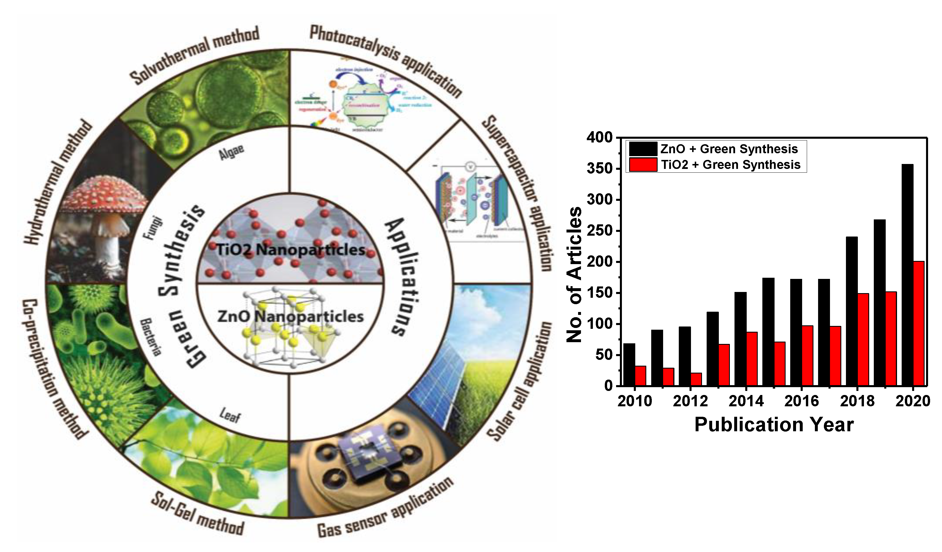
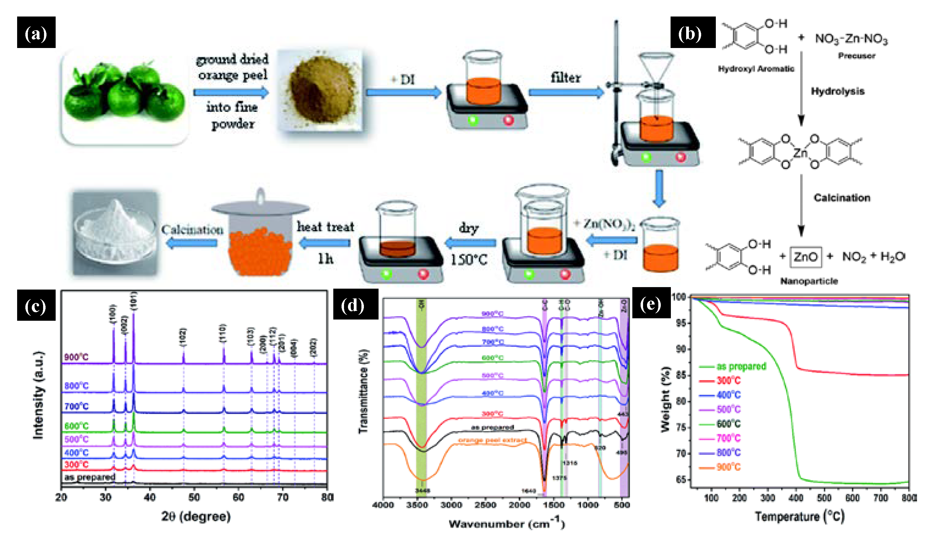
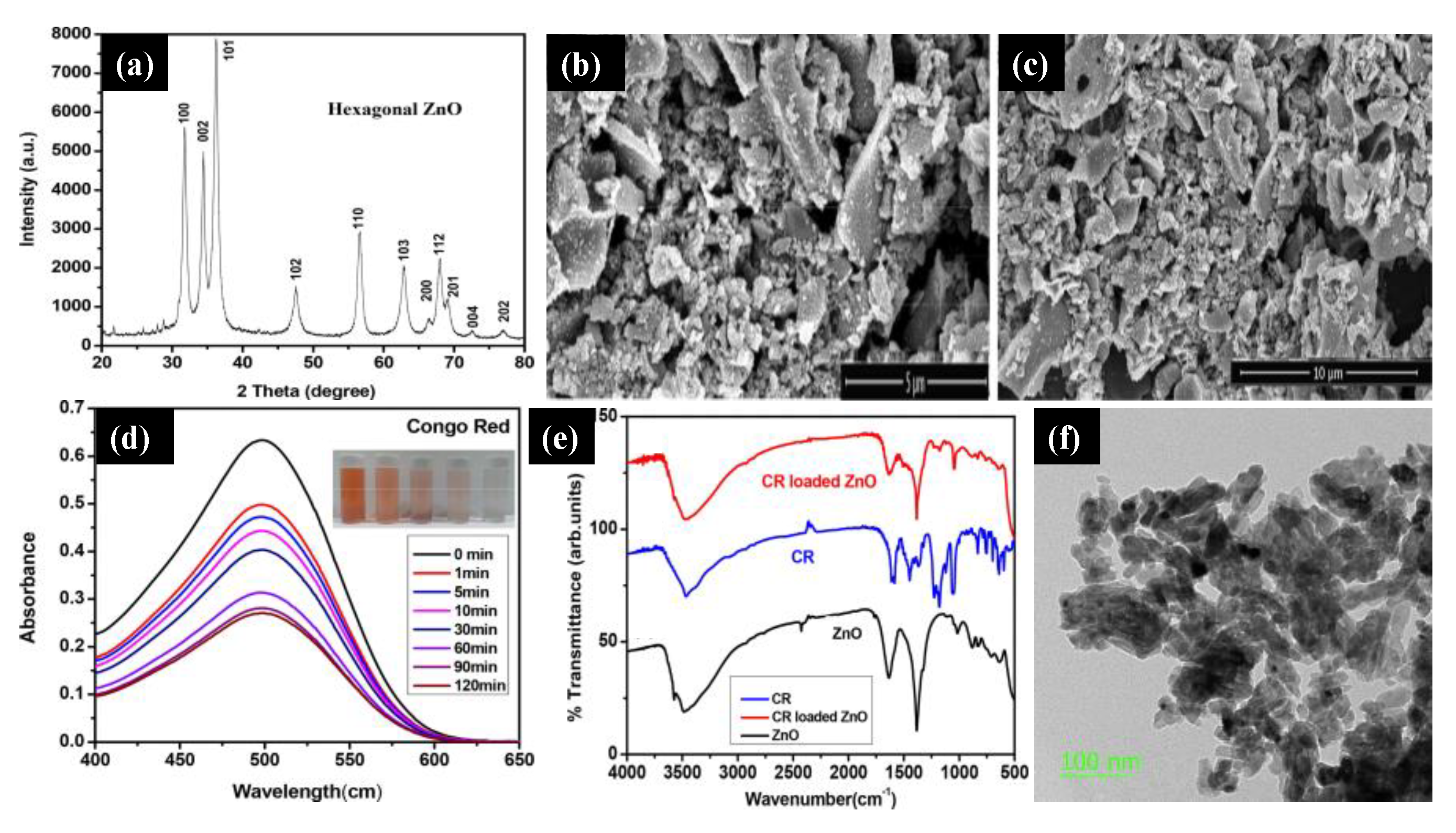
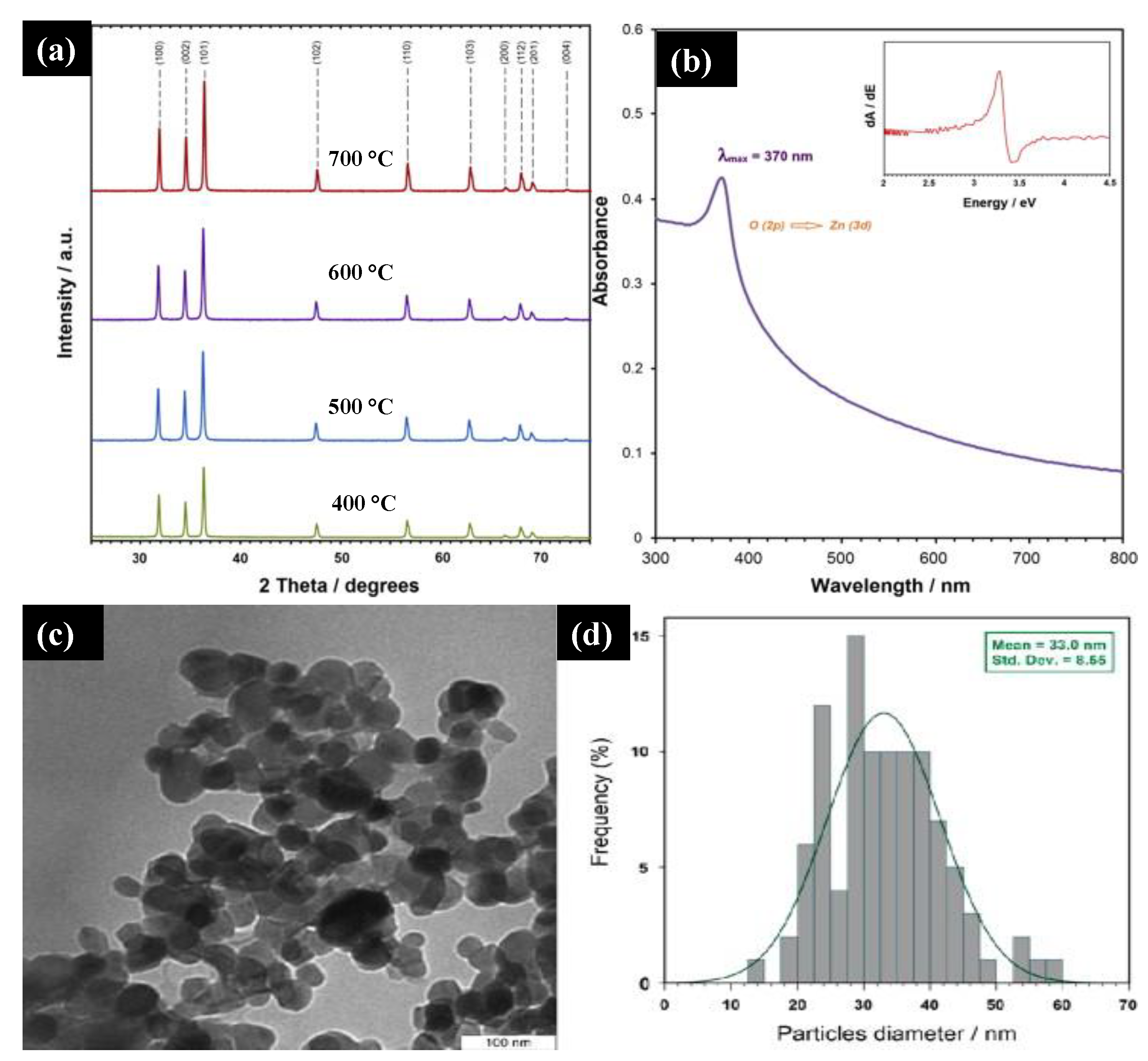
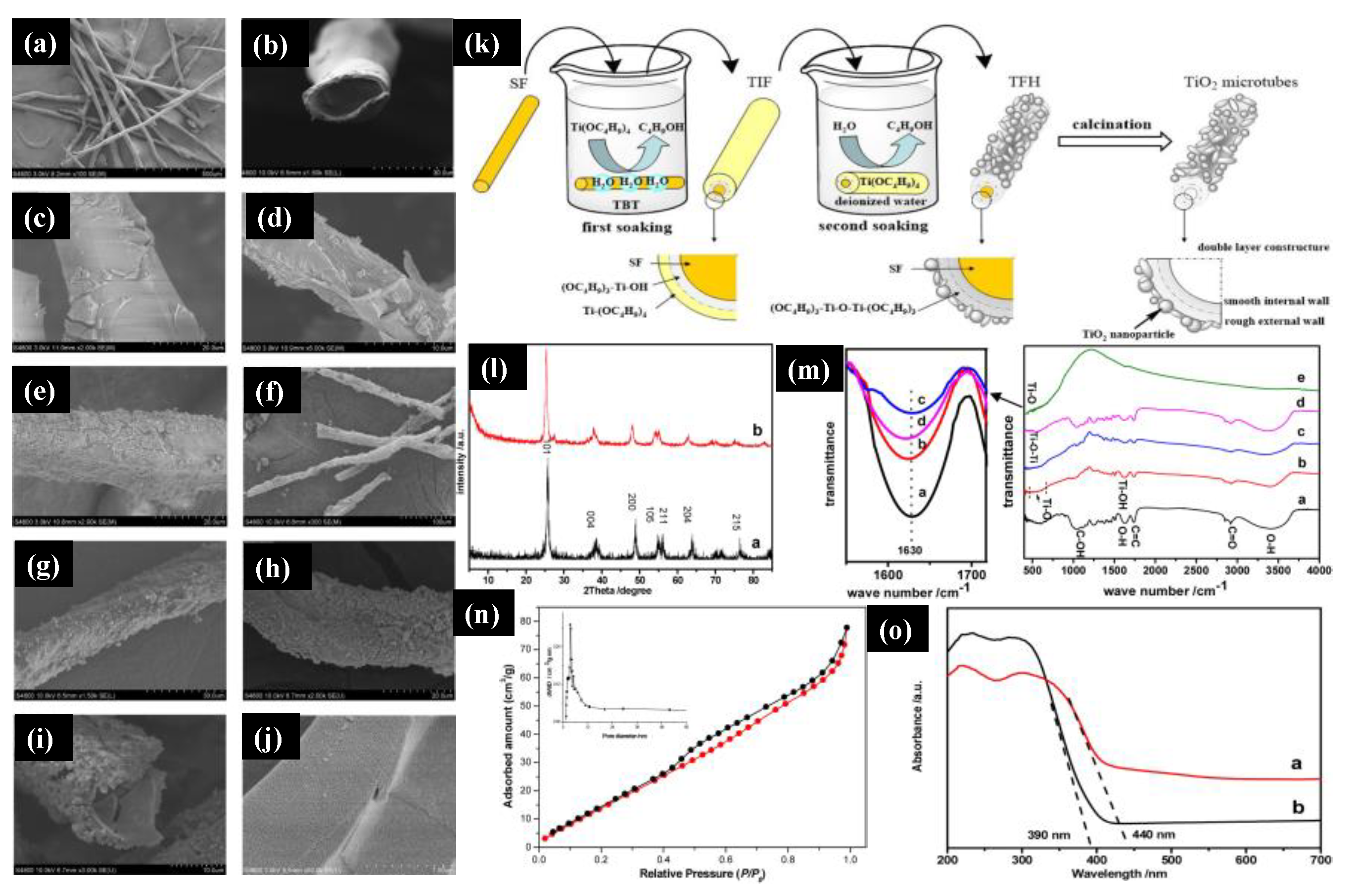
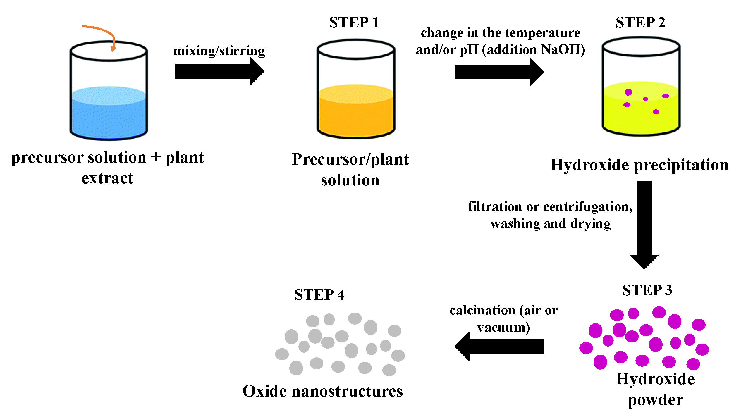
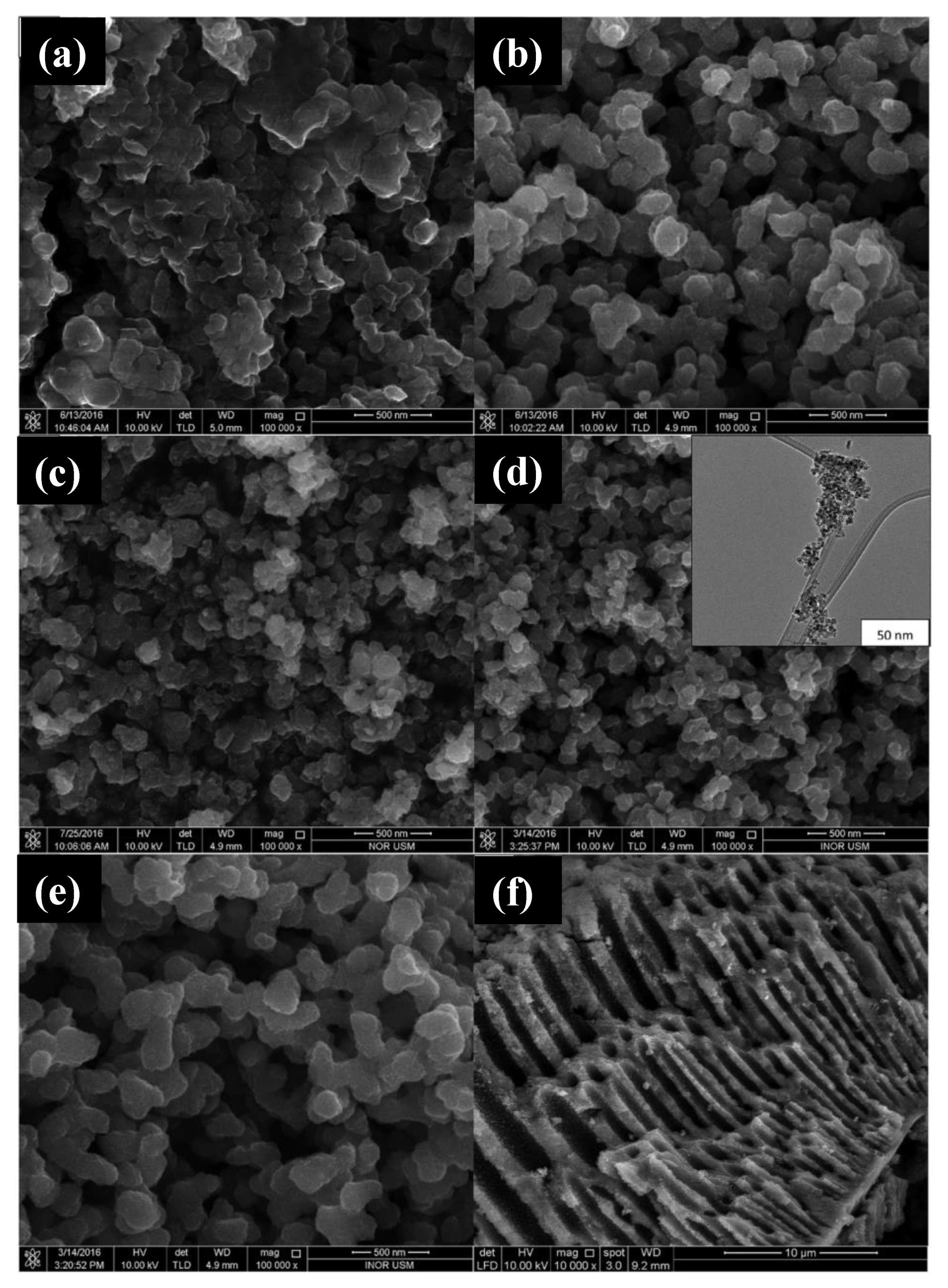
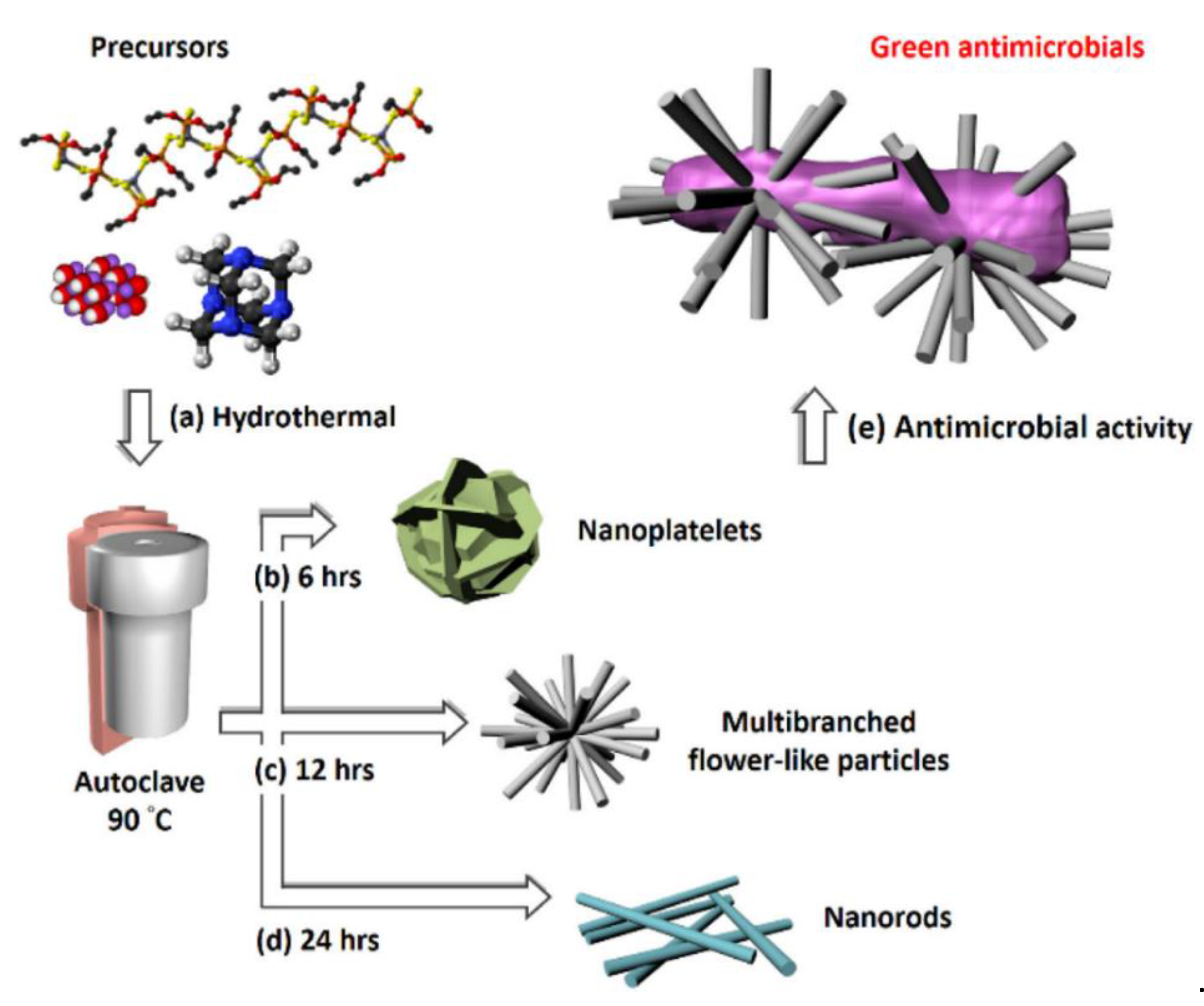
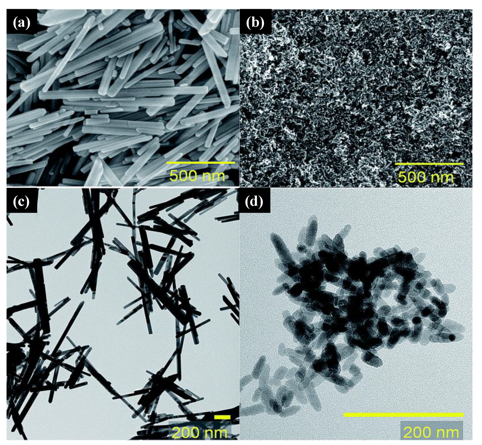
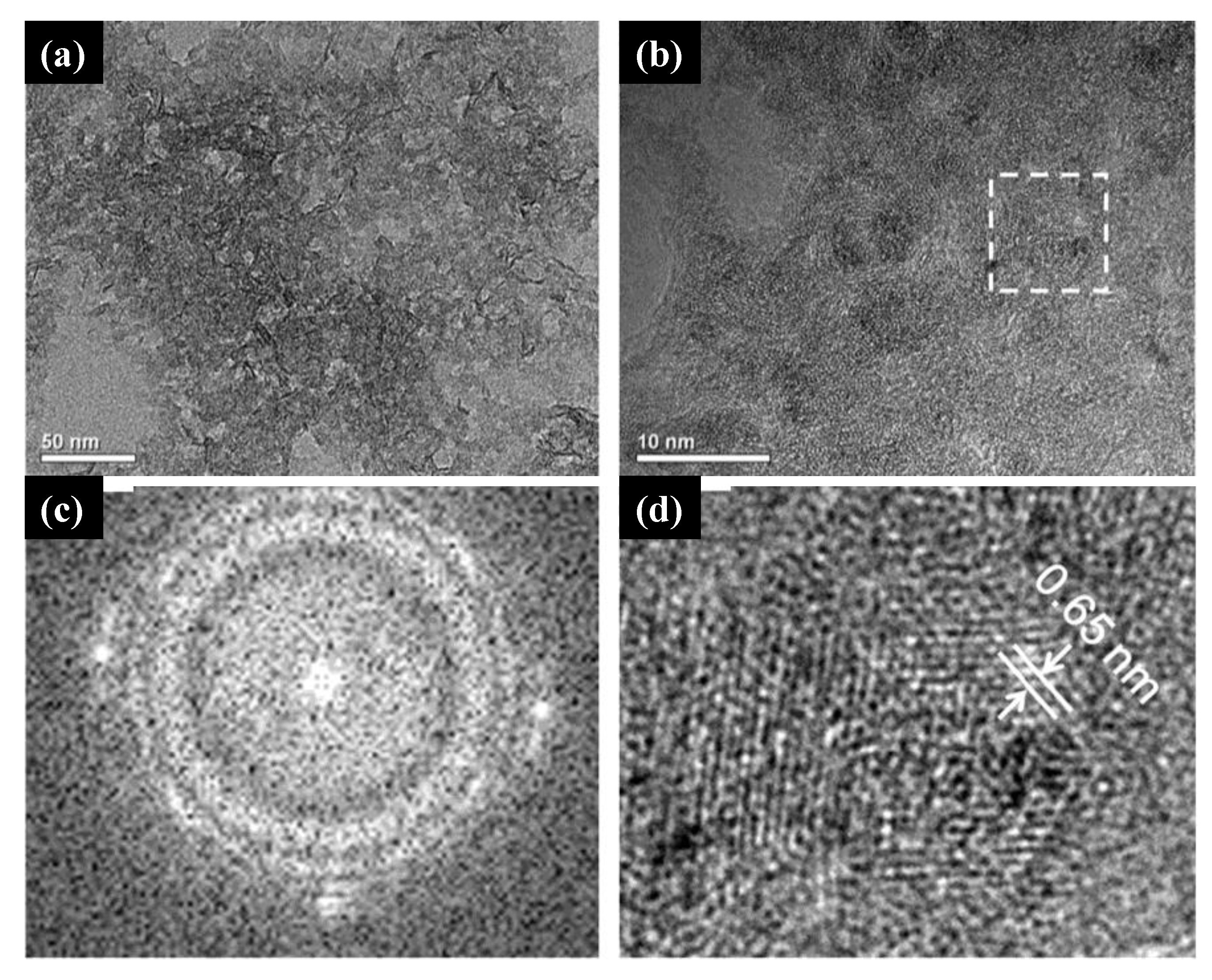
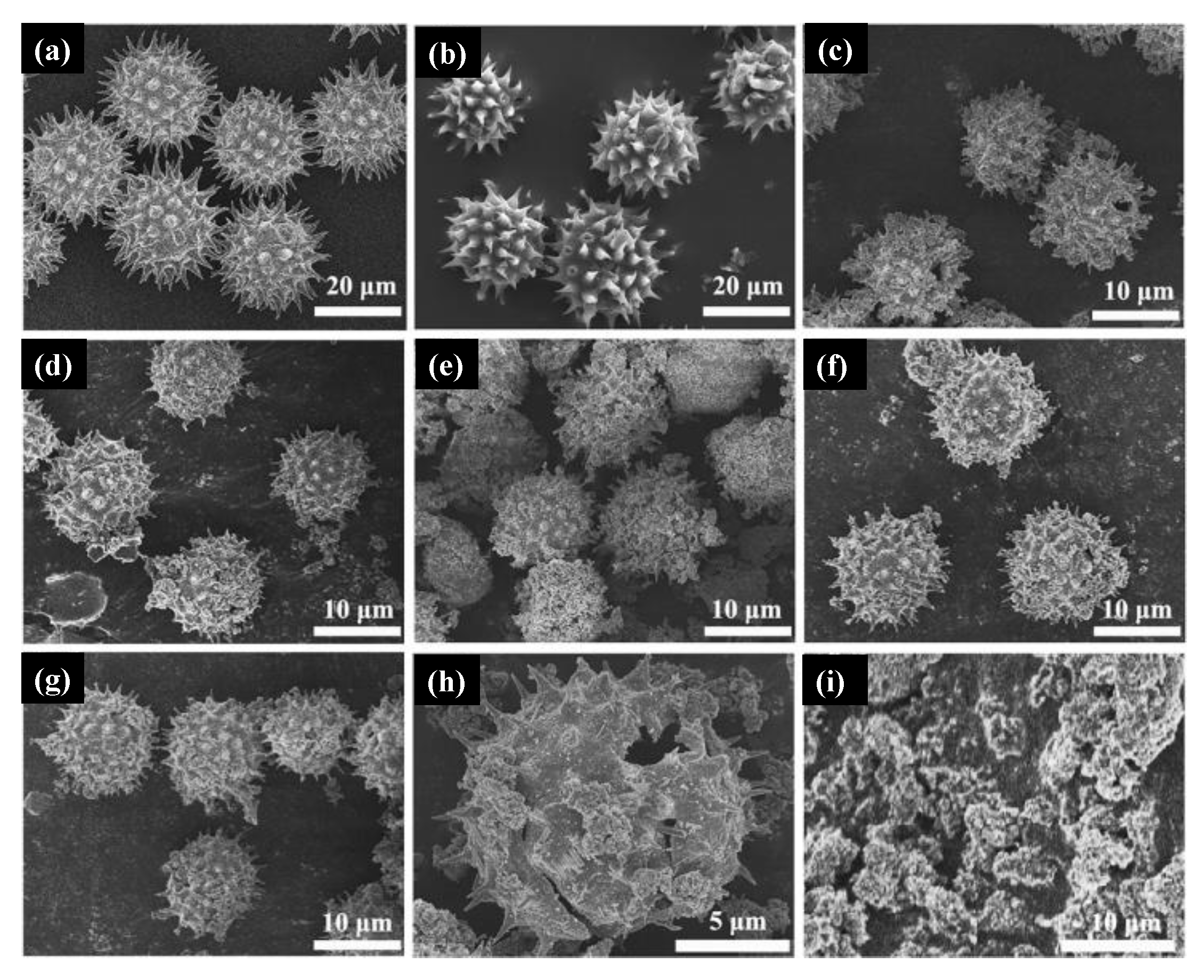
Table 1.
Classification of five different types of “gels” essential to a material’s sol-gel synthesis [35].
Table 1.
Classification of five different types of “gels” essential to a material’s sol-gel synthesis [35].
| Type of Gel | Bonding | Source | Gel Schematic |
|---|---|---|---|
| Colloidal | Particles connected by Van der Waals or hydrogen bonding | Metal oxides or hydroxide sols |  |
| Metal-oxane polymer | Inorganic polymers connected via covalent or intermolecular bonding | Hydrolysis or condensation of metal alkoxides, e.g., SiO2 from tetramethyl orthosilicate |  |
| Metal complex | Weakly interconnected metal complexes | Concentrated metal complex solution, e.g., aqueous metal citrate or ethanolic metal urea often form resins or glassy solids rather than gels |  |
| Polymer complex I in situ polymerizable complex (“Pechini” method) | Organic polymers interconnected by covalent or coordinate bonding | Polyesterification between polyhydroxy alcohol (e.g., ethylene glycol) and carboxylic acid with metal complex (e.g., metal-citrate) |  |
| Polymer complex II coordinating and crosslinking polymers | Organic polymers interconnected by coordinate and intermolecular bonding | Coordinating polymer (e.g., alginate) and metal salt solution (typically aqueous) |  |
Table 2.
Biosynthesis approaches to synthesize ZnO NPs from different plant sources.
| Morphology/Shape | Plant Source | Zinc Precursor | Reference |
|---|---|---|---|
| Quasi-spherical | Agathosma betulina | Zinc nitrate hexahydrate | [43] |
| Hexagonal | Allium sativum, Allium cepa, and Petroselinum crispum | Zinc nitrate hexahydrate | [44] |
| Spherical | Aloe barbadensis miller | Zinc nitrate solution | [45] |
| Spherical, oval, and hexagonal | Aloe vera | ZnSO4 | [46] |
| Not Reported | Aloe vera | Zinc nitrate, sodium hydroxide | [47] |
| Hexagonal | Anchusa italic | Zinc acetate dehydrate | [48] |
| Hexagonal | Anisochilus carnosus | Zinc nitrate hexahydrate | [49] |
| Spherical | Artocarpus gomezianus | Zinc nitrate hexahydrate | [50] |
| Spherical | Aspalathus linearis | ZnNO3, ZnCl2, and Zn-ammonium hydrate | [51] |
| Spherical | Azadirachta indica | Zinc nitrate hexahydrate | [52] |
| Various morphologies | Azadirachta indica | Zinc nitrate hexahydrate | [53] |
| Rod-shaped | Black tea | Zinc acetate dehydrate | [54] |
| Spherical | Boswellia ovalifoliolata | Zinc nitrate solution | [55] |
| Spherical and granular | Calotropis procera | Zinc acetate dehydrate | [56] |
| Hexagonal | Caralluma fimbriata | Zinc nitrate hexahydrate | [57] |
| Nano-flowers | Carica papaya | Zinc nitrate | [58] |
| Hexagonal | Carica papaya | Zinc nitrate solution | [59] |
| Flower-shaped NPs | Carissa edulis | Zinc nitrate hexahydrate | [60] |
| Spherical | Cassia fistula | Zinc nitrate hexahydrate | [61] |
| Spherical | Citrus aurantifolia | Zinc acetate dehydrate | [62] |
| Pyramid-like | Citrus aurantifolia | Zinc acetate dehydrate | [63] |
| Hexagonal | Coffee | Zinc acetate dehydrate | [64] |
| Polyhedron | Corymbia citriodora | Zinc nitrate solution | [65] |
| NPs | Heritiera fomes and Sonneratia apetala | Zinc chloride | [66] |
| NPs | Jacaranda mimosifolia | Zinc gluconate hydrate | [67] |
| Spherical and hexagonal | L. leschenaultiana | Zinc acetate dehydrate | [68] |
| Spherical | Limonia acidissima L. | Zinc nitrate solution | [69] |
| NR | Mimosa pudica | Zinc acetate dehydrate | [64] |
| Needle-like | Nephelium lappaceum | Zinc nitrate hexahydrate | [70] |
| Spherical | Nephelium lappaceum L. | Zinc nitrate hexahydrate | [71] |
| Hexagonal | Ocimum basilicum L. var. purpurascens | Zinc nitrate hexahydrate | [72] |
| Spherical | Parthenium hysterophorous | Zinc nitrate solution | [73] |
| Spherical and hexagonal | Parthenium hysterophorus L. | Zinc nitrate solution | [74] |
| Spherical | Phyllanthus niruri | Zinc nitrate solution | [75] |
| Triangular | Physalis alkekengi L. | Zinc contaminated soil | [76] |
| Spherical and hexagonal | Plectranthus amboinicus | Zinc nitrate solution | [77] |
| Rod-shaped | Plectranthus amboinicus | Zinc nitrate hexahydrate | [78] |
| Spherical | Polygala tenuifolia | Zinc nitrate hexahydrate | [79] |
| Spherical | Pongamia pinnata | Zinc nitrate hexahydrate | [80] |
| Spherical | Rosa canina | Zinc nitrate solution | [81] |
| Columnar | Sedum alfredii | ZnSO4 | [82] |
| Hexagonal | Solanum nigrum | Zinc nitrate solution | [83] |
| Spherical | Terminalia chebula | Zinc nitrate hexahydrate | [84] |
| Spherical | Tribulus terrestris | Zinc oxide powder | [85] |
| Not Reported | Trifolium pratense | ZnO powder | [86] |
| Spherical | Vitex negundo | Zinc nitrate hexahydrate | [87] |
| Spherical | Vitex trifolia L. | Zinc nitrate hexahydrate | [88] |
Table 3.
Green sol-gel synthesis approach for the preparation of TiO2 nanostructures.
| Material | Green Synthesis | Reactant | Reference |
|---|---|---|---|
| TiO2 | Lagenaria siceraria and Pithecellobium dulce leaf | Titanium tetraisopropoxide, isopropanol, acetic acid, and ethanol | [92] |
| TiO2 | A. altissima leaf extracts | Titanic acid and water | [93] |
| TiO2 | Leaf extract of L. siceraria | Titanium (IV)-isopropoxide, ammonia, glacial acetic acid, and ethanol | [94] |
| TiO2 | Jatropha curcas L. | TiCl4, ammonia | [95] |
| TiO2 | Acanthophyllum laxiusculum SchimanCzeika roots | Titanium tetraisopropoxide(TTIP), 2-propanol, nitric acid | [96] |
| TiO2 | Pista Shell, Tamarind Seed, Corn Pith | Isopropanol, titanium tetraisopropoxide, acetic acid (2%) | [97] |
| TiO2 | Green tea extract powder | Titanium isopropoxide, isopropanol | [98] |
Table 4.
Green co-precipitation syntheses of ZnO nanostructures mediated by plant extract.
| Morphology | Zinc Precursor | Plant/Part Used | Role of Biocomponents | Reference |
|---|---|---|---|---|
| Spherical NPs | Zinc acetate dihydrate | Azadirachta indica/leaf | Reducing and stabilizing agent | [111] |
| Flower-like structures | Zinc acetate dehydrate | Laurus nobilis/leaf | Reducing and capping agent | [112] |
| Quasi-spherical NPs | Hydrated zinc nitrate | Agathosma betulina/leaf | Oxidizing/reducing chemical agent | [43] |
| Spherical NPs | Zinc nitrate hexahydrate | Tabernaemontana divaricata/leaf | Capping and chelating agents | [113] |
| Spherical NPs | Zinc acetate dihydrate | Carica papaya/leaf | Capping and reducing agent | [114] |
| Spherical NPs | Zinc nitrate hexahydrate | NepheliumLappaceum L./fruit | Natural ligation agent | [71] |
| Nanoflowers | Zinc chloride | Typha latifolia. L./leaf | Reduction agent | [115] |
| Flower-like structure | Zinc acetate | Kalopanax septemlobus/barks | Reducing and capping agent | [116] |
| Rod-like and spherical NPs | Zinc nitrate | Bambusa vulgaris and Artabotrys hexapetalu/leaf | Reducing agent | [117] |
| Flower-like structure, cauliflower-like, and nanoflowers | Zinc nitrate hexahydrate | Zea mays, Artocarpus heterophyllus, Punica granatum/husk, peel and peel | Capping agent | [118] |
| Flower-like nanostructures | Zinc acetate | Cyanometra ramiflora/leaf | Reducing agent | [119] |
| Spherical NPs | Hydrated zinc chloride | Broccoli/leaf | Capping agent | [120] |
| Nanoflowers | Zinc acetate | Citrullus lanatus/rind | Reducing agent | [121] |
| Tetrameric structured NPs | Zinc nitrate hexahydrate | Amomum longiligulare/fruit | Reducing and stabilizing agent | [122] |
| Hexagonal NPs | Zinc nitrate tetrahydrate | Andrographis paniculate/leaf | Reducing agent | [123] |
| Leaf-like nanostructures | Zinc nitrate | Rubus coreanus/fruit | Reducing and capping agent | [124] |
Table 5.
Parameters involved in the process of dye photodegradation by TiO2 and ZnO nanostructures synthesized by green routes. The dyes methylene blue, rhodamine B, malachite green, methyl orange, Congo red and crystal violet are indicated by the abbreviations MB, RhB, MG, MO, CR and CV, respectively.
| Morphology/Material Phase | Green Synthesis Method | Radiation | Dye | Dye Concentration | Catalyst Concentration | pH | Exposure Time (min) | Efficiency (%) | Reference |
|---|---|---|---|---|---|---|---|---|---|
| Nanorods/TiO2 anatase | Microwave | Artificial sunlight | RhB | 10−5 M (50 mL) | 50 mg/10 mL | dye pH | 120 | >98% | [182] |
| Spherical NPs/TiO2 anatase | Co-precipitation * | Solar light | Coralline red | 5 mg/100 mL | 10 mg/100 mL | 8 | 140 | 92.17% | [103] |
| Meso/macro-porous nanostructures | Precipitation * | Sunlight | MB | 20 mg/L | - | dye pH | 135 | >95% | [183] |
| Spherical NPs/TiO2 anatase | Continuous ultrasonic stimulation | UV light | MB | 10 ppm | 1 g/L | dye pH | 150 | 92.5% | [184] |
| Elliptical NPs/TiO2 anatase | Sol-gel * | Visible light | MB, fuchsine, CV, and Rhodamine 6G | 10 mg/L (100 mL) | 0.1 g | dye pH | 180 | 88–99% | [185] |
| Dandelion-like structures/TiO2 anatase-rutile | Hydrothermal | UV light | MB | 10 mg/L (40 mL) | 20 mg–40 mL | dye pH | 650 | >97% | [132] |
| Spherical NPs/TiO2 rutile | Microwave * | Sunlight | MB, MO, CV, and alizarin red | 1 mg/100 mL | 10 mg/50 mL | dye pH | 360 | 77.3–92.5% | [186] |
| Spherical NPs/TiO2 anatase | Co-precipitation * | UV light | Reactive Green-19 | 6.7 mM | 0.030 g/100 mL | 3.5, 10.5 | 120 | 98.88% | [94] |
| Non-spherical NPs/TiO2 anatase | Sol-gel | UV light | MO | 20 ppm (100 mL) | 0.1 g | dye pH | 150 | 94% | [187] |
| Spherical structures/TiO2 anatase | Precipitation | Sunlight | MB | 6–40 ppm (200 mL) | 0.05–0.40 g | dye pH | 120 | 100% | [110] |
| Nanoflowers/ZnO wurtzite | Co-precipitation * | UV light | MB, MG, CR, and Eosin Y | 15 mg/L | 5 mg/L | dye pH | 90 | 100% | [188] |
| Spherical and hexagonal prismatic NPs and nanosheets/ZnO wurtzite | Co-precipitation | Visible light | RhB | 5 × 10⁻⁶ M (2 mL) | 1 mg/2 mL | dye pH | 120 | 75–84% | [125] |
| Leaf-like structures/ZnO wurtzite | Co-precipitation * | Dark condition | MG | 10 mg/L (90 mL) | 5 mg/90 mL | dye pH | 240 | ~80% | [124] |
| Hollow microspheres/ZnO wurtzite | Hydrothermal * | UV light | MG | 10 mg/L (200 mL) | 1 g/L | 5 | 60 | ~90% | [189] |
| Nanosheets/ZnO wurtzite | Hydrothermal | UV light | MB | 1 × 10⁻⁵ M (200 mL) | 0.05 g/200 mL | dye pH | 50 | 99.2% | [190] |
| Flower-like nanostructures/ZnO wurtzite | Co-precipitation * | UV light | MB | 50 µM | 0.5–1.0 g/ml | dye pH | 30 | 97.5% | [116] |
| Quasi-hexagonal NPs/ZnO wurtzite | Microwave * | UV light | MB | 5 mg/L (100 mL) | 30 mg (100 mL) | 3–11 | 40 | 70–100% | [191] |
| Spherical NPs/ZnO wurtzite | Mechanically assisted metathesis reaction | UV light | MB | 10 mg/L (100 mL) | 10 mg/100 mL | dye pH | 120 | 78% | [192] |
| Hollow nanospheres/ZnO wurtzite | Hydrothermal | UV and visible light | CR | 20 ppm (50 mL) | 25 mg/50 mL | 5–9 | 90 | 99% | [193] |
| Spongy cave-like structures/ZnO wurtzite | Solution combustion * | UV and sun light | MB | 5 ppm (100 mL) | 50 mg/100 mL | 2–12 | 90 | ~18–100% | [194] |
| Mysorepak-like, canine teeth, hollow pyramid, and aggregated hexagonal/ZnO wurtzite | Combustion * | UV light | MB | 5–20 ppm (100 mL) | 50–200 mg/100 mL | 2–12 | 150 | 85–100% | [195] |
| Quasi-spherical NPs/ZnO wurtzite | Co-precipitation * | Sunlight | MB | 1 × 10⁻⁵ M (100 mL) | 100 mg/100 mL | dye pH | 90 | 100% | [113] |
| Spherical NPs/ZnO wurtzite | Sol-gel | Visible light | Direct blue 129 | 20 mg/L (50 mL) | 30–60 mg/50 mL | dye pH | 105 | ~60–95% | [196] |
| Spherical NPs/ZnO wurtzite | Hydrothermal * | UV light | MB and MO | 10 mg/L (50 mL) | 1–30 mg/50 mL | dye pH | 50–60 | 96.6–98.2% | [197] |
| Spherical and rod-like NPs/ZnO wurtzite | Co-precipitation * | Visible light | RhB | 10 mg/L | 1 g | dye pH | 180 | 88–92% | [117] |
| Sponge-like structures/ZnO wurtzite | Combustion * | UV and sun light | MB and MG | 5–25 ppm (100 mL) | 50–200 mg/100 mL | 2–12 | 120–150 | ~10–100% | [198] |
| NPs/ZnO wurtzite | Combustion * | UV light | Rose Bengal | 2–40 ppm (250 mL) | 20–80 mg/250 mL | 6–10 | 90 | ~70–90% | [199] |
| Porous NPs/ZnO wurtzite | Solution combustion * | UV e sun light | MB | 5–20 ppm (100 mL) | 50–200 mg/100 mL | 2–12 | 120 | ~3–99% | [50] |
| Spherical NPs/ZnO wurtzite | Combustion * | UV light | CR | 10–40 ppm (250 mL) | 20–80 mg/250 mL | 6–10 | 60 | 70–90% | [200] |
| Hexagonal NPs/ZnO wurtzite | Solution combustion * | UV and sun light | MB | 5–20 ppm (100 mL) | 100 mg/100 mL | 3–12 | 40–50 | 90–100% | [201] |
| Nanoflowers/ZnO wurtzite | Co-precipitation * | Sunlight | RhB | 10 µM (100 mL) | 20 mg/100 mL | dye pH | 200 | 98% | [201] |
| Sphere-like nanostructures | Co-precipitation * | UV light | MB | 50 µM | 50 mg | dye pH | 210 | 98.6% | [119] |
| Spherical NPs/ZnO wurtzite | Hydrothermal * | UV light | MB and MO | 10 mg/L (50mL) | 5–30 mg/50 mL | dye pH | 50 | 96.6–98.2% | [202] |
| Spherical NPs/ZnO wurtzite | Sol-gel * | UV light | MB, MO, and Methyl red | 5–25 ppm (50 mL) | 50 mg/50 mL | dye pH | 35 | 60–100% | [197] |
| Spherical morphology/ZnO wurtzite | Solvothermal * | Visible light | MB | 20 mg/L (100 mL) | 100 mg/100 mL | 4.0–9.8 | 30 | 7.6–96.8% | [203] |
| Nanoflowers/ZnO wurtzite | Co-precipitation | Sunlight | Indigo carmine | - | 50 mg | dye pH | 120 | 83% | [204] |
| Quasi-spherical NPs/ZnO wurtzite | Combustion * | UV light | MB | 5 × 10⁻⁵ M (30 mL) | 20 mg/30 mL | 5–12 | 120 | 40–96% | [115] |
| Plates, bullets, flower, prismatic tip, and closed pinecone nanostructures/ZnO wurtzite | Solution combustion * | UV and sun light | MB | 10 ppm (250 mL) | 60 mg/250 mL | dye pH | 60 | 85–92% | [205] |
The * in the second column indicates that the green syntheses were aided by plant extracts.
Publisher’s Note: MDPI stays neutral with regard to jurisdictional claims in published maps and institutional affiliations. |
© 2021 by the authors. Licensee MDPI, Basel, Switzerland. This article is an open access article distributed under the terms and conditions of the Creative Commons Attribution (CC BY) license (https://creativecommons.org/licenses/by/4.0/).
Share and Cite
MDPI and ACS Style
Gonçalves, R.A.; Toledo, R.P.; Joshi, N.; Berengue, O.M. Green Synthesis and Applications of ZnO and TiO2 Nanostructures. Molecules 2021, 26, 2236. https://doi.org/10.3390/molecules26082236
AMA Style
Gonçalves RA, Toledo RP, Joshi N, Berengue OM. Green Synthesis and Applications of ZnO and TiO2 Nanostructures. Molecules. 2021; 26(8):2236. https://doi.org/10.3390/molecules26082236
Chicago/Turabian StyleGonçalves, Rosana A., Rosimara P. Toledo, Nirav Joshi, and Olivia M. Berengue. 2021. "Green Synthesis and Applications of ZnO and TiO2 Nanostructures" Molecules 26, no. 8: 2236. https://doi.org/10.3390/molecules26082236