Next Article in Journal
miRNAs, Mesenchymal Stromal Cells and Major Neoplastic and Inflammatory Skin Diseases: A Page Being Written: A Systematic Review
Next Article in Special Issue
Upregulated Expression of ERBB2/HER2 in Multiple Myeloma as a Predictor of Poor Survival Outcomes
Previous Article in Journal
GPR19 Coordinates Multiple Molecular Aspects of Stress Responses Associated with the Aging Process
Previous Article in Special Issue
Simultaneously Targeting Two Coupled Signalling Molecules in the Mesenchymal Stem Cell Support Efficiently Sensitises the Multiple Myeloma Cell Line H929 to Bortezomib
 
 
ijms-logo
Article Menu

Article Menu

Article

Polymorphisms within Autophagy-Related Genes as Susceptibility Biomarkers for Multiple Myeloma: A Meta-Analysis of Three Large Cohorts and Functional Characterization

by
Esther Clavero
1,†,
José Manuel Sanchez-Maldonado
2,3,†,
Angelica Macauda
4,
Rob Ter Horst
5,6,
Belém Sampaio-Marques
7,
Artur Jurczyszyn
8,
Alyssa Clay-Gilmour
9,10,
Angelika Stein
4,
Michelle A. T. Hildebrandt
11,
Niels Weinhold
12,13,
Gabriele Buda
14,
Ramón García-Sanz
15,
Waldemar Tomczak
16,
Ulla Vogel
17,
Andrés Jerez
18,
Daria Zawirska
19,
Marzena Wątek
20,21,
Jonathan N. Hofmann
22,
Stefano Landi
23,
John J. Spinelli
24,25,
Aleksandra Butrym
26,27,
Abhishek Kumar
28,29,
Joaquín Martínez-López
30,
Sara Galimberti
14,
María Eugenia Sarasquete
15,
Edyta Subocz
31,
Elzbieta Iskierka-Jażdżewska
32,
Graham G. Giles
33,34,35,
Malwina Rybicka-Ramos
36,
Marcin Kruszewski
37,
Niels Abildgaard
38,
Francisco García Verdejo
39,
Pedro Sánchez Rovira
39,
Miguel Inacio da Silva Filho
40,
Katalin Kadar
41,
Małgorzata Razny
42,
Wendy Cozen
43,
Matteo Pelosini
44,
Manuel Jurado
1,3,45,
Parveen Bhatti
46,47,
Marek Dudzinski
48,
Agnieszka Druzd-Sitek
49,
Enrico Orciuolo
14,
Yang Li
5,50,
Aaron D. Norman
10,51,
Jan Maciej Zaucha
52,
Rui Manuel Reis
53,54,
Miroslaw Markiewicz
48,
Juan José Rodríguez Sevilla
55,
Vibeke Andersen
56,
Krzysztof Jamroziak
57,
Kari Hemminki
58,59,
Sonja I. Berndt
22,
Vicent Rajkumar
60,
Grzegorz Mazur
61,
Shaji K. Kumar
60,
Paula Ludovico
7,
Arnon Nagler
62,
Stephen J. Chanock
22,
Charles Dumontet
63,
Mitchell J. Machiela
22,
Judit Varkonyi
64,
Nicola J. Camp
65,
Elad Ziv
66,
Annette Juul Vangsted
67,
Elizabeth E. Brown
68,
Daniele Campa
23,
Celine M. Vachon
10,
Mihai G. Netea
5,69,
Federico Canzian
4,
Asta Försti
70,71 and
Juan Sainz
2,3,72,*
add Show full author list remove Hide full author list
1
Hematology Department, Virgen de las Nieves University Hospital, 18012 Granada, Spain
2
Genomic Oncology Area, GENYO, Centre for Genomics and Oncological Research, Pfizer/University of Granada/Andalusian Regional Government, PTS, 18016 Granada, Spain
3
Instituto de Investigación Biosanataria IBs, Granada, 18014 Granada, Spain
4
Genomic Epidemiology Group, German Cancer Research Center (DKFZ), 69120 Heidelberg, Germany
5
Department of Internal Medicine and Radboud Center for Infectious Diseases, Radboud University Medical Center, 6525 GA Nijmegen, The Netherlands
6
CeMM Research Center for Molecular Medicine of the Austrian Academy of Sciences, 1090 Vienna, Austria
7
Life and Health Sciences Research Institute (ICVS), School of Medicine, University of Minho, 4710-057 Braga, Portugal
8
Plasma Cell Dyscrasias Center, Department of Hematology, Jagiellonian University Medical College, 31-066 Kraków, Poland
9
Department of Biostatistics and Epidemiology, Arnold School of Public Health, University of South Carolina, Greenville, SC 29208, USA
10
Division of Epidemiology, Department of Health Sciences Research, Mayo Clinic, Rochester, MN 55902, USA
11
Department of Lymphoma–Myeloma, Division of Cancer Medicine, The University of Texas MD Anderson Cancer Center, Houston, TX 77030, USA
12
Myeloma Institute, University of Arkansas for Medical Sciences, Little Rock, AR 72205, USA
13
Department of Internal Medicine V, University of Heidelberg, 69120 Heidelberg, Germany
14
Haematology Unit, Department of Clinical and Experimental Medicine, University of Pisa/AOUP, 56126 Pisa, Italy
15
Diagnostic Laboratory Unit in Hematology, University Hospital of Salamanca, IBSAL, CIBERONC, Centro de Investigación del Cáncer-IBMCC (USAL-CSIC), 37007 Salamanca, Spain
16
Department of Hematooncology and Bone Marrow Transplantation, Medical University of Lublin, 20-059 Lublin, Poland
17
National Research Centre for the Working Environment, DK-2100 Copenhagen, Denmark
18
Department of Hematology, Experimental Hematology Unit, Vall d’Hebron Institute of Oncology (VHIO), University Hospital Vall d’Hebron, 08035 Barcelona, Spain
19
Department of Hematology, University Hospital, 30-688 Kraków, Poland
20
Holycross Medical Oncology Center, 25-735 Kielce, Poland
21
Institute of Hematology and Transfusion Medicine, 00-791 Warsaw, Poland
22
Division of Cancer Epidemiology and Genetics, National Cancer Institute, National Institutes of Health, Bethesda, MD 20892, USA
23
Department of Biology, University of Pisa, 56126 Pisa, Italy
24
Division of Population Oncology, BC Cancer, Vancouver, BC V5Z 4E6, Canada
25
School of Population and Public Health, University of British Columbia, Vancouver, BC V6T 1Z4, Canada
26
Department of Cancer Prevention and Therapy, Wroclaw Medical University, 50-367 Wroclaw, Poland
27
Alfred Sokolowski Specialist Hospital in Walbrzych Oncology Support Centre for Clinical Trials, 58-309 Walbrzych, Poland
28
Institute of Bioinformatics, International Technology Park, Bangalore 560066, India
29
Manipal Academy of Higher Education (MAHE), Manipal 576104, India
30
Hospital 12 de Octubre, Complutense University, CNIO, CIBERONC, 28041 Madrid, Spain
31
Department of Hematology, Military Institute of Medicine, 04-141 Warsaw, Poland
32
Department of Hematology, Medical University of Lodz, 90-419 Lodz, Poland
33
Cancer Epidemiology Division, Cancer Council Victoria, Melbourne, VIC 3004, Australia
34
Centre for Epidemiology and Biostatistics, School of Population and Global Health, The University of Melbourne, Melbourne, VIC 3010, Australia
35
Precision Medicine, School of Clinical Sciences at Monash Health, Monash University, Clayton, VIC 3168, Australia
36
Department of Hematology, Specialist Hospital No. 1 in Bytom, Academy of Silesia, Faculty of Medicine, 40-055 Katowice, Poland
37
Department of Hematology, University Hospital No. 2, 85-168 Bydgoszcz, Poland
38
Department of Hematology, Odense University Hospital, DK-5000 Odense, Denmark
39
Department of Medical Oncology, Complejo Hospitalario de Jaén, 23007 Jaén, Spain
40
Division of Molecular Genetic Epidemiology, German Cancer Research Center (DKFZ), Im Neuenheimer Feld 580, D-69120 Heidelberg, Germany
41
St Johns Hospital, 62769 Budapest, Hungary
42
Department of Hematology, Rydygier Hospital, 31-826 Cracow, Poland
43
Division of Hematology/Oncology, Department of Medicine, School of Medicine, Department of Pathology, School of Medicine, Susan and Henry Samueli College of Health Sciences, Chao Family Comprehensive Cancer Center, University of California at Irvine, Irvine, CA 92697, USA
44
U.O. Dipartimento di Ematologia, Azienda USL Toscana Nord Ovest, 57124 Livorno, Italy
45
Department of Medicine, University of Granada, 18012 Granada, Spain
46
Cancer Control Research, BC Cancer, Vancouver, BC V5Z 4E6, Canada
47
Program in Epidemiology, Public Health Sciences Division, Fred Hutchinson Cancer Research Center, Seattle, WA 98109, USA
48
Department of Hematology, Institute of Medical Sciences, College of Medical Sciences, University of Rzeszow, 35-310 Rzeszow, Poland
49
Department of Lymphoproliferative Diseases, Maria Skłodowska Curie National Research Institute of Oncology, 02-781 Warsaw, Poland
50
Centre for Individualised Infection Medicine (CiiM) & TWINCORE, Joint Ventures between the Helmholtz-Centre for Infection Research (HZI) and the Hannover Medical School (MHH), 30625 Hannover, Germany
51
Genetic Epidemiology and Risk Assessment Program, Mayo Clinic Comprehensive Cancer Center, Division of Biomedical Statistics and Informatics, Department of Health Sciences Research, Mayo Clinic, Rochester, MN 55902, USA
52
Department of Hematology and Transplantology, Medical University of Gdansk, 80-210 Gdansk, Poland
53
Life and Health Sciences Research Institute (ICVS), School of Health Sciences, University of Minho, 4710-057 Braga, Portugal and ICVS/3B’s-PT Government Associate Laboratory, 4710-057 Braga/Guimarães, Portugal
54
Molecular Oncology Research Center, Barretos Cancer Hospital, Barretos 14784-400, Brazil
55
Department of Hematology, Hospital del Mar, 08003 Barcelona, Spain
56
Molecular Diagnostics and Clinical Research Unit, Institute of Regional Health Research, University Hospital of Southern Denmark, DK-6200 Aabenraa, Denmark
57
Department of Hematology, Transplantology and Internal Medicine, Medical University of Warsaw, 02-097 Warsaw, Poland
58
Division of Cancer Epidemiology, German Cancer Research Center (DKFZ), Im Neuenheimer Feld 280, 69120 Heidelberg, Germany
59
Faculty of Medicine and Biomedical Center in Pilsen, Charles University in Prague, 30605 Pilsen, Czech Republic
60
Division of Hematology, Department of Internal Medicine, Mayo Clinic, Rochester, MN 55902, USA
61
Department of Internal Diseases, Occupational Medicine, Hypertension and Clinical Oncology, Wroclaw Medical University, 50-368 Wroclaw, Poland
62
Hematology Division, Chaim Sheba Medical Center, Tel Hashomer 52621, Israel
63
UMR INSERM 1052/CNRS 5286, University of Lyon, Hospices Civils de Lyon, 69008 Lyon, France
64
Semmelweis University, 1083 Budapest, Hungary
65
Division of Hematology, Huntsman Cancer Institute, University of Utah, Salt Lake City, UT 84112, USA
66
Department of Medicine, University of California San Francisco Helen Diller Family Comprehensive Cancer Center, San Francisco, CA 94143, USA
67
Department of Hematology, Rigshospitalet, Copenhagen University, DK-2100 Copenhagen, Denmark
68
Department of Pathology, Heersink School of Medicine, The University of Alabama at Birmingham, Birmingham, AL 35294, USA
69
Department for Immunology & Metabolism, Life and Medical Sciences Institute (LIMES), University of Bonn, 53115 Bonn, Germany
70
Division of Pediatric Neurooncology, German Cancer Research Center (DKFZ), German Cancer Consortium (DKTK), 69120 Heidelberg, Germany
71
Hopp Children’s Cancer Center (KiTZ), 69120 Heidelberg, Germany
72
Department of Biochemistry and Molecular Biology I, University of Granada, 18071 Granada, Spain
*
Author to whom correspondence should be addressed.
These authors contributed equally to this work.
Int. J. Mol. Sci. 2023, 24(10), 8500; https://doi.org/10.3390/ijms24108500
Submission received: 4 March 2023 / Revised: 10 April 2023 / Accepted: 21 April 2023 / Published: 9 May 2023
(This article belongs to the Special Issue Multiple Myeloma: Molecular Mechanism and Targeted Therapy)

Abstract

:

Simple Summary

We investigated the influence of autophagy-related variants in modulating Multiple Myeloma (MM) risk through a meta-analysis of germline genetic data on 234 autophagy-related genes from three independent study populations including 13,387 subjects of European ancestry (6863 MM patients and 6524 controls) and examined the functional mechanisms behind the observed associations. We identified SNPs within the six CD46, IKBKE, PARK2, ULK4, ATG5, and CDKN2A loci associated with MM risk and observed that their effect on disease risk was mediated by specific subsets of immune cells, as well as vitamin D3-, MCP-2-, and IL20-dependent mechanisms.

Abstract

Multiple myeloma (MM) arises following malignant proliferation of plasma cells in the bone marrow, that secrete high amounts of specific monoclonal immunoglobulins or light chains, resulting in the massive production of unfolded or misfolded proteins. Autophagy can have a dual role in tumorigenesis, by eliminating these abnormal proteins to avoid cancer development, but also ensuring MM cell survival and promoting resistance to treatments. To date no studies have determined the impact of genetic variation in autophagy-related genes on MM risk. We performed meta-analysis of germline genetic data on 234 autophagy-related genes from three independent study populations including 13,387 subjects of European ancestry (6863 MM patients and 6524 controls) and examined correlations of statistically significant single nucleotide polymorphisms (SNPs; p < 1 × 10⁻⁹) with immune responses in whole blood, peripheral blood mononuclear cells (PBMCs), and monocyte-derived macrophages (MDM) from a large population of healthy donors from the Human Functional Genomic Project (HFGP). We identified SNPs in six loci, CD46, IKBKE, PARK2, ULK4, ATG5, and CDKN2A associated with MM risk (p = 4.47 × 10⁻⁴−5.79 × 10⁻¹⁴). Mechanistically, we found that the ULK4rs6599175 SNP correlated with circulating concentrations of vitamin D3 (p = 4.0 × 10⁻⁴), whereas the IKBKErs17433804 SNP correlated with the number of transitional CD24+CD38+ B cells (p = 4.8 × 10⁻⁴) and circulating serum concentrations of Monocyte Chemoattractant Protein (MCP)-2 (p = 3.6 × 10⁻⁴). We also found that the CD46rs1142469 SNP correlated with numbers of CD19+ B cells, CD19+CD3 B cells, CD5+IgD cells, IgM cells, IgDIgM cells, and CD4CD8 PBMCs (p = 4.9 × 10⁻⁴−8.6 × 10⁻⁴) and circulating concentrations of interleukin (IL)-20 (p = 0.00082). Finally, we observed that the CDKN2Ars2811710 SNP correlated with levels of CD4+EMCD45RO+CD27 cells (p = 9.3 × 10⁻⁴). These results suggest that genetic variants within these six loci influence MM risk through the modulation of specific subsets of immune cells, as well as vitamin D3, MCP-2, and IL20-dependent pathways.

1. Introduction

Multiple myeloma (MM) is a relatively common and incurable hematological malignancy arising from post–germinal mature B cells and it is characterized by the presence of proliferating plasma cells in the bone marrow that secrete specific monoclonal immunoglobulin (also called M-protein) [1]. Given the exacerbated production of immunoglobulin, MM patients often suffer from a concomitant decrease in normal immunoglobulins [2] that causes immune dysfunction, increases susceptibility to opportunistic infections, and impacts on disease severity and prognosis [3,4].
Although the high production of monoclonal immunoglobulins occurring in MM invariably results in the presence of a high amount of unfolded or misfolded proteins that might be toxic for MM cells in the bone marrow, recent studies demonstrated that plasma cells are able to exploit molecular pathways to protect themselves from damage caused by toxic proteins [5]. These molecular pathways include the activation of unfolded protein response (UPR) [6,7,8], heat protein chaperones [9], aggresome formation [10], and the induction of cellular autophagy [5]. Autophagy is a lysosome-dependent catabolic degradation process by which cells remove toxic aggregated cytosolic proteins and malfunctioning organelles. It has been well-documented that autophagy not only is an autonomous mechanism that modulates cell homeostasis at basal level, but it is also involved in B-cell development and proliferation [11], cell survival [12,13], apoptosis [14,15], tumorigenesis, anti-tumoral immune responses [16], and resistance to chemotherapeutic agents. In this regard, a growing number of studies have suggested that autophagy shapes anti-tumoral immune responses by acting at multiple levels [17]. This includes the promotion of signals to activate phagocytosis of tumor cells [18,19], as well as signals to induce myeloid cell recruitment [20], MHC-class-I and -II presentation [21], B- and T-cell activation, development, maintenance [11,22], and self-tolerance [23]. Strikingly, experimental studies have demonstrated that the inhibition of autophagy enhances the sensitivity of MM cells to a number of anticancer agents and induces MM cell death [24,25]. Based on these findings, it has been suggested that autophagy could be targeted to treat MM [5,24,26] and recent studies and clinical trials have indeed confirmed the therapeutic implications of new and less toxic autophagy inhibitors in MM, used in combination with current anti-MM drugs [27,28,29].
In support of a possible role of autophagy in MM, it was reported that aberrant expression of autophagy-related genes is associated with cancer development [30] and that multiple activators of autophagy or specific autophagy-related genes are commonly found in cancer-associated regions [31]. Interestingly, it was also described that expression of autophagy-related genes influences the response to conventional treatments in MM [32,33,34] and, thereby, disease progression [35,36], arguing that they may be useful to predict disease risk [37]. However, despite these findings suggesting a key role of autophagy in the etiology of MM and the existence of a genetic component controlling this catalytic process in MM, so far only ULK4, ATG5, and CDKN2A polymorphisms have been suggested to have an impact on the risk of MM [38,39]. Therefore, it is vital to perform a systematic analysis of autophagy-related markers to identify new susceptibility variants for MM and validate those associations already reported.
In this context, the aim of this study was to comprehensively evaluate the impact of common genetic variation of 234 autophagy-related genes in determining the risk of developing MM. We also assessed the influence of the most promising markers on modulating immune responses in whole blood, peripheral blood mononuclear cells (PBMCs), and monocyte-derived macrophages (MDM) from a large population of healthy donors from the Human Functional Genomic Project (HFGP). Additionally, we measured the autophagy flux in an independent population.

2. Results

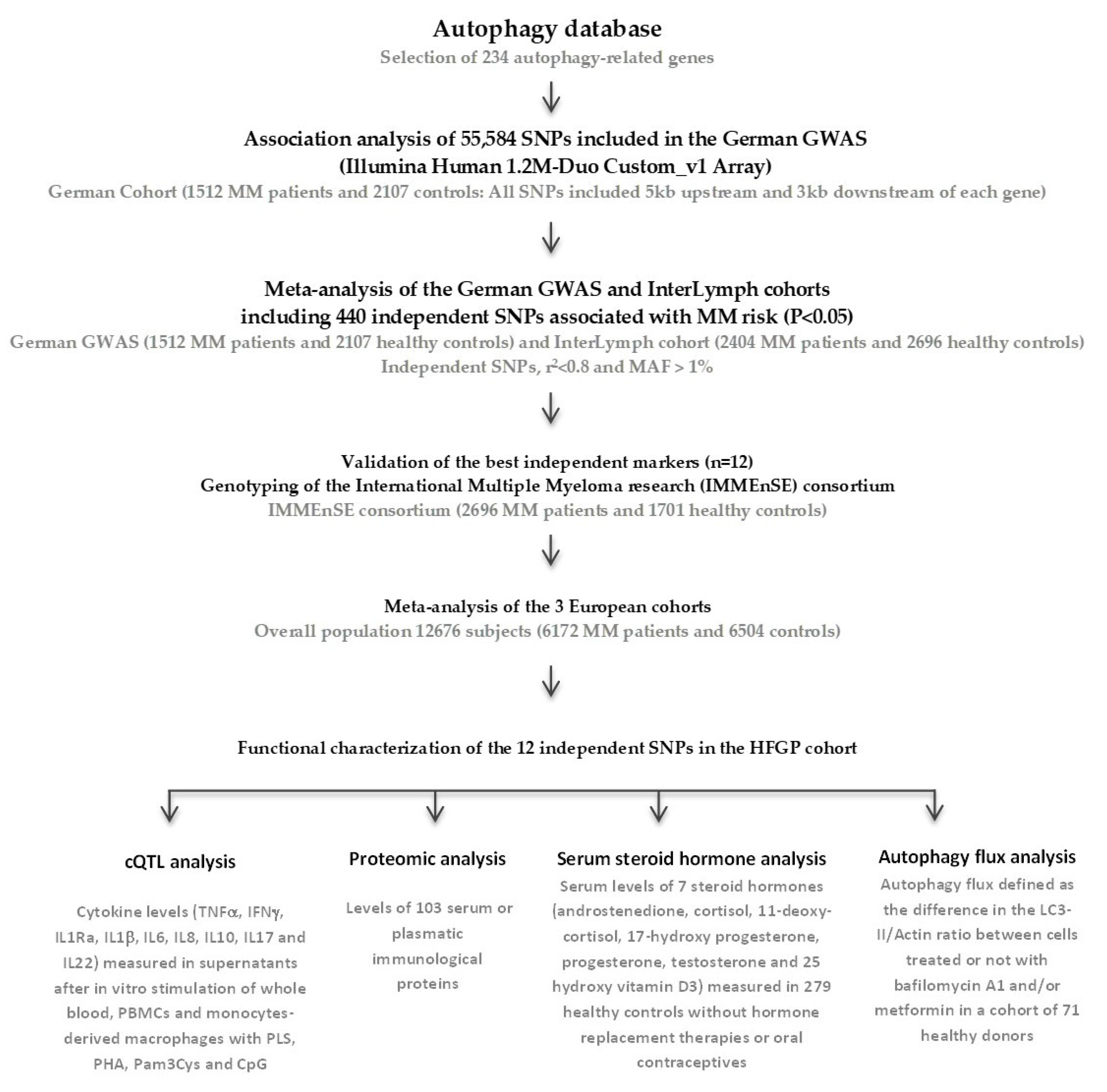
2.1. Association of Autophagy-Related Polymorphisms with the Risk of Developing MM

This study included 8719 individuals (3916 MM cases and 4803 controls) from the German GWAS consisting of 1512 MM patients and 2107 controls and the InterLymph MM GWAS that included 2404 MM cases and 2696 healthy controls. Selected polymorphisms showed no deviation from HWE (p < 0.001), either in the German GWAS or in the InterLymph MM GWAS. The association analysis of the German cohort showed that 440 independent SNPs (r2 < 0.1) were significantly associated with MM risk at p ≤ 0.05. The association of these SNPs with MM risk was then validated through meta-analysis with data from the InterLymph MM GWAS. The meta-analysis of these large independent studies confirmed the association of 12 genetic variants within the ATG5, CD46, CDKN2A, CTSD, HSPB8, IKBKE, PARK2, RPTOR, ULK4, and USP10 loci with MM risk (Table 1), with loci in ULK4 and ATG5 reaching the highest level of statistical significance.
Although the lack of significant heterogeneity between both study populations suggested that the association found for these 12 SNPs might represent true associations, we decided to replicate these findings in a third independent population ascertained through the IMMEnSE consortium that included 2696 MM cases and 1701 controls. Results are reported in Table 2. After correction for multiple testing (pBonferroni = 1.14 × 10⁻⁴), we could confirm the previously reported association for ULK4, ATG5, and CDKN2A polymorphisms. Importantly, we also found that the association of the IKBKErs17433804 SNP with the risk of MM was observed after multiple testing correction, which suggests that this gene could represent a new susceptibility locus for MM. In addition, although the associations were borderline significant after correction for multiple comparisons, we found of interest the associations of CD46 and PARK2 variants with the risk of developing MM. Although these associations need to be further validated, it was important to confirm that, with the exception of the CDKN2A, all the association signals included several SNPs (LD blocks), which reinforced the role of these loci in determining disease risk (Supplementary Figure S1A–F).

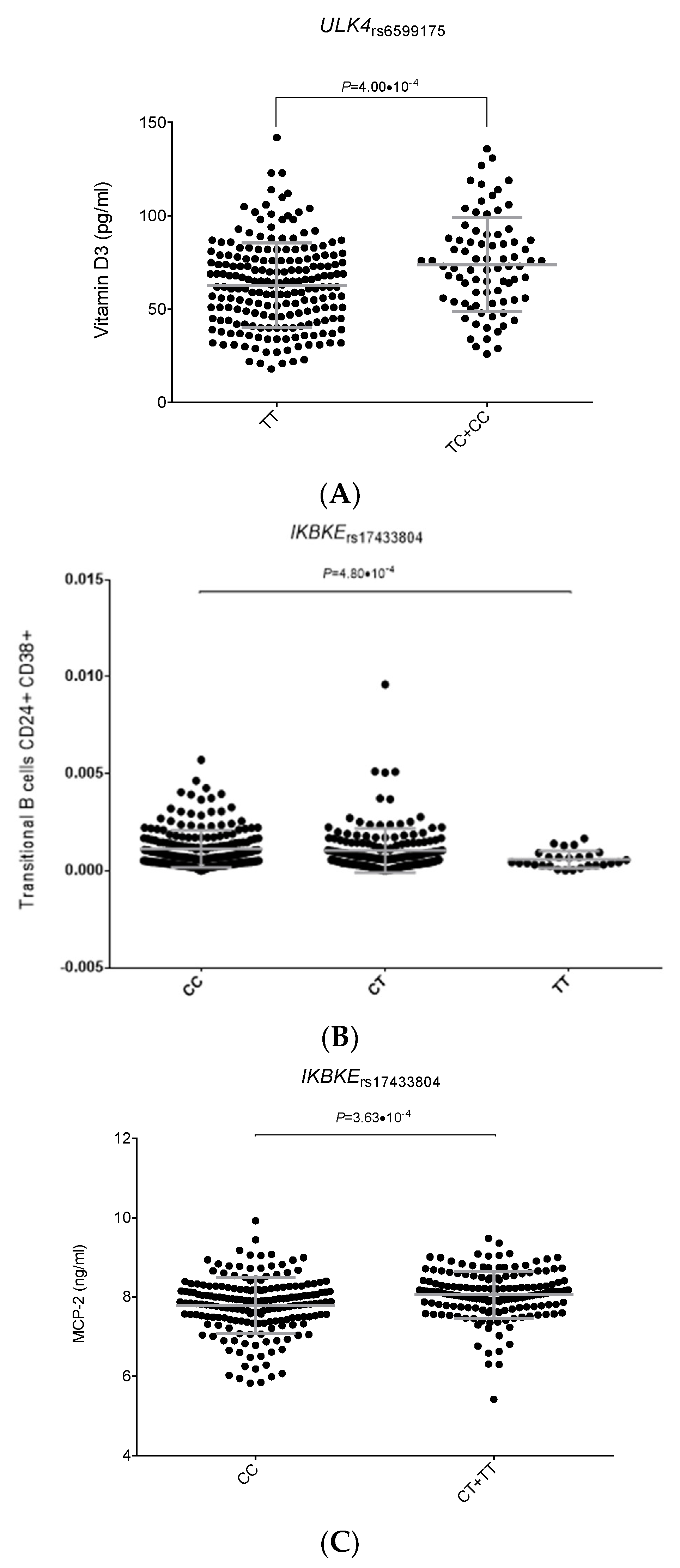
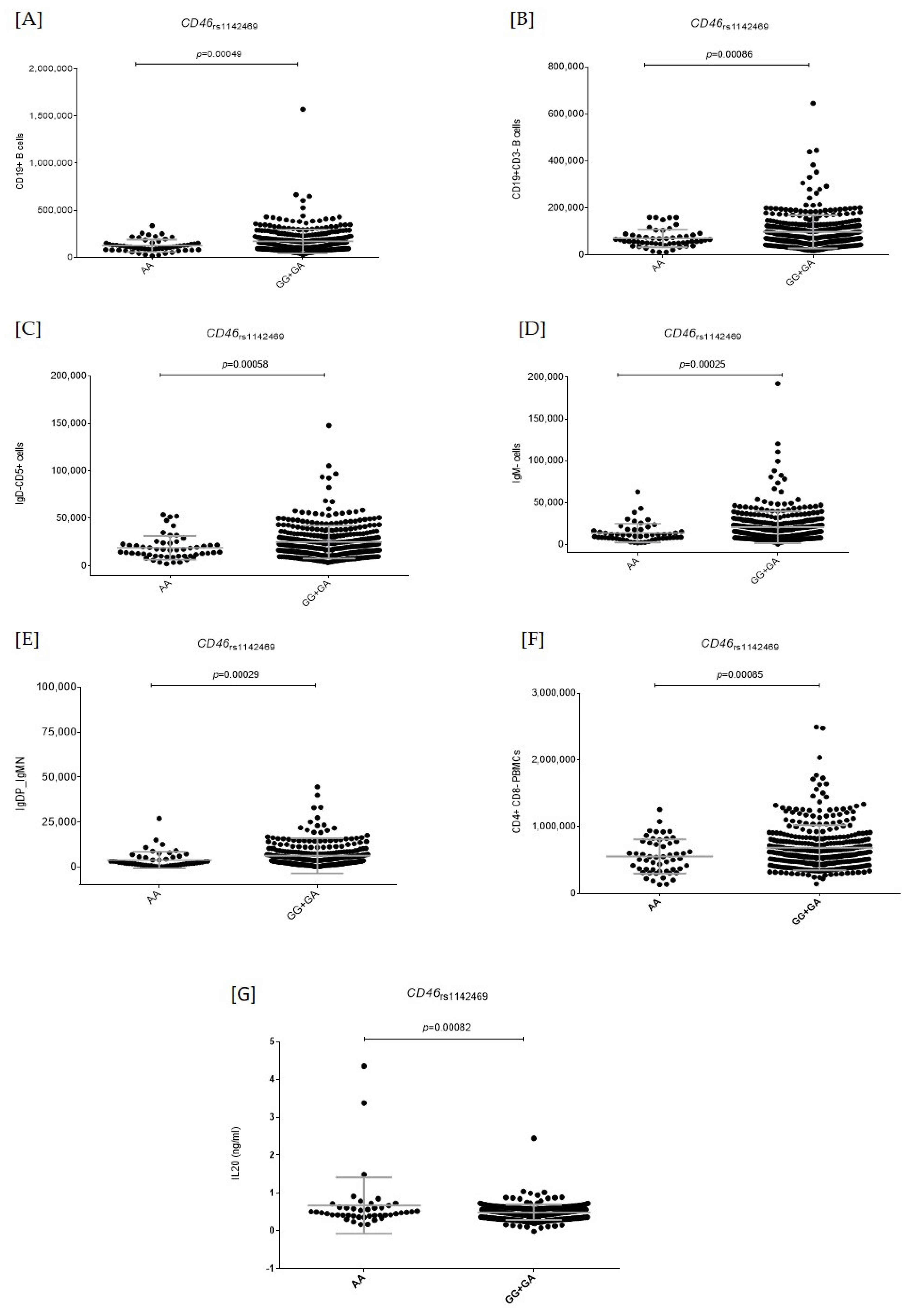
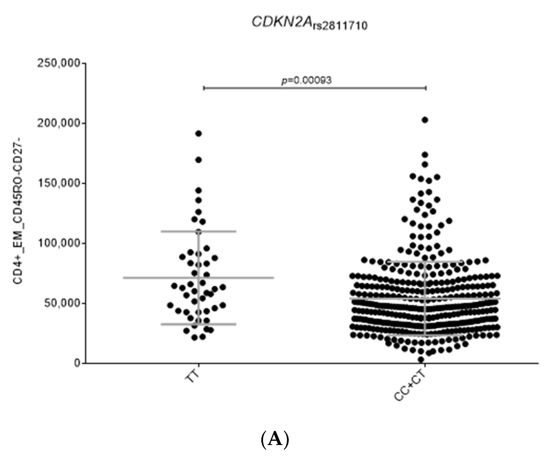
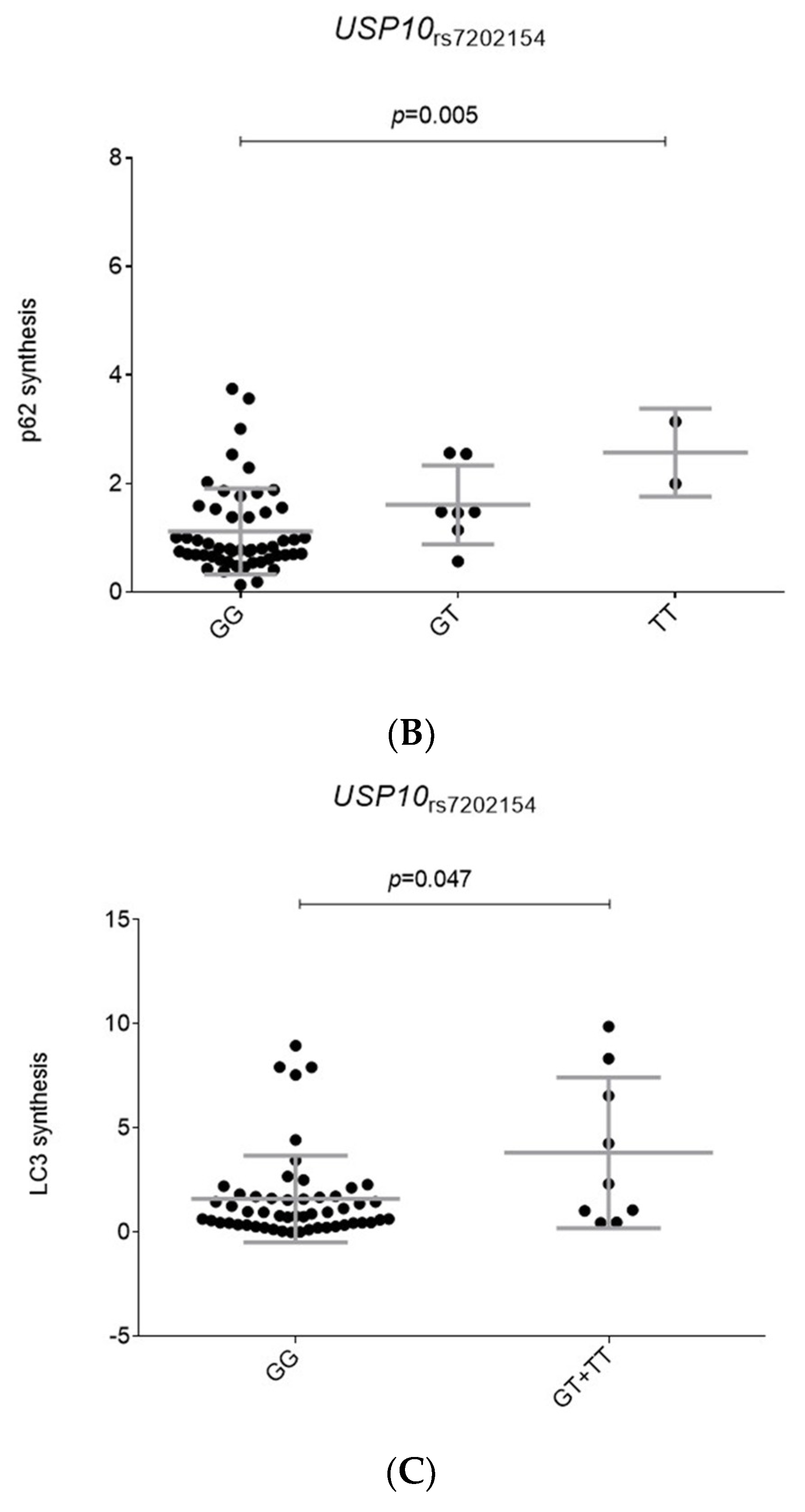
2.2. Functional Relevance of Autophagy-Related SNPs

Given the relatively strong association of the genetic variants within the CD46, IKBKE, PARK2, ULK4, ATG5, and CDKN2A loci on MM risk, we explored whether these variants could modulate host immune responses, serum steroid hormones, circulating immunological proteins, and blood-derived cell populations using data from the HFGP population. Although the genetic association of the USP10rs7202154 SNP with MM risk was modest, we decided to include it in the functional analysis due to the known role of USP proteins on the modulation of MM cell apoptosis.
Interestingly, we found a significant association between the ULK4rs6599175 SNP and circulating concentrations of vitamin D3 (p = 4.0 × 10⁻⁴, Figure 1A), which suggests a possible involvement of this SNP in modulating MM risk through a vitamin D-dependent mechanism. We also observed that the presence of the IKBKErs17433804C allele was associated with increased numbers of transitional CD24+CD38+ B cells (p = 4.8 × 10⁻⁴, Figure 1B), whereas carriers of the IKBKErs17433804C/C genotype showed decreased circulating serum concentrations of MCP-2 (p = 3.6 × 10⁻⁴; Figure 1C), a chemotactic molecule involved in the activation of multiple immune cells and linked to MM cell migration.
We also found that homozygous carriers of the CD46rs1142469A allele, which was associated with an increased risk of MM, had decreased numbers of CD19+ B cells, CD19+CD3 B cells, CD5+IgD cells, IgM cells, IgDIgM cells, and CD4CD8 PBMCs compared with those subjects carrying the G allele (p = 0.00025–0.00086; Figure 2A–F).
Moreover, we found that homozygous carriers of the CD46rs1142469A risk allele had increased circulating concentrations of IL20 compared with those subjects carrying the G allele (p = 0.00082; Figure 2G), which suggests a role of this angiogenic cytokine in modulating MM risk.
In addition, we observed that carriers of the CDKN2Ars2811710C allele had decreased levels of CD4+EMCD45RO+CD27 cells (p = 0.00093; Figure 3A), which constitutes a subset of memory T cells that does not require co-stimulation for T-cell receptors to display a high antigen recall response. Finally, we found that carriers of the USP10rs7202154G/G genotype had decreased synthesis of p62 and LC3-II compared to non-carriers (p = 0.005 and p = 0.047; Figure 3B,C). Given that p62 serves as a useful marker for the induction of autophagy, clearance of protein aggregates, and the inhibition of autophagy and LC3-II serves to track the binding of p62 and subsequent recruitment of autophagosomes, this result might suggest that this genetic variant could be involved in determining autophagy activation. Given that the association of the USP10 SNP with autophagy flux markers (LC3-II/Actin ratio) did not remain statistically significant after correction for multiple testing, additional studies are needed to confirm whether the weak association of this SNP with MM risk might be mediated by the regulation of the autophagy flux. No functional effect on host immune responses or autophagy flux was detected for the remaining selected SNPs, which suggests that the effect of these variants on MM risk is not mediated by these biological processes.

3. Discussion

To the best of our knowledge this is the first population-based case-control study comprehensively assessing the role of autophagy-related SNPs in modulating the risk of developing MM. After a comprehensive meta-analysis of three large independent studies, we found three novel significant associations between MM risk and SNPs within CD46, IKBKE, and PARK2, in addition to confirming the known associations with MM-susceptibility genes ULK4, ATG5, and CDKN2A loci.
As the most interesting finding, we found an association of the CD46rs1142469 SNP with the risk of developing MM. The CD46 locus is located on chromosome 1q32 and has been involved in pathogen recognition and in the differentiation of CD4+ cells into T-regulatory 1 cells that suppress immune responses by secreting IL10. The CD46rs1142469 SNP is an intronic variant that acts as eQTL for the CD46 in multiple tissues [40,41] and lymphoblastoid cell lines [41] and it is in LD with three neighboring SNPs that confirm the association signal of the CD46 locus with MM risk. Our functional results also showed that homozygous carriers of the CD46rs1142469A risk allele had decreased numbers of CD19+ B cells, CD19+CD3 B cells, CD5+IgD, IgM cells, IgD, IgM cells, and CD4, CD8 PBMCs, but also increased circulating concentrations of IL20. This latter observation is consistent with previous studies, which reported that IL20 is highly expressed in MM patients and correlates with levels of angiogenic cytokines and bone marrow microvascular density [42]. Although none of these functional results survived after correction for multiple testing and therefore need to be replicated, they are consistent with previous studies suggesting a relevant role of the CD46 locus in MM pathogenesis. Considering that the CD46rs1142469 SNP also alters binding sites for Hoxa7 [43], a transcription factor that is frequently dysregulated in MM patients and plays a role in modulating hematopoiesis and cell differentiation [44], it seems conceivable to suggest that the CD46rs1142469 SNP could influence the risk of developing MM by modulating not only CD46 expression, but also the number of specific subsets of B and T cells and IL20 and Hoxa7-mediated signaling pathways.
We also found, for the first time, that carriers of the IKBKErs17433804C allele had an increased risk of developing MM. Two additional neighboring SNPs in the same LD block confirmed the association of IKBKE SNPs with disease risk. The IKBKE locus is located at chromosome 1q32.1 and it acts as a modulator of multiple immune processes [45] including the necrosis factor (NF)-κB non-canonical pathway [46], a key process in MM pathogenesis. IKBKE expression is almost restricted to lymphoid tissue and Westra et al. reported that the IKBKErs17433804 SNP is an eQTL marker for IKBKE in peripheral blood (p = 9.65 × 10⁻⁹) [43]. In addition, it has been described that the IKBKE locus is frequently mutated in MM patients [47], which reinforces the idea of a role of this gene in determining MM risk. Interestingly, we also found that carriers of the IKBKErs17433804C allele had increased numbers of transitional CD24 + CD38+ B cells, which is a type of B cell linked to MM tumorigenesis. It has been reported that CD24+ cells are upregulated in MM and they have an increased expression of CXCR4 [48], which is a well-known chemokine receptor that plays a pivotal role in proliferation, invasion, dissemination, and drug resistance in MM [49]. However, CD24 expression has been also found to be associated to a more apoptotic and less tumorigenic phenotype by impaired capability to migrate and to create colonies as compared with CD24 MM cells [48]. These controversial results might indicate that CD24 might have a dual effect on MM by contributing to disease onset but also by preventing disease progression. In line with this hypothesis, Gross Even-Zohar et al. reported that patients having high expression of CD24+ MM cells had longer overall survival and progression free survival [50]. In addition, we found that carriers of the IKBKErs17433804C allele had decreased circulating serum concentrations of MCP-2, which has been linked to chemotaxis of myeloid and lymphoid cells, but also to other important processes such as autophagy, leukocyte behavior, cell adhesion and polarization, cell secretion, and cell survival of MM cells [51]. A recent study demonstrated that MCP-1, another chemokine from the MCP family, was increased in MM patients and correlated with clinical characteristics and enhanced angiogenesis in MM [52]. In line with these results, another study demonstrated that the inhibition of MCP-2, as well as its receptor CCR2, by neutralizing antibodies reduced migration of MM cells [53]. Altogether, these findings suggest that the IKBKE locus might influence MM risk by modulating absolute numbers of specific subsets of B-cells and MCP-2-mediated immune responses and MM cell migration. Nonetheless, it is also important to consider that the presence of this variant alters regulatory motifs for multiple transcription factors including AP-1, p300, TCF4, Myc, and PRMD1 [43]. These transcription factors which are highly expressed in MM [54,55] have been involved in B cell development and plasma cell differentiation, MM cell proliferation, cell survival, and drug resistance in the bone marrow microenvironment [56,57,58], and they even exhibit antitumoral activity in MM pathogenesis [54,59]. Even though the above reported results shed some light on the role of the IKBKE locus in MM, additional functional studies are still warranted to establish the exact mechanism by which the IKBKE locus is linked to MM onset and disease progression.
Another novel finding was the association between the PARK2rs1884158 SNP (and four neighboring SNPs) and the risk of MM. In addition to its role in autophagy, PARK2 is involved in modulating stress response, mitochondrial biogenesis, stability of G1/S cyclins [60], cell growth (acting as tumor suppressor), and mitophagy, where it purges damaged organelles from the vital mitochondrial network [61]. Although the PARK2 gene was associated with the risk of solid tumors [62], little is known about its role in MM. Our functional experiments did not show any positive correlation between the PARK2rs1884158 SNP and host immune responses and, to the best of our knowledge, no functional roles for this SNP have been reported in Haploreg or eQTL browsers. Therefore, additional studies are still warranted to replicate the association of the PARK2rs1884158 SNP with MM risk and, subsequently, to characterize its functional role in MM pathogenesis.
As well as the novel association of CD46, IKBKE, and PARK2 SNPs with MM risk, we could validate the association of SNPs within well-known MM susceptibility genes such as ULK4, ATG5, and CDKN2A [39,63]. The strongest association was found for the ULK4rs6599175 polymorphism within the ULK4 gene. Each copy of the ULK4rs6599175C allele increased the risk of developing MM by 31%. This result agreed with a previous study by Broderick et al. (2011) [63]. The ULK4 gene is located at chromosome 3p22.1 and encodes for a member of the unc-51-like serine/threonine kinase (STK) family that plays a role in modulating hypertension [64] and cardiologic disorders [65]. According to GTEx portal data and other eQTL studies [40], the ULK4rs6599175C allele strongly correlates with higher levels of ULK4 mRNA expression levels in multiple tissues including whole blood (p = 1.4 × 10−59). However, outside of mRNA expression levels, little is known about the functional role of this genetic variant to modulate the risk of MM. In this regard, we found that carriers of the ULK4rs6599175C risk allele correlated with increased serum concentrations of vitamin D3 in healthy donors, which might suggest that the effect of the ULK4rs6599175C allele to increase the risk of MM might be mediated by a vitamin D-dependent mechanism likely affecting cell differentiation and proliferation, angiogenesis, immune responses, and apoptosis either in normal or malignant tissues [66,67,68]. In addition, this SNP has been linked to monoclonal gammopathy of undetermined significance (MGUS), a non-cancerous precursor condition that precedes MM [69], suggesting that the ULK4rs6599175 SNP might exert its biological effect during the earliest stages of plasma cell dyscrasia progression from early to late-stage disease. Additional studies are required to decipher the interplay between the ULK4 locus and circulating vitamin D3 levels in the context of MM risk and progression.
On the other hand, the ATG5 is located on chromosome 6q21 and it has been related to autophagy vesicle formation and apoptosis [70]. A previous study reported the association between the ATG5rs9372120 SNP (a variant in strong LD with the rs2299864) and MM risk [39], but also MGUS [69], which suggests that the ATG5 locus might also exert its biological effect on the risk of developing MM by acting at early and non-neoplastic stages. Although we could not find any correlation between the ATG5rs2299864 SNP and host immune responses or autophagy flux, it was reported that this variant is located among histone marks for primary T helper naive and memory cells and primary T CD8+ naive cells from peripheral blood and that it disrupts regulatory motifs for several transcription factors including DMRT3, which has an altered methylation profile during early stages of myelomagenesis [71].
Finally, the CDKN2A gene is located on chromosome 9p21.3 and encodes for a cyclin-dependent kinase inhibitor 2A that has been involved in p53 stabilization, cell cycle arrest, and cell proliferation. Unlike the ULK4rs6599175 or ATG5rs2299864 SNPs, the CDKN2Ars2811710 SNP has not been linked to MGUS susceptibility, which suggests that the CDKN2Ars2811710 SNP might be involved in promoting the accumulation of tumorigenic MM cells rather than acting in earlier stages. In fact, several germline mutations within the CDKN2A have been consistently associated with MM predisposition [72], but also improved patient overall survival [73]. Interestingly, we found that carriers of the CDKN2Ars2811710C allele, which is associated with an increased risk of MM, had decreased numbers of CD4+EMCD45RO+ CD27 T memory cells, which are cells that display a high antigen recall response and were reported to be poorly represented in MM patients compared to controls [74]. In addition, the CDKN2Ars2811710 SNP was associated with the expression of CDKN2B and STK16 mRNAs in monocytes that are involved in the modulation of cell cycle arrest through the TFG signaling pathway [75]. The CDKN2Ars2811710 SNP maps among histone marks for primary T helper memory cells and primary T regulatory cells from peripheral blood and alters binding sites for key transcription factors such as HDAC2, HNF1, OTX, and p300 that play important roles in epigenetic repression and transcriptional regulation, cell cycle progression, and developmental events, and are even involved in influencing overall survival [76].
It is worth mentioning that this study has both strengths and limitations. The major strengths of our study were the comprehensive analysis of autophagy-related SNPs and the inclusion of three large independent European populations of European ancestry including a total of 12,676 participants. Furthermore, we comprehensively analyzed the impact of autophagy-related SNPs in modulating blood cell counts, steroid hormones, serum and plasma metabolites, and immune responses in a large study of healthy subjects ascertained through the HFGP. Another important strength of this study was the experimental analysis assessing the effect of autophagy SNPs in modulating the autophagy flux in PBMCs left untreated or treated with metformin or bafilomycin. This approach allowed us to investigate, for the first time, the impact of susceptibility loci for MM on the autophagy flux. A limitation of this study was its multicentric nature that placed inevitable limitations such as the impossibility of uniformly collected mutation and/or cytogenetic profiles for a significant proportion of patients analyzed. In addition, this study included a subset of German participants, which overlapped with the GWAS used for discovering the ULK4 MM susceptibility locus. Finally, given that all study participants included in this study were of European ancestry, we could not determine the impact of autophagy variants in other ethnic or ancestral populations. Additional studies using other ethnic and/or ancestry populations are now warranted to confirm the results described herein.

4. Materials and Methods

A workflow diagram of the study is included in Figure 4.

4.1. Study Populations

The discovery population consisted of 3169 subjects from GWAS conducted by the German-Speaking Multiple Myeloma Multicenter Study Group (GMMG), followed by meta-analysis with an independent GWAS on 5100 subjects by the International Lymphoma Epidemiology Consortium (InterLymph; https://epi.grants.cancer.gov/interlymph/; accessed on 12 February 2020). International Myeloma Working Group (IMWG) criteria were used by physicians to establish the diagnosis of MM [77,78]. Ethical approval for these respective studies was obtained from each participating institution and all participanting subjects provided written informed consent. The German GWAS coordinated by the University Clinic, Heidelberg (ISRCTN06413384: GMMG-HD3 http://www.isrctn.com/search?q=ISRCTN06413384, accessed on 12 February 2020; ISRCTN64455289: GMMG-HD4 http://www.isrctn.com/search?q=ISRCTN64455289, accessed on 12 February 2020; and ISRCTN05745813: GMMG-HD5 http://www.isrctn.com/search?q=ISRCTN05745813, accessed on 12 February 2020) included 1512 MM patients that were genotyped using Illumina Human OmniExpress-12 v1.0 arrays and 2107 healthy individuals enrolled into the Heinz Nixdorf Recall (HNR) study that were genotyped using either the Illumina HumanOmni1-Quad_v1 or OmniExpress-12 v1.0 arrays [39]. The InterLymph MM GWAS included 2404 MM and 2696 controls that were genotyped using Affymetrix, Human660W-quad Beadchip, and Illumina arrays 610 Quad, Omni5, OmniExpress Beadchip, and OncoArray [69]. Both German and InterLymph GWAS were independently subjected to rigorous standard quality controls prior to imputation using the IMPUTE2 v2.3 software or the Michigan imputation server (based on the Haplotype Reference Consortium), respectively [79]. After imputation, each site filtered the data to include only high-quality imputed variants (information score > 0.8), and further quality-control checks were implemented including checks for missingness, duplicates, abnormal heterozygosity, cryptic relatedness, population outliers (evaluated by principal components analyses using Eigenstrat software), and genomic inflation (λ = 1.00) [39,69].

4.2. SNP Selection

A total of 234 autophagy-related genes were selected on the basis of their presence in the autophagy database (http://autophagy.lu/index.html, accessed on 12 February 2020; Supplementary Table S1). Estimates of association with MM risk for all genotyped or imputed SNPs within or near these genes (5 Kb up-stream and 3 Kb downstream) were extracted from the German GWAS [39]. After testing Hardy-Weinberg Equilibrium (HWE) in the control group by a standard observed-expected chi-square (χ2) test (p < 10−5), 3018 SNPs were selected according to their level of association with MM risk (p ≤ 0.05), but also considering linkage disequilibrium values (r2 < 0.8) and minor allele frequency (MAF) of > 0.01. Among them, 440 SNPs were independent (r2 < 0.1) according to LDLink information (https://ldlink.nci.nih.gov/?tab=snpclip, accessed on 12 February 2020) and, therefore, the multiple testing significance threshold was set to 1.14 × 10⁻⁴ (0.05/440 independent SNPs; Supplementary Table S2).

4.3. Replication Cohort, Genotyping, and Meta-Analysis

Following the same approach, these SNPs were then tested for association with MM risk in the InterLymph MM GWAS and a meta-analysis of both study populations was conducted using METAL [80]. The I2 statistic was used to assess statistical heterogeneity between the studies and the pooled odds ratio (OR) was computed using the fixed-effect model for SNPs showing non-significant heterogeneity and the random-effect model for SNPs reporting significant heterogeneity. After the meta-analysis of both populations, 12 SNPs were advanced for replication in the International Multiple Myeloma (IMMEnSE) consortium, consisting of 2696 MM patients and 1701 controls. As for the GWAS populations, the Ethical Committee of the Faculty of Medicine of the University of Heidelberg approved the IMMEnSE study protocol and written informed consent from each study participant was obtained [81].
Genotyping of selected SNPs in the IMMEnSE study was carried out at DKFZ (German Cancer Research Center, Heidelberg, Germany) using KASPar® (LGC Genomics, Hoddesdon, UK) or Taqman® SNP Genotyping assays (Thermo Fisher Scientific, Foster City, CA, USA) according to previously reported protocols [82]. For internal quality control, ~5% of samples were randomly selected and included as duplicates. Concordance between the original and the duplicate samples for the 12 SNPs tested was ≥99.0%. All SNPs showed genotype frequencies in the control population similar to those found in the 1000 Genomes database and were in HWE.

4.4. Functional Effect of the Autophagy-Related Variants on Immune Responses

To provide insight into the functional role of the most interesting autophagy SNPs in modulating immune responses, we tested if any of these genetic markers correlated with cytokine expression quantitative trait loci (cQTL) data from in vitro stimulation experiments, but also absolute numbers of 91 blood-derived cell populations and 103 serum or plasmatic inflammatory proteins quantified in the approximately 500 volunteers from the 500 Functional Genomics cohort from the Human Functional Genomics Project (HFGP; http://www.humanfunctionalgenomics.org/site/, accessed on 12 February 2020). The HFGP study was approved by the Arnhem-Nijmegen Ethical Committee (no. 42561.091.12) and biological specimens were collected after informed consent was obtained.
cQTL data included cytokine levels (IFNγ, IL1β, IL6, TNFα, IL17, and IL22) after the stimulation of peripheral blood mononuclear cells (PBMCs), monocyte-derived macrophages (MDM), or whole blood from 408 healthy subjects with LPS (1 or 100 ng/mL; Sigma Aldrich, St. Louis, MO, USA), PHA (10 µg/mL, Sigma, St. Louis, MO, USA), Pam3Cys (10 µg/mL, EMC microcollections, Tübingen, Germany), or CpG (100 ng/mL, InvivoGen, San Diego, CA, USA). Concentrations of human IFNγ, IL1β, IL6, TNFα, IL17, and IL22 were determined using specific commercial ELISA kits (PeliKine Compact, Amsterdam, or R&D Systems), in accordance with the manufacturers’ instructions. When values were below or above the detection limit of the ELISA, the corresponding limit was used. After log transformation, linear regression analyses adjusted for age and sex were used to determine the correlation of the selected SNPs with cQTL data. Details on PBMC isolation, macrophage differentiation, and stimulation assays have been reported elsewhere [83].
To evaluate the impact of autophagy SNPs on cell-level variation, 91 blood-derived cell populations were measured by 10-color flow cytometry (Navios flow cytometer, Beckman Coulter, Miami, FL, USA) after blood sampling (2–3 h), and cell count analysis was performed using Kaluza software (Beckman Coulter, v.1.3). In order to reduce inter-experimental noise and increase statistical power, cell count analysis was performed by calculating parental and grandparental percentages, which were defined as the percentage of a certain cell type within the subpopulation of the cells from which it was isolated [48]. Detailed laboratory protocols for cell isolation, reagents, gating, and flow cytometry analysis have been reported elsewhere [84]. The accession number for the raw flow cytometry data and analyzed data files are available upon request to the authors (accessed on 13 December 2019; Supplementary Table S3).
A proteomic analysis was also performed in serum and plasma samples from the HFGP study. Circulating proteins were measured using the commercial Olink® Inflammation panel (Olink, Sweden) that resulted in the measurement of 103 different biomarkers (Supplementary Table S4). Protein levels were expressed on a log2-scale as normalized protein expression values and normalized using bridging samples to correct for batch variation.
All analyses were performed using R software (http://www.r-project.org/, accessed on 12 February 2020), using custom scripts in the R programming language based on existing functions such as lm (stats). In order to account for multiple comparisons, we used Bonferroni corrected significant thresholds of 0.00069 (0.05/12 SNPs/6 cytokines), 4.57 × 10⁻⁵ (0.05/12 SNPs/91 cell-derived populations), and 4.05 × 10⁻⁵ (0.05/12 SNPs/103 proteins) for the cQTL, cell variability, and proteomic analyses, respectively.

4.5. Correlation between Autophagy-Related SNPs and Steroid Hormone Levels

In addition to the immunological data, we also assessed the association of autophagy SNPs with data on steroid hormone levels (androstenedione, cortisol, 11-deoxy-cortisol, 17-hydroxy progesterone, progesterone, testosterone, and 25 hydroxy vitamin D3) from 279 healthy controls of the HFGP study without hormone replacement or oral contraceptive therapies. Serum steroid hormone levels were determined by ELISA following the manufacturer’s instructions. Correlation between levels of seven serum steroid hormones and autophagy-related SNPs was evaluated by linear regression analysis adjusted for age and sex. The significance threshold was set to p = 7.1 × 10⁻⁴, considering the number of independent SNPs (n = 12) and the number of hormones tested (n = 7).

4.6. Impact of Autophagy-Related Variants on the Autophagy Flux

In order to accurately determine the role of autophagy SNPs in modulating autophagy, we investigated their impact on the autophagy flux in a population of 68 European healthy donors. For that purpose, we isolated peripheral blood mononuclear cells (PBMCs) from whole blood by density gradient centrifugation using Histopaque®, and we treated them for 2 h with 10 µM of bafilomycin A1 or 10 mM of metformin to inhibit or induce autophagy, respectively. A total of 5 × 10⁵ PBMCs were plated in each well for stimulatory and inhibitory experiments and treated with metformin or bafilomycin A1 alone or in combination. Untreated cells were used as experimental controls. After treatment, cells were harvested and protein extraction was performed with 50 µL of lysis buffer (1% NP-40, 500 mM Tris HCL, 2.5 M NaCl, 20 mM EDTA, phosphatase and protease inhibitors—from Roche—at pH 7.2). Then 20 µg of the total protein were resolved in a 12% SDS gel and transferred to a nitrocellulose membrane for 10 min in a Trans-Blot Turbo transfer system. Membranes were then blocked for 1 h using Tris-buffered saline (TBS) with 0.1% Tween 20 (TBST) containing 5% BSA and incubated overnight at 4 °C with the polyclonal primary antibodies at 1:1000 in 1% BSA (Rabbit anti-LC3A/B Antibody, Cell-Signaling and Mouse anti-Actin antibody, Merck Millipore, Darmstadt, Germany). After washing with tris-buffered saline-tween (TBS-T), nitrocellulose membranes were incubated with the corresponding secondary antibodies (IgG anti-Rabbit for LC3A/B and IgG anti-Mouse for Actin). Protein levels were detected after incubation with SuperSignal West Femto Maximum Sensitivity Substrate (Thermofisher, Waltham, MA, USA) or Clarity Western ECL Substrate (Bio-Rad, Hercules, CA, USA). Digital images of the Western blots were obtained in a ChemiDoc XRS System (Bio-Rad) with Quantity One software V4.6.5 (Bio-Rad). Autophagy flux was determined as the difference in the LC3-II/Actin ratio between cells treated or not with bafilomycin A1 and/or metformin. Linear regression analyses adjusted for age and sex were used to determine the correlation between autophagy-related SNPs and autophagy flux values. A significance threshold of p = 0.0028 was set according to the number of SNPs tested (n = 12) and the treatments administrated in vitro (n = 2).

4.7. In Silico Functional Analysis

Haploreg (http://www.broadinstitute.org/mammals/haploreg/haploreg.php, accessed on 12 February 2020) and ENCODE annotation data (https://genome.ucsc.edu/ENCODE) were also used to predict the functional role of the most interesting autophagy SNPs. We also analyzed whether selected SNPs could be expression quantitative loci (eQTL) for different cell types and tissues using data from public eQTL browsers such as GTex portal (https://gtexportal.org/home/, accessed on 12 February 2020), Blood eQTL browser (https://genenetwork.nl/bloodeqtlbrowser/, accessed on 12 February 2020), and Haploreg [43].

5. Conclusions

This study identifies novel associations for genetic polymorphisms within the CD46, IKBKE, and PARK2 loci and MM risk and validates previously reported associations for SNPs within the ULK4, ATG5, and CDKN2A genes. Although the biological effect of the CD46, IKBKE, ULK4, and CDKN2A SNPs seemed to be mediated by alterations of absolute numbers of certain B- and T-cell subsets and MCP-2-, IL20-, and vitamin D3-dependent mechanisms affecting cell differentiation and proliferation, angiogenesis, immune responses, and apoptosis, we could not identify the biological mechanisms underscoring the ATG5 association.

Supplementary Materials

The following supporting information can be downloaded at: https://www.mdpi.com/article/10.3390/ijms24108500/s1.

Author Contributions

J.S. and A.F. designed and coordinated the study. E.C., J.M.S.-M., A.M. and J.S. were involved in the generation of genetic data from the CRuCIAL cohort and drafted the manuscript. J.S. obtained funding. J.M.S.-M., F.C., D.C. and J.S. performed data quality control and genetic association analyses. R.T.H., Y.L. and M.G.N. provided the functional raw data from the HFGP cohort and J.S. and R.T.H. performed the statistical analysis of functional data. A.J. (Artur Jurczyszyn), A.C.-G., A.S., M.A.T.H., N.W., G.B., R.G.-S., W.T., U.V., A.J. (Andrés Jerez), D.Z., M.W., J.N.H., S.L., J.J.S., A.B., A.K., J.M.-L., S.G., M.E.S., E.S., E.I.-J., G.G.G., M.R.-R., M.K., N.A., F.G.V., P.S.R., M.I.d.S.F., K.K., M.R., W.C., M.P., M.J., P.B., M.D., A.D.-S., E.O., A.D.N., J.M.Z., R.M.R., M.M., J.J.R.S., V.A., K.J., K.H., S.I.B., V.R., G.M., S.K.K., A.N., S.J.C., C.D., M.J.M., J.V., N.J.C., E.Z., A.J.V., E.E.B., C.M.V. and A.F. provided biological samples and clinical and demographic data of CLL patients and/or healthy controls from the InterLymph and/or CRuCIAL consortia. A.C.-G., S.I.B., V.R., E.E.B., C.M.V., K.H. and A.F. provided GWAS data from the GMMG or InterLymph cohorts. B.S.-M. and P.L. performed the autophagy flux experiments. All authors have read and agreed to the published version of the manuscript.

Funding

This work was supported by the European Union’s Horizon 2020 research and innovation program, N° 856620 and by grants from the Instituto de Salud Carlos III and FEDER (Madrid, Spain; PI17/02256 and PI20/01845), Consejería de Transformación Económica, Industria, Conocimiento y Universidades and FEDER (PY20/01282), from the CRIS foundation against cancer, from the Cancer Network of Excellence (RD12/10 Red de Cáncer), from the Dietmar Hopp Foundation and the German Ministry of Education and Science (BMBF: CLIOMMICS [01ZX1309]), and from National Cancer Institute of the National Institutes of Health under award numbers: R01CA186646, U01CA249955 (EEB). This work was also funded d by Portuguese National funds, through the Foundation for Science and Technology (FCT)—project UIDB/50026/2020 and UIDP/50026/2020 and by the project NORTE-01-0145-FEDER-000055, supported by Norte Portugal Regional Operational Programme (NORTE 2020), under the PORTUGAL 2020 Partnership Agreement, through the European Regional Development Fund (ERDF).

Institutional Review Board Statement

This study was conducted according to the guidelines of the Declaration of Helsinki and approved by the Institutional Review Boards of all institutions participating in patient recruitment.

Informed Consent Statement

Informed consent was obtained from all subjects involved in this study.

Data Availability Statement

The genotype data used in the present study are available by the corresponding authors upon reasonable request. Functional data used in this project have been meticulously catalogued and archived in the BBMRI-NL data infrastructure (https://hfgp.bbmri.nl/, accessed on 12 February 2020) using the MOLGENIS open-source platform for scientific data. This allows flexible data querying and download, including sufficiently rich metadata and interfaces for machine processing (R statistics, REST API) and using FAIR principles to optimize Findability, Accessibility, Interoperability, and Reusability.

Acknowledgments

We kindly thank all individuals who agreed to participate in this study, as well as all cooperating physicians and students.

Conflicts of Interest

The authors declare no conflict of interest.

References

  1. Palumbo, A.; Anderson, K. Multiple myeloma. N. Engl. J. Med. 2011, 364, 1046–1060. [Google Scholar] [CrossRef] [PubMed]
  2. Kyle, R.A.; Gertz, M.A.; Witzig, T.E.; Lust, J.A.; Lacy, M.Q.; Dispenzieri, A.; Fonseca, R.; Rajkumar, S.V.; Offord, J.R.; Larson, D.R.; et al. Review of 1027 patients with newly diagnosed multiple myeloma. Mayo Clin. Proc. 2003, 78, 21–33. [Google Scholar] [CrossRef] [PubMed]
  3. Kastritis, E.; Zagouri, F.; Symeonidis, A.; Roussou, M.; Sioni, A.; Pouli, A.; Delimpasi, S.; Katodritou, E.; Michalis, E.; Michael, M.; et al. Preserved levels of uninvolved immunoglobulins are independently associated with favorable outcome in patients with symptomatic multiple myeloma. Leukemia 2014, 28, 2075–2079. [Google Scholar] [CrossRef] [PubMed]
  4. Bradwell, A.; Harding, S.; Fourrier, N.; Mathiot, C.; Attal, M.; Moreau, P.; Harousseau, J.L.; Avet-Loiseau, H. Prognostic utility of intact immunoglobulin Ig’kappa/Ig’lambda ratios in multiple myeloma patients. Leukemia 2013, 27, 202–207. [Google Scholar] [CrossRef]
  5. Hoang, B.; Benavides, A.; Shi, Y.; Frost, P.; Lichtenstein, A. Effect of autophagy on multiple myeloma cell viability. Mol. Cancer Ther. 2009, 8, 1974–1984. [Google Scholar] [CrossRef]
  6. Obeng, E.A.; Carlson, L.M.; Gutman, D.M.; Harrington, W.J., Jr.; Lee, K.P.; Boise, L.H. Proteasome inhibitors induce a terminal unfolded protein response in multiple myeloma cells. Blood 2006, 107, 4907–4916. [Google Scholar] [CrossRef]
  7. Lee, A.H.; Iwakoshi, N.N.; Anderson, K.C.; Glimcher, L.H. Proteasome inhibitors disrupt the unfolded protein response in myeloma cells. Proc. Natl. Acad. Sci. USA 2003, 100, 9946–9951. [Google Scholar] [CrossRef]
  8. Landowski, T.H.; Megli, C.J.; Nullmeyer, K.D.; Lynch, R.M.; Dorr, R.T. Mitochondrial-mediated disregulation of Ca2+ is a critical determinant of Velcade (PS-341/bortezomib) cytotoxicity in myeloma cell lines. Cancer Res. 2005, 65, 3828–3836. [Google Scholar] [CrossRef]
  9. Mitsiades, C.S.; Mitsiades, N.S.; McMullan, C.J.; Poulaki, V.; Kung, A.L.; Davies, F.E.; Morgan, G.; Akiyama, M.; Shringarpure, R.; Munshi, N.C.; et al. Antimyeloma activity of heat shock protein-90 inhibition. Blood 2006, 107, 1092–1100. [Google Scholar] [CrossRef]
  10. Catley, L.; Weisberg, E.; Kiziltepe, T.; Tai, Y.T.; Hideshima, T.; Neri, P.; Tassone, P.; Atadja, P.; Chauhan, D.; Munshi, N.C.; et al. Aggresome induction by proteasome inhibitor bortezomib and alpha-tubulin hyperacetylation by tubulin deacetylase (TDAC) inhibitor LBH589 are synergistic in myeloma cells. Blood 2006, 108, 3441–3449. [Google Scholar] [CrossRef]
  11. Arsov, I.; Adebayo, A.; Kucerova-Levisohn, M.; Haye, J.; MacNeil, M.; Papavasiliou, F.N.; Yue, Z.; Ortiz, B.D. A role for autophagic protein beclin 1 early in lymphocyte development. J. Immunol. 2011, 186, 2201–2209. [Google Scholar] [CrossRef]
  12. Shimizu, S.; Kanaseki, T.; Mizushima, N.; Mizuta, T.; Arakawa-Kobayashi, S.; Thompson, C.B.; Tsujimoto, Y. Role of Bcl-2 family proteins in a non-apoptotic programmed cell death dependent on autophagy genes. Nat. Cell Biol. 2004, 6, 1221–1228. [Google Scholar] [CrossRef]
  13. Kroemer, G.; Levine, B. Autophagic cell death: The story of a misnomer. Nat. Rev. Mol. Cell Biol. 2008, 9, 1004–1010. [Google Scholar] [CrossRef]
  14. Yu, L.; Alva, A.; Su, H.; Dutt, P.; Freundt, E.; Welsh, S.; Baehrecke, E.H.; Lenardo, M.J. Regulation of an ATG7-beclin 1 program of autophagic cell death by caspase-8. Science 2004, 304, 1500–1502. [Google Scholar] [CrossRef]
  15. Schwarten, M.; Mohrluder, J.; Ma, P.; Stoldt, M.; Thielmann, Y.; Stangler, T.; Hersch, N.; Hoffmann, B.; Merkel, R.; Willbold, D. Nix directly binds to GABARAP: A possible crosstalk between apoptosis and autophagy. Autophagy 2009, 5, 690–698. [Google Scholar] [CrossRef]
  16. Ma, Y.; Galluzzi, L.; Zitvogel, L.; Kroemer, G. Autophagy and cellular immune responses. Immunity 2013, 39, 211–227. [Google Scholar] [CrossRef]
  17. Kuballa, P.; Nolte, W.M.; Castoreno, A.B.; Xavier, R.J. Autophagy and the immune system. Annu. Rev. Immunol. 2012, 30, 611–646. [Google Scholar] [CrossRef]
  18. Deretic, V. Autophagy as an innate immunity paradigm: Expanding the scope and repertoire of pattern recognition receptors. Curr. Opin. Immunol. 2012, 24, 21–31. [Google Scholar] [CrossRef]
  19. Deretic, V. Autophagy: An emerging immunological paradigm. J. Immunol. 2012, 189, 15–20. [Google Scholar] [CrossRef]
  20. Ma, Y.; Adjemian, S.; Mattarollo, S.R.; Yamazaki, T.; Aymeric, L.; Yang, H.; Portela Catani, J.P.; Hannani, D.; Duret, H.; Steegh, K.; et al. Anticancer chemotherapy-induced intratumoral recruitment and differentiation of antigen-presenting cells. Immunity 2013, 38, 729–741. [Google Scholar] [CrossRef]
  21. Dengjel, J.; Schoor, O.; Fischer, R.; Reich, M.; Kraus, M.; Muller, M.; Kreymborg, K.; Altenberend, F.; Brandenburg, J.; Kalbacher, H.; et al. Autophagy promotes MHC class II presentation of peptides from intracellular source proteins. Proc. Natl. Acad. Sci. USA 2005, 102, 7922–7927. [Google Scholar] [CrossRef] [PubMed]
  22. Jia, W.; He, Y.W. Temporal regulation of intracellular organelle homeostasis in T lymphocytes by autophagy. J. Immunol. 2011, 186, 5313–5322. [Google Scholar] [CrossRef] [PubMed]
  23. Nedjic, J.; Aichinger, M.; Emmerich, J.; Mizushima, N.; Klein, L. Autophagy in thymic epithelium shapes the T-cell repertoire and is essential for tolerance. Nature 2008, 455, 396–400. [Google Scholar] [CrossRef] [PubMed]
  24. Yun, Z.; Zhichao, J.; Hao, Y.; Ou, J.; Ran, Y.; Wen, D.; Qun, S. Targeting autophagy in multiple myeloma. Leuk. Res. 2017, 59, 97–104. [Google Scholar] [CrossRef]
  25. Dykstra, K.M.; Allen, C.; Born, E.J.; Tong, H.; Holstein, S.A. Mechanisms for autophagy modulation by isoprenoid biosynthetic pathway inhibitors in multiple myeloma cells. Oncotarget 2015, 6, 41535–41549. [Google Scholar] [CrossRef]
  26. Desantis, V.; Saltarella, I.; Lamanuzzi, A.; Mariggio, M.A.; Racanelli, V.; Vacca, A.; Frassanito, M.A. Autophagy: A New Mechanism of Prosurvival and Drug Resistance in Multiple Myeloma. Transl. Oncol. 2018, 11, 1350–1357. [Google Scholar] [CrossRef]
  27. Escalante, A.M.; McGrath, R.T.; Karolak, M.R.; Dorr, R.T.; Lynch, R.M.; Landowski, T.H. Preventing the autophagic survival response by inhibition of calpain enhances the cytotoxic activity of bortezomib in vitro and in vivo. Cancer Chemother. Pharmacol. 2013, 71, 1567–1576. [Google Scholar] [CrossRef]
  28. Vogl, D.T.; Stadtmauer, E.A.; Tan, K.S.; Heitjan, D.F.; Davis, L.E.; Pontiggia, L.; Rangwala, R.; Piao, S.; Chang, Y.C.; Scott, E.C.; et al. Combined autophagy and proteasome inhibition: A phase 1 trial of hydroxychloroquine and bortezomib in patients with relapsed/refractory myeloma. Autophagy 2014, 10, 1380–1390. [Google Scholar] [CrossRef]
  29. Tucci, M.; Stucci, S.; Savonarola, A.; Resta, L.; Cives, M.; Rossi, R.; Silvestris, F. An imbalance between Beclin-1 and p62 expression promotes the proliferation of myeloma cells through autophagy regulation. Exp. Hematol. 2014, 42, 897–908.e1. [Google Scholar] [CrossRef]
  30. Klionsky, D.J.; Petroni, G.; Amaravadi, R.K.; Baehrecke, E.H.; Ballabio, A.; Boya, P.; Bravo-San Pedro, J.M.; Cadwell, K.; Cecconi, F.; Choi, A.M.K.; et al. Autophagy in major human diseases. EMBO J. 2021, 40, e108863. [Google Scholar] [CrossRef]
  31. Liang, C.; Jung, J.U. Autophagy genes as tumor suppressors. Curr. Opin. Cell Biol. 2010, 22, 226–233. [Google Scholar] [CrossRef]
  32. Huang, X.; Cao, W.; Yao, S.; Chen, J.; Liu, Y.; Qu, J.; Li, Y.; Han, X.; He, J.; Huang, H.; et al. NEDD4L binds the proteasome and promotes autophagy and bortezomib sensitivity in multiple myeloma. Cell Death Dis. 2022, 13, 197. [Google Scholar] [CrossRef]
  33. Jaganathan, S.; Malek, E.; Vallabhapurapu, S.; Vallabhapurapu, S.; Driscoll, J.J. Bortezomib induces AMPK-dependent autophagosome formation uncoupled from apoptosis in drug resistant cells. Oncotarget 2014, 5, 12358–12370. [Google Scholar] [CrossRef]
  34. Di Lernia, G.; Leone, P.; Solimando, A.G.; Buonavoglia, A.; Saltarella, I.; Ria, R.; Ditonno, P.; Silvestris, N.; Crudele, L.; Vacca, A.; et al. Bortezomib Treatment Modulates Autophagy in Multiple Myeloma. J. Clin. Med. 2020, 9, 552. [Google Scholar] [CrossRef]
  35. Cheng, Y.; Qi, F.; Li, L.; Qin, Z.; Li, X.; Wang, X. Autophagy-related genes are potential diagnostic and prognostic biomarkers in prostate cancer. Transl. Androl. Urol. 2020, 9, 2616–2628. [Google Scholar] [CrossRef]
  36. Wang, L.; Fang, D.; Liu, Y. Autophagy-related genes are potential diagnostic biomarkers for dermatomyositis. Ann. Transl. Med. 2022, 10, 228. [Google Scholar] [CrossRef]
  37. Zhu, F.X.; Wang, X.T.; Zeng, H.Q.; Yin, Z.H.; Ye, Z.Z. A predicted risk score based on the expression of 16 autophagy-related genes for multiple myeloma survival. Oncol. Lett. 2019, 18, 5310–5324. [Google Scholar] [CrossRef]
  38. Went, M.; Sud, A.; Forsti, A.; Halvarsson, B.M.; Weinhold, N.; Kimber, S.; van Duin, M.; Thorleifsson, G.; Holroyd, A.; Johnson, D.C.; et al. Identification of multiple risk loci and regulatory mechanisms influencing susceptibility to multiple myeloma. Nat. Commun. 2018, 9, 3707, Correction in Nat. Commun. 2019, 10, 213. [Google Scholar] [CrossRef]
  39. Mitchell, J.S.; Li, N.; Weinhold, N.; Forsti, A.; Ali, M.; van Duin, M.; Thorleifsson, G.; Johnson, D.C.; Chen, B.; Halvarsson, B.M.; et al. Genome-wide association study identifies multiple susceptibility loci for multiple myeloma. Nat. Commun. 2016, 7, 12050. [Google Scholar] [CrossRef]
  40. Consortium, G.T. Human genomics. The Genotype-Tissue Expression (GTEx) pilot analysis: Multitissue gene regulation in humans. Science 2015, 348, 648–660. [Google Scholar] [CrossRef]
  41. Lappalainen, T.; Sammeth, M.; Friedlander, M.R.; t Hoen, P.A.; Monlong, J.; Rivas, M.A.; Gonzalez-Porta, M.; Kurbatova, N.; Griebel, T.; Ferreira, P.G.; et al. Transcriptome and genome sequencing uncovers functional variation in humans. Nature 2013, 501, 506–511. [Google Scholar] [CrossRef] [PubMed]
  42. Alexandrakis, M.G.; Pappa, C.A.; Kokonozaki, M.; Boula, A.; Vyzoukaki, R.; Staphylaki, D.; Papadopoulou, A.; Androulakis, N.; Tsirakis, G.; Sfiridaki, A. Circulating serum levels of IL-20 in multiple myeloma patients: Its significance in angiogenesis and disease activity. Med. Oncol. 2015, 32, 42. [Google Scholar] [CrossRef] [PubMed]
  43. Westra, H.J.; Peters, M.J.; Esko, T.; Yaghootkar, H.; Schurmann, C.; Kettunen, J.; Christiansen, M.W.; Fairfax, B.P.; Schramm, K.; Powell, J.E.; et al. Systematic identification of trans eQTLs as putative drivers of known disease associations. Nat. Genet. 2013, 45, 1238–1243. [Google Scholar] [CrossRef]
  44. Hudlebusch, H.R.; Lodahl, M.; Johnsen, H.E.; Rasmussen, T. Expression of HOXA genes in patients with multiple myeloma. Leuk. Lymphoma 2004, 45, 1215–1217. [Google Scholar] [CrossRef] [PubMed]
  45. Fitzgerald, K.A.; McWhirter, S.M.; Faia, K.L.; Rowe, D.C.; Latz, E.; Golenbock, D.T.; Coyle, A.J.; Liao, S.M.; Maniatis, T. IKKepsilon and TBK1 are essential components of the IRF3 signaling pathway. Nat. Immunol. 2003, 4, 491–496. [Google Scholar] [CrossRef] [PubMed]
  46. Chau, T.L.; Gioia, R.; Gatot, J.S.; Patrascu, F.; Carpentier, I.; Chapelle, J.P.; O’Neill, L.; Beyaert, R.; Piette, J.; Chariot, A. Are the IKKs and IKK-related kinases TBK1 and IKK-epsilon similarly activated? Trends Biochem. Sci. 2008, 33, 171–180. [Google Scholar] [CrossRef]
  47. Leich, E.; Weissbach, S.; Klein, H.U.; Grieb, T.; Pischimarov, J.; Stuhmer, T.; Chatterjee, M.; Steinbrunn, T.; Langer, C.; Eilers, M.; et al. Multiple myeloma is affected by multiple and heterogeneous somatic mutations in adhesion- and receptor tyrosine kinase signaling molecules. Blood Cancer J. 2013, 3, e102. [Google Scholar] [CrossRef]
  48. Gilad, N.; Zukerman, H.; Pick, M.; Gatt, M.E. The role of CD24 in multiple myeloma tumorigenicity and effects of the microenvironment on its expression. Oncotarget 2019, 10, 5480–5491. [Google Scholar] [CrossRef]
  49. Ullah, T.R. The role of CXCR4 in multiple myeloma: Cells’ journey from bone marrow to beyond. J. Bone Oncol. 2019, 17, 100253. [Google Scholar] [CrossRef]
  50. Gross Even-Zohar, N.; Pick, M.; Hofstetter, L.; Shaulov, A.; Nachmias, B.; Lebel, E.; Gatt, M.E. CD24 Is a Prognostic Marker for Multiple Myeloma Progression and Survival. J. Clin. Med. 2022, 11, 2913. [Google Scholar] [CrossRef]
  51. Gschwandtner, M.; Derler, R.; Midwood, K.S. More Than Just Attractive: How CCL2 Influences Myeloid Cell Behavior Beyond Chemotaxis. Front. Immunol. 2019, 10, 2759. [Google Scholar] [CrossRef]
  52. Valkovic, T.; Babarovic, E.; Lucin, K.; Stifter, S.; Aralica, M.; Seili-Bekafigo, I.; Duletic-Nacinovic, A.; Jonjic, N. Plasma Levels of Monocyte Chemotactic Protein-1 Are Associated with Clinical Features and Angiogenesis in Patients with Multiple Myeloma. Biomed. Res. Int. 2016, 2016, 7870590. [Google Scholar] [CrossRef]
  53. Broek, I.V.; Asosingh, K.; Vanderkerken, K.; Straetmans, N.; Van Camp, B.; Van Riet, I. Chemokine receptor CCR2 is expressed by human multiple myeloma cells and mediates migration to bone marrow stromal cell-produced monocyte chemotactic proteins MCP-1, -2 and -3. Br. J. Cancer 2003, 88, 855–862. [Google Scholar] [CrossRef]
  54. Sukhdeo, K.; Mani, M.; Zhang, Y.; Dutta, J.; Yasui, H.; Rooney, M.D.; Carrasco, D.E.; Zheng, M.; He, H.; Tai, Y.T.; et al. Targeting the beta-catenin/TCF transcriptional complex in the treatment of multiple myeloma. Proc. Natl. Acad. Sci. USA 2007, 104, 7516–7521. [Google Scholar] [CrossRef]
  55. Gyory, I.; Fejer, G.; Ghosh, N.; Seto, E.; Wright, K.L. Identification of a functionally impaired positive regulatory domain I binding factor 1 transcription repressor in myeloma cell lines. J. Immunol. 2003, 170, 3125–3133. [Google Scholar] [CrossRef]
  56. Fan, F.; Podar, K. The Role of AP-1 Transcription Factors in Plasma Cell Biology and Multiple Myeloma Pathophysiology. Cancers 2021, 13, 2326. [Google Scholar] [CrossRef]
  57. Jovanovic, K.K.; Roche-Lestienne, C.; Ghobrial, I.M.; Facon, T.; Quesnel, B.; Manier, S. Targeting MYC in multiple myeloma. Leukemia 2018, 32, 1295–1306. [Google Scholar] [CrossRef]
  58. Jin, Z.; Zhou, S.; Ye, H.; Jiang, S.; Yu, K.; Ma, Y. The mechanism of SP1/p300 complex promotes proliferation of multiple myeloma cells through regulating IQGAP1 transcription. Biomed. Pharmacother. 2019, 119, 109434. [Google Scholar] [CrossRef]
  59. Viziteu, E.; Grandmougin, C.; Goldschmidt, H.; Seckinger, A.; Hose, D.; Klein, B.; Moreaux, J. Chetomin, targeting HIF-1alpha/p300 complex, exhibits antitumour activity in multiple myeloma. Br. J. Cancer 2016, 114, 519–523. [Google Scholar] [CrossRef]
  60. Gong, Y.; Zack, T.I.; Morris, L.G.; Lin, K.; Hukkelhoven, E.; Raheja, R.; Tan, I.L.; Turcan, S.; Veeriah, S.; Meng, S.; et al. Pan-cancer genetic analysis identifies PARK2 as a master regulator of G1/S cyclins. Nat. Genet. 2014, 46, 588–594. [Google Scholar] [CrossRef]
  61. Tanaka, A. Parkin-mediated selective mitochondrial autophagy, mitophagy: Parkin purges damaged organelles from the vital mitochondrial network. FEBS Lett. 2010, 584, 1386–1392. [Google Scholar] [CrossRef] [PubMed]
  62. Kay, D.M.; Stevens, C.F.; Hamza, T.H.; Montimurro, J.S.; Zabetian, C.P.; Factor, S.A.; Samii, A.; Griffith, A.; Roberts, J.W.; Molho, E.S.; et al. A comprehensive analysis of deletions, multiplications, and copy number variations in PARK2. Neurology 2010, 75, 1189–1194. [Google Scholar] [CrossRef] [PubMed]
  63. Broderick, P.; Chubb, D.; Johnson, D.C.; Weinhold, N.; Forsti, A.; Lloyd, A.; Olver, B.; Ma, Y.; Dobbins, S.E.; Walker, B.A.; et al. Common variation at 3p22.1 and 7p15.3 influences multiple myeloma risk. Nat. Genet. 2011, 44, 58–61. [Google Scholar] [CrossRef] [PubMed]
  64. Levy, D.; Ehret, G.B.; Rice, K.; Verwoert, G.C.; Launer, L.J.; Dehghan, A.; Glazer, N.L.; Morrison, A.C.; Johnson, A.D.; Aspelund, T.; et al. Genome-wide association study of blood pressure and hypertension. Nat. Genet. 2009, 41, 677–687. [Google Scholar] [CrossRef]
  65. Folkersen, L.; van’t Hooft, F.; Chernogubova, E.; Agardh, H.E.; Hansson, G.K.; Hedin, U.; Liska, J.; Syvanen, A.C.; Paulsson-Berne, G.; Franco-Cereceda, A.; et al. Association of genetic risk variants with expression of proximal genes identifies novel susceptibility genes for cardiovascular disease. Circ. Cardiovasc. Genet. 2010, 3, 365–373. [Google Scholar] [CrossRef]
  66. Cross, H.S.; Lipkin, M.; Kallay, E. Nutrients regulate the colonic vitamin D system in mice: Relevance for human colon malignancy. J. Nutr. 2006, 136, 561–564. [Google Scholar] [CrossRef]
  67. Pendas-Franco, N.; Aguilera, O.; Pereira, F.; Gonzalez-Sancho, J.M.; Munoz, A. Vitamin D and Wnt/beta-catenin pathway in colon cancer: Role and regulation of DICKKOPF genes. Anticancer Res. 2008, 28, 2613–2623. [Google Scholar]
  68. Rohan, J.N.; Weigel, N.L. 1Alpha,25-dihydroxyvitamin D3 reduces c-Myc expression, inhibiting proliferation and causing G1 accumulation in C4-2 prostate cancer cells. Endocrinology 2009, 150, 2046–2054. [Google Scholar] [CrossRef]
  69. Clay-Gilmour, A.I.; Hildebrandt, M.A.T.; Brown, E.E.; Hofmann, J.N.; Spinelli, J.J.; Giles, G.G.; Cozen, W.; Bhatti, P.; Wu, X.; Waller, R.G.; et al. Coinherited genetics of multiple myeloma and its precursor, monoclonal gammopathy of undetermined significance. Blood Adv. 2020, 4, 2789–2797. [Google Scholar] [CrossRef]
  70. Yousefi, S.; Perozzo, R.; Schmid, I.; Ziemiecki, A.; Schaffner, T.; Scapozza, L.; Brunner, T.; Simon, H.U. Calpain-mediated cleavage of Atg5 switches autophagy to apoptosis. Nat. Cell Biol. 2006, 8, 1124–1132. [Google Scholar] [CrossRef]
  71. Heuck, C.J.; Mehta, J.; Bhagat, T.; Gundabolu, K.; Yu, Y.; Khan, S.; Chrysofakis, G.; Schinke, C.; Tariman, J.; Vickrey, E.; et al. Myeloma is characterized by stage-specific alterations in DNA methylation that occur early during myelomagenesis. J. Immunol. 2013, 190, 2966–2975. [Google Scholar] [CrossRef] [PubMed]
  72. Dilworth, D.; Liu, L.; Stewart, A.K.; Berenson, J.R.; Lassam, N.; Hogg, D. Germline CDKN2A mutation implicated in predisposition to multiple myeloma. Blood 2000, 95, 1869–1871. [Google Scholar] [CrossRef] [PubMed]
  73. Shah, V.; Boyd, K.D.; Houlston, R.S.; Kaiser, M.F. Constitutional mutation in CDKN2A is associated with long term survivorship in multiple myeloma: A case report. BMC Cancer 2017, 17, 718. [Google Scholar] [CrossRef] [PubMed]
  74. Kay, N.E.; Leong, T.; Bone, N.; Kyle, R.A.; Greipp, P.R.; Van Ness, B.; Oken, M.M. T-helper phenotypes in the blood of myeloma patients on ECOG phase III trials E9486/E3A93. Br. J. Haematol. 1998, 100, 459–463. [Google Scholar] [CrossRef] [PubMed]
  75. Zeller, T.; Wild, P.; Szymczak, S.; Rotival, M.; Schillert, A.; Castagne, R.; Maouche, S.; Germain, M.; Lackner, K.; Rossmann, H.; et al. Genetics and beyond--the transcriptome of human monocytes and disease susceptibility. PLoS ONE 2010, 5, e10693. [Google Scholar] [CrossRef] [PubMed]
  76. Rios-Tamayo, R.; Lupianez, C.B.; Campa, D.; Hielscher, T.; Weinhold, N.; Martinez-Lopez, J.; Jerez, A.; Landi, S.; Jamroziak, K.; Dumontet, C.; et al. A common variant within the HNF1B gene is associated with overall survival of multiple myeloma patients: Results from the IMMEnSE consortium and meta-analysis. Oncotarget 2016, 7, 59029–59048. [Google Scholar] [CrossRef]
  77. International Myeloma Working Group. Criteria for the classification of monoclonal gammopathies, multiple myeloma and related disorders: A report of the International Myeloma Working Group. Br. J. Haematol. 2003, 121, 749–757. [Google Scholar] [CrossRef]
  78. Rajkumar, S.V.; Dimopoulos, M.A.; Palumbo, A.; Blade, J.; Merlini, G.; Mateos, M.V.; Kumar, S.; Hillengass, J.; Kastritis, E.; Richardson, P.; et al. International Myeloma Working Group updated criteria for the diagnosis of multiple myeloma. Lancet Oncol. 2014, 15, e538–e548. [Google Scholar] [CrossRef]
  79. Howie, B.N.; Donnelly, P.; Marchini, J. A flexible and accurate genotype imputation method for the next generation of genome-wide association studies. PLoS Genet. 2009, 5, e1000529. [Google Scholar] [CrossRef]
  80. Willer, C.J.; Li, Y.; Abecasis, G.R. METAL: Fast and efficient meta-analysis of genomewide association scans. Bioinformatics 2010, 26, 2190–2191. [Google Scholar] [CrossRef]
  81. Martino, A.; Sainz, J.; Buda, G.; Jamroziak, K.; Reis, R.M.; Garcia-Sanz, R.; Jurado, M.; Rios, R.; Szemraj-Rogucka, Z.; Marques, H.; et al. Genetics and molecular epidemiology of multiple myeloma: The rationale for the IMMEnSE consortium (review). Int. J. Oncol. 2012, 40, 625–638. [Google Scholar] [CrossRef]
  82. Manuel Sanchez-Maldonado, J.; Martinez-Bueno, M.; Canhao, H.; Ter Horst, R.; Munoz-Pena, S.; Moniz-Diez, A.; Rodriguez-Ramos, A.; Escudero, A.; Sorensen, S.B.; Hetland, M.L.; et al. NFKB2 polymorphisms associate with the risk of developing rheumatoid arthritis and response to TNF inhibitors: Results from the REPAIR consortium. Sci. Rep. 2020, 10, 4316. [Google Scholar] [CrossRef]
  83. Li, Y.; Oosting, M.; Smeekens, S.P.; Jaeger, M.; Aguirre-Gamboa, R.; Le, K.T.T.; Deelen, P.; Ricano-Ponce, I.; Schoffelen, T.; Jansen, A.F.M.; et al. A Functional Genomics Approach to Understand Variation in Cytokine Production in Humans. Cell 2016, 167, 1099–1110.e14. [Google Scholar] [CrossRef]
  84. Aguirre-Gamboa, R.; Joosten, I.; Urbano, P.C.M.; van der Molen, R.G.; van Rijssen, E.; van Cranenbroek, B.; Oosting, M.; Smeekens, S.; Jaeger, M.; Zorro, M.; et al. Differential Effects of Environmental and Genetic Factors on T and B Cell Immune Traits. Cell Rep. 2016, 17, 2474–2487. [Google Scholar] [CrossRef]
Figure 1. Functional impact of the ULK4rs6599175 and IKBKErs17433804 SNPs [A-C]. (A) Vitamin D3 levels (pg/mL) according to the ULK4rs6599175 SNP; (B) Numbers of transitional CD24+CD38+ B cells according to the IKBKErs17433804 SNP; (C) Serum levels of MCP-2 according to the IKBKErs17433804 SNP.
Ijms 24 08500 g001
Figure 2. Functional impact of the CD46rs1142469 SNP (AG). (A) Numbers of CD19+ B cells according to the CD46rs1142469 SNP; (B) Numbers of CD19+CD3 B cells according to the CD46rs1142469 SNP; (C) Numbers of IgDCD5+ cells according to the CD46rs1142469 SNP; (D) Numbers of IgM cells according to the CD46rs1142469 SNP; (E) Numbers of IgD+IgM cells according to the CD46rs1142469 SNP; (F) Numbers of CD4+CD8 PBMCs according to the CD46rs1142469 SNP; and (G) Serum IL20 levels (ng/mL) according to the CD46rs1142469 SNP.
Ijms 24 08500 g002
Figure 3. Functional impact of the CDKN2Ars2811710 and USP10rs7202154 SNPs. (A) Numbers of CD4+ Effector Memory CD45ROCD27 cells according to the CDKN2Ars2811710 SNP; (B) p62 synthesis according to the USP10rs7202154 SNP; (C) LC3 synthesis according to the USP10rs7202154 SNP.
Ijms 24 08500 g003aIjms 24 08500 g003b
Figure 4. Flow diagram of the study.
Ijms 24 08500 g004
Table 1. Association analysis of autophagy-related SNPs in the discovery cohorts.
German GWAS
(n = 3619)
InterLymph GWAS
(n = 5100)
Meta-Analysis
(n = 8719)
SNPGeneA1OR (95%CI)pOR (95%CI)pOR (95%CI)pPHet
rs2299864ATG5T1.18 (1.03–1.33)0.0151.24 (1.12–1.36)1.96 × 10−51.22 (1.13–1.32)1.48 × 10−60.545
rs1142469CD46A1.14 (1.02–1.25)0.0201.11 (1.02–1.21)0.0121.12 (1.05–1.20)9.68 × 10−40.694
rs2811710CDKN2AC1.15 (1.03–1.29)0.0111.11 (1.02–1.20)0.01761.12 (1.05–1.20)5.28 × 10−40.623
rs143309009CTSDG2.22 (1.46–3.38)2.1 × 10−41.24 (0.89–1.72)0.21141.55 (1.20–2.01)9.14 × 10−40.032
rs11064698HSPB8T1.53 (1.05–2.24)0.0271.16 (1.06–1.27)0.00121.18 (1.08–1.28)2.46 × 10−40.163
rs12739461IKBKET1.18 (1.05–1.32)0.00531.10 (1.01–1.20)0.0281.13 (1.05–1.21)6.36 × 10−40.337
rs2297546IKBKEG1.22 (1.09–1.37)7.2 × 10−41.09 (1.01–1.18)0.0371.13 (1.06–1.21)2.25 × 10−40.110
rs17433804IKBKEC1.24 (1.09–1.41)0.00111.08 (1.00–1.18)0.0661.13 (1.05–1.21)7.51 × 10−40.086
rs1884158PARK2T1.21 (1.08–1.35)0.000811.09 (1.00–1.18)0.0491.14 (1.06–1.21)3.07 × 10−40.141
rs34048269RPTORA1.30 (1.14–1.48)1.3 × 10−41.06 (0.97–1.17)0.1991.14 (1.05–1.23)0.00240.013
rs6599175ULK4C1.33 (1.16–1.53)5.7 × 10−51.25 (1.13–1.39)3.6 × 10−51.28 (1.18–1.39)4.00 × 10−90.482
rs7202154USP10G1.31 (1.06–1.62)0.0111.23 (1.05–1.44)0.01261.26 (1.11–1.43)3.83 × 10−40.640
Abbreviations: SNP, single nucleotide polymorphism; A1, effect-allele.
Table 2. Association estimates of the 12 autophagy SNPs representing susceptibility regions for MM.
German GWAS
(n = 3619)
InterLymph GWAS
(n = 5100)
IMMEnSE
(n = 3957)
Meta-Analysis
(n = 12676)
Gene_SNPOR (95%CI)pOR (95%CI)pOR (95%CI)pOR (95%CI)PMetaPHet
ATG5_rs2299864T1.18 (1.03–1.33)0.0151.24 (1.12–1.36)1.96 × 10−51.07 (0.92–1.23)0.401.18 (1.11–1.27)1.3 × 10⁻⁶ Ϯ0.254
CD46_rs1142469A1.14 (1.02–1.25)0.0201.11 (1.02–1.21)0.0121.09 (0.96–1.23)0.191.12 (1.05–1.18)2.2 × 10−40.851
CDKN2A_rs2811710C1.15 (1.03–1.29)0.0111.11 (1.02–1.20)0.01761.18 (1.06–1.32)0.0031.14 (1.08–1.20)7.0 × 10⁻⁶ Ϯ0.666
CTSD_rs143309009G2.22 (1.46–3.38)2.1 × 10−41.24 (0.89–1.72)0.21140.72 (0.47–1.10)0.131.26 (1.01–1.57)0.0420.001
HSPB8_rs11064698T1.53 (1.05–2.24)0.0271.16 (1.06–1.27)0.00120.84 (0.72–0.99)0.0351.09 (1.01–1.18)0.0290.001
IKBKE_rs12739461T1.18 (1.05–1.32)0.00531.10 (1.01–1.20)0.0281.01 (0.90–1.14)0.881.10 (1.03–1.16)0.00220.179
IKBKE_rs2297546G1.22 (1.09–1.37)7.2 × 10−41.09 (1.01–1.18)0.0371.00 (0.90–1.12)0.941.10 (1.04–1.16)0.00130.047
IKBKE_rs17433804C1.24 (1.09–1.41)0.00111.08 (1.00–1.18)0.0661.16 (1.02–1.31)0.0241.14 (1.07–1.21)4.6 × 10⁻⁵ Ϯ0.213
PARK2_rs1884158T1.21 (1.08–1.35)0.000811.09 (1.00–1.18)0.0491.04 (0.91–1.19)0.561.11 (1.05–1.18)4.5 × 10−40.184
RPTOR_rs34048269A1.30 (1.14–1.48)1.3 × 10−41.06 (0.97–1.17)0.1991.04 (0.91–1.19)0.561.11 (1.04–1.19)0.00170.024
ULK4_rs6599175C1.33 (1.16–1.53)5.7 × 10−51.25 (1.13–1.39)3.6 × 10−51.37 (1.21–1.56)2.6 × 10−61.31 (1.22–1.40)5.8 × 10⁻¹⁴ Ϯ0.522
USP10_rs7202154G1.31 (1.06–1.62)0.0111.23 (1.05–1.44)0.01260.93 (0.76–1.15)0.501.16 (1.04–1.29)0.00750.048
Abbreviations: SNP, single nucleotide polymorphism; A1, effect allele; Ϯ Significant after correction for multiple testing.
Disclaimer/Publisher’s Note: The statements, opinions and data contained in all publications are solely those of the individual author(s) and contributor(s) and not of MDPI and/or the editor(s). MDPI and/or the editor(s) disclaim responsibility for any injury to people or property resulting from any ideas, methods, instructions or products referred to in the content.

