
Levofloxacin Cocrystal/Salt with Phthalimide and Caffeic Acid as Promising Solid-State Approach to Improve Antimicrobial Efficiency


 , , and
Abstract
:1. Introduction
2. Results and Discussion
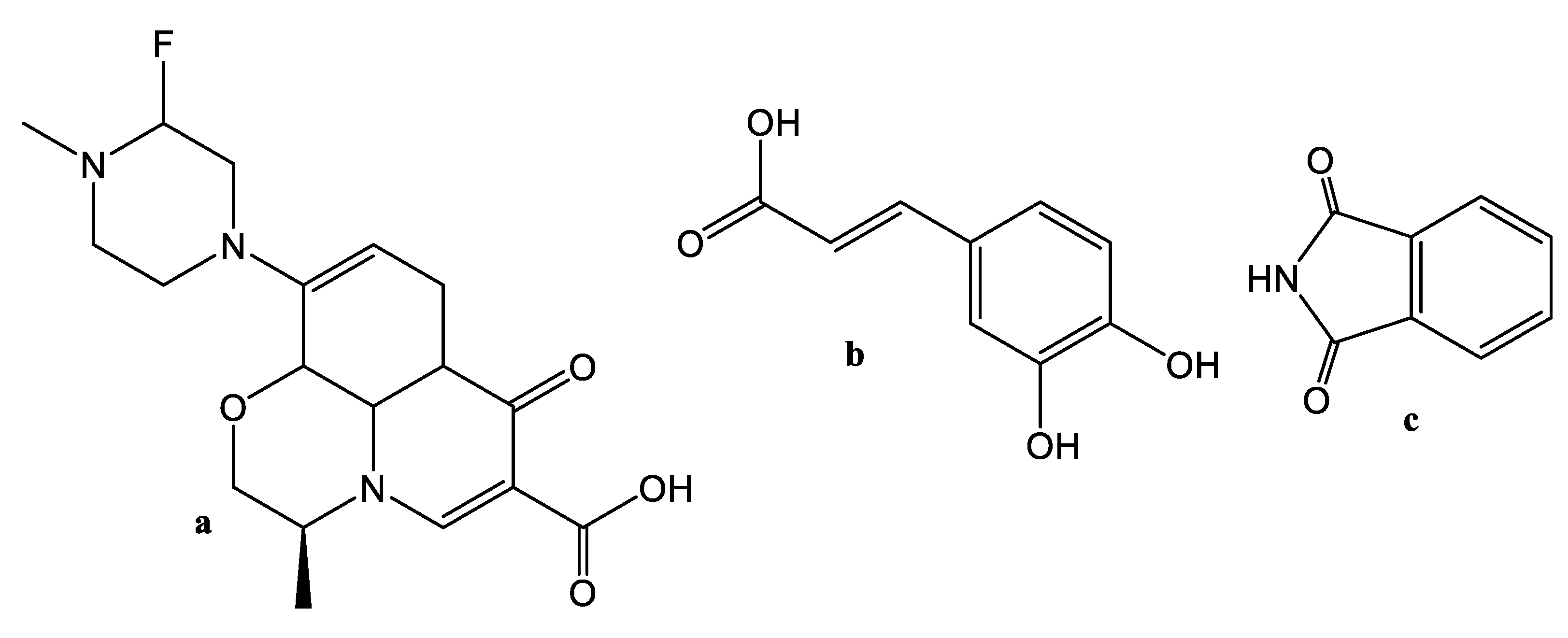
2.1. Theoretical Considerations
2.2. Construction of Binary Phase Diagram and Determination of Melting Point
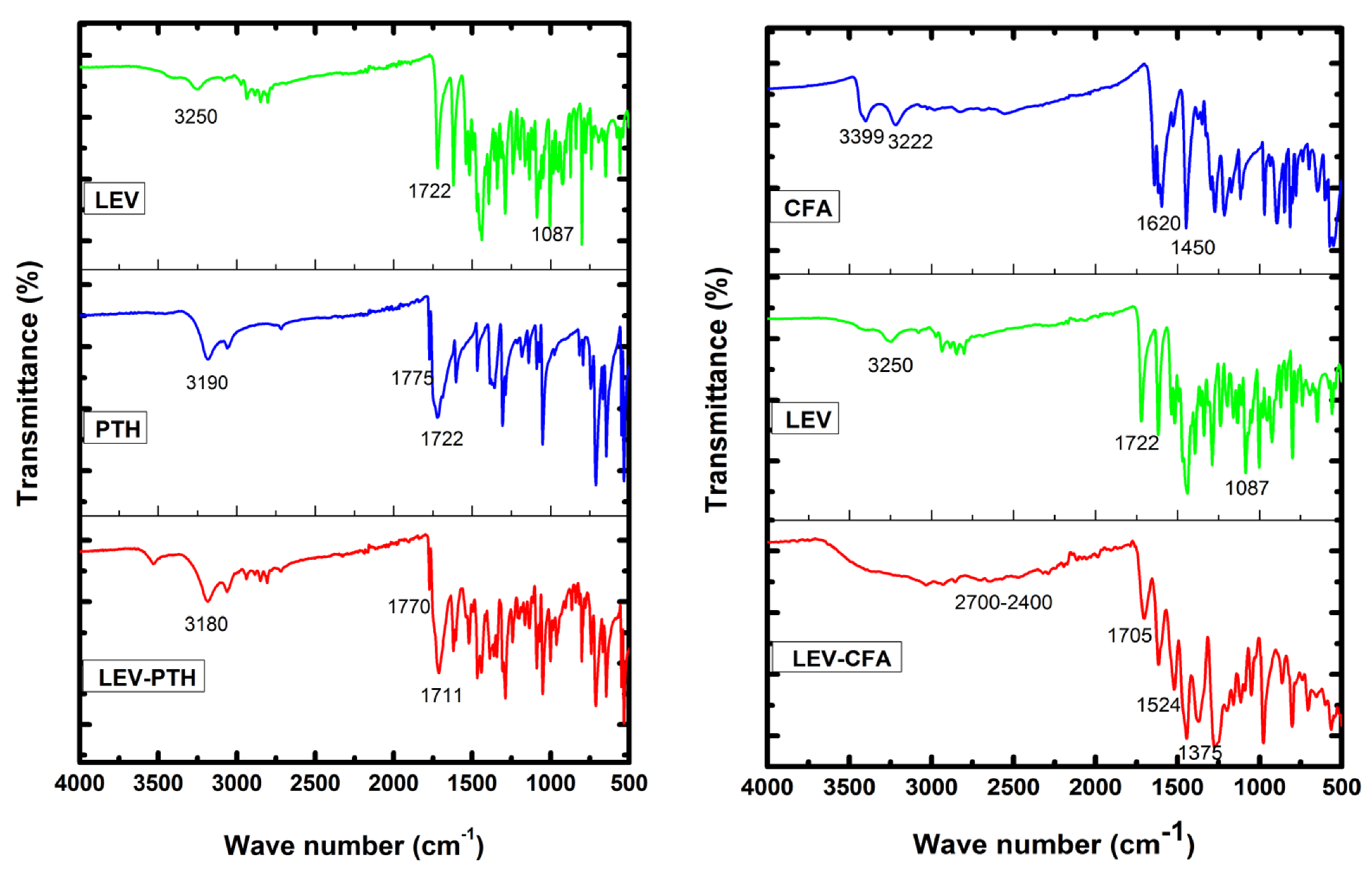
2.3. FT-IR Analysis
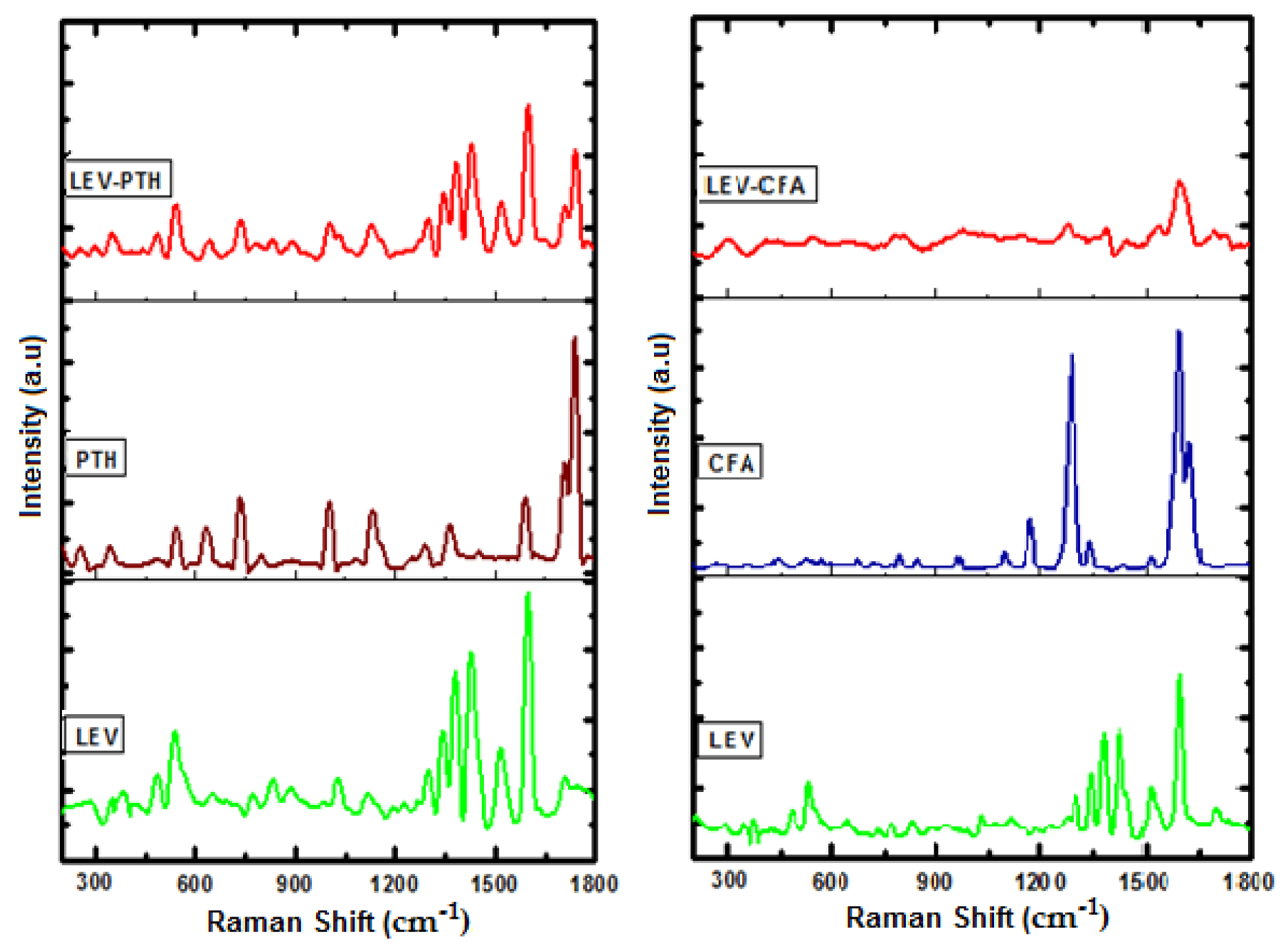
2.4. Raman Analysis
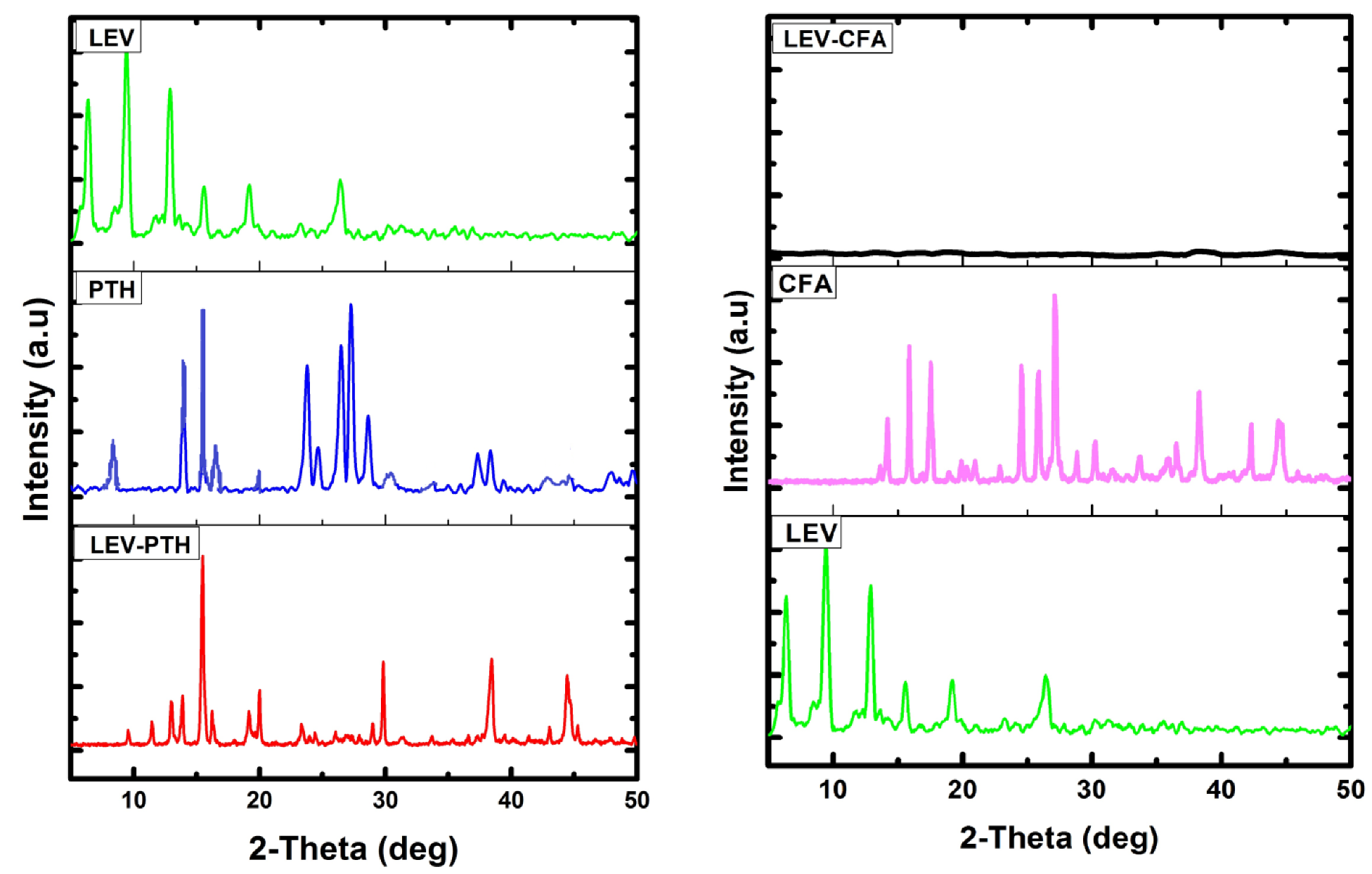
2.5. PXRD Analysis
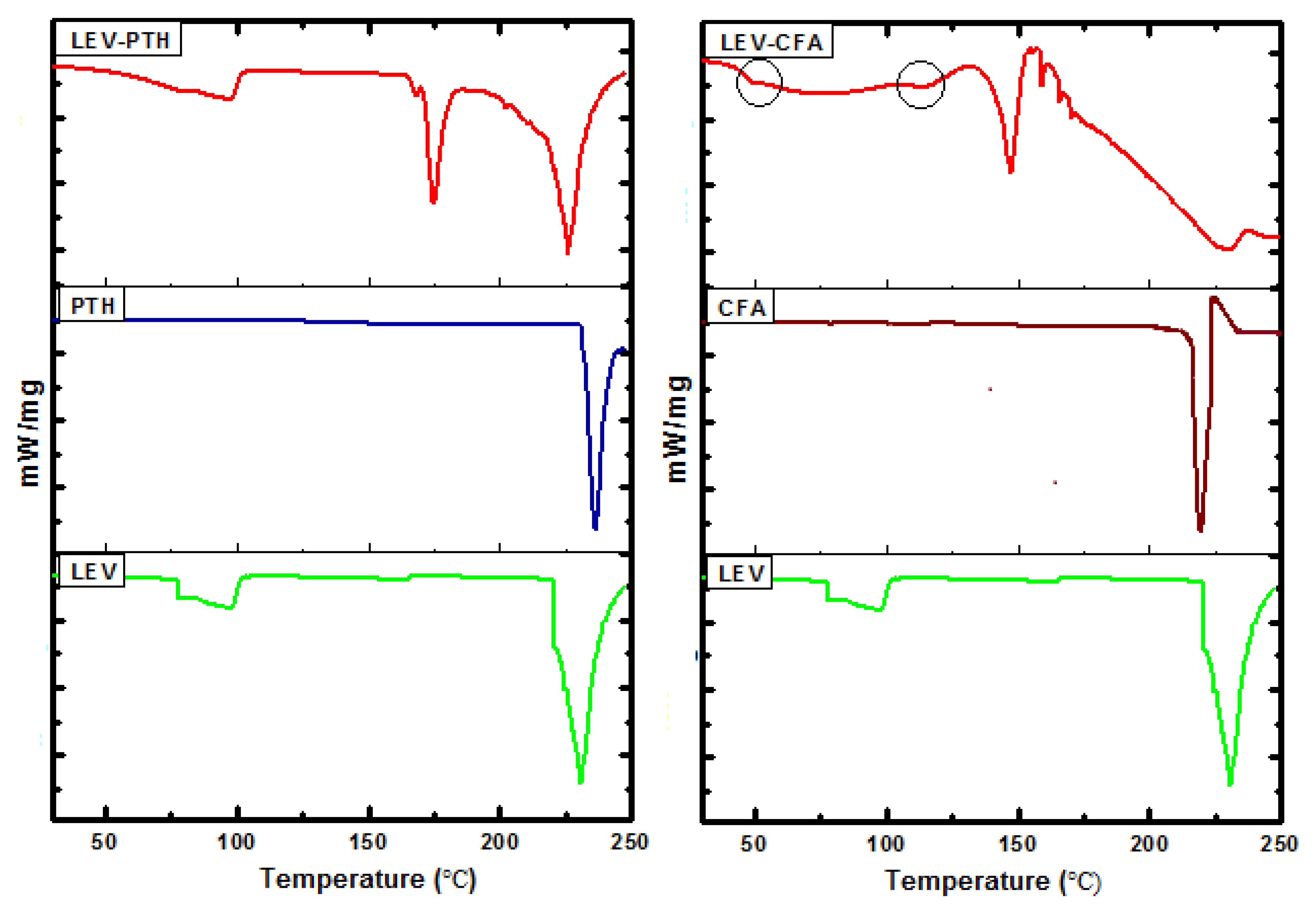
2.6. DSC Analysis
2.7. TG Analysis
2.8. 1H-NMR Studies
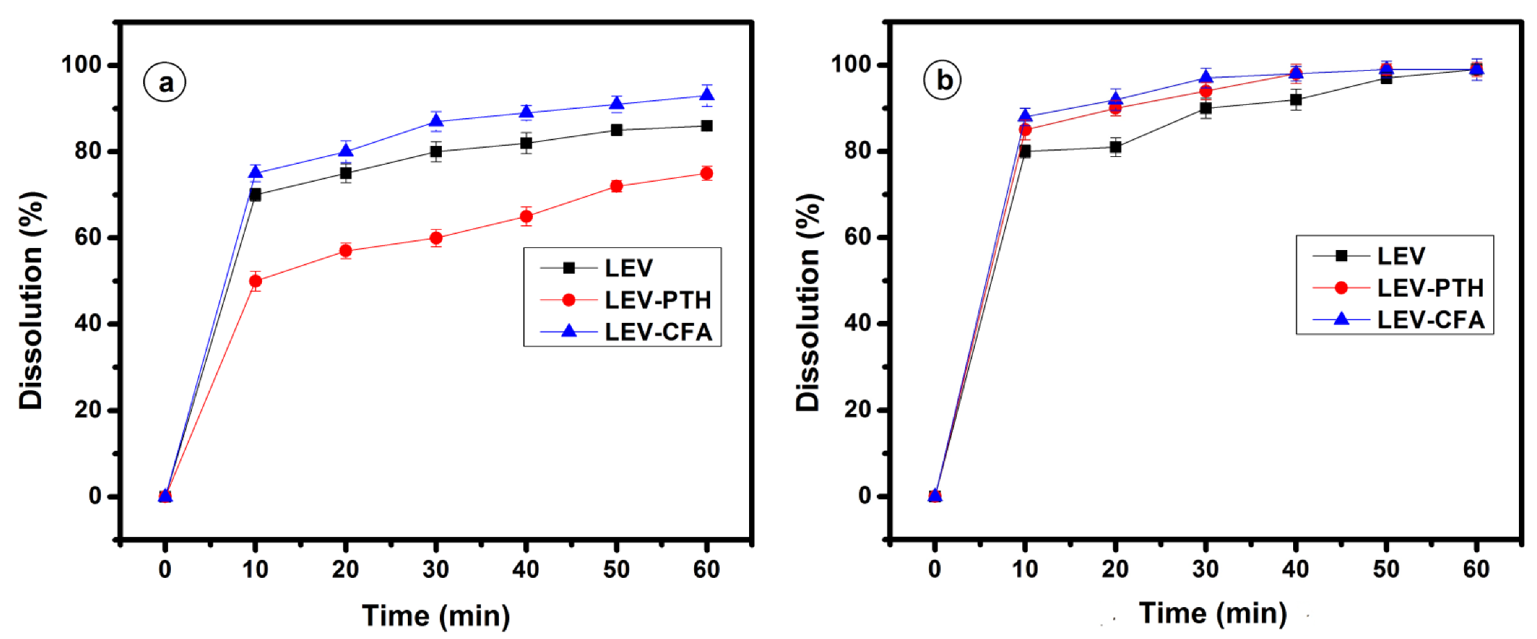
2.9. In Vitro Powder Dissolution Study
2.10. In Vitro Antimicrobial Study
3. Experimental Section
3.1. Materials
3.2. Cocrystal/Salt Synthesis
3.3. Construction of Binary Phase Diagram and Determination of Melting Point
3.4. Characterization of Cocrystal/Coamorphous
3.5. In Vitro Powder Dissolution Study
3.6. In Vitro Antibacterial Study
4. Conclusions and Future Work
Supplementary Materials
Author Contributions
Funding
Institutional Review Board Statement
Informed Consent Statement
Data Availability Statement
Acknowledgments
Conflicts of Interest
References
- Vioglio, P.C.; Chierotti, M.R.; Gobetto, R. Pharmaceutical Aspects of Salt and Cocrystal Forms of APIs and Characterization Challenges. Adv. Drug Deliv. Rev. 2017, 117, 86–110. [Google Scholar] [CrossRef] [PubMed]
- Rajput, L.; Sanphui, P.; Desiraju, G.R. New Solid Forms of the Anti-HIV Drug Etravirine: Salts, Cocrystals, and Solubility. Cryst. Growth Des. 2013, 13, 3681–3690. [Google Scholar] [CrossRef]
- Sathisaran, I.; Dalvi, S.V. Engineering Cocrystals of Poorlywater-Soluble Drugs to Enhance Dissolution in Aqueous Medium. Pharmaceutics 2018, 10, 108. [Google Scholar] [CrossRef] [PubMed] [Green Version]
- Aakeröy, C.B.; Grommet, A.B.; Desper, J. Co-Crystal Screening of Diclofenac. Pharmaceutics 2011, 3, 601–614. [Google Scholar] [CrossRef]
- Martínez-Jiménez, C.; Cruz-Angeles, J.; Videa, M.; Martínez, L.M. Co-Amorphous Simvastatin-Nifedipine with Enhanced Solubility for Possible Use in Combination Therapy of Hypertension and Hypercholesterolemia. Molecules 2018, 23, 2161. [Google Scholar] [CrossRef] [Green Version]
- Newman, A.; Reutzel-Edens, S.M.; Zografi, G. Coamorphous Active Pharmaceutical Ingredient–Small Molecule Mixtures: Considerations in the Choice of Coformers for Enhancing Dissolution and Oral Bioavailability. J. Pharm. Sci. 2018, 107, 5–17. [Google Scholar] [CrossRef] [Green Version]
- Karagianni, A.; Kachrimanis, K.; Nikolakakis, I. Co-Amorphous Solid Dispersions for Solubility and Absorption Improvement of Drugs: Composition, Preparation, Characterization and Formulations for Oral Delivery. Pharmaceutics 2018, 10, 98. [Google Scholar] [CrossRef] [Green Version]
- Islam, N.U.; Khan, E.; Umar, M.N.; Shah, A.; Zahoor, M.; Ullah, R.; Bari, A. Enhancing Dissolution Rate and Antibacterial Efficiency of Azithromycin through Drug-Drug Cocrystals with Paracetamol. Antibiotics 2021, 10, 939. [Google Scholar] [CrossRef]
- Duggirala, N.K.; Perry, M.L.; Almarsson, Ö.; Zaworotko, M.J. Pharmaceutical Cocrystals: Along the Path to Improved Medicines. Chem. Commun. 2016, 52, 640–655. [Google Scholar] [CrossRef]
- Surov, A.O.; Manin, A.N.; Voronin, A.P.; Drozd, K.V.; Simagina, A.A.; Churakov, A.V.; Perlovich, G.L. Pharmaceutical Salts of Ciprofloxacin with Dicarboxylic Acids. Eur. J. Pharm. Sci. 2015, 77, 112–121. [Google Scholar] [CrossRef]
- Mesallati, H.; Conroy, D.; Hudson, S.; Tajber, L. Preparation and Characterization of Amorphous Ciprofloxacin-Amino Acid Salts. Eur. J. Pharm. Biopharm. 2017, 121, 73–89. [Google Scholar] [CrossRef] [Green Version]
- Chadha, R.; Singh, P.; Khullar, S.; Mandal, S.K. Ciprofloxacin Hippurate Salt: Crystallization Tactics, Structural Aspects, and Biopharmaceutical Performance. Cryst. Growth Des. 2016, 16, 4960–4967. [Google Scholar] [CrossRef]
- Bandari, S.; Dronam, V.R.; Eedara, B.B. Development and Preliminary Characterization of Levofloxacin Pharmaceutical Cocrystals for Dissolution Rate Enhancement. J. Pharm. Investig. 2017, 47, 583–591. [Google Scholar] [CrossRef]
- Wagh, V.D.; Ghadlinge, S.V. Taste Masking Methods and Techniques in Oral Pharmaceuticals: Current Perspectives. J. Pharm. Res. 2009, 2, 1049–1054. [Google Scholar]
- Shinozaki, T.; Ono, M.; Higashi, K.; Moribe, K. A Novel Drug-Drug Cocrystal of Levofloxacin and Metacetamol: Reduced Hygroscopicity and Improved Photostability of Levofloxacin. J. Pharm. Sci. 2019, 108, 2383–2390. [Google Scholar] [CrossRef]
- Tønnesen, H.H. Formulation and Stability Testing of Photolabile Drugs. Int. J. Pharm. 2001, 225, 1–14. [Google Scholar] [CrossRef]
- Bhalekar, M.R.; Harinarayana, D.; Madgulkar, A.R.; Pandya, S.J.; Jain, D.K. Improvement of Photostability in Formulation: A Review. Asian J. Chem. 2008, 20, 5095. [Google Scholar]
- Zheng, K.; Gao, S.; Chen, M.; Li, A.; Wu, W.; Qian, S.; Pang, Q.; Zheng, K.; Gao, S.; Chen, M.; et al. Color Tuning of an Active Pharmaceutical Ingredient through Cocrystallization: A Case Study of a Metronidazole-Pyrogallol Cocrystal. CrystEngComm 2020, 22, 1404–1413. [Google Scholar] [CrossRef]
- Mesallati, H.; Umerska, A.; Paluch, K.J.; Tajber, L. Amorphous Polymeric Drug Salts as Ionic Solid Dispersion Forms of Ciprofloxacin. Mol. Pharm. 2017, 14, 2209–2223. [Google Scholar] [CrossRef] [Green Version]
- Shemchuk, O.; D’Agostino, S.; Fiore, C.; Sambri, V.; Zannoli, S.; Grepioni, F.; Braga, D. Natural Antimicrobials Meet a Synthetic Antibiotic: Carvacrol/Thymol and Ciprofloxacin Cocrystals as a Promising Solid-State Route to Activity Enhancement. Cryst. Growth Des. 2020, 20, 6796–6803. [Google Scholar] [CrossRef]
- Lima, V.N.; Oliveira-Tintino, C.D.M.; Santos, E.S.; Morais, L.P.; Tintino, S.R.; Freitas, T.S.; Geraldo, Y.S.; Pereira, R.L.S.; Cruz, R.P.; Menezes, I.R.A.; et al. Antimicrobial and Enhancement of the Antibiotic Activity by Phenolic Compounds: Gallic Acid, Caffeic Acid and Pyrogallol. Microb. Pathog. 2016, 99, 56–61. [Google Scholar] [CrossRef]
- Rezaei, Z.; Moghimi, S.; Javaheri, R.; Asadi, M.; Mahdavi, M.; Shabani, S.; Edraki, N.; Firuzi, O.; Safavi, M.; Amini, M.; et al. Synthesis and Biological Evaluation of 1,3,4-Thiadiazole Linked Phthalimide Derivatives as Anticancer Agents. Lett. Drug Des. Discov. 2017, 14, 1138–1144. [Google Scholar] [CrossRef]
- Stewart, S.G.; Spagnolo, D.; Polomska, M.E.; Sin, M.; Karimi, M.; Abraham, L.J. Synthesis and TNF Expression Inhibitory Properties of New Thalidomide Analogues Derived via Heck Cross Coupling. Bioorg. Med. Chem. Lett. 2007, 17, 5819–5824. [Google Scholar] [CrossRef]
- Noguchi, T.; Fujimoto, H.; Sano, H.; Miyajima, A.; Miyachi, H.; Hashimoto, Y. Angiogenesis Inhibitors Derived from Thalidomide. Bioorg. Med. Chem. Lett. 2005, 15, 5509–5513. [Google Scholar] [CrossRef]
- Kocyigit, U.M.; Budak, Y.; Gürdere, M.B.; Tekin, Ş.; Köprülü, T.K.; Ertürk, F.; Özcan, K.; Gülçin, İ.; Ceylan, M. Synthesis, Characterization, Anticancer, Antimicrobial and Carbonic Anhydrase Inhibition Profiles of Novel (3aR,4S,7R,7aS)-2-(4-((E)-3-(3-Aryl)Acryloyl) Phenyl)-3a,4,7,7a-Tetrahydro-1H-4,7-Methanoisoindole-1,3(2H)-Dione Derivatives. Bioorg. Chem. 2017, 70, 118–125. [Google Scholar] [CrossRef]
- Meng, X.B.; Han, D.; Zhang, S.N.; Guo, W.; Cui, J.R.; Li, Z.J. Synthesis and Anti-Inflammatory Activity of N-Phthalimidomethyl 2,3-Dideoxy- and 2,3-Unsaturated Glycosides. Carbohydr. Res. 2007, 342, 1169–1174. [Google Scholar] [CrossRef]
- Kuang, W.J.; Ji, S.C.; Xu, S.M.; Lan, P.; Liao, A.P.; Zhou, J.Y.; Zhang, J.Y. Thermodynamic and Crystallization of Lamotrigine Cocrystal. Cryst. Growth Des. 2019, 19, 6603–6610. [Google Scholar] [CrossRef]
- Deng, Y.; Chen, B.; Qi, Y.; Magdalou, J.; Wang, H.; Chen, L. The Effects of Levofloxacin on Rabbit Anterior Cruciate Ligament Cells in Vitro. Toxicol. Appl. Pharmacol. 2011, 257, 67–73. [Google Scholar] [CrossRef] [PubMed]
- Drozak, J.; Miecznik, A.; Jarzyna, R.; Bryla, J. The Inhibition of Gluconeogenesis by Gatifloxacin May Contribute to Its Hypoglycaemic Action. Eur. J. Pharmacol. 2008, 594, 39–43. [Google Scholar] [CrossRef] [PubMed]
- Tasso, L.; De Andrade, C.; Dalla Costa, T. Pharmacokinetic/Pharmacodynamic Modelling of the Bactericidal Activity of Free Lung Concentrations of Levofloxacin and Gatifloxacin against Streptococcus Pneumoniae. Int. J. Antimicrob. Agents 2011, 38, 307–313. [Google Scholar] [CrossRef] [PubMed] [Green Version]
- Koeppe, M.O.; Cristofoletti, R.; Fernandes, E.F.; Storpirtis, S.; Junginger, H.E.; Kopp, S.; Midha, K.K.; Shah, V.P.; Stavchansky, S.; Dressman, J.B.; et al. Biowaiver Monographs for Immediate Release Solid Oral Dosage Forms: Levofloxacin. J. Pharm. Sci. 2011, 100, 1628–1636. [Google Scholar] [CrossRef]
- Singh, S.S.; Thakur, T.S. New Crystalline Salt Forms of Levofloxacin: Conformational Analysis and Attempts towards the Crystal Structure Prediction of the Anhydrous Form. CrystEngComm 2014, 16, 4215–4230. [Google Scholar] [CrossRef]
- Jang, W.H.; Yoo, D.H.; Park, S.W. Prevalence of and Risk Factors for Levofloxacin-Resistant E. Coli Isolated from Outpatients with Urinary Tract Infection. Korean J. Urol. 2011, 52, 554–559. [Google Scholar] [CrossRef] [Green Version]
- Caliskan, R.; Tokman, H.B.; Erzin, Y.; Saribas, S.; Yuksel, P.; Bolek, B.K.; Sevuk, E.O.; Demirci, M.; Yılmazli, O.; Akgul, O.; et al. Antimicrobial Resistance of Helicobacter Pylori Strains to Five Antibiotics, Including Levofloxacin, in Northwestern Turkey. Rev. Soc. Bras. Med. Trop. 2015, 48, 278–284. [Google Scholar] [CrossRef]
- Kitaoka, H.; Wada, C.; Moroi, R.; Hakusui, H. Effect of Dehydration on the Formation of Levofloxacin Pseudopolymorphs. Chem. Pharm. Bull. 1995, 43, 649–653. [Google Scholar] [CrossRef] [Green Version]
- Gorman, E.M.; Samas, B.; Munson, E.J. Understanding the Dehydration of Levofloxacin Hemihydrate. J. Pharm. Sci. 2012, 101, 3319–3330. [Google Scholar] [CrossRef]
- Freitas, J.T.J.; De Melo, C.C.; Viana, O.M.M.S.; Ferreira, F.F.; Doriguetto, A.C. Crystal Structure of Levofloxacin Anhydrates: A High-Temperature Powder X-Ray Diffraction Study Versus Crystal Structure Prediction. Cryst. Growth Des. 2018, 18, 3558–3568. [Google Scholar] [CrossRef]
- Asghar, A.; Tan, Y.C.; Zahoor, M.; Zainal Abidin, S.A.; Yow, Y.Y.; Khan, E.; Lahiri, C. A Scaffolded Approach to Unearth Potential Antibacterial Components from Epicarp of Malaysian Nephelium Lappaceum L. Sci. Rep. 2021, 11, 13859. [Google Scholar] [CrossRef] [PubMed]
- Bartlett, J.G.; Gilbert, D.N.; Spellberg, B. Seven Ways to Preserve the Miracle of Antibiotics. Clin. Infect. Dis. 2013, 56, 1445–1450. [Google Scholar] [CrossRef] [PubMed]
- Ali, S.; Perveen, S.; Ali, M.; Shah, M.R.; Khan, E.; Sharma, A.S.; Li, H.; Chen, Q. Nano-Conjugates of Cefadroxil as Efficient Antibacterial Agent Against Staphylococcus Aureus ATCC 11632. J. Clust. Sci. 2020, 31, 811–821. [Google Scholar] [CrossRef]
- Levy, S.B.; Bonnie, M. Antibacterial Resistance Worldwide: Causes, Challenges and Responses. Nat. Med. 2004, 10, S122–S129. [Google Scholar] [CrossRef]
- Brown, E.D.; Wright, G.D. Antibacterial Drug Discovery in the Resistance Era. Nature 2016, 529, 336–343. [Google Scholar] [CrossRef]
- De Kraker, M.E.A.; Davey, P.G.; Grundmann, H. Mortality and Hospital Stay Associated with Resistant Staphylococcus Aureus and Escherichia Coli Bacteremia: Estimating the Burden of Antibiotic Resistance in Europe. PLoS Med. 2011, 8, e1001104. [Google Scholar] [CrossRef] [Green Version]
- Li, X.; Bai, H.; Yang, Y.; Yoon, J.; Wang, S.; Zhang, X. Supramolecular Antibacterial Materials for Combatting Antibiotic Resistance. Adv. Mater. 2019, 31, 1805092. [Google Scholar] [CrossRef]
- Payne, D.J.; Gwynn, M.N.; Holmes, D.J.; Pompliano, D.L. Drugs for Bad Bugs: Confronting the Challenges of Antibacterial Discovery. Nat. Rev. Drug Discov. 2007, 6, 29–40. [Google Scholar] [CrossRef]
- Shemchuk, O.; Braga, D.; Grepioni, F.; Turner, R.J. Co-Crystallization of Antibacterials with Inorganic Salts: Paving the Way to Activity Enhancement. RSC Adv. 2020, 10, 2146–2149. [Google Scholar] [CrossRef]
- Theuretzbacher, U. Global Antibacterial Resistance: The Never-Ending Story. J. Glob. Antimicrob. Resist. 2013, 1, 63–69. [Google Scholar] [CrossRef]
- Basavoju, S.; Boström, D.; Velaga, S.P. Indomethacin-Saccharin Cocrystal: Design, Synthesis and Preliminary Pharmaceutical Characterization. Pharm. Res. 2008, 25, 530–541. [Google Scholar] [CrossRef]
- Qiao, N.; Li, M.; Schlindwein, W.; Malek, N.; Davies, A.; Trappitt, G. Pharmaceutical Cocrystals: An Overview. Int. J. Pharm. 2011, 419, 1–11. [Google Scholar] [CrossRef]
- Fleischman, S.G.; Kuduva, S.S.; McMahon, J.A.; Moulton, B.; Bailey Walsh, R.D.; Rodríguez-Hornedo, N.; Zaworotko, M.J. Crystal Engineering of the Composition of Pharmaceutical Phases: Multiple-Component Crystalline Solids Involving Carbamazepine. Cryst. Growth Des. 2003, 3, 530–541. [Google Scholar] [CrossRef]
- Yu, H.; Zhang, Y.; Xing, C.; Wang, Y.; Zhang, H.; Gong, N.; Lu, Y.; Du, G. Venlafaxine Caffeic Acid Salt: Synthesis, Structural Characterization, and Hypoglycemic Effect Analysis. ACS Omega 2021, 6, 13895–13903. [Google Scholar] [CrossRef] [PubMed]
- Bhogala, B.R.; Basavoju, S.; Nangia, A. Tape and Layer Structures in Cocrystals of Some Di- and Tricarboxylic Acids with 4,4′-Bipyridines and Isonicotinamide. From Binary to Ternary Cocrystals. CrystEngComm 2005, 7, 551–562. [Google Scholar] [CrossRef]
- Golovnev, N.N.; Molokeev, M.S.; Lesnikov, M.K.; Atuchin, V.V. Two Salts and the Salt Cocrystal of Ciprofloxacin with Thiobarbituric and Barbituric Acids: The Structure and Properties. J. Phys. Org. Chem. 2018, 31, e3773. [Google Scholar] [CrossRef] [Green Version]
- Childs, S.L.; Stahly, G.P.; Park, A. The Salt-Cocrystal Continuum: The Influence of Crystal Structure on Ionization State. Mol. Pharm. 2007, 4, 323–338. [Google Scholar] [CrossRef] [Green Version]
- Ahmad, I.; Bano, R.; Sheraz, M.A.; Ahmed, S.; Mirza, T.; Ansari, S.A. Photodegradation of Levofloxacin in Aqueous and Organic Solvents: A Kinetic Study. Acta Pharm. 2013, 63, 223–229. [Google Scholar] [CrossRef] [Green Version]
- Kushwaha, N.; Kaushik, D. Recent Advances and Future Prospects of Phthalimide Derivatives. J. Appl. Pharm. Sci. 2016, 6, 159–171. [Google Scholar] [CrossRef] [Green Version]
- Lee, P.T.; Harfield, J.C.; Crossley, A.; Pilgrim, B.S.; Compton, R.G. Significant Changes in PKa between Bulk Aqueous Solution and Surface Immobilized Species: Ortho-Hydroquinones. RSC Adv. 2013, 3, 7347–7354. [Google Scholar] [CrossRef]
- Nugrahani, I.; Utami, D.; Nugraha, Y.P.; Uekusa, H.; Hasianna, R.; Darusman, A.A. Cocrystal Construction between the Ethyl Ester with Parent Drug of Diclofenac: Structural, Stability, and Anti-Inflammatory Study. Heliyon 2019, 5, e02946. [Google Scholar] [CrossRef] [Green Version]
- Évora, A.O.L.; Castro, R.A.E.; Maria, T.M.R.; Ramos Silva, M.; Canotilho, J.; Eusébio, M.E.S. Lamotrigine: Design and Synthesis of New Multicomponent Solid Forms. Eur. J. Pharm. Sci. 2019, 129, 148–162. [Google Scholar] [CrossRef]
- Yamashita, H.; Hirakura, Y.; Yuda, M.; Teramura, T.; Terada, K. Detection of Cocrystal Formation Based on Binary Phase Diagrams Using Thermal Analysis. Pharm. Res. 2013, 30, 70–80. [Google Scholar] [CrossRef]
- Nugrahani, I.; Tjengal, B.; Gusdinar, T.; Horikawa, A.; Uekusa, H. A Comprehensive Study of a New 1.75 Hydrate of Ciprofloxacin Salicylate: SCXRD Structure Determination, Solid Characterization, Water Stability, Solubility, and Dissolution Study. Crystals 2020, 10, 349. [Google Scholar] [CrossRef]
- Al-Sabagh, A.M.; El-Hamouly, S.H.; Khidr, T.T.; El-Ghazawy, R.A.; Higazy, S.A. Synthesis of Phthalimide and Succinimide Copolymers and Their Evaluation as Flow Improvers for an Egyptian Waxy Crude Oil. Egypt. J. Pet. 2013, 22, 381–393. [Google Scholar] [CrossRef] [Green Version]
- Tosovic, J. Spectroscopic Features of Caffeic Acid: Theoretical Study. Kragujev. J. Sci. 2017, 39, 99–108. [Google Scholar] [CrossRef] [Green Version]
- Świsłocka, R. Spectroscopic (FT-IR, FT-Raman, UV Absorption, 1H and 13C NMR) and Theoretical (in B3LYP/6-311++G** Level) Studies on Alkali Metal Salts of Caffeic Acid. Spectrochim. Acta-Part A Mol. Biomol. Spectrosc. 2013, 100, 21–30. [Google Scholar] [CrossRef]
- Pandey, N.K.; Sehal, H.R.; Garg, V.; Gaur, T.; Kumar, B.; Singh, S.K.; Gulati, M.; Gowthamarajan, K.; Bawa, P.; Rajesh, S.Y.; et al. Stable Co-Crystals of Glipizide with Enhanced Dissolution Profiles: Preparation and Characterization. AAPS PharmSciTech 2017, 18, 2454–2465. [Google Scholar] [CrossRef]
- Reddy, J.S.; Ganesh, S.V.; Nagalapalli, R.; Dandela, R.; Solomon, K.A.; Kumar, K.A.; Goud, N.R.; Nangia, A. Fluoroquinolone Salts with Carboxylic Acids. J. Pharm. Sci. 2011, 100, 3160–3176. [Google Scholar] [CrossRef]
- Nicolov, M.; Ghiulai, R.M.; Voicu, M.; Mioc, M.; Duse, A.O.; Roman, R.; Ambrus, R.; Zupko, I.; Moaca, E.A.; Coricovac, D.E.; et al. Cocrystal Formation of Betulinic Acid and Ascorbic Acid: Synthesis, Physico-Chemical Assessment, Antioxidant, and Antiproliferative Activity. Front. Chem. 2019, 7, 92. [Google Scholar] [CrossRef]
- Elbagerma, M.A.; Edwards, H.G.M.; Munshi, T.; Scowen, I.J. Identification of a New Cocrystal of Citric Acid and Paracetamol of Pharmaceutical Relevance. CrystEngComm 2011, 13, 1877–1884. [Google Scholar] [CrossRef] [Green Version]
- Sathisaran, I.; Dalvi, S.V. Cocrystallization of Carbamazepine with Amides: Cocrystal and Eutectic Phases with Improved Dissolution. J. Mol. Struct. 2019, 1193. [Google Scholar] [CrossRef]
- Garbacz, P.; Paukszta, D.; Sikorski, A.; Wesolowski, M. Structural Characterization of Co-Crystals of Chlordiazepoxide with p-Aminobenzoic Acid and Lorazepam with Nicotinamide by Dsc, x-Ray Diffraction, Ftir and Raman Spectroscopy. Pharmaceutics 2020, 12, 648. [Google Scholar] [CrossRef]
- Vrbanec, T.; Šket, P.; Merzel, F.; Smrkolj, M.; Grdadolnik, J. Spectroscopic Characterization of Omeprazole and Its Salts. J. Spectrosc. 2017, 2017, 6505706. [Google Scholar] [CrossRef] [Green Version]
- Brittain, H.G. Vibrational Spectroscopic Studies of Cocrystals and Salts. 3. Cocrystal Products Formed by Benzenecarboxylic Acids and Their Sodium Salts. Cryst. Growth Des. 2010, 10, 1990–2003. [Google Scholar] [CrossRef]
- De Almeida, A.C.; Ferreira, P.O.; Torquetti, C.; Ekawa, B.; Carvalho, A.C.S.; dos Santos, E.C.; Caires, F.J. Mechanochemical Synthesis, Characterization and Thermal Study of New Cocrystals of Ciprofloxacin with Pyrazinoic Acid and p-Aminobenzoic Acid. J. Therm. Anal. Calorim. 2020, 140, 2293–2303. [Google Scholar] [CrossRef]
- Sathisaran, I.; Dalvi, S.V. Crystal Engineering of Curcumin with Salicylic Acid and Hydroxyquinol as Coformers. Cryst. Growth Des. 2017, 17, 3974–3988. [Google Scholar] [CrossRef]
- Wu, S.; Shen, H.; Li, K.; Yu, B.; Xu, S.; Chen, M.; Gong, J.; Hou, B.H. Agglomeration Mechanism of Azithromycin Dihydrate in Acetone-Water Mixtures and Optimization of the Powder Properties. Ind. Eng. Chem. Res. 2016, 55, 4905–4910. [Google Scholar] [CrossRef]
- Shiozawa, R.; Inoue, Y.; Murata, I.; Kanamoto, I. Effect of Antioxidant Activity of Caffeic Acid with Cyclodextrins Using Ground Mixture Method. Asian J. Pharm. Sci. 2018, 13, 24–33. [Google Scholar] [CrossRef]
- Ali, A.M.A.; Ali, A.A.; Maghrabi, I.A. Clozapine-Carboxylic Acid Plasticized Co-Amorphous Dispersions: Preparation, Characterization and Solution Stability Evaluation. Acta Pharm. 2015, 65, 133–146. [Google Scholar] [CrossRef] [PubMed] [Green Version]
- Su, M.; Xia, Y.; Shen, Y.; Heng, W.; Wei, Y.; Zhang, L.; Gao, Y.; Zhang, J.; Qian, S. A Novel Drug-Drug Coamorphous System without Molecular Interactions: Improve the Physicochemical Properties of Tadalafil and Repaglinide. RSC Adv. 2019, 10, 565–583. [Google Scholar] [CrossRef] [PubMed] [Green Version]
- Shayanfar, A.; Jouyban, A. Drug-Drug Coamorphous Systems: Characterization and Physicochemical Properties of Coamorphous Atorvastatin with Carvedilol and Glibenclamide. J. Pharm. Innov. 2013, 8, 218–228. [Google Scholar] [CrossRef]
- Lekšić, E.; Pavlović, G.; Meštrović, E. Cocrystals of Lamotrigine Based on Coformers Involving Carbonyl Group Discovered by Hot-Stage Microscopy and DSC Screening. Cryst. Growth Des. 2012, 12, 218–228. [Google Scholar] [CrossRef]
- Pinho, E.; Henriques, M.; Soares, G. Caffeic Acid Loading Wound Dressing: Physicochemical and Biological Characterization. Ther. Deliv. 2014, 5, 1063–1075. [Google Scholar] [CrossRef] [Green Version]
- Lubach, J.W.; Xu, D.; Segmuller, B.E.; Munson, E.J. Investigation of the Effects of Pharmaceutical Processing upon Solid-State NMR Relaxation Times and Implications to Solid-State Formulation Stability. J. Pharm. Sci. 2007, 96, 777–787. [Google Scholar] [CrossRef]
- Mazzeo, P.P.; Carraro, C.; Monica, A.; Capucci, D.; Pelagatti, P.; Bianchi, F.; Agazzi, S.; Careri, M.; Raio, A.; Carta, M.; et al. Designing a Palette of Cocrystals Based on Essential Oil Constituents for Agricultural Applications. ACS Sustain. Chem. Eng. 2019, 7, 17929–17940. [Google Scholar] [CrossRef]
- Rajbhar, P.; Sahu, A.K.; Gautam, S.S.; Prasad, R.K.; Singh, V.; Nair, S.K. Formulation and Evaluation of Clarithromycin Co- Crystals Tablets Dosage Forms to Enhance the Bioavailability. Pharma Innov. J. TPI 2016, 5, 5. [Google Scholar]
- Nugrahani, I.; Asyarie, S.; Soewandhi, S.N.; Ibrahim, S. The Antibiotic Potency of Amoxicillin-Clavulanate Co-Crystal. Int. J. Pharmacol. 2007, 3, 475–481. [Google Scholar] [CrossRef] [Green Version]


| Sample | Melting Point (°C) |
|---|---|
| Levofloxacin | 224–226 |
| phthalimide | 234–236 |
| Caffiec acid | 220–224 |
| Cocrystal (LEV-PTH) | 172–176 |
| Salt (LEV-CFA) | 150–152 |
| Bacterial Strain | Sample Amount (µg) | LEV (100%) Zone of Inhibition (mm) | LEV-PTH Cocrystal (71%) Zone of Inhibition (mm) | LEV-CFA Amorphous Salt (66.7%) Zone of Inhibition (mm) |
|---|---|---|---|---|
| K. pneumonia | 5 | 28.16 ± 0.76 | 30.4 ± 0.36 | 33.96 ± 0.25 |
| 2.5 | 23.33 ± 0.57 | 24.16 ± 0.47 | 28.30 ± 0.26 | |
| 1.25 | 17.63 ± 0.77 | 19.93 ± 0.60 | 21.76 ± 0.25 | |
| 0.62 | 12.26 ± 0.30 | 14.36 ± 0.35 | 18.00 ± 0.30 | |
| E. coli | 5 | 25.033 ± 0.25 | 26.33 ± 0.35 | 31.66 ± 0.35 |
| 2.5 | 18.13 ± 0.32 | 21.3 ± 0.30 | 26.23 ± 0.25 | |
| 1.25 | 14.36 ± 0.40 | 15.43 ± 45 | 21.30 ± 0.30 | |
| 0.62 | 12.3 ± 0.26 | 14.03 ± 15 | 18.23 ± 0.20 | |
| S. typhi | 5 | 27.03 ± 0.65 | 30.03 ± 0.25 | 32.80 ± 0.20 |
| 2.5 | 22.16 ± 0.37 | 22.93 ± 0.50 | 27.93 ± 0.40 | |
| 1.25 | 17.96 ± 0.15 | 19.30 ± 30 | 22.73 ± 0.25 | |
| 0.62 | 11.06 ± 0.20 | 14.26 ± 25 | 17.03 ± 0.25 |
| Sample | MIC (μg/mL) | ||
|---|---|---|---|
| E. coli | S. typhi | K. pneumonia | |
| Pure LEV (100%) | 32 | 128 | 64 |
| LEV-PTH cocrystal (71%) | 16 | 64 | 32 |
| LEV-CFA salt (66.7%) | 16 | 16 | 16 |
Publisher’s Note: MDPI stays neutral with regard to jurisdictional claims in published maps and institutional affiliations. |
© 2022 by the authors. Licensee MDPI, Basel, Switzerland. This article is an open access article distributed under the terms and conditions of the Creative Commons Attribution (CC BY) license (https://creativecommons.org/licenses/by/4.0/).
Share and Cite
Islam, N.U.; Umar, M.N.; Khan, E.; Al-Joufi, F.A.; Abed, S.N.; Said, M.; Ullah, H.; Iftikhar, M.; Zahoor, M.; Khan, F.A. Levofloxacin Cocrystal/Salt with Phthalimide and Caffeic Acid as Promising Solid-State Approach to Improve Antimicrobial Efficiency. Antibiotics 2022, 11, 797. https://doi.org/10.3390/antibiotics11060797
Islam NU, Umar MN, Khan E, Al-Joufi FA, Abed SN, Said M, Ullah H, Iftikhar M, Zahoor M, Khan FA. Levofloxacin Cocrystal/Salt with Phthalimide and Caffeic Acid as Promising Solid-State Approach to Improve Antimicrobial Efficiency. Antibiotics. 2022; 11(6):797. https://doi.org/10.3390/antibiotics11060797
Chicago/Turabian StyleIslam, Noor Ul, Muhammad Naveed Umar, Ezzat Khan, Fakhria A. Al-Joufi, Shaymaa Najm Abed, Muhammad Said, Habib Ullah, Muhammad Iftikhar, Muhammad Zahoor, and Farhat Ali Khan. 2022. "Levofloxacin Cocrystal/Salt with Phthalimide and Caffeic Acid as Promising Solid-State Approach to Improve Antimicrobial Efficiency" Antibiotics 11, no. 6: 797. https://doi.org/10.3390/antibiotics11060797