
Pharmacological Evaluation of the Anticancer Activity of Extracts and Fractions of Lannea barteri Oliv. (Anacardiaceae) on Adherent Human Cancer Cell Lines

 , and
, and
Abstract
:
1. Introduction
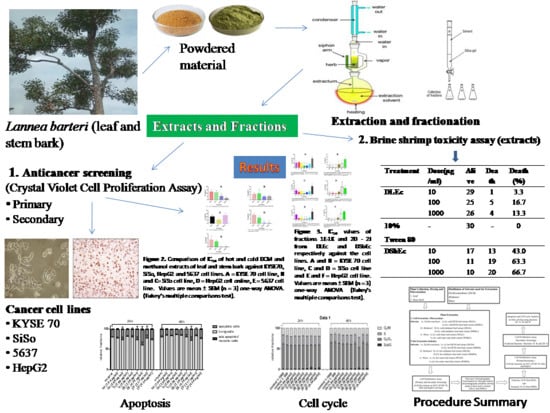
2. Results
2.1. Yield of Extracts and Fractions
2.2. Phytochemical Constituents of Extracts and Most Active Chromatography Fractions
2.3. The RP-HPLC Chromatograms of the Active Fractions from the Cold DCM Extracts of Leaf (1E–1J) and Stem Bark (2D–2I)
2.4. Effect of Extracts of on 5637, KYSE 70 and SiSo Cancer Cell Lines Proliferation in Primary Screening
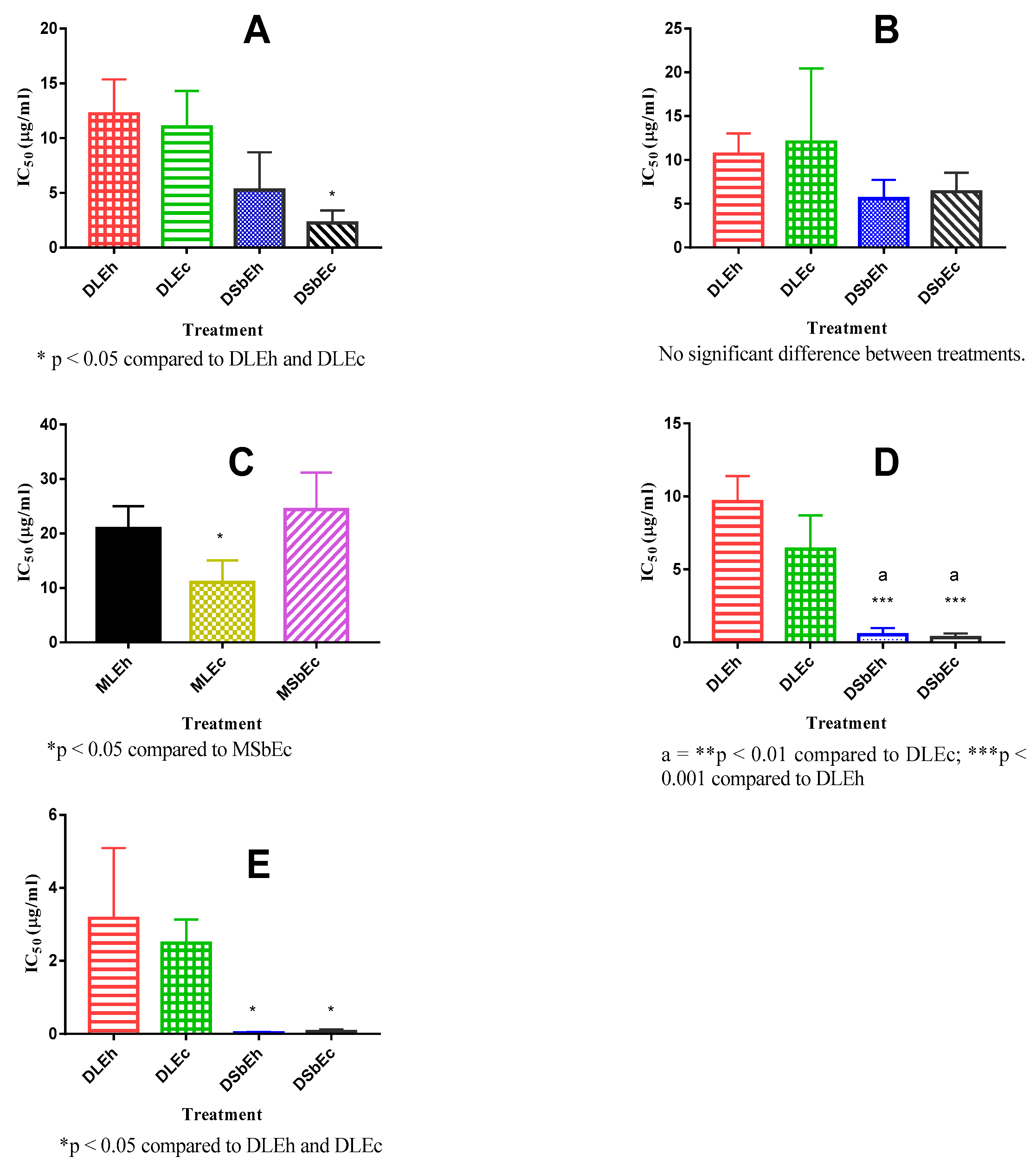
2.5. Effects of Extracts on KYSE 70 and SiSo Cell Lines in Secondary Screening
2.6. Effect of Column Chromatography Fractions of DLEc (fractions 1A–K) and DSbEc (fractions 2A–I) on 5637, KYSE 70, SiSo and HepG2 Cell Lines Proliferation in Primary Screening
2.7. Effect of Column Chromatography Fractions of DLEc (Fractions 1E–1K) and DSbEc (Fractions 2D–2I) on KYSE 70 and SiSo and HepG2 Cell Lines Proliferation in Secondary Screening
2.8. Effect of Column Chromatography Fractions 1H, 1I 2E, 2F on Apoptosis Induction in SiSo Cancer Cell Line
2.9. Effect of Fractions 1H, 1I 2E, 2F on Cell Cycle Progression in SiSo Cancer Cell Line
2.10. Toxic effect of Extracts on Brine Shrimp
3. Discussion
4. Materials
4.1. Equipment and Apparatuses
4.2. Chemicals and Solvents
4.3. Cancer Cell Lines
4.4. Brine Shrimp Larvae
5. Methods
5.1. Plant Collection and Preparation
5.2. Distillation of Solvents Used for Extraction and Fractionation
5.3. Cold Extraction of the Plant Materials
5.4. Sohxlet (hot) Extraction of the Plant Materials
5.5. Thin Layer Chromatography (TLC) of the Most Active Extract
5.6. Column Chromatography of DLEc and DSbEc with Gradient Elution Method
5.7. High Performance Liquid Chromatographic (HPLC) Separation
5.8. Phytochemical Analysis of DLEc and DSbEc
5.9. BiologicalStudies
5.9.1. Crystal Violet cell Proliferation Assay
Primary Screening
Secondary Screening
5.9.2. Brine Shrimp Toxicity Assay (BSCA)
5.9.3. Determination of Mechanism of Cytotoxic Action of Most Active Fractions on SiSo Cancer Cell Line
Apoptosis Measurement
Cell Cycle Analysis: Propidium Iodide (PI) Staining
Supplementary Materials
Author Contributions
Funding
Acknowledgments
Conflicts of Interest
References
- Majid, A.; Mahmoud, R.; Hedayatollah, S. A systematic review of medicinal plants with anti-cancer activity. J. Cancer Sci. Ther. 2015, 7. [Google Scholar] [CrossRef]
- Arbonnier, M. Arbres, arbustes et lianes des zones sèchesd’Afrique de l’Ouest, 2nd ed.; (CIRAD)-MNHN: Paris, France, 2002; p. 573. [Google Scholar]
- Blench, R.; Dendo, M. Fulfulde names for plants and trees in Nigeria, Cameroun, Chad and Niger, Latin-Fulfulde index Circulation Version. Available online: https://www.researchgate.net/publication/237253671 (accessed on 28 January 2019).
- Burkill, H.M. The useful plants of west Tropical Africa; Royal Botanic Gardens: Kew, London, UK, 1985; p. 319. [Google Scholar]
- Lannea barteri-Useful Tropical Plants. Available online: http://tropical.theferns.info/viewtropical.php?id=Lannea+barteri (accessed on 28 January 2019).
- Irvine, V.R. Woody plants of Ghana with special reference to their uses; Oxford University Press: London, UK, 1961; p. 868. [Google Scholar]
- Kone, W.M.; Soro, D.; Dro, B.; Yao, K.; Kamanzi, K. Chemical composition, antioxidant, antimicrobial and acetylcholinesterase inhibitory properties of Lannea barteri (Anacardiaceae). AJBAS 2011, 5, 1516–1523. [Google Scholar]
- Brice, B.J.; Benson, B.B.; Fernique, K.K.; Mida, K.G.R.; Christian, K.K.; Nathalle, G.K.; Akhanovna, M.-B.J.; Yves-alain, B. Lannea barteri Engl. (Anacardiaceae) plant used in the treatment of urinary tract infections in Ivory Coast: Biological and chemical studies of the aqueous extract. Int. J. Pharm. Pharm. Sci. 2018, 10, 64–69. [Google Scholar] [CrossRef]
- Garba, K.; Yaro, A.; Ya’U, J. Anticonvulsant effects of ethanol stem bark extract of Lannea barteri (Anacardiaceae) in mice and chicks. J. Ethnopharmacol. 2015, 172, 227–231. [Google Scholar] [CrossRef] [PubMed]
- Mbaoji, F.N.; Nweze, J.A. Antioxidant and hepatoprotective potentials of active fractions of Lannea barteri Oliv. (Anacardiaceae) in rat. Heliyon (under review).
- Mbaoji, F.N.; Peter, I.E.; Onwuka, A.M. Wound healing, anti-inflammatory and antimicrobial activities of methanol leaf extract and fractions of Lannea barteri Oliv. Engl. (Anacardiaceae) in rats. Drug Discovery 2020, 14, 25–32. [Google Scholar]
- National Cancer Institute (NCI). NCI-60 screening methodology. 2015. Available online: http:// https://dtp.cancer.gov/discovery_development/nci-60/methodology.htm (accessed on 1 October 2019).
- López-Sáez, J.F.; De La Torre, C.; Pincheira, J.; Giménez-Martín, G. Cell proliferation and cancer. Histol. Histopathol. 1998, 13, 1197–1214. [Google Scholar] [PubMed]
- Lage, O.M.; Ramos, M.C.; Calisto, R.; Almeida, E.; Vasconcelos, V.; Vicente, F. Current Screening Methodologies in Drug Discovery for Selected Human Diseases. Mar. Drugs 2018, 16, 279. [Google Scholar] [CrossRef] [PubMed] [Green Version]
- Feoktistova, M.; Geserick, P.; Leverkus, M. crystal violet assay for determining viability of cultured cells. Cold Spring Harb. Protoc. 2016, 2016. [Google Scholar] [CrossRef] [PubMed]
- Burger, A.M.; Fiebig, H.-H. Preclinical screening for new anticancer agents. In Handbook of anticancer pharmacokinetics and pharmacodynamics, 2nd ed.; Rudek, M.A., Chau, C.H., Figg, W.D., McLeod, H.L., Eds.; Humana press: New York, NY, USA, 2014; Volume IX, 836, pp. 145–150. [Google Scholar]
- Shimada, Y.; Imamura, M.; Wagata, T.; Yamaguchi, N.; Tobe, T. Characterisation of 21 newly established oesophageal cancer cell lines. Cancer 1992, 69, 227–284. [Google Scholar] [CrossRef]
- Quazi, S.S.; Fatema, A.C.; Mir, M. Brine shrimp lethality assay. Bangladesh J. Pharmacol. 2017, 12, 186–189. [Google Scholar]
- Definitions.net. ec50. (n.d). Definitions.net. 2019. Available online: https://www.definitions.net > definition (accessed on 3 June 2019).
- Renehan, A.G.; Booth, C.; Potten, C.S. What is apoptosis, and why is it important? BMJ 2001, 322, 1536–1538. [Google Scholar] [CrossRef] [PubMed] [Green Version]
- van der Mark, V.A.; Elferink, R.P.O.; Paulusma, C.C. P4 ATPases: Flippases in Health and Disease. Int. J. Mol. Sci. 2013, 14, 7897–7922. [Google Scholar] [CrossRef] [PubMed] [Green Version]
- Geske, F.J.; Nelson, A.C.; Lieberman, R.; Strange, R.; Sun, T.; Gerschenson, L.E. DNA repair is activated in early stages of p53-induced apoptosis. Cell Death Differ. 2013, 7, 393–401. [Google Scholar] [CrossRef] [PubMed] [Green Version]
- Olson, R.J.; Zettler, E.R.; Durand, M.D. Phytoplankton Analysis Using Flow Cytometry. Handb. Methods Aquat. Microb. Ecol. 2018, 175–186. [Google Scholar]
- McNaught, A.D.; Wilkinson, A. IUPAC Compendium of Chemical Terminology Retention factor, k in Column Chromatography, 2nd ed.; Royal Society of Chemistry: Cambridge, UK, 2006; Online corrected version: (2006). [Google Scholar]
- Harborne, A.J. Phytochemical Methods. A Guide to Modern Techniques of Plant Analysis; Chapman and Hall, Ltd.: London, UK, 1973; pp. 49–279. [Google Scholar]
- Trease, G.E.; Evans, W.C. Pharmacognosy, 13th ed.; Bailliere and Tindall: London, UK, 1989; pp. 176–180. [Google Scholar]
- Bracht, K.; Grünert, R.; Bednarski, P.J. Correlations between the activities of 19 anti-tumor agents and the intracellular glutathione concentrations in a panel of 14 human cancer cell lines: Comparisons with the National Cancer Institute data. Anti-Cancer Drugs 2006, 17, 41–51. [Google Scholar] [CrossRef] [PubMed]
- Mothana, R.A.A.; Grünert, R.; Lindequist, U.; Bednarski, P.J. Study of the anticancer potential of Yemeni plants used in folk medicine. Die Pharmazie 2007, 62, 305–307. [Google Scholar] [PubMed]
- Meyer, B.N.; Ferrigni, N.R.; Putnam, J.E.; Jacobsen, L.B.; Nichols, D.E.; McLaughlin, J.L. Brine shrimp: A convenient general bioassay for active plant constituents. Planta Med. 1998, 45, 31–34. [Google Scholar] [CrossRef] [PubMed]
- Gupta, M.P.; Monge, A.; Karikas, G.A.; Lopez de Cerain, A.; Solis, P.N.; de Leon, E.; Trujillo, M.; Suarez, O.; Wilson, F.; Montenegro, G.; et al. Screening of Panamanian medicinal plants for brine shrimp toxicity, crown gall tumour inhibition, cytotoxicity and DNA interaction. IJPPR 1996, 34, 19–27. [Google Scholar]
- Behnisch-Cornwell, S.; Laubenstein, G.; Bednarski, P.J. Studies of the inhibitory activities of tiopronin and mercaptosuccinic acid on glutathione peroxidase and their cytotoxic and antioxidant properties. Die Pharmazie 2019, 74, 536–542. [Google Scholar] [PubMed]
- Kang, H.K.; Joel, M.S. Assaying cell cycle status using flow cytometry. Curr. Protoc. Mol. Boil. 2015, 111, 28.6.1–28.6.111. [Google Scholar]
Sample Availability: No sample available. |


| Extract | Yield | |
|---|---|---|
| mg | % | |
| DLEc | 40.1 | 1.86 |
| DSbEc | 46.1 | 1.15 |
| DLEh | 235.2 | 2.35 |
| DSbEh | 216.1 | 2.37 |
| MLEc | 195.0 | 9.75 |
| MSbEc | 78.5 | 3.93 |
| MLEh | 748.7 | 17.49 |
| MSbEh | 912.0 | 10.03 |
| WLEc | 90.4 | 4.52 |
| WSbEc | 47.0 | 2.35 |
| WLEh | 718.8 | 7.17 |
| WSbEh | 227.3 | 2.27 |
| Solvent Ratio (n-hexane: ethyl acetate) | DLEc | DSbEc |
|---|---|---|
| Rf | ||
| 9:1 | 0.25 | 0.30 |
| 8:2 | 0.38 | 0.32 |
| 7:3 | 0.47 | 0.50 |
| 6:4 | 0.50 | 0.55 |
| 5:5 | 0.68 | 0.70 |
| 4:6 | 0.44 | 0.38 |
| 3:7 | 0.40 | 0.30 |
| 2:8 | 0.26 | 0.28 |
| 1:9 | 0.22 | 0.25 |
| Fractions | Pooled Samples | Weight (mg) | Yield (%) |
|---|---|---|---|
| 1A | 8–13 | 3.30 | 0.66 |
| 1B | 14–20 | 15.50 | 3.10 |
| 1C | 21–28 | 27.80 | 5.56 |
| 1D | 29–37 | 13.40 | 2.68 |
| 1E | 38–42 | 113.70 | 22.74 |
| 1F | 43–47 | 31.70 | 6.34 |
| 1G | 48–56 | 13.60 | 2.72 |
| 1H | 57–85 | 14.90 | 2.98 |
| 1I | 86–102 | 7.80 | 1.56 |
| 1J | 103–187 | 41.00 | 8.20 |
| 1K | 188–192 | 39.40 | 7.00 |
| 2A | 1–7 | 8.20 | 1.64 |
| 2 B | 8–24 | 9.30 | 1.86 |
| 2 C | 25–40 | 13.00 | 2.60 |
| 2 D | 41–44 | 29.60 | 5.92 |
| 2 E | 45–51 | 48.20 | 9.64 |
| 2 F | 52–61 | 69.80 | 13.96 |
| 2 G | 62–77 | 59.50 | 11.90 |
| 2 H | 78–105 | 54.10 | 10.82 |
| 2 I | 106–109 | 84.00 | 16.80 |
| 2 J | 110–140 | 8.80 | 1.76 |
| Extract | Cell Line | ||
|---|---|---|---|
| 5637 | KYSE 70 | SiSo | |
| T/C corr. (%) | |||
| DLEc | 14.76 | 5.83 | 4.62 |
| DSbEc | 7.82 | 3.03 | 2.56 |
| DLEh | 2.01 | 5.18 | 4.21 |
| DSbEh | -3.12 | 4.63 | 2.67 |
| MLEc | 1.77 | >50 | 20.95 |
| MSbEc | 3.64 | >50 | 24.33 |
| MLEh | >50 | >50 | >50 |
| MSbEh | >50 | >50 | >50 |
| WLEc | >50 | >50 | >50 |
| WSbEc | >50 | >50 | >50 |
| WLEh | >50 | >50 | >50 |
| WSbEh | >50 | >50 | >50 |
| Extract | Cell Line | |
|---|---|---|
| KYSE 70 | SiSo | |
| IC50 (µg/mL) | ||
| DLEc | 11.03 ± 2.68 | 12.06 ± 8.39 |
| DSbEc | 2.26 ± 0.92 | 6.37 ± 2.15 |
| DLEh | 12.23 ± 3.12 | 10.68 ± 2.36 |
| DSbEh | 5.28 ± 3.43 | 5.62 ± 3.00 |
| MLEc | >50 | 11.01 ± 4.04 |
| MSbEc | >50 | 23.85 ± 9.41 |
| MLEh | >50 | 20.93 ± 4.09 |
| MSbEh | >50 | >50 |
| Fraction | Cell line | |||
|---|---|---|---|---|
| 5637 | KYSE 70 | SiSo | HepG2 | |
| T/C corr. (%) | ||||
| 1A | 33.67 | 85.26 | 108.84 | 99.21 |
| 1B | 3.06 | 62.12 | 100.00 | 20.58 |
| 1C | 3.06 | 69.29 | 116.02 | 44.33 |
| 1D | −9.18 | 61.57 | −10.50 | 37.99 |
| 1E | -11.22 | −0.22 | −4.18 | −7.39 |
| 1F | −1.02 | −2.22 | −4.50 | −10.95 |
| 1G | −9.66 | −2.95 | −6.75 | −12.40 |
| 1H | −6.90 | −4.15 | −6.59 | 14.25 |
| 1I | −7.38 | −4.04 | −6.27 | 14.12 |
| 1J | −8.18 | −3.82 | −6.11 | 14.38 |
| 1K | 15.54 | −4.42 | −2.73 | 10.55 |
| 2A | 22.94 | 71.51 | 23.79 | 83.51 |
| 2B | −14.94 | 51.64 | 21.57 | 48.15 |
| 2C | −16.91 | 12.01 | 6.11 | −1.58 |
| 2D | −16.21 | −4.91 | −0.07 | −14.64 |
| 2E | −15.01 | −5.13 | −7.24 | −14.51 |
| 2F | −17.01 | −4.80 | −7.07 | −14.51 |
| 2G | −13.91 | −5.02 | −6.43 | −13.98 |
| 2H | −8.91 | −4.80 | −6.01 | −14.64 |
| 2I | −13.17 | −4.04 | −5.95 | −11.21 |
| 2J | −9.34 | 62.55 | 22.51 | 10.82 |
| Treatment | Dose (µg/mL) | Alive | Dead | Dead (%) |
|---|---|---|---|---|
| DLEc | 10 | 29 | 1 | 3.3 |
| 100 | 25 | 5 | 16.7 | |
| 1000 | 26 | 4 | 13.3 | |
| 10% Tween 80 | - | 30 | - | 0 |
| DSbEc | 10 | 17 | 13 | 43.0 |
| 100 | 11 | 19 | 63.3 | |
| 1000 | 10 | 20 | 66.7 |
© 2020 by the authors. Licensee MDPI, Basel, Switzerland. This article is an open access article distributed under the terms and conditions of the Creative Commons Attribution (CC BY) license (http://creativecommons.org/licenses/by/4.0/).
Share and Cite
Mbaoji, F.N.; Behnisch-Cornwell, S.; Ezike, A.C.; Nworu, C.S.; Bednarski, P.J. Pharmacological Evaluation of the Anticancer Activity of Extracts and Fractions of Lannea barteri Oliv. (Anacardiaceae) on Adherent Human Cancer Cell Lines. Molecules 2020, 25, 849. https://doi.org/10.3390/molecules25040849
Mbaoji FN, Behnisch-Cornwell S, Ezike AC, Nworu CS, Bednarski PJ. Pharmacological Evaluation of the Anticancer Activity of Extracts and Fractions of Lannea barteri Oliv. (Anacardiaceae) on Adherent Human Cancer Cell Lines. Molecules. 2020; 25(4):849. https://doi.org/10.3390/molecules25040849
Chicago/Turabian StyleMbaoji, Florence N., Steven Behnisch-Cornwell, Adaobi C. Ezike, Chukwuemeka S. Nworu, and Patrick J. Bednarski. 2020. "Pharmacological Evaluation of the Anticancer Activity of Extracts and Fractions of Lannea barteri Oliv. (Anacardiaceae) on Adherent Human Cancer Cell Lines" Molecules 25, no. 4: 849. https://doi.org/10.3390/molecules25040849