Review

The Impact of Anthocyanins and Iridoids on Transcription Factors Crucial for Lipid and Cholesterol Homeostasis

Department of Pharmacology, Wroclaw Medical University, Jana Mikulicza-Radeckiego 2, 50-345 Wroclaw, Poland
*
Author to whom correspondence should be addressed.
Int. J. Mol. Sci. 2021, 22(11), 6074; https://doi.org/10.3390/ijms22116074
Submission received: 30 April 2021 / Revised: 29 May 2021 / Accepted: 3 June 2021 / Published: 4 June 2021
(This article belongs to the Special Issue Natural Product Pharmacology)

Abstract

:
Nutrition determines our health, both directly and indirectly. Consumed foods affect the functioning of individual organs as well as entire systems, e.g., the cardiovascular system. There are many different diets, but universal guidelines for proper nutrition are provided in the WHO healthy eating pyramid. According to the latest version, plant products should form the basis of our diet. Many groups of plant compounds with a beneficial effect on human health have been described. Such groups include anthocyanins and iridoids, for which it has been proven that their consumption may lead to, inter alia, antioxidant, cholesterol and lipid-lowering, anti-obesity and anti-diabetic effects. Transcription factors directly affect a number of parameters of cell functions and cellular metabolism. In the context of lipid and cholesterol metabolism, five particularly important transcription factors can be distinguished: liver X receptor (LXR), peroxisome proliferator-activated receptor-α (PPAR-α), peroxisome proliferator-activated receptor-γ (PPAR-γ), CCAAT/enhancer binding protein α (C/EBPα) and sterol regulatory element-binding protein 1c (SREBP-1c). Both anthocyanins and iridoids may alter the expression of these transcription factors. The aim of this review is to collect and systematize knowledge about the impact of anthocyanins and iridoids on transcription factors crucial for lipid and cholesterol homeostasis.

1. Introduction

It is a truism to say that nutrition directly reflects our health. Many scientific publications have proven the beneficial effects of a proper diet on, among others, blood parameters, skin condition or the work of key metabolic organs such as the pancreas, liver or kidneys. Moreover, although, globally, store shelves are filled with highly processed, heavily sweetened and high-saturated-fat products, natural and organic ingredients are becoming more and more popular. Various campaigns are organized—sometimes national or nationwide—calling for populations to adopt an appropriate dietary regimen. Furthermore, the mass media, celebrities and public figures often promote a healthy lifestyle, frequently by setting an example.
Each individual has their own unique dietary habits. Countless factors influence our ways of eating. An important role is played by cultural, financial or geographic aspects. Various types of diets are known, such as basic, alternative, therapeutic, experimental and many more. Occasionally, new diets are proposed. They are based on, for example, including or excluding a certain dietary group from everyday consumption, a specific caloric balance or the content of basic nutrients—proteins, fats or carbohydrates. Regardless of the scientific basis for creating a given diet or the effectiveness of achieving its goal, the WHO pyramid of healthy eating remains a universal determinant of proper nutrition. According to its new version, plant products should form the basis of our diet and be consumed in the largest quantities.
Transcription factors are proteins that possess the ability to bind DNA in the promoter or enhancer sequence at a specific site or region, where they regulate the transcription process. Transcription factors are essentially classified by three different aspects: mechanism of action, regulatory function and sequence homology in their DNA-binding domains. Due to the fact that they help to "turn on" or "turn off" individual genes, they directly affect a number of parameters of cell functions and cellular metabolism. Thus, in a broader context, transcription factors affect the work of different organs, e.g., the liver, or entire systems, e.g., cardiovascular [1,2,3,4,5].
Five transcription factors play a crucial role in lipid and cholesterol metabolism: liver X receptor (LXR), peroxisome proliferator-activated receptor-α (PPAR-α), peroxisome proliferator-activated receptor-γ (PPAR-γ), CCAAT/enhancer binding protein α (C/EBPα) and sterol regulatory element-binding protein 1c (SREBP-1c). There are many groups of plant substances that influence the expression of these factors, and thus also the genes whose functioning they regulate. This impact was confirmed in numerous studies—both tissue and animal as well as human models. Among the most frequently mentioned compounds exhibiting such effects, the following are worth mentioning: polyphenols, phytoestrogens, soy proteins, mono- and polyunsaturated fatty acids, alkaloids, saponins, and also anthocyanins and iridoids. The latter two are the subject of our particular interest. Studies summarizing the current knowledge on the effect of, e.g., polyphenols or unsaturated fatty acids, on the transcription factors associated with lipid and cholesterol turnovers are quite common. However, to the best of our knowledge, there is no publication collecting data on this subject in the case of anthocyanins and iridoids. Therefore, we decided to assemble and systematize information on the impact of anthocyanins and iridoids on transcription factors crucial for lipid and cholesterol metabolism.

2. Major Transcription Factors Involved in Lipid and Cholesterol Metabolism

Replication, transcription and translation—at the cellular level—play a key role in the reproduction, development and maintenance of life processes in all organisms. The genome contains all information regarding the structure and function of bodily proteins. The process of protein formation is indirectly controlled by a specific group of proteins and nuclear receptors, described as transcription factors (TFs). The transcription process never enfolds the whole DNA strand, but only the fragment that encrypts a protein that is currently required by the body. This phenomenon is determined, among others, by transcription factors. Their task, apart from the transcription induction itself, is to direct the appropriate signal to the promoter site of the gene encoding the desired protein [6,7]. It was found that a dysfunction of transcriptional factors is related to a third of human developmental disorders. Moreover, TFs occur numerously among the group of oncogenes [3].
Transcription factors control initiation or elongation processes. Certain TFs may affect both of these processes. They typically bind cofactors—protein complexes that are associated with activation (coactivators) and repression (corepressors) but do not have DNA-binding capabilities of their own. Most TFs are thought to contribute to transcription initiation precisely by recruiting coactivators [2].
Transcription factors, as mentioned earlier, can be divided according to various criteria, but the simplest classification differentiates them into two basic groups: general transcription factors (GTFs) and specific transcription factors (STFs). GTFs, which always remain active, forming the pre-initiation complex, are responsible for inducing the transcription process. Six general factors can be distinguished: TFIIA, TFIIB, TFIID, TFIIE TFIIF, and TFIIH. In turn, specific factors accelerate or inhibit the transcription of target genes in response to signals sent to the cell nucleus. These factors are activated or suppressed depending on the demand for specific proteins [6,7,8].
STFs attach to the DNA fragment within the promoter, which leads to facilitating or hindering the recognition of the binding sequence by the pre-initiation complex. This conflation takes place by using small binding proteins, called either enhancers or silencers, i.e., short DNA sequences that are located even up to several thousand base pairs away from the promoter. The attachment of the transcription factor to the enhancer results in accelerated transcription, while silencers cause the opposite effect [2,9]. This is why STFs are a very engaging research target and therapeutically promising factors for many substances, including various food compounds.
To date, over 1000 various transcription factors have been described in humans [3]. Five of them play a key role in lipid and cholesterol metabolism: liver X receptor (LXR), peroxisome proliferator-activated receptor-α (PPAR-α), peroxisome proliferator-activated receptor-γ (PPAR-γ), CCAAT/enhancer binding protein α (C/EBPα) and sterol regulatory element-binding protein 1c (SREBP-1c).
Liver X receptor is a ligand-activated transcription factor of the nuclear receptor superfamily, playing a critical role in the regulation of the expression of major genes involved in cholesterol, lipid and glucose homeostasis. Two isoforms of liver X receptor can be distinguished: LXRα and LXRβ. It was proven that LXRs’ reverse the transportation of cholesterol to peripheral tissues and transport the excess cholesterol into the liver, reduce cholesterol absorption by intestinal epithelial cells and directly induce the expression of the essential transcription factor for lipid and cholesterol synthesis in the liver, sterol regulatory element-binding protein 1c [10,11]. One of the first direct LXR target genes identified was ATP-binding cassette transporter A1 (ABCA1). LXR activation leads to the robust upregulation of ABCA1 in macrophages, the intestine and in the liver, and the efflux of cholesterol to apolipoprotein A1, which results in the formation of high-density lipoproteins (HDL) [10,12]. LXRs are responsible for the stimulation of HDL synthesis [13].
Cholesterol is, above all, an essential constituent of the cell membrane but also represents a precursor for the synthesis of several hormones and bile acids that play significant roles in various physiological processes. Incorrect maintenance of cholesterol homeostasis, including absorption, biosynthesis, transmission and efflux, may contribute to severe disorders, such as atherosclerosis, metabolic syndrome, cardiovascular disease (CVD) or cholelithiasis [14]. Therefore, LXR as a cholesterol sensor play a critical role in its metabolism and protection against atherosclerosis [15,16,17]. It is worth highlighting that synthetic LXR agonists may exert undesirable effects on plasma and hepatic triglycerides through activating another transcriptional factor—hepatic sterol regulatory element-binding protein 1c [18]. For this reason, natural LXR agonists, which could be deprived of these side effects, are urgently needed.
Peroxisome proliferator-activated receptors (PPARs) are a superfamily of nuclear receptors functioning as ligand-activated transcription factors. PPARs include PPAR-α, PPAR-δ and PPAR-γ subtypes. PPARs play a relevant role in the regulation of glucose and lipid metabolism. PPAR-α, highly expressed in the liver, is closely related to fatty acid β-oxidation in the liver and in brown adipose tissue and also modulates both acute and chronic inflammation [11,19]. To a lesser extent, it occurs in the cardiovascular system, muscles and kidneys. PPAR-α regulates the transcription of multiple genes, including acyl-CoA-oxidase, carnitine palmitoyl transferase (CPT) and several CYP4As [20]. PPAR-δ is expressed in almost all the tissues of the body. PPAR-γ is a major regulator expressed in fat tissue that promotes fat storage in white adipose tissue and also, to a lesser extent, in the liver, muscles and cardiovascular system [11]. In the case of overweight or obesity, white adipose tissue plays a pivotal role in the development of oxidative stress, inflammation and depletion of n-3 long-chain polyunsaturated fatty acids [21].
Two essential transcription factors that adjust adipocyte differentiation and regulate the expression of adipogenic and lipogenic genes are PPAR-γ and CCAAT/enhancer binding protein α (C/EBPα) [22]. CCAAT/enhancer binding protein α is a member of a family of six transcription factors, named from C/EBPα to C/EBPζ. They interact with the CCAAT (cytosine–cytosine–adenosine–adenosine–thymidine) sequence, present in several gene promoters. C/EBPs are pivotal in adipogenesis (C/EBPβ and C/EBPδ are early adipogenic transcription factors, while isoform α regulates the final stages of adipogenesis) and the development of osteoporosis. C/EBPα is one of the inductors of PPAR-γ expression [23,24]. Importantly, from a potential therapeutic perspective, the suppressor of PPAR-γ, C/EBPα, and also SREBP-1 expression, is AMP-activated protein kinase (AMPK) [25,26]. AMPK is a key factor controlling cellular energy homeostasis. It influences hepatic lipid metabolism through modulating the downstream acetyl-CoA carboxylase (ACC) and carnitine palmitoyl transferase-1 (CPT-1) pathway [27].
Another crucial transcription factor that enhances lipogenesis and adipogenesis is sterol regulatory element-binding protein 1c (SREBP-1c). There are two isoforms of sterol regulatory element-binding protein 1, SREBP-1a and SREBP-1c. SREBP-1a is expressed in the intestine and spleen, whereas SREBP-1c is mainly expressed in the liver, muscle and adipose tissue. SREBP-1c regulates genes required for glucose metabolism, fatty acid and lipid production and facilitates the storage of fatty acids as triglycerides [28,29,30]. Its expression is induced i.a. by insulin. A high-fat diet (HFD) is another key factor triggering the overexpression of both SREBP-1c and PPAR-γ in the liver and adipose tissue [31,32].

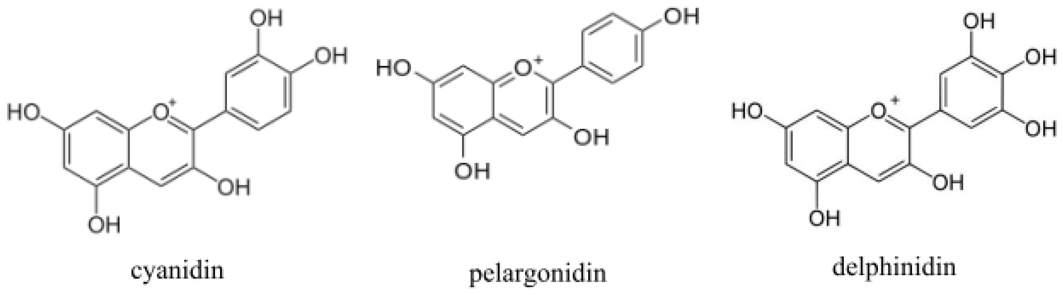
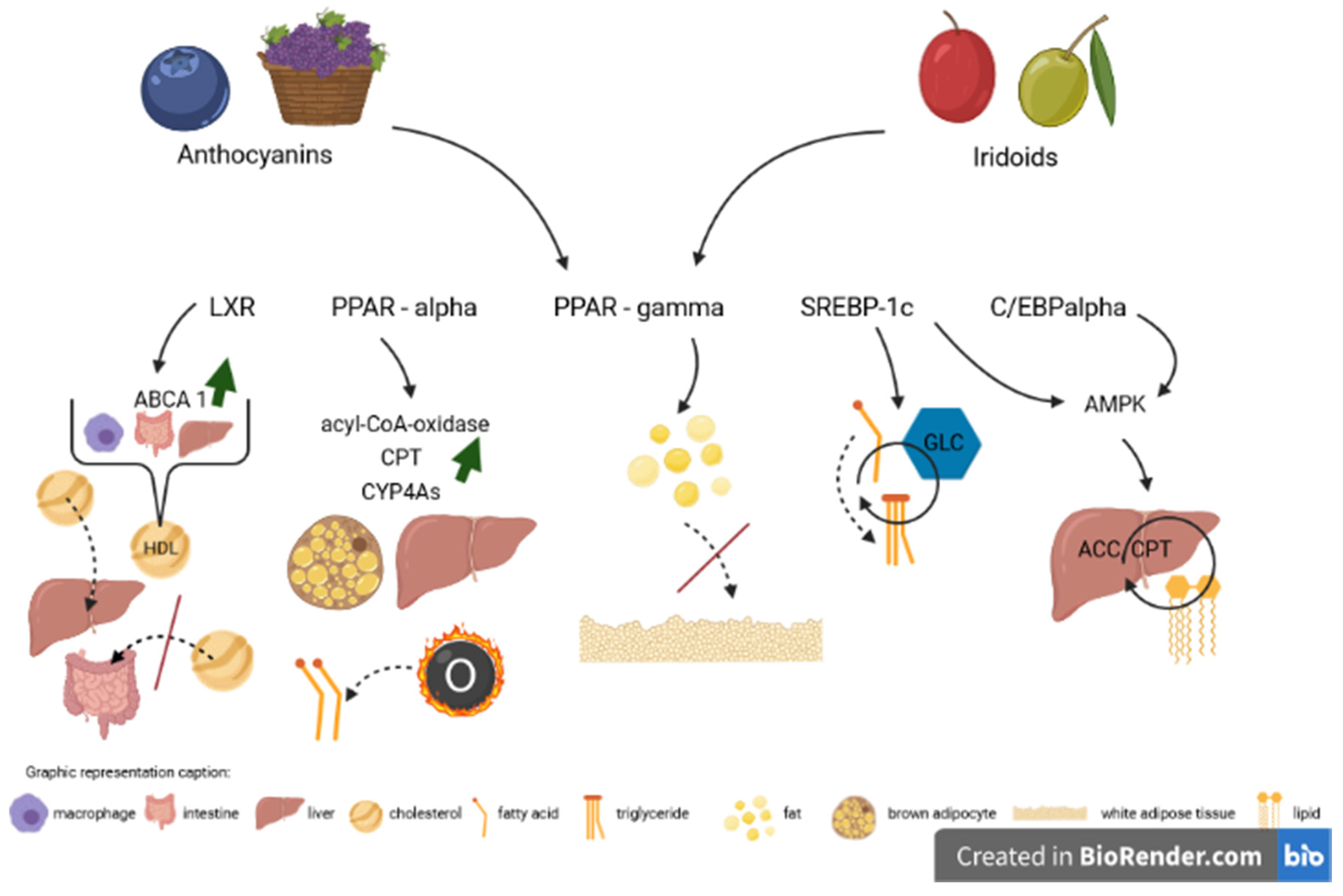
3. Impact of Anthocyanins

Anthocyanins are a group of natural substances that have garnered increasing interest in recent years. Anthocyanins are natural polyphenolic glycosidic phytopigments (mainly red, blue or purple), occurring primarily in fruits and flowers, that are biochemically related to flavonoids. Structurally, anthocyanins are aliphatic or aromatic three-ring compounds with one or more sugar molecules, and sometimes with a sugar attached aryl group. The colored aglycons are the anthocyanidins (usually cyanidin, pelargonidin or delphinidin) [33] (Figure 1). Fruits, which contain anthocyanins, are the sources of 90% of their habitual consumption [34]. Although anthocyanins are common in fruits, their daily intake by individual people in western countries is often relatively low [35]. The most common sources of these compounds are blueberries, blackberries, cranberries, acai berries, chokeberries, purple sweet potatoes, cherries, redcurrants or black soybeans. Anthocyanins possess potential anti-oxidative, antimicrobial, anti-inflammatory, anti-aging, lipid-lowering, anti-diabetic, anti-cancerous and anti-obesity bioactivities [36,37].
Anthocyanins suppress lipid accumulation in adipocytes due to the broad inhibition of the transcription factors regulating lipogenesis. This may partially explain the mechanism by which anthocyanins exert their anti-obesity effect. Anthocyanins may also lessen the process of triglyceride and lipid accumulation during adipocyte differentiation [38]. They reduce the gene and protein expression levels of lipogenic transcription factors such as liver X receptor α, sterol regulatory element-binding protein 1c, peroxisome proliferator-activated receptor-γ and CCAAT/enhancer binding protein α. A fortiori, target genes’ and proteins’ expression of these TFs is also distinctly suppressed by anthocyanins [38].
One of the most common anthocyanidins (aglicons of anthocyanins) is cyanidin. Cyanidin is natural compound abundant in fruits and vegetables, i.a. hawthorn, hibiscus or blueberry [33]. Jia et al. [13] showed that cyanidin is a direct ligand for both LXRα and LXRβ. Moreover, cyanidin affects all three PPAR subtypes, with the greatest affinity to PPAR-α [39]. Cyanidin-3-O-β-glucoside (C3G) activates liver X receptor (LXR)-ATP-binding cassette transporter-dependent cholesterol efflux [40,41] and phosphorylation of cellular AMPK in human hepatoma (HepG2) cells [27,42,43]. It was proven that C3G decreases the expression of adipogenic transcription factors involved in hepatic lipid metabolism, such as SREBP-1c, C/EBPα and PPAR-γ, both in the HepG2 cells and in the livers of supplemented mice [44]. The decreasing influence of anthocyanins on the gene expression of SREBP-1c and C/EBPα was also validated in other studies [43,45,46,47].
Nonetheless, the issue of anthocyanins’ effects on PPAR-α activation still requires more research. There have been reports that anthocyanins have a strong affinity and a significant impact on the expression of this receptor [39,41,45,48], while Rimando et al.’s [49] results on the hamster model stand in opposition to the above findings.
Anthocyanins have been reported to have beneficial effects on obesity and obesity-related metabolic disorders (e.g., insulin resistance and dyslipidemia). Anthocyanins elicited from, e.g., purple corn, sweet cherry, hibiscus or black soybean, in mice or 3T3-L1 preadipocyte models, prevented adipocyte differentiation, lipid accumulation and low-density lipoprotein (LDL) oxidation and protected against diet-induced hepatic steatosis by reducing PPAR-γ transcriptional activity [50,51,52,53]. A very abundant source of anthocyanins is the fruits of Aronia melanocarpa (AM). Park et al. [54] conducted an interesting AM study both on cells and mice. It appeared that AM extract significantly reduced free fatty acid-induced lipid droplet accumulation in vitro. Moreover, the synthesis of PPAR-γ2 was inhibited in vivo, the transcriptional activity of PPAR-γ2 was downregulated in vitro and the mRNA expression of PPAR-γ2 and its target genes, adipocyte protein 2 and lipoprotein lipase, was lowered both in vitro and in vivo.
However, in a recently conducted human study, berry supplementation triggered an inhibition of nuclear factor-kappa B (NF-κB)-dependent gene expression, accompanied by an enhancement in PPAR-γ expression. Nevertheless, an improvement in selected features of metabolic syndrome and related cardiovascular risk factors was observed [55]. Therefore, it can be hypothesized that stimulation or inhibition of PPAR-γ expression by anthocyanins may have both positive and negative consequences. Stimulation of this transcription factor may contribute to the reduction of inflammation while acting as a lipogenic, whereas the inhibition of PPAR-γ expression brings beneficial effects in the context of lipid homeostasis. Overall, it may lead to a partial prevention of obesity-induced atherosclerosis by attenuating inflammatory responses. It is also worth remembering that there are differences in fat metabolism between rodents and humans, which also may translate into disparities in the results of individual research models.
In 2019, Park et al. [37] confirmed most of the properties attributed to anthocyanins in earlier studies. They proved that another common anthocyanin—Delphinidin-3-O-glucoside (D3G)—significantly inhibited the accumulation of lipids in a dose-dependent manner without displaying cytotoxicity. In the 3T3-L1 adipocytes, D3G lowered the expression of major adipogenic and lipogenic transcription factors, i.e., PPAR-γ, SREBP-1 and C/EBPα. In addition, D3G enhanced the phosphorylation of AMPK and ACC—a fatty acid oxidation enzyme. These results once again supported the previously presented hypothesis that anthocyanins attenuate adipogenesis and promote lipid metabolism and are potential therapeutic agents in the treatment of obesity. Similar findings were obtained with a sole aglicon, delphinidin [56].
Anthocyanins represent also an interesting therapeutic option in diabetic nephropathy. Renal damage occurring in this disorder is mainly caused by oxidative stress and lipotoxicity. Db/db mice treated with anthocyanins show decreased albuminuria, ameliorated intra-renal lipid concentrations and improvement of glomerular matrix expansion and inflammation related to an increase in AMPK phosphorylation, upregulation of PPAR-α and PPAR-γ and decrease in SREBP-1 activity [48]. The studies describing the impact of anthocyanins on the discussed transcription factors are summarized in Table 1.

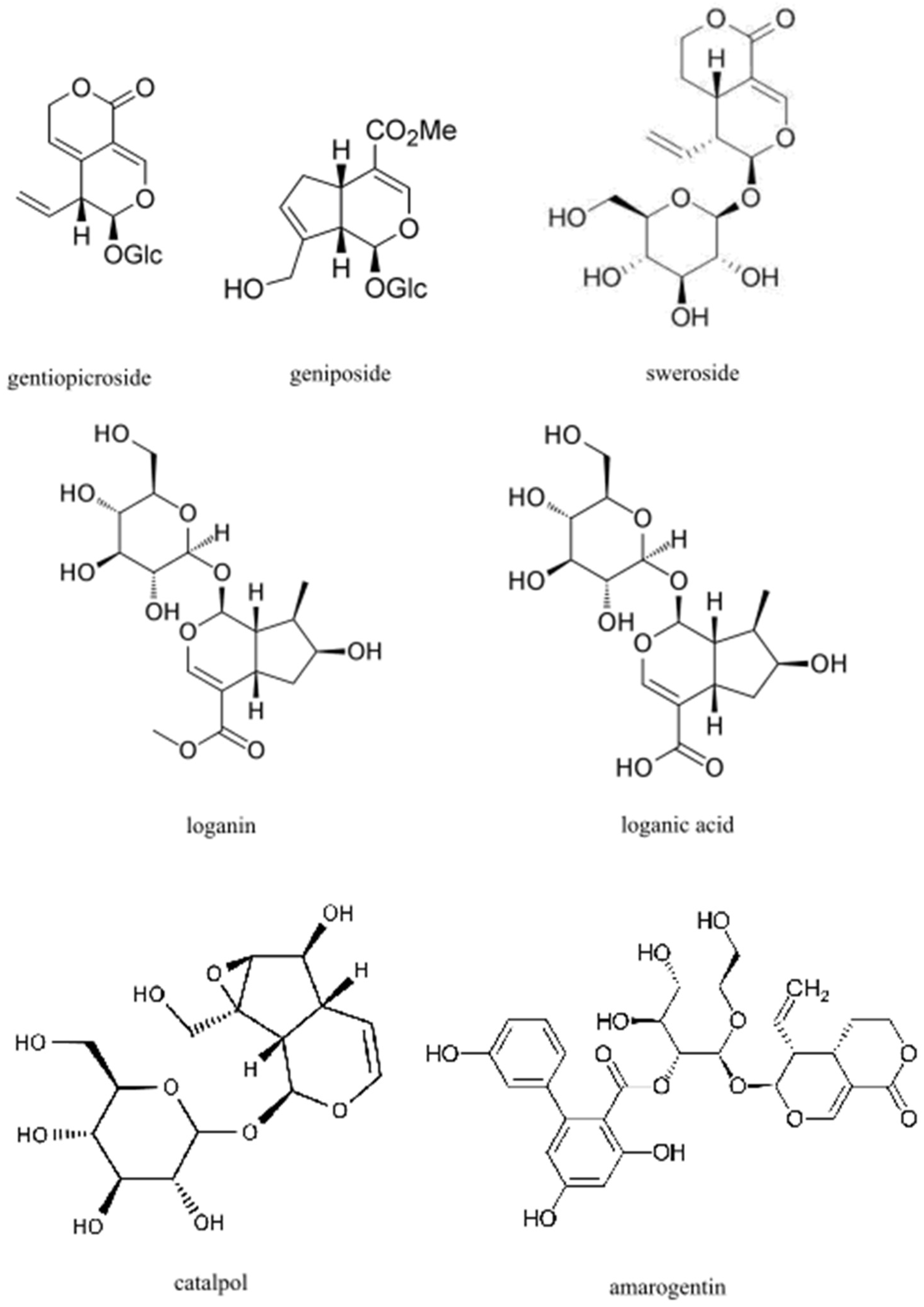
4. Impact of Iridoids

Iridoids are a large group of organic monoterpenoids, occurring in plants usually as glycosides with a glucose moiety attached to C-1 in the pyrene ring. The basic structural feature of iridoids is a bicyclic H-5/H-9β, β-cis-fusedcyclopentanopyran ring system. Iridoids are found in the green parts of plants, mainly in the leaves and young stems, and sometimes in fruits and sprouts. They are found in many plant families, e.g., Apocynaceae, Gentianaceae, Lamiaceae, Loganiaceae, Rubiaceae, Scrophulariaceae and Verbenaceae. Among the most commonly mentioned iridoids in the therapeutic context, the following stand out above all: gentiopicroside, geniposide, sweroside, loganin, loganic acid, catalpol and amarogentin [33] (Figure 2). In foods consumed in western diets, they are rarely present. However, they can be found in some raw fruits (e.g., olives, cornelian cherry, honeysuckle berries and cranberries) and in products derived from these fruits [57,58,59,60]. Iridoids possess potential cardiovascular, hypoglycemic, hypolipidemic, antihepatotoxic, choleretic, anti-inflammatory, antispasmodic, antitumor, antiviral, immunomodulatory and purgative bioactivities [61].
MicroRNAs are short, non-coding, single-stranded RNA molecules which have been reported to play a key role in the adjustment of lipid and lipoprotein metabolism. Increased levels of some miRNAs have been observed in the circulation in the course of hyperlipidemia [62]. It was proven that genipin upregulates the expression of miR-142a-5p, which binds to SREBP-1c and consequently leads to the inhibition of lipogenesis, which results in decreased body weight, lipid serum levels and hepatic lipid accumulation in HFD mice [63]. However, it is worth noting that there are a lot of miRNAs involved in fat metabolism, and some variants grow while others decline in the course of hyperlipidemia.
Zhu et al. [64] conducted an interesting study whose assumptions were based on providing HFD rats with three different doses of an iridoid fraction isolated from Valeriana jatamansi. Compared to the model group, a decrease in weight gain as well as a decrease in triglyceride and an increase in HDL-C serum levels were noticed regardless of the dose. All doses enhanced the expression of the PPAR-α receptor and reduced the expression of the SREBP-1c protein in the liver. In addition, depending on the dose, alterations in liver X receptor-α expression were observed. Hypolipidemic and antioxidant activity of iridoids through the stimulation of PPAR-α was noted also in different studies [36,65,66,67,68].
Cornus alternifolia (CA) is a tree that is common mainly in Eastern Asia and North America. Extracts of this plant have been used in traditional Chinese medicine as tonics, analgesics and diuretic drugs, while, in the United States, CA is grown as an ornamental plant. Cornus alternifolia leaves are a valuable source of iridoid compounds. It was shown that one of them—kaempferol-3-O-β-glucopyranoside—exhibited significant agonistic activities for PPAR-α, PPAR-γ and LXR dose-dependently in human hepatoma (HepG2) cells and Chinese hamster ovary cells (CHO) [69]. Several other iridoid substances isolated from CA leaves also influenced the expression of these transcription factors, but it is kaempferol-3-O-β-glucopyranoside which presents the most promising curative potential [69]. Fraxinus is a tree genus from which valuable quantities of iridoid compounds can also be obtained. Fraxinus compounds show significant inhibitory activity on adipocyte differentiation in the 3T3-L1 cells, reduce fat and triglyceride accumulation in differentiated 3T3-L1 cells without affecting cell viability and suppress the induction of C/EBPα, C/EBPβ and PPAR-γ transcription factors, concomitantly activating the PPAR-α receptor [70,71]. Similar effects were noticed in the case of iridoids of olive trees [72].
As with anthocyanins, iridoids, e.g., catalpol, may be a useful therapeutic option for the treatment of diabetes complications [73]. Gentiopicroside, a compound that belongs to the sub-group of seco-iridoids, ameliorated dyslipidemia and improved nerve blood flow through regulating the PPAR-γ/AMPK/ACC signal pathway in a diabetic peripheral neuropathy rat model [74]. Moreover, gentiopicroside in both acute and chronic alcohol-induced mouse hepatosteatosis (the latter stage of alcoholic liver disease, ALD) models mitigated the upregulation of SREBP-1 and downregulation of PPAR-α, among others, by activation of AMPK [75]. Alleviation of SREBP-1 expression was observed also in a nonalcoholic fatty liver disease model, namely by swertiamarin [76].
Iridoids and seco-iridoids such as catalpol, geniposide, harpagoside, loganin and oleuropein have been found to exhibit essential neuroprotective effects in Alzheimer’s disease (AD) and Parkinson’s disease (PD). One of the proposed mechanisms by which the process of neurogedeneration is slowed down is by increasing the PPAR-γ receptor expression, which contributes to the clearance of amyloid-β in the apolipoprotein E-mediated pathway in the brain [77].
For the prevention and treatment of obesity and dyslipidemia, a potentially useful iridoid compound is loganic acid (LA). Park et al. [78] demonstrated the antiadipogenic effects of LA in vitro and in vivo. Loganic acid treatment significantly decreased the adipocyte differentiation of 3T3-L1 preadipocytes in a dose-dependent manner and substantially lowered the expression of key adipogenesis transcription factors such as peroxisome proliferator-activated receptor-γ and CCAAT/enhancer-binding protein α. Moreover, a diminution of body weight gain, total fat increase, fatty hepatocyte deposition in the liver and adipocyte enlargement in the abdominal visceral fat tissues were noticed in an ovariectomy-induced obesity mice model compared to the comparative group. Loganic acid is also an important active ingredient found in cornelian cherry fruits. Cornus mas fruits, abundant in substantial amounts of both anthocyanins and iridoids, may exert positive additive or synergistic effects on dyslipidemia and atherosclerosis. In a rabbit model of diet-induced dyslipidemia and atherosclerosis, Sozański et al. [36] demonstrated that both anthocyanins and, to a lesser extent, loganic acid increased the expression of PPAR-α and PPAR-γ receptors in the liver and decreased intima thickness and the intima/media ratio in the thoracic aorta. The studies describing the impact of iridoids on the discussed transcription factors are summarized in Table 2.

5. Future Perspectives

Improper nutrition is a compelling problem nowadays. The share of plant products in the diets of many people should be much more extensive. In this context, compounds with proven therapeutic effects on the body, such as anthocyanins and iridoids, may play an increasingly important role in the prevention and adjunctive or combined treatment of many human diseases. Many studies confirmed the curative effects and mechanisms of action of these groups of substances. Unfortunately, the majority of these studies were performed on animals, and first and foremost, so the paramount goal for the near future is to conduct more clinical studies to confirm the positive impact of anthocyanins and iridoids on the human body. New applications for the above-mentioned compounds should also be sought. Interestingly, most of the conducted research pertains to the influence of anthocyanins and iridoids on high-fat-diet models. It would certainly be a challenging option to consider a study, or a whole series, verifying the impact of these substances on multiple parameters in a low-fat diet.

6. Conclusions

In conclusion, the current knowledge clearly points to the benefits of consuming foods comprising anthocyanins and iridoids. Both groups of substances modulate the expression of crucial transcription factors in lipid and cholesterol homeostasis, i.e., liver X receptor, peroxisome proliferator-activated receptor-α, peroxisome proliferator-activated receptor-γ, CCAAT/enhancer binding protein α and sterol regulatory element-binding protein 1c and their target genes (Figure 3). Therefore, anthocyanins and iridoids present an essential therapeutic option in disorders proceeding with disturbances in lipid and cholesterol metabolism, such as obesity, atherosclerosis, diabetes, metabolic syndrome and many more.

Author Contributions

Conceptualization, M.D. and T.S.; literature search, M.D.; writing—original draft preparation, M.D.; writing—review and editing, A.M., A.S. and T.S.; visualization, M.D. All authors have read and agreed to the published version of the manuscript.

Funding

This research received no external funding.

Conflicts of Interest

The authors declare no conflict of interest.

Abbreviations

ABCA1ATP-binding cassette transporter A1
HDLhigh-density lipoprotein
acyl-CoA-oxidaseacyl coenzyme A oxidase
CPTcarnitine palmitoyl transferase
CYP4Ascytochrome P450 4A subfamily enzymes
GLCglucose
AMPKAMP-activated protein kinase
ACCacetyl coenzyme A carboxylase
Oβ-oxidation

References

  1. Papavassiliou, A.G. Molecular Medicine. Transcription Factors. N. Engl. J. Med. 1995, 332, 45–47. [Google Scholar] [CrossRef] [PubMed]
  2. Lee, T.I.; Young, R.A. Transcriptional Regulation and its Misregulation in Disease. Cell 2013, 152, 1237–1251. [Google Scholar] [CrossRef] [PubMed] [Green Version]
  3. Vaquerizas, J.M.; Kummerfeld, S.K.; Teichmann, S.A.; Luscombe, N.M. A Census of Human Transcription Factors: Function, Expression and Evolution. Nat. Rev. Genet. 2009, 10, 252–263. [Google Scholar] [CrossRef]
  4. Papavassiliou, K.A.; Papavassiliou, A.G. Transcription Factor Drug Targets. J. Cell Biochem. 2016, 9999, 1–5. [Google Scholar] [CrossRef]
  5. Ortega, E.; Rengachari, S.; Ibrahim, Z.; Hoghoughi, N.; Gaucher, J.; Holehouse, A.S.; Khochbin, S.; Panne, D. Transcription Factor Dimerization Activates the p300 Acetyltransferase. Nature 2019, 562, 538–544. [Google Scholar] [CrossRef]
  6. Lee, T.I.; Young, R.A. Transcription of Eukaryotic Protein-Coding Genes. Annu. Rev. Genet. 2000, 34, 77–137. [Google Scholar] [CrossRef]
  7. Lemon, B.; Tjian, R. Orchestrated Response: A Symphony of Transcription Factors for Gene Control. Genes Dev. 2000, 14, 2551–2569. [Google Scholar] [CrossRef] [Green Version]
  8. Orphanides, G.; Lagrange, T.; Reinberg, D. The General Transcription Factors of RNA Polymerase II. Genes Dev. 1996, 10, 2657–2683. [Google Scholar] [CrossRef] [PubMed] [Green Version]
  9. Bartnik, E. Struktura i działanie genów eukariotycznych. In Genetyka Molekularna; Węgleński, P., Ed.; Wydawnictwa Naukowe PWN: Warszawa, Poland, 2002; p. 497. [Google Scholar]
  10. Cao, G.; Liang, Y.; Broderick, C.L.; Oldham, B.A.; Beyer, T.P.; Schmidt, R.J.; Zhang, Y.; Stayrook, K.R.; Suen, C.; Otto, K.A.; et al. Antidiabetic Action of Liver X Receptor Agonist Mediated by Inhibition of Hepatic Gluconeogenesis. J. Biol. Chem. 2003, 278, 1131–1136. [Google Scholar] [CrossRef] [PubMed] [Green Version]
  11. Shuang, R.; Rui, X.; Wenfang, L. Phytosterols and Dementia. Plant Foods Hum. Nutr. 2016, 71, 347–354. [Google Scholar] [CrossRef]
  12. Sozański, T.; Kucharska, A.Z.; Szumny, A.; Magdalen, J.; Bielska, K.; Merwid-Ląd, A.; Woźniak, A.; Dzimira, S.; Piórecki, N.; Trocha, M. The Protective Effect of the Cornus Mas Fruits (Cornelian Cherry) on Hypertriglyceridemia and Atherosclerosis Through PPARα Activation in Hypercholesterolemic Rabbits. Phytomedicine 2014, 21, 1774–1784. [Google Scholar] [CrossRef]
  13. Jia, Y.; Hoang, M.H.; Jun, H.J.; Lee, J.H.; Lee, S.J. Cyanidin, a Natural Flavonoid, Is an Agonistic Ligand for Liver X Receptor Alpha and Beta and Reduces Cellular Lipid Accumulation in Macrophages and Hepatocytes. Bioorganic Med. Chem. Lett. 2013, 23, 4185–4190. [Google Scholar] [CrossRef] [PubMed]
  14. Guo, S.; Li, L.; Yin, H. Cholesterol Homeostasis and Liver X Receptor (LXR) in Atherosclerosis. Cardiovasc. Hematol. Disord. Drug Targets 2018, 18, 27–33. [Google Scholar] [CrossRef]
  15. Chen, Z.; Liu, J.; Fu, Z.; Ye, C.; Zhang, R.; Song, Y.; Zhang, Y.; Li, H.; Ying, H.; Liu, H. 24(S)-Saringosterol from Edible Marine Seaweed Sargassum fusiforme is a Novel Selective LXRbeta Agonist. J. Agric. Food Chem. 2014, 62, 6130–6137. [Google Scholar] [CrossRef]
  16. Hoang, M.H.; Jia, Y.; Jun, H.J.; Lee, J.H.; Lee, B.Y.; Lee, S.J. Fucosterol is a Selective Liver X Receptor Modulator that Regulates the Expression of Key Genes in Cholesterol Homeostasis in Macrophages, Hepatocytes, and Intestinal Cells. J. Agric. Food Chem. 2012, 60, 11567–11575. [Google Scholar] [CrossRef] [PubMed]
  17. El Kharrassi, Y.; Samadi, M.; Lopez, T.; Nury, T.; El Kebbaj, R.; Andreoletti, P.; El Hajj, H.I.; Vamecq, J.; Moustaid, K.; Latruffe, N.; et al. Biological Activities of Schottenol and Spinasterol, Two Natural Phytosterols Present in Argan Oil and in Cactus Pear Seed Oil, on Murine Microglial BV2 cells. Biochem. Biophys. Res. Commun. 2014, 446, 798–804. [Google Scholar] [CrossRef]
  18. Bogie, J.; Hoeks, C.; Schepers, M.; Tiane, A.; Cuypers, A.; Leijten, F.; Chintapakorn, Y.; Suttiyut, T.; Pornpakakul, S.; Struik, D.; et al. Dietary Sargassum fusiforme Improves Memory and Reduces Amyloid Plaque Load in an Alzheimer’s Disease Mouse Model. Sci. Rep. 2019, 9, 4908. [Google Scholar] [CrossRef] [Green Version]
  19. Liu, H.; Zhong, H.; Leng, L.; Jiang, Z. Effects of Soy Isoflavone on Hepatic Steatosis in High Fat-Induced Rats. J. Clin. Biochem. Nutr. 2017, 61, 85–90. [Google Scholar] [CrossRef]
  20. Ronis, M.J. Effects of Soy Containing Diet and Isoflavones on Cytochrome P450 Enzyme Expression and Activity. Drug Metab. Rev. 2016, 48, 331–341. [Google Scholar] [CrossRef]
  21. Illesca, P.; Valenzuela, R.; Espinosa, A.; Echeverría, F.; Soto-Alarcon, S.; Ortiz, M.; Videla, L.A. Hydroxytyrosol Supplementation Ameliorates the Metabolic Disturbances in White Adipose Tissue From Mice Fed a High-Fat Diet Through Recovery of Transcription Factors Nrf2, SREBP-1c, PPAR-γ and NF-κB. Biomed. Pharmacother. 2019, 109, 2472–2481. [Google Scholar] [CrossRef] [PubMed]
  22. Wakil, S.J.; Abu-Elheiga, L.A. Fatty Acid Metabolism: Target for Metabolic Syndrome. J. Lipid Res. 2009, 50, S138–S143. [Google Scholar] [CrossRef] [Green Version]
  23. Ramji, D.P.; Foka, P. CCAAT/enhancer-Binding Proteins: Structure, Function and Regulation. Biochem. J. 2002, 365, 561–575. [Google Scholar] [CrossRef] [Green Version]
  24. Clarke, S.L.; Robinson, C.E.; Gimble, J.M. CCAAT/enhancer Binding Proteins Directly Modulate Transcription From the Peroxisome Proliferator-Activated Receptor Gamma 2 Promoter. Biochem. Biophys. Res. Commun. 1997, 240, 99–103. [Google Scholar] [CrossRef]
  25. Chan, C.Y.; Wei, L.; Castro-Munozledo, F.; Koo, W.L. (−)-Epigallocatechin-3-gallate Blocks 3T3-L1 Adipose Conversion by Inhibition of Cell Proliferation and Suppression of Adipose Phenotype Expression. Life Sci. 2011, 89, 779–785. [Google Scholar] [CrossRef] [PubMed]
  26. Angin, Y.; Beauloye, C.; Horman, S.; Bertrand, L. Regulation of Carbohydrate Metabolism, Lipid Metabolism, and Protein Metabolism by AMPK. In AMP-Activated Protein Kinase; Cordero, M., Viollet, B., Eds.; Springer International Publishing: Cham, Switzerland, 2016; pp. 23–43. [Google Scholar]
  27. Guo, H.; Liu, G.; Zhong, R.; Wang, Y.; Wang, D.; Xia, M. Cyanidin-3-O-β-glucoside Regulates Fatty Acid Metabolism via an AMP-activated Protein Kinase-dependent Signaling Pathway in Human HepG2 cells. Lipids Health Dis. 2012, 11, 10. [Google Scholar] [CrossRef] [PubMed] [Green Version]
  28. Bengoechea-Alonso, M.T.; Ericsson, J. SREBP in Signal Transduction: Cholesterol Metabolism and Beyond. Curr. Opin. Cell Biol. 2007, 19, 215–222. [Google Scholar] [CrossRef]
  29. Chen, G.; Liang, G.; Ou, J.; Goldstein, J.L.; Brown, M.S. Central Role of Liver X Receptor in Insulin-mediated Activation of SREBP-1c Transcription and Stimulation of Fatty Acid Synthesis in Liver. Proc. Natl. Acad. Sci. USA 2004, 101, 11245–11250. [Google Scholar] [CrossRef] [PubMed] [Green Version]
  30. Ferré, P.; Foufelle, F. Hepatic Steatosis: A Role for de novo Lipogenesis and the Transcription Factor SREBP-1c. Diabetes Obes. Metab. 2010, 12, 83–92. [Google Scholar] [CrossRef] [PubMed]
  31. Yamazaki, T.; Li, D.; Ikaga, R. Effective Food Ingredients for Fatty Liver: Soy Protein β-Conglycinin and Fish Oil. Int. J. Mol. Sci. 2018, 19, 4107. [Google Scholar] [CrossRef] [PubMed] [Green Version]
  32. Terzo, S.; Caldara, G.F.; Ferrantelli, V.; Puleio, R.; Cassata, G.; Mulè, F.; Amato, A. Pistachio Consumption Prevents and Improves Lipid Dysmetabolism by Reducing the Lipid Metabolizing Gene Expression in Diet-Induced Obese Mice. Nutrients 2018, 10, 1857. [Google Scholar] [CrossRef] [Green Version]
  33. Danielewski, M.; Matuszewska, A.; Nowak, B.; Kucharska, A.Z.; Sozański, T. The Effects of Natural Iridoids and Anthocyanins on Selected Parameters of Liver and Cardiovascular System Functions. Oxidative Med. Cell Longev. 2020, 2020, 2735790. [Google Scholar] [CrossRef] [PubMed]
  34. Cassidy, A.; Bertoia, M.; Chiuve, S.; Flint, A.; Forman, J.; Rimm, E.B. Habitual Intake of Anthocyanins and Flavanones and Risk of Cardiovascular Disease in Men. Am. J. Clin. Nutr. 2016, 104, 587–594. [Google Scholar] [CrossRef] [PubMed] [Green Version]
  35. Wu, X.; Beecher, G.R.; Holden, J.M.; Haytowitz, D.B.; Gebhardt, S.E.; Prior, R.L. Concentrations of Anthocyanins in Common Foods in the United States and Estimation of Normal Consumption. J. Agric. Food Chem. 2006, 54, 4069–4075. [Google Scholar] [CrossRef] [PubMed]
  36. Sozański, T.; Kucharska, A.Z.; Rapak, A.; Szumny, D.; Trocha, M.; Merwid-Ląd, A.; Dzimira, S.; Piasecki, T.; Piórecki, N.; Magdalan, J.; et al. Iridoid-loganic Acid Versus Anthocyanins From the Cornus Mas Fruits (Cornelian Cherry): Common and Different Effects on Diet-Induced Atherosclerosis, PPARs Expression and Inflammation. Atherosclerosis 2016, 254, 151–160. [Google Scholar] [CrossRef]
  37. Park, M.; Sharma, A.; Lee, H.J. Anti-Adipogenic Effects of Delphinidin-3- O- β-Glucoside in 3T3-L1 Preadipocytes and Primary White Adipocytes. Molecules 2019, 24, 1848. [Google Scholar] [CrossRef] [Green Version]
  38. Lee, B.; Lee, M.; Lefevre, M.; Kim, H.R. Anthocyanins Inhibit Lipogenesis During Adipocyte Differentiation of 3T3-L1 Preadipocytes. Plant Foods Hum. Nutr. 2014, 69, 137–141. [Google Scholar] [CrossRef]
  39. Jia, Y.; Kim, J.Y.; Jun, H.J.; Kim, S.J.; Lee, J.H.; Hoang, M.H.; Kim, H.S.; Chang, H.I.; Hwang, K.Y.; Um, S.J.; et al. Cyanidin is an Agonistic Ligand for Peroxisome Proliferator-Activated Receptor-alpha Reducing Hepatic Lipid. Biochim. Biophys. Acta 2013, 1831, 698–708. [Google Scholar] [CrossRef]
  40. Fu, Y.; Wei, Z.; Zhou, E.; Zhang, N.; Yang, Z. Cyanidin-3-O-β-glucoside Inhibits Lipopolysaccharide-Induced Inflammatory Response in Mouse Mastitis Model. J. Lipid Res. 2014, 55, 1111–1119. [Google Scholar] [CrossRef] [Green Version]
  41. Du, C.; Shi, Y.; Ren, Y.; Wu, H.; Yao, F.; Wei, J.; Wu, M.; Hou, Y.; Duan, H. Anthocyanins Inhibit High-Glucose-Induced Cholesterol Accumulation and Inflammation by Activating LXRα Pathway in HK-2 Cells. Drug Des. Dev. Ther. 2015, 9, 5099–5113. [Google Scholar]
  42. Hwang, Y.P.; Choi, J.H.; Han, E.H.; Kim, H.G.; Wee, J.H.; Jung, K.O.; Jung, K.H.; Kwon, K.I.; Jeong, T.C.; Chung, Y.C.; et al. Purple Sweet Potato Anthocyanins Attenuate Hepatic Lipid Accumulation through Activating Adenosine Monophosphate-activated Protein Kinase in Human HepG2 Cells and Obese Mice. Nutr. Res. 2011, 31, 896–906. [Google Scholar] [CrossRef]
  43. Chang, J.J.; Hsu, M.J.; Huang, H.P.; Chung, D.J.; Chang, Y.C.; Wang, C.J. Mulberry Anthocyanins Inhibit Oleic Acid Induced Lipid Accumulation by Reduction of Lipogenesis and Promotion of Hepatic Lipid Clearance. J. Agric. Food Chem. 2013, 61, 6069–6076. [Google Scholar] [CrossRef]
  44. Park, M.; Yoo, J.H.; Lee, Y.S.; Lee, H.J. Lonicera caerulea Extract Attenuates Non-Alcoholic Fatty Liver Disease in Free Fatty Acid-Induced HepG2 Hepatocytes and in High Fat Diet-Fed Mice. Nutrients 2019, 11, 494. [Google Scholar] [CrossRef] [Green Version]
  45. de Sousa, A.R.; de Castro Moreira, M.E.; Lopes Toledo, R.C.; Anjos Benjamin, L.D.; Vieira Queiroz, V.A.; Paranho Veloso, M.; de Souza Reis, K.; Duarte Martino, H.S. Extruded Sorghum (Sorghum Bicolor L.) Reduces Metabolic Risk of Hepatic Steatosis in Obese Rats Consuming a High Fat Diet. Food Res. Int. 2018, 112, 48–55. [Google Scholar] [CrossRef] [PubMed] [Green Version]
  46. Park, S.; Kang, S.; Jeong, D.Y.; Jeong, S.Y.; Park, J.J.; Yun, H.S. Cyanidin and Malvidin in Aqueous Extracts of Black Carrots Fermented With Aspergillus Oryzae Prevent the Impairment of Energy, Lipid and Glucose Metabolism in Estrogen-Deficient Rats by AMPK Activation. Genes Nutr. 2015, 10, 455. [Google Scholar] [CrossRef]
  47. Khan, M.I.; Shin, J.H.; Shin, T.S.; Kim, M.Y.; Cho, N.J.; Kim, J.D. Anthocyanins From Cornus Kousa Ethanolic Extract Attenuate Obesity in Association With Anti-Angiogenic Activities in 3T3-L1 Cells by Down-Regulating Adipogeneses and Lipogenesis. PLoS ONE 2018, 13, e0208556. [Google Scholar] [CrossRef] [PubMed]
  48. Koh, E.S.; Lim, J.H.; Kim, M.Y.; Chung, S.; Shin, S.J.; Choi, B.S.; Kim, H.W.; Hwang, S.Y.; Kim, S.W.; Park, C.W.; et al. Anthocyanin-rich Seoritae Extract Ameliorates Renal Lipotoxicity via Activation of AMP-activated Protein Kinase in Diabetic Mice. J. Transl. Med. 2015, 13, 203. [Google Scholar] [CrossRef] [PubMed] [Green Version]
  49. Rimando, A.M.; Khan, S.I.; Mizuno, C.S.; Ren, G.; Mathews, S.T.; Kim, H.; Yokoyama, W. Evaluation of PPARα Activation by Known Blueberry Constituents. J. Sci. Food Agric. 2016, 96, 1666–1671. [Google Scholar] [CrossRef]
  50. Luna-Vital, D.; Weiss, M.; de Mejia, E.G. Anthocyanins From Purple Corn Ameliorated Tumor Necrosis Factor-α-Induced Inflammation and Insulin Resistance in 3T3-L1 Adipocytes via Activation of Insulin Signaling and Enhanced GLUT4 Translocation. Mol. Nutr. Food Res. 2017, 61, 1700362. [Google Scholar] [CrossRef] [PubMed]
  51. Song, H.; Wu, T.; Xu, D.; Chu, Q.; Lin, D.; Zheng, X. Dietary Sweet Cherry Anthocyanins Attenuates Diet-Induced Hepatic Steatosis by Improving Hepatic Lipid Metabolism in Mice. Nutrition 2016, 32, 827–833. [Google Scholar] [CrossRef] [PubMed]
  52. Kao, E.S.; Tseng, T.H.; Lee, H.J.; Chan, K.C.; Wang, C.J. Anthocyanin Extracted From Hibiscus Attenuate Oxidized LDL-mediated Foam Cell Formation Involving Regulation of CD36 Gene. Chem. Biol. Interact. 2009, 179, 212–218. [Google Scholar] [CrossRef]
  53. Kim, H.K.; Kim, J.N.; Han, S.N.; Nam, J.H.; Na, H.N.; Ha, T.J. Black Soybean Anthocyanins Inhibit Adipocyte Differentiation in 3T3-L1 Cells. Nutr. Res. 2012, 32, 770–777. [Google Scholar] [CrossRef] [PubMed]
  54. Park, C.H.; Kim, J.H.; Lee, E.B.; Hur, W.; Kwon, O.J.; Park, H.J.; Yoon, S.K. Aronia melanocarpa Extract Ameliorates Hepatic Lipid Metabolism through PPARγ2 Downregulation. PLoS ONE 2017, 12, e0169685. [Google Scholar] [CrossRef] [Green Version]
  55. Aboonabi, A.; Aboonabi, A. Anthocyanins Reduce Inflammation and Improve Glucose and Lipid Metabolism Associated With Inhibiting Nuclear factor-kappaB Activation and Increasing PPAR-γ Gene Expression in Metabolic Syndrome Subjects. Free Radic. Biol. Med. 2020, 150, 30–39. [Google Scholar] [CrossRef] [PubMed]
  56. Rahman, N.; Jeon, M.; Kim, Y.S. Delphinidin, a Major Anthocyanin, Inhibits 3T3-L1 Pre-Adipocyte Differentiation Through Activation of Wnt/β-catenin Signaling. Biofactors 2016, 42, 49–59. [Google Scholar]
  57. Turner, A.; Chen, S.N.; Nikolic, D.; van Breemen, R.; Farnsworth, N.R.; Pauli, G.F. Coumaroyl Iridoids and a Depside from Cranberry (Vaccinium macrocarpon). J. Nat. Prod. 2007, 70, 253–258. [Google Scholar] [CrossRef] [Green Version]
  58. Xie, P.J.; Huang, L.X.; Zhang, C.H.; Zhang, Y.L. Phenolic Compositions, and Antioxidant Performance of Olive Leaf and Fruit (Olea europaea L.) Extracts and their Structure–Activity Relationships. J. Funct. Foods 2015, 16, 460–471. [Google Scholar] [CrossRef]
  59. Kucharska, A.Z.; Szumny, A.; Sokół-Łętowska, A.; Piórecki, N.; Klymenko, S.V. Iridoids and Anthocyanins in Cornelian Cherry (Cornus mas L.) Cultivars. J. Food Compos. Anal. 2015, 40, 95–102. [Google Scholar] [CrossRef]
  60. Kucharska, A.Z.; Fecka, I. Identification of Iridoids in Edible Honeysuckle Berries (Lonicera caerulea L. var. kamtschatica Sevast.) by UPLC-ESI-qTOF-MS/MS. Molecules 2016, 21, 1157. [Google Scholar] [CrossRef] [PubMed] [Green Version]
  61. Ghisalberti, E.L. Biological and Pharmacological Activity of Naturally Occurring Iridoids and Secoiridoids. Phytomedicine 1998, 5, 147–163. [Google Scholar] [CrossRef]
  62. Desgagne, V.; Bouchard, L.; Guerin, R. MicroRNAs in Lipoprotein and Lipid Metabolism: From Biological Function to Clinical Application. Clin. Chem. Lab. Med. 2017, 55, 667–686. [Google Scholar] [CrossRef] [PubMed] [Green Version]
  63. Zhong, H.; Chen, K.; Feng, M.; Shao, W.; Wu, J.; Chen, K.; Liang, T.; Liu, C. Genipin Alleviates High-Fat Diet-Induced Hyperlipidemia and Hepatic Lipid Accumulation in Mice via miR-142a-5p/SREBP-1c Axis. FEBS J. 2018, 285, 501–517. [Google Scholar] [CrossRef] [PubMed]
  64. Zhu, J.; Xu, K.; Zhang, X.; Cao, J.; Jia, Z.; Yang, R.; Ma, C.; Chen, C.; Zhang, T.; Yan, Z. Studies on the Regulation of Lipid Metabolism and its Mechanism of the Iridoids Rich Fraction in Valeriana jatamansi Jones. Biomed. Pharmacother. 2016, 84, 1891–1898. [Google Scholar] [CrossRef] [PubMed]
  65. Malliou, F.; Andreadou, I.; Gonzalez, F.J.; Lazou, A.; Xepapadaki, E.; Vallianou, I.; Lambrinidis, G.; Mikros, E.; Marselos, M.; Skaltsounis, A.L.; et al. The Olive Constituent Oleuropein, as a PPARα Agonist, Markedly Reduces Serum Triglycerides. J. Nutr. Biochem. 2018, 59, 17–28. [Google Scholar] [CrossRef] [PubMed]
  66. Ma, T.; Huang, C.; Zong, G.; Zha, D.; Meng, X.; Li, J.; Tang, W. Hepatoprotective Effects of Geniposide in a Rat Model of Nonalcoholic Steatohepatitis. J. Pharm. Pharmacol. 2011, 63, 587–593. [Google Scholar] [CrossRef] [PubMed]
  67. Patel, T.P.; Rawal, K.; Soni, S.; Gupta, S. Swertiamarin Ameliorates Oleic Acid Induced Lipid Accumulation and Oxidative Stress by Attenuating Gluconeogenesis and Lipogenesis in Hepatic Steatosis. Biomed. Pharmacother. 2016, 83, 785–791. [Google Scholar] [CrossRef]
  68. Yang, Q.; Shu, F.; Gong, J.; Ding, P.; Cheng, R.; Li, J.; Tong, R.; Ding, L.; Sun, H.; Huang, W.; et al. Sweroside Ameliorates NAFLD in High-Fat Diet Induced Obese Mice Through the Regulation of Lipid Metabolism and Inflammatory Response. J. Ethnopharmacol. 2020, 255, 112556. [Google Scholar] [CrossRef]
  69. He, Y.Q.; Ma, G.Y.; Peng, J.N.; Ma, Z.Y.; Hamann, M.T. Liver X Receptor and Peroxisome Proliferator-Activated Receptor Agonist from Cornus alternifolia. Biochim. Biophys. Acta 2012, 1820, 1021–1026. [Google Scholar] [CrossRef] [Green Version]
  70. Choi, K.M.; Shin, E.; Liu, Q.; Yoo, H.S.; Kim, Y.C.; Sung, S.H.; Hwang, B.Y.; Lee, M.K. Hydroxyframoside B, a Secoiridoid of Fraxinus Rhynchophylla, Inhibits Adipocyte Differentiation in 3T3-L1 Cells. Planta Med. 2011, 77, 1020–1023. [Google Scholar] [CrossRef] [PubMed]
  71. Bai, N.; He, K.; Ibarra, A.; Bily, A.; Roller, M.; Chen, X.; Rühl, R. Iridoids From Fraxinus Excelsior With Adipocyte Differentiation-Inhibitory and PPARalpha Activation Activity. J. Nat. Prod. 2010, 73, 2–6. [Google Scholar] [CrossRef]
  72. Drira, R.; Chen, S.; Sakamoto, K. Oleuropein and Hydroxytyrosol Inhibit Adipocyte Differentiation in 3 T3-L1 Cells. Life Sci. 2011, 89, 708–716. [Google Scholar] [CrossRef] [Green Version]
  73. Bai, Y.; Zhu, R.; Tian, Y.; Li, R.; Chen, B.; Zhang, H.; Xia, B.; Zhao, D.; Mo, F.; Zhang, D.; et al. Catalpol in Diabetes and Its Complications: A Review of Pharmacology, Pharmacokinetics, and Safety. Molecules 2019, 24, 3302. [Google Scholar] [CrossRef] [Green Version]
  74. Lu, Y.; Yao, J.; Gong, C.; Wang, B.; Zhou, P.; Zhou, S.; Yao, X. Gentiopicroside Ameliorates Diabetic Peripheral Neuropathy by Modulating PPAR- Γ/AMPK/ACC Signaling Pathway. Cell Physiol. Biochem. 2018, 50, 585–596. [Google Scholar] [CrossRef]
  75. Li, X.; Zhang, Y.; Jin, Q.; Xia, K.L.; Jiang, M.; Cui, B.W.; Wu, Y.L.; Song, S.Z.; Lian, L.H.; Nan, J.X. Liver Kinase B1/AMP-activated Protein Kinase-Mediated Regulation by Gentiopicroside Ameliorates P2X7 Receptor-Dependent Alcoholic Hepatosteatosis. Br. J. Pharmacol. 2018, 175, 1451–1470. [Google Scholar] [CrossRef] [Green Version]
  76. Yang, Y.; Li, J.; Wei, C.; He, Y.; Cao, Y.; Zhang, Y.; Sun, W.; Qiao, B.; He, J. Amelioration of Nonalcoholic Fatty Liver Disease by Swertiamarin in Fructose-Fed Mice. Phytomedicine 2019, 59, 152782. [Google Scholar] [CrossRef]
  77. Dinda, B.; Dinda, M.; Kulsi, G.; Chakraborty, A.; Dinda, S. Therapeutic Potentials of Plant Iridoids in Alzheimer’s and Parkinson’s Diseases: A Review. Eur. J. Med. Chem. 2019, 169, 185–199. [Google Scholar] [CrossRef]
  78. Park, E.; Kim, J.; Yeo, S.; Kim, G.; Ko, E.H.; Lee, S.W.; Li, W.Y.; Choi, C.W.; Jeong, S.Y. Antiadipogenic Effects of Loganic Acid in 3T3-L1 Preadipocytes and Ovariectomized Mice. Molecules 2018, 23, 1663. [Google Scholar] [CrossRef] [Green Version]
Figure 1. Chemical structures of main anthocyanins’ aglycons.
Ijms 22 06074 g001
Figure 2. Chemical structures of main iridoids.
Ijms 22 06074 g002
Figure 3. Influence of anthocyanins and iridoids on main transcription factors involved in cholesterol and lipid metabolism. Accessed on 28 May 2021.
Ijms 22 06074 g003
Table 1. List of studies describing the impact of anthocyanins on discussed transcription factors.
Authors and Date of PublicationResearch ModelCompounds Used in StudyObserved Changes
Aboonabi et al., 2020 [55]humanberry anthocyanin supplementsPPAR-γ Ijms 22 06074 i001
Chang et al., 2013 [43]HepG2 cellsmulberry anthocyanin extractSREBP-1c Ijms 22 06074 i001 PPAR-α Ijms 22 06074 i002
de Sousa et al., 2018 [45]ratsextruded sorghum flourSREBP-1c Ijms 22 06074 i001 PPAR-α Ijms 22 06074 i002
Du et al., 2015 [41]HK-2 cellscyanidin-3-O-β-glucoside, cyanidinLXRα Ijms 22 06074 i002 PPAR-α Ijms 22 06074 i002
Fu et al., 2014 [40]mice, mice mammary epithelial cellscyanidin-3-O-β-glucosideLXRα Ijms 22 06074 i002
Hwang et al., 2011 [42]micepurple sweet potato anthocyanin fractionSREBP-1c Ijms 22 06074 i001
Jia et al., 2013 [39]HepG2 cells, CHO-K1 cellscyanidinPPAR-α Ijms 22 06074 i002 PPAR-δ Ijms 22 06074 i002
PPAR-γ Ijms 22 06074 i002
Jia et al., 2013 [13]macrophages, hepatocytescyanidinLXRα, LXRβ Ijms 22 06074 i002
SREBP-1c Ijms 22 06074 i002
Kao et al., 2009 [52]mouse macrophage J774A.1 cellshibiscus anthocyanin extractPPAR-γ Ijms 22 06074 i001
Khan et al., 2018 [47]3T3-L1 cellsC. kousa anthocyanin ethanolic leaf extractPPAR-γ Ijms 22 06074 i001 C/EBPα Ijms 22 06074 i001
Kim et al., 2012 [53]3T3-L1 cellsblack soybean anthocyanin extractPPAR-γ Ijms 22 06074 i001
Koh et al., 2015 [48]miceSeoritae anthocyanin extractPPAR-α Ijms 22 06074 i002 PPAR-γ Ijms 22 06074 i002
SREBP-1c Ijms 22 06074 i001
Lee et al., 2014 [38]3T3-L1 cellsgrape anthocyanin isolateLXRα Ijms 22 06074 i001 PPAR-γ Ijms 22 06074 i001
C/EBPα Ijms 22 06074 i001 SREBP-1c Ijms 22 06074 i001
Luna-Vital et al., 2017 [50]3T3-L1 cellspurple corn pericarp anthocyanin extract, pure anthocyaninsPPAR-γ Ijms 22 06074 i001
Park et al., 2015 [46]ratsunfermented and fermented black carrot extractSREBP-1c Ijms 22 06074 i001 PPAR-α Ijms 22 06074 i001
Park et al., 2017 [54]mice, FL83B cellsA. melanocarpa spray-dried ethanol extractPPAR-γ Ijms 22 06074 i001
Park et al., 2019 [44]HepG2 cellshoneyberry extractSREBP-1c Ijms 22 06074 i001 PPAR-γ Ijms 22 06074 i001
C/EBPα Ijms 22 06074 i001 PPAR-α Ijms 22 06074 i002
Park et al., 2019 [37]3T3-L1 cells, primary white adipocytesdelphinidin-3-O-β-glucosidePPAR-γ Ijms 22 06074 i001 C/EBPα Ijms 22 06074 i001
SREBP-1c Ijms 22 06074 i001
Rahman et al., 2016 [56]3T3-L1 cellsdelphinidinPPAR-γ Ijms 22 06074 i001 C/EBP Ijms 22 06074 i001
Rimando et al., 2016 [49]hamstersblueberry peel extract Ijms 22 06074 i003 PPAR-α
Song et al., 2016 [51]micesweet cherry anthocyaninsPPAR-γ Ijms 22 06074 i001
Sozański et al., 2014 [12]rabbitscornelian cherry fruits lyophilisatePPAR-α Ijms 22 06074 i002
Sozański et al., 2016 [36]rabbitsmixture of anthocyaninsPPAR-α Ijms 22 06074 i002 PPAR-γ Ijms 22 06074 i002
Ijms 22 06074 i002— up regulation, Ijms 22 06074 i001—down regulation, Ijms 22 06074 i003—unchanged.
Table 2. List of studies describing the impact of iridoids on discussed transcription factors.
Authors and Date of PublicationResearch ModelCompounds Used in StudyObserved Changes
Bai et al., 2010 [71]3T3-L1 cellsaqueous extract and compounds isolated from the seeds of F. excelsiorPPAR-α Ijms 22 06074 i002
Choi et al., 2011 [70]3T3-L1 cellshydroxyframoside BC/EBPα Ijms 22 06074 i001 C/EBPβ Ijms 22 06074 i001
PPAR-γ Ijms 22 06074 i001
Drira et al., 2011 [72]3T3-L1 cellsoleuropeinPPAR-γ Ijms 22 06074 i001 C/EBPα Ijms 22 06074 i001
SREBP-1c Ijms 22 06074 i001
He et al., 2012 [69]HepG2 cells, CHO cellsleaf extract of C. alternifolia, incl. Kaempferol-3-O-β-glucopyranosidePPAR-α Ijms 22 06074 i002 PPAR-γ Ijms 22 06074 i002
LXRα Ijms 22 06074 i002
Li et al., 2018 [75]mice, HepG2 cells, macrophagesgentiopicrosideSREBP-1c Ijms 22 06074 i001 PPAR-α Ijms 22 06074 i002
Lu et al., 2018 [74]ratsgentiopicrosidePPAR-γ Ijms 22 06074 i002
Ma et al., 2011 [66]ratsgeniposidePPAR-α Ijms 22 06074 i002
Malliou et al., 2018 [65]miceoleuropeinPPAR-α Ijms 22 06074 i002
Park et al., 2018 [78]3T3-L1 cells, miceloganic acidPPAR-γ Ijms 22 06074 i001 C/EBPα Ijms 22 06074 i001
Patel et al., 2016 [67]HepG2 cellsswertiamarinSREBP-1c Ijms 22 06074 i001 PPAR-α Ijms 22 06074 i002
Sozański et al., 2014 [12]rabbitscornelian cherry fruits lyophilisatePPAR-α Ijms 22 06074 i002
Sozański et al., 2016 [36]rabbitsloganic acidPPAR-α Ijms 22 06074 i002 PPAR-γ Ijms 22 06074 i002
Yang et al., 2019 [76]miceswertiamarinSREBP-1c Ijms 22 06074 i001
Yang et al., 2020 [68]miceswerosidePPAR-α Ijms 22 06074 i002
Zhong et al., 2018 [63]mice, primary hepatocytesgenipinSREBP-1c Ijms 22 06074 i001
Zhu et al., 2016 [64]ratsiridoids rich fraction in V. jatamansiLXRα Ijms 22 06074 i001 SREBP-1c Ijms 22 06074 i001
PPAR-α Ijms 22 06074 i002
Ijms 22 06074 i002—up regulation, Ijms 22 06074 i001—down regulation.
Publisher’s Note: MDPI stays neutral with regard to jurisdictional claims in published maps and institutional affiliations.

