YAP drives cutaneous squamous cell carcinoma formation and progression
- The Francis Crick Institute, United Kingdom
- University of Massachusetts Medical School, United States
- University of Southampton, United Kingdom
Figures
Figure 1 with 1 supplement
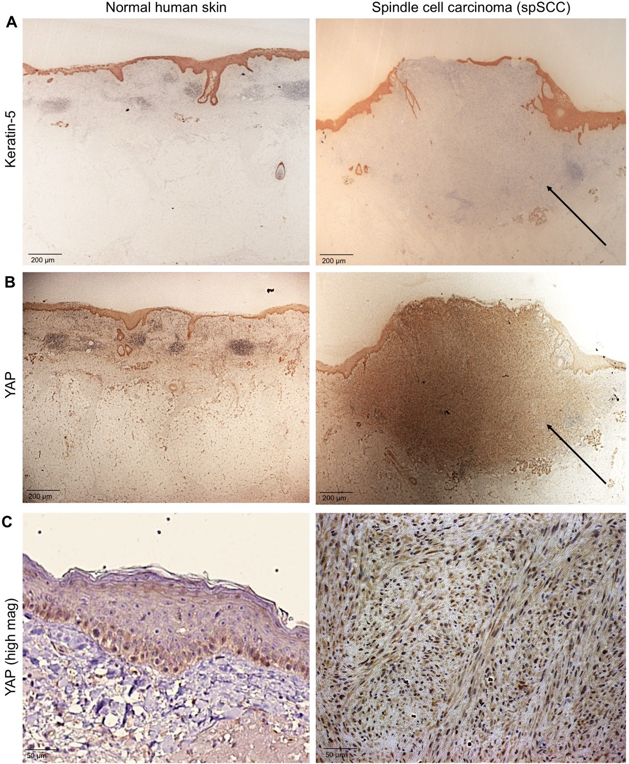
YAP is nuclear localised in human spindle cell carcinoma.
(A) Histological sections of normal human skin and spindle cell carcinoma patient tumour stained for the epithelial marker Keratin-5 (brown immunostain). Scale bar 200 μM. (B) Histological sections of normal human skin and spindle cell carcinoma patient tumour stained for YAP (brown immunostain). Scale bar 200 μM. (C) High magnification view of (B) showing nuclear localisation of YAP protein in spindle cell carcinoma (brown immunostain). Sections are co-stained for eosin (blue). Scale bar 200 μM.
Figure 1—figure supplement 1
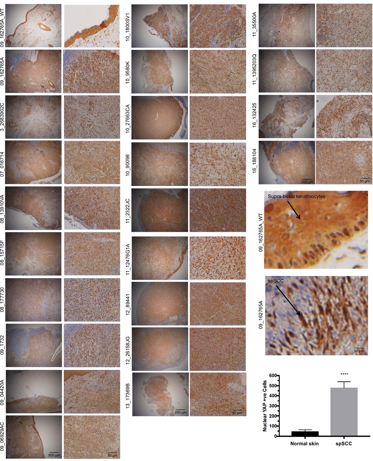
A panel of human spSCC tumours are characterised by widespread nuclear YAP localisation.
https://doi.org/10.7554/eLife.33304.003
Figure 2
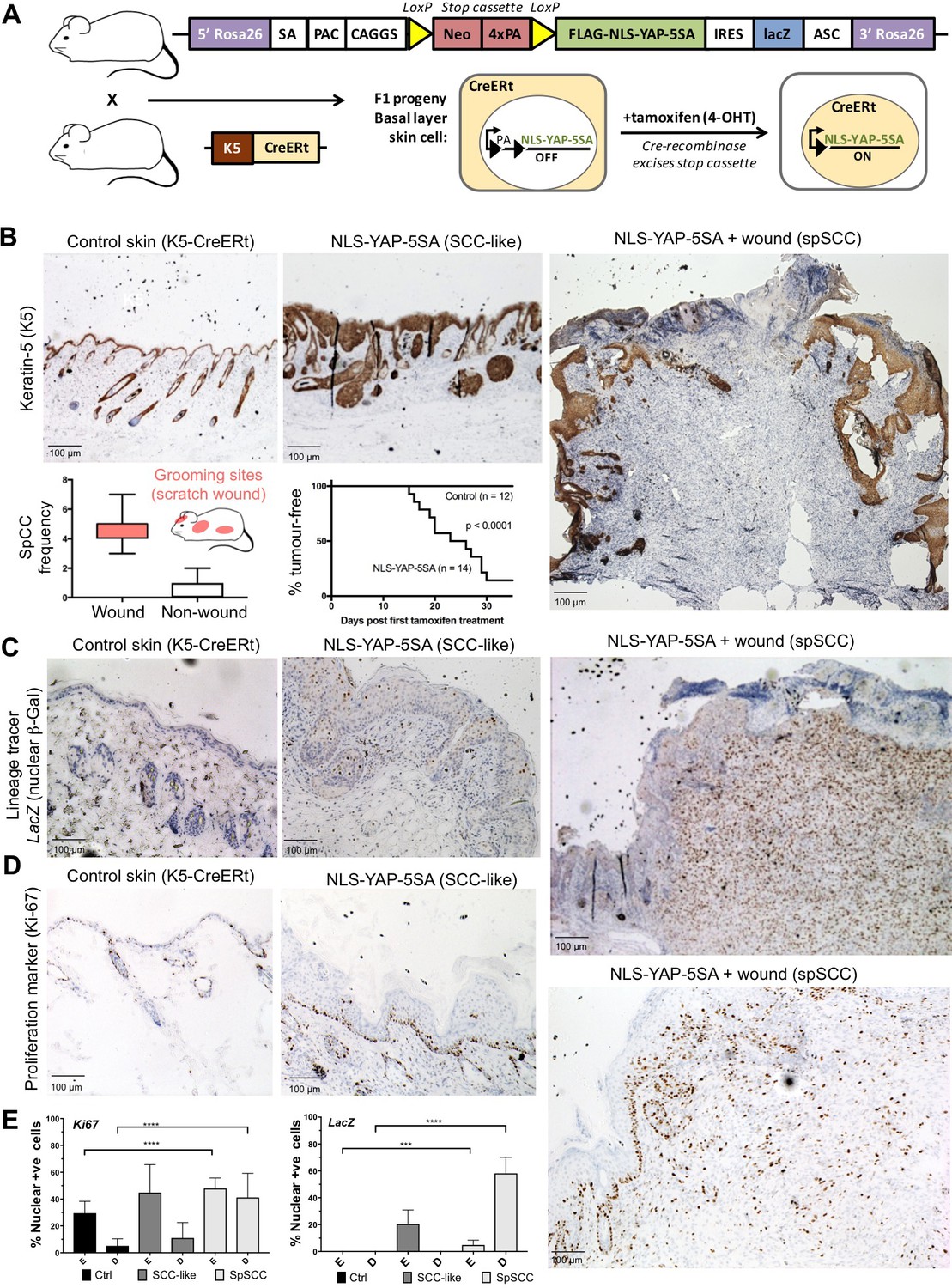
Nuclear YAP drives formation of both SCC and spSCC in mice.
(A) Skin-specific expressison of nuclear YAP was achieved by crossing K5-CreERt mice to a Lox-Stop-Lox cassette for conditional expression of nuclear YAP and a LacZ lineage tracer in basal layer skin cells. (B) Expression of nuclear YAP drives formation of both SCC-like overgrowths (Keratin-5 positive) and spSCC-like tumours (mostly Keratin-5 negative). Multiple spSCC tumours arise per animal, but only in areas subject to scratch wounding. Note the disruption in the continuity of Keratin-5 positive epithelial layer above the spSCC tumour, indicative of a wound-induced tumour (n = 20). Kaplan-Meier analysis shows rapid induction of tumours in NLS-YAP-5SA expressing skin. (C) Lineage tracing with LacZ (encoding nuclear beta-Gal immunostained in brown) induced with the K5-CreERt line indicates that both SCC and spSCC tumours arise from the K5-positive basal layer of the skin (n = 22). (D) Proliferation of cells was measured by staining for the mitotic marker Ki-67 in control, SCC and spSCC samples (n = 25). Scale bars 100 μM. (E) Quantification of C and D in epidermal (E) vs dermal (D) compartments.
Figure 3
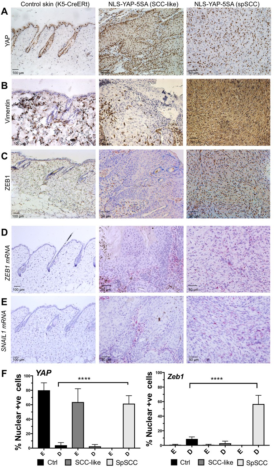
YAP-driven mouse spSCC formation involves transcriptional induction of ZEB1 expression and EMT.
(A) YAP immunostaining of control skin as well as NLS-YAP-5SA driven SCC and spSCC tumours (n = 32). (B) Vimentin (mesenchymal marker) immunostaining of control skin as well as NLS-YAP-5SA driven SCC and spSCC tumours. Note strong induction in spSCC (n = 30). (C) ZEB1 (EMT transcription factor) immunostaining of control skin as well as NLS-YAP-5SA driven SCC and spSCC tumours. Note strong induction in spSCC (n = 33). (D) ZEB1 mRNA in situ hybridisation of control skin as well as NLS-YAP-5SA driven SCC and spSCC tumours. Note strong induction in spSCC (n = 29). (E) SNAIL1 mRNA in situ hybridisation of control skin as well as NLS-YAP-5SA driven SCC and spSCC tumours. Note strong induction in spSCC (n = 8). (F) Quantitation of YAP and ZEB1 marker expression in samples from wild-type skin, SCC-like, and spSCC-like mouse skin tumours (n > 30 samples for each case). Scale bars 50–100 μM.
Figure 4
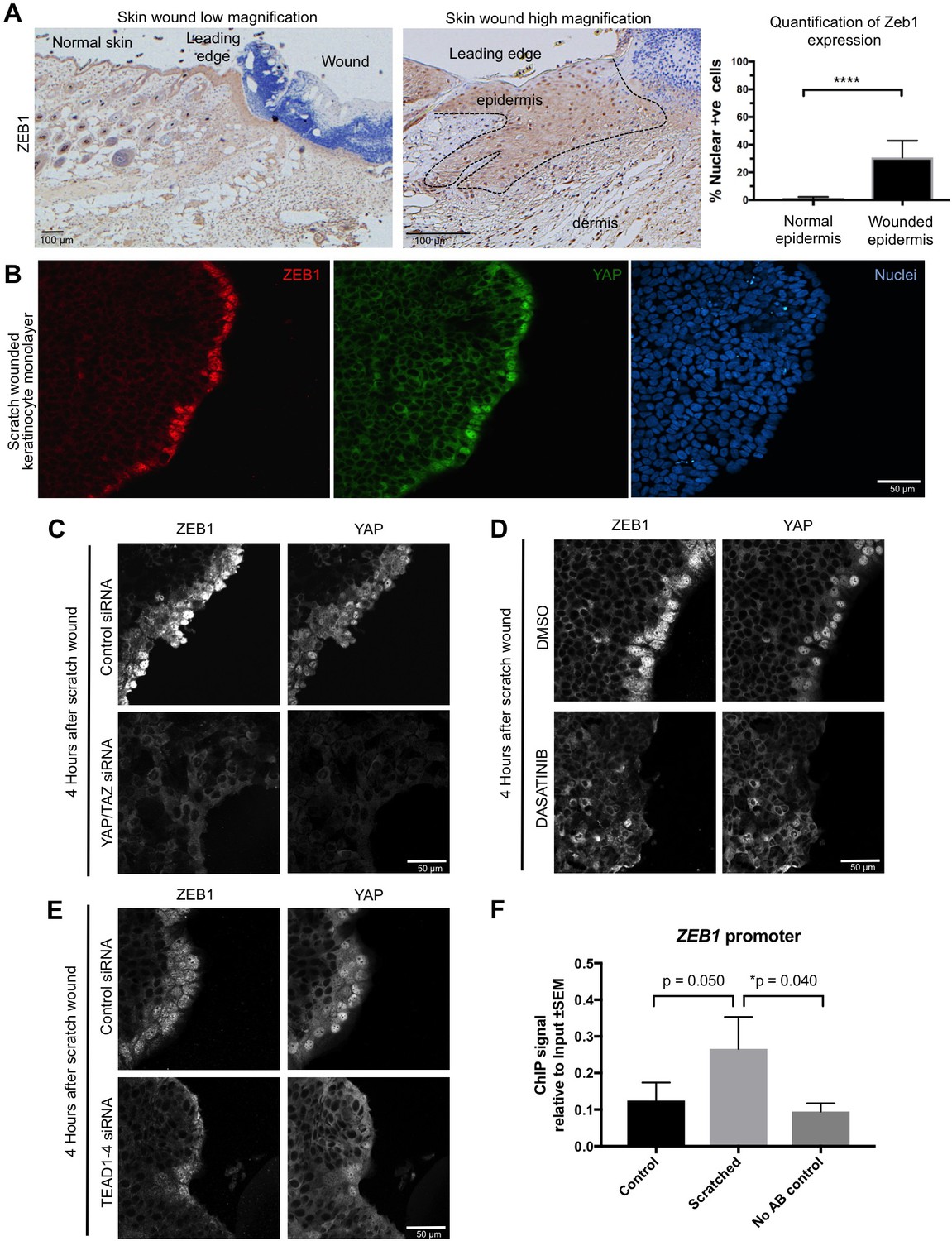
YAP promotes ZEB1 expression after epidermal wounding to drive EMT.
(A) Punch wounding of mouse skin induces ZEB1 immunostaining in some leading edge cells. Scale bars 100 μM. (B) Scratch wounding of skin keratinocytes in culture induces ZEB1 and YAP immunostaining in leading edge cells. Scale bar 50 μM. (C) Induction of ZEB1 at the leading edge is prevented by transfection with siRNAs against YAP/TAZ. Scale bar 50 μM. (D) Induction of ZEB1 at the leading edge is prevented by treatment with Dasatinib, a Src-family kinase inhibitor that prevents YAP activation. Scale bar 50 μM. (E) Induction of ZEB1 at the leading edge is prevented by treatment with siRNAs against TEAD1-4. Scale bar 50 μM. (F) Chromatin Immunoprecipitation of TEAD1 at an upstream enhancer of the ZEB1 gene in keratinocytes before or after scratch wounding. The weak enrichment may be caused by the small percentage of ZEB1-expressing cells in this experiment. Data were analysed by a Mann-Whitney Test n = 9 samples per experimental condition.
Figure 5 with 1 supplement
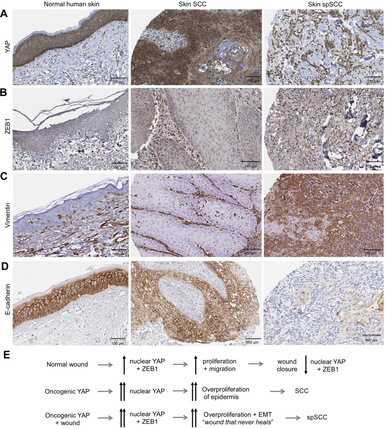
Human spSCC is characterised by co-expression of YAP and ZEB1.
(A) YAP immunostaining of normal human skin, SCC and spSCC-like tumours. Note strong nuclear localisation in spindle-shaped spSCC tumour cells. (B) ZEB1 immunostaining of normal human skin, SCC and spSCC-like tumours. Note strong expression in spindle-shaped spSCC tumour cells. (C) Vimentin immunostaining of normal human skin, SCC and spSCC-like tumours. Note strong expression in spindle-shaped spSCC tumour cells. (D) E-cadherin immunostaining of normal human skin, SCC and spSCC-like tumours. Note absence of expression in spindle-shaped spSCC tumour cells. (E) Model comparing normal wound healing with SCC and spSCC formation. Scale bars 100 μM.
Figure 5—figure supplement 1
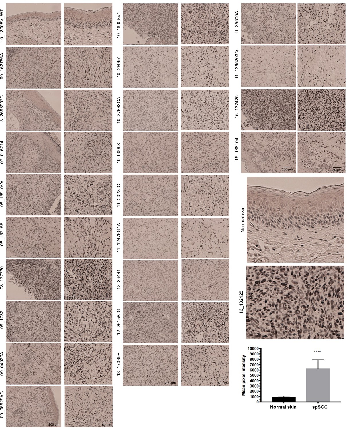
A panel of human spSCC tumours are characterised by widespread nuclear Zeb1 localisation.
https://doi.org/10.7554/eLife.33304.008
Author response image 1
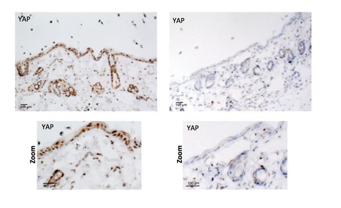
Validation of Rabbit anti-YAP antibody in knockout skin.
https://doi.org/10.7554/eLife.33304.011Tables
Key resources table
| Reagent type (species) or resource | Designation | Source or reference | Identifiers | Additional information |
|---|---|---|---|---|
| Cell Line (Human) | HaCAT | Cell Services (Francis Crick Institute) Pubmed ID: 26989177 | (CLS Cat# 300493/p800_HaCaT, RRID:CVCL_0038) | |
| Antibody (Rabbit monoclonal) | anti-Vimentin | Abcam | (Abcam Cat# ab92547, RRID:AB_10562134) | 1/600 IHC |
| Antibody (Rabbit polyclonal) | anti-ZEB1 | Proteintech | (Proteintech Group Cat# 21544–1-AP, RRID:AB_10734325) | 1/500 IHC/1/100 IF |
| Antibody (Rabbit monoclonal) | anti-Keratin-5 | Abcam | (Abcam Cat# ab52635, RRID:AB_869890) | 1/500 IHC |
| Antibody (Rabbit polyclonal) | anti-beta- galactosidase | Acris | (Acris Antibodies GmbH Cat# R1064P, RRID:AB_973264) | 1/5000 IHC |
| Antibody (Rabbit polyclonal) | anti-E-Cadherin | Santa Cruz | (Santa Cruz Biotechnology Cat# sc-7870, RRID:AB_2076666) | 1/75 IHC |
| Antibody (Rabbit monoclonal) | anti-YAP | Cell Signalling Technology | (Cell Signaling Technology Cat# 14074, RRID:AB_2650491) | 1/400 O/N IHC |
| Antibody (Rabbit monoclonal) | anti-Ki67 | Abcam | (Abcam Cat# ab16667, RRID:AB_302459) | 1/350 IHC |
| Antibody (Mouse monoclonal) | anti-TEAD-1 | BD Biosciences | (BD Biosciences Cat# 610922, RRID:AB_398237) | 12.5 per 200 ug chromatin input CHIP |
| Antibody (Mouse monoclonal) | anti-YAP | Santa Cruz | (Santa Cruz Biotechnology Cat# sc-101199, RRID:AB_1131430) | 1/100 IF |
| Transfection reagent | Lipofectamine RNAiMAX | Thermo Fisher | Cat no: 13778075 | |
| siRNA | TEAD 1 | Dhamacon | Cat no: M-012603-01-0005 | 80 nM Final |
| siRNA | TEAD 2 | Dhamacon | M-012611-00-0005 | 80 nM Final |
| siRNA | TEAD 3 | Dhamacon | M-012604-01-0005 | 80 nM Final |
| siRNA | TEAD 4 | Dhamacon | M-019570-03-0005 | 80 nM Final |
| siRNA | YAP | Dhamacon | M-012200-00-0005 | 80 nM Final |
| Human Protein Atlas | Various | Pubmed ID: 16774037 | https://www.proteinatlas.org/ | |
| Human cancer samples | Vimentin/YAP | University of Southamption/ Gareth Thomas | ||
| Chemical compound, drug | Dasatinib | Selleck Biochem | S1021 | 5 uM Final |
| Mouse strain | Rosa26-YAP5SA | Junhao Mao (University of Massachusetts Medical School) | mixed background | |
| Mouse strain | K5-CreERT2 | Ian Rosewell (Francis Crick Institute) | mixed background | |
| Mouse strain | Yapfl/fl Tazfl/fl | Axel Behrens (Francis Crick Institute) | mixed background | |
| Chemical compound, drug | 4-Hydroxytamoxifen | Sigma | H7904 | topical application of 200 ul oF 1.0 mg per 0.1 mL 4’OHT in DMSO on dorsal skin 5x consecutive days |
| Chemical compound, drug | Tamoxifen | Sigma | T5648 | IP 5 ul/g body weight of a 20 mg/ml solution in corn oil 5x consecutive days |
| RNA target probe | RNAscope Probe - Mm-Zeb1 | ACD | 451201 | |
| RNA target probe | RNAscope Probe - Mm-Snai1 | ACD | 451211 | |
| RNA target probe | RNAscope Probe - Mm-Snai2 | ACD | 451191 |
Additional files
-
Transparent reporting form
- https://doi.org/10.7554/eLife.33304.009
Download links
A two-part list of links to download the article, or parts of the article, in various formats.
Downloads (link to download the article as PDF)
Open citations (links to open the citations from this article in various online reference manager services)
Cite this article (links to download the citations from this article in formats compatible with various reference manager tools)
YAP drives cutaneous squamous cell carcinoma formation and progression
eLife 7:e33304.
https://doi.org/10.7554/eLife.33304


