World Journal of Emergency Medicine ›› 2014, Vol. 5 ›› Issue (1): 57-62.doi: 10.5847/wjem.j.issn.1920-8642.2014.01.010
• Original Articles • Previous Articles Next Articles
Shou-peng Li, Ji-yuan Han( ), Peng Sun, Guo-yan Wu, Xiang-yan Bai
Received:2013-08-26
Accepted:2014-02-01
Online:2014-03-15
Published:2014-03-15
Contact:
Ji-yuan Han
E-mail:jiyuanhan@126.com
Shou-peng Li, Ji-yuan Han, Peng Sun, Guo-yan Wu, Xiang-yan Bai. Effect of SP-A/B in lipoic acid on acute paraquat poisoning[J]. World Journal of Emergency Medicine, 2014, 5(1): 57-62.
Add to citation manager EndNote|Ris|BibTeX
URL: http://wjem.com.cn//EN/10.5847/wjem.j.issn.1920-8642.2014.01.010
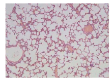
Figure 1.
The results of HE staining in the NS group (original magnification×100).
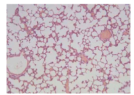
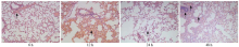
Figure 2.
The results of HE staining in the LA group (original magnification×100).
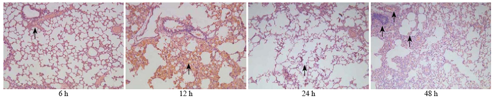
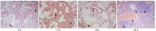
Figure 3.
The results of HE staining in the PQ group (original magnification×100).
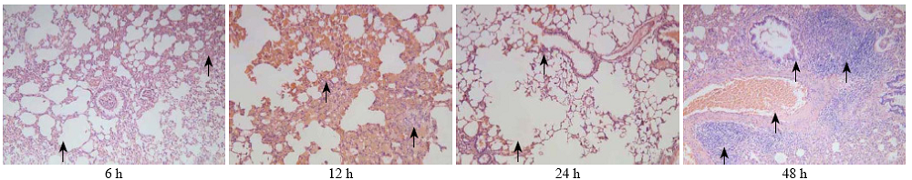
Table 1
Comparison of MDA levels between the NS, PQ, and LA groups
| Groups | 3 h | 6 h | 12 h | 24 h | 48 h | F | P< |
|---|---|---|---|---|---|---|---|
| NS | 3.904±0.200 | 3.904±0.200 | 3.904±0.200 | 3.904±0.200 | 3.904±0.200 | 0 | |
| PQ | 4.476±0.300 | 5.620±0.402 | 7.633±0.497 | 8.220±0.395 | 10.197±1.213 | 65.938 | 0.01 |
| LA | 4.194±0.440 | 5.131±0.236 | 6.592±0.454 | 7.408±0.400 | 8.744±0.787 | 80.502 | 0.01 |
| F | 3.772 | 50.551 | 152.092 | 260.045 | 87.925 | ||
| P | 0.163 | 0.001 | 0.001 | 0.001 | 0.001 |
Table 2
Comparison of SOD levels between the NS, PQ, and LA groups
| Groups | 3 h | 6 h | 12 h | 24 h | 48 h | F | P< |
|---|---|---|---|---|---|---|---|
| NS | 175.189±11.047 | 175.189±11.047 | 175.189±11.047 | 175.189±11.047 | 175.189±11.047 | 0 | |
| PQ | 155.290±18.195 | 149.808±19.581 | 132.696±7.071 | 127.345±8.913 | 117.764±14.205 | 7.025 | 0.01 |
| LA | 162.591±8.331 | 151.976±9.709 | 139.179±2.886 | 132.381±6.414 | 123.292±5.663 | 29.789 | 0.01 |
| F | 1.983 | 9.402 | 111.5882 | 49.329 | 41.485 | ||
| P | 0.172 | 0.002 | 0.001 | 0.001 | 0.001 |
Table 3
Comparison of TNF-α levels between the NS, PQ, and LA groups
| Groups | 3 h | 6 h | 12 h | 24 h | 48 h | F | P< |
|---|---|---|---|---|---|---|---|
| NS | 13.838±0.684 | 13.838±0.684 | 13.838±0.684 | 13.838±0.684 | 13.838±0.684 | 0 | |
| PQ | 16.782±1.047 | 23.743±1.002 | 31.688±1.978 | 38.281±2.999 | 44.990±2.260 | 19.543 | 0.01 |
| LA | 14.339±2.550 | 21.828±1.762 | 28.139±1.762 | 31.742±1.436 | 40.666±1.576 | 10.340 | 0.01 |
| F | 66.166 | 207.620 | 379.197 | 288.939 | 695.125 | ||
| P | 0.001 | 0.001 | 0.001 | 0.001 | 0.001 |
Table 4
Comparison of SP-A mRNA levels between the NS, PQ, and LA groups
| Groups | 3 h | 6 h | 12 h | 24 h | 48 h | F | P< |
|---|---|---|---|---|---|---|---|
| NS | 1.03±0.015 | 1.03±0.015 | 1.03±0.015 | 1.03±0.015 | 1.03±0.015 | 0 | |
| PQ | 0.72±0.035 | 0.47±0.032 | 0.25±0.051 | 0.13±0.046 | 0.09±0.041 | 24.749 | 0.05 |
| LA | 0.94±0.041 | 0.85±0.019 | 0.43±0.029 | 0.30±0.065 | 0.18±0.034 | 32.673 | 0.01 |
| F | 248.804 | 534.160 | 147.765 | 236.630 | 349.001 | ||
| P | 0.001 | 0.001 | 0.001 | 0.001 | 0.001 |
Table 5
Comparison of SP-B mRNA levels between the NS, PQ, and LA groups
| Groups | 3 h | 6 h | 12 h | 24 h | 48 h | F | P< |
|---|---|---|---|---|---|---|---|
| NS | 1.06±0.025 | 1.06±0.025 | 1.06±0.025 | 1.06±0.025 | 1.06±0.025 | 0 | |
| PQ | 0.54±0.044 | 0.39±0.041 | 0.34±0.037 | 0.21±0.036 | 0.12±0.043 | 64.269 | 0.05 |
| LA | 0.91±0.036 | 0.72±0.056 | 0.45±0.051 | 0.29±0.021 | 0.24±0.056 | 93.421 | 0.01 |
| F | 632.007 | 918.273 | 131.827 | 219.255 | 196.001 | ||
| P | 0.001 | 0.001 | 0.001 | 0.001 | 0.001 |
| 1 |
Suntres ZE. Role of antioxidants in paraquat toxicity. Toxicology 2002; 180:65-77.
doi: 10.1016/s0300-483x(02)00382-7 pmid: 12324200 |
| 2 |
Dinis-Oliveira RJ, Remião F, Duarte JA, Ferreira R, Sánchez Navarro A, Bastos ML, et al. P-glycoprotein induction:an antidotal pathway for paraquat-induced lung toxicity. Free Radic Biol Med 2006; 41:1213-1224.
doi: 10.1016/j.freeradbiomed.2006.06.012 pmid: 17015168 |
| 3 | Dinis-Oliveira RJ, Duarte JA, Remião F, Sánchez-Navarro A, Bastos ML, Carvalho F. Single high dose dexamethasone treatment decreases the pathological score and increases the survival rate of paraquat-inroxicated rats. Toxicoligy 2006; 227:73-85. |
| 4 |
Bus JS, Aust SD, Gibson JE. Superoxide and singlet oxygen catalyzed lipid peroxidation as a possible mechanism for paraquat (methyl viologen) toxicity. Biochem Biophys Res Commun 1974; 58:749-755.
doi: 10.1016/s0006-291x(74)80481-x pmid: 4365647 |
| 5 |
Chen CM, Lua AC. Lung toxicity of paraquat in the rat. J Toxicol Environ Health A 2000; 60:477-487.
doi: 10.1080/00984100050079548 pmid: 12607909 |
| 6 |
Youngman RJ, Elstner EF. Oxygen species in paraquat toxicity: the crypto-OH radical. FEBS Letters 1981; 129:265-268.
doi: 10.1016/0014-5793(81)80180-9 pmid: 6269879 |
| 7 |
Meng XX, Wang RL, Gao S, Xie H, Tan JT, Qian YB. Effect of ulinastatin on paraquat-induced-oxidative stress in human type II alveolar epithelial cells. World J Emerg Med 2013; 4:133-137.
pmid: 25215107 |
| 8 |
Sommer D, Fakata KL, Swanson SA, Stemmer PM. Modulation of the phosphatase activity of calcineurin by oxidants and antioxidants in vitro. Eur J Biochem 2000; 267:2312-2322.
doi: 10.1046/j.1432-1327.2000.01240.x pmid: 10759856 |
| 9 |
Yi X, Maeda N. Endogenous production of lipoic acid is essential for mouse development. Mol Cell Biol 2005; 25:8387-8392.
doi: 10.1128/MCB.25.18.8387-8392.2005 pmid: 16135825 |
| 10 | Kim DJ, Kim KH, Lee HH, Lee SJ, Ha JY, Yoon HJ, et al. Crystal structure of lipoate-protein ligase A bound with the activated intermediate: insights into interaction with lipoyl domains. Biol Chem 2005; 280:38081-38089. |
| 11 | Carswell EA, Old LJ, Kassel RL, Green S, Fiore N, Williamson B. An endotoxin-induced serum factor that causes necrosis of tumors. Proc Natl Acad Sci U S A 1975; 72:3666-3670. |
| 12 |
Cheng ZQ, Han JY, Sun P, Weng YY, Chen J, Wu GY, et al. Edaravone attenuates paraquat-induced lung injury by inhibiting oxidative stress in human type II alveolar epithelial cells. World J Emerg Med 2012; 3:55-59.
doi: 10.5847/wjem.j.issn.1920-8642.2012.01.010 pmid: 25215040 |
| 13 |
Graczyk M, Przybyszewski M, Kuźmiński A, Tlappa J, Mućka J, Napiórkowska K, et al. Role of E-selectin and platelet endothelial cell adhesion molecule 1 in gastritis in food allergy patients. Postepy Dermatol Alergol 2013; 30:271-276.
doi: 10.5114/pdia.2013.38354 pmid: 24353485 |
| 14 |
Chaudhary P, Marracci GH, Bourdette DN. Lipoic acid inhibits expression of ICAM-1 and VCAM-1 by CNS endothelial cells and T cell migration into the spinal cord in experimental autoimmune encephalomyelitis. J Neuroimmunol 2006; 175:87-96. Epub 2006 Apr 27.
doi: 10.1016/j.jneuroim.2006.03.007 pmid: 16644024 |
| 15 | Kunt T, Forst T, Wilhelm A, Tritschler H, Pfuetzner A, Harzer O, et al. Alpha-lipoic acid reduces expression of vascular cell adhesion molecule-1 and endothelial adhesion of human monocytes after stimulation with advanced glycation end products. Clin Sci (Lond) 1999; 96:75-82. |
| 16 |
Zhang WJ, Wei H, Hagen T, Frei B. Alpha-lipoic acid attenuates LPS-induced inflammatory responses by activating the phosphoinositide 3-kinase/Akt signaling pathway. Proc Natl Acad Sci USA 2007; 104:4077-4082.
doi: 10.1073/pnas.0700305104 pmid: 17360480 |
| 17 | Salinthone S, Schillace RV, Marracci GH, Bourdette DN, Carr DW. Lipoic acid stimulates cAMP production via the EP2 and EP4 prostanoid receptors and inhibits IFN gamma synthesis and cellular cytotoxicity in NK cells. Neuroimmunol 2008; 199:46-55. |
| 18 | Wollin SD, Jones PJ. Alpha-lipoic acid and cardiovascular disease. Nutr 2003; 133:3327-3330. |
| 19 | Pearson FG, Zhao Fengrui [translation]. Thoracic Surgery. Liaoning Education Press. 1999: 42. |
| 20 |
Shi J, Hu CL, Gao YF, Liao XX, Xu H. The relationship between platelet endothelial cell adhesion molecule-1 and paraquat-induced lung injury in rabbits. World J Emerg Med 2012; 3:60-64.
doi: 10.5847/wjem.j.issn.1920-8642.2012.01.011 pmid: 25296667 |
| 21 |
Xie LX, Liu YN, Zhao XW, Chen LA, Zhao XM, Liu YY. The changes in effective local blood perfusion and compensatory ventilation in different lung areas of acute respiratory distress syndrome rabbits model. Zhonghua Nei Ke Za Zhi 2004; 43:522-526.
pmid: 15312408 |
| [1] | Jian-hua Yi, Zhao-cai Zhang, Mei-bian Zhang, Xin He, Hao-ran Lin, Hai-wen Huang, Hai-bin Dai, Yu-wen Huang. Role of epithelial-to-mesenchymal transition in the pulmonary fibrosis induced by paraquat in rats [J]. World Journal of Emergency Medicine, 2021, 12(3): 214-220. |
| [2] | Yun-fei Jiang, Jian Kang, Pei-pei Huang, Jia-xi Yao, Zhong-he Wang, Lei Jiang, Jun Wang, Li Qiao, Bao-li Zhu, Hao Sun, Jin-song Zhang. Evaluation of gastric lavage efficiency and utility using a rapid quantitative method in a swine paraquat poisoning model [J]. World Journal of Emergency Medicine, 2020, 11(3): 174-181. |
| [3] | Jia-jun Xu, Jian-tao Zhen, Li Tang, Qing-ming Lin. Intravenous injection of Xuebijing attenuates acute kidney injury in rats with paraquat intoxication [J]. World Journal of Emergency Medicine, 2017, 8(1): 61-64. |
| [4] | Ming Wei, Yan-jie Gong, Ling Tu, Jia Li, Ying-hong Liang, Yi-hua Zhang. Expression of phosphatidylinositol-3 kinase and effects of inhibitor Wortmannin on expression of tumor necrosis factor-α in severe acute pancreatitis associated with acute lung injury [J]. World Journal of Emergency Medicine, 2015, 6(4): 299-304. |
| [5] | Zhi-wei Liu, Hai-ying Wang, Lan Guan, Bin Zhao. Regulatory effects of hydrogen sulfide on alveolar epithelial cell endoplasmic reticulum stress in rats with acute lung injury [J]. World Journal of Emergency Medicine, 2015, 6(1): 67-73. |
| [6] | Xiao-xiao Meng, Rui-lan Wang, Shan Gao, Hui Xie, Jiu-ting Tan, Yong-bin Qian. Effect of ulinastatin on paraquat-induced-oxidative stress in human type II alveolar epithelial cells [J]. World Journal of Emergency Medicine, 2013, 4(2): 133-137. |
| [7] | Yin-song Jiang, Yu-ying Ma, Zhan-qing Wang, Guang-jun Li. Therapeutic effects of smecta or smectite powder on rats with paraquat toxication [J]. World Journal of Emergency Medicine, 2013, 4(2): 144-150. |
| [8] | Zhi-jian Zhang, Li-bo Peng, Ya-juan Luo, Cong-yang Zhou. Prospective experimental studies on the renal protective effect of ulinastatin after paraquat poisoning [J]. World Journal of Emergency Medicine, 2012, 3(4): 299-304. |
| [9] | Zhi-qiang Cheng, Ji-yuan Han, Peng Sun, Yu-ying Weng, Jiao Chen, Guo-yan Wu, Hong-xia Ma. Edaravone attenuates paraquat-induced lung injury by inhibiting oxidative stress in human type II alveolar epithelial cells [J]. World Journal of Emergency Medicine, 2012, 3(1): 55-59. |
| [10] | Jing Shi, Chun-lin Hu, Yu-feng Gao, Xiao-xing Liao, Hope Xu. The relationship between platelet endothelial cell adhesion molecule-1 and paraquat-induced lung injury in rabbits [J]. World Journal of Emergency Medicine, 2012, 3(1): 60-64. |
| [11] | Kai Yin, Sheng-chun Dang, Jian-xin Zhang. Relationship between expression of triggering receptor-1 on myeloid cells in intestinal tissue and intestinal barrier dysfunction in severe acute pancreatitis [J]. World Journal of Emergency Medicine, 2011, 2(3): 216-221. |
| [12] | Chang-bin Li, Xin-hua Li, Zhen Wang, Cheng-hua Jiang, Ai Peng. Serum paraquat concentration detected by spectrophotometry in patients with paraquat poisoning [J]. World Journal of Emergency Medicine, 2011, 2(3): 179-184. |
| [13] | Hui-li Zhang, Yuan-fei Liu, Xu-rui Luo, Wei-hua Tan, Liang Huang. Saturated hydrogen saline protects rats from acute lung injury induced by paraquat [J]. World Journal of Emergency Medicine, 2011, 2(2): 149-153. |
| [14] | Jian-qiang Wang, Chun Pan, Lin Liu, Liang Jin, Yi Yang, Hai-bo Qiu. Effect of post recruitment maneuver ventilation by different tidal volume on lung vascular endothelial diastole function in rats with acute lung injury [J]. World Journal of Emergency Medicine, 2011, 2(2): 141-148. |
| [15] | Xie Juan, Yi-ming Lu, Jin-dong Shi, Xing-qi Deng, Wei Long. Visfatin levels in patients with severe pneumonia [J]. World Journal of Emergency Medicine, 2011, 2(2): 132-136. |
| Viewed | ||||||
|
Full text |
|
|||||
|
Abstract |
|
|||||
