Hydrogen-Rich Green Diamond Color-Treated by Multi-Step Processing
ABSTRACT
A cut diamond of intense yellowish green color has been characterized using microscopy and spectroscopic techniques. The diamond has been unambiguously identified as color-treated. The simultaneous presence of multiple centers related to irradiation and annealing—including H1a, H1b, NV0, NV–, H3, H4, GR1, and H2—was revealed. UV-Vis-NIR absorption spectroscopy showed that the diamond owes its color to the two major bands related to H3 and GR1. The combination of these spectroscopic features in one diamond has not been reported in the gemological literature, suggesting that this diamond was subjected to a complex treatment procedure that is not frequently applied. Taking into account the thermal stability of the defects involved and the defect transformations at high temperatures, two possible treatment procedures explaining the observed combination of spectroscopic features are proposed.
INTRODUCTION
Although a number of color centers such as GR1 (neutral vacancies), H3 (N-V-N defects), and some hydrogen- and nickel-related centers can impart green coloration to diamonds (Breeding et al., 2018), diamonds with a natural saturated green color are among the rarest. In the laboratory, attempts have been made to artificially induce green color in diamonds. This can be done with high-energy electron irradiation of yellowish diamonds (Collins, 1982; Wang et al., 2018), followed by prolonged annealing at a temperature of 1400°C for colorless type Ia diamonds (Collins, 2001), or using high-pressure, high-temperature (HPHT) annealing for brown type Ia diamonds (Collins et al., 2000; Collins, 2001, 2003). A multi-step processing that probably involved irradiation, annealing, and re-irradiation was documented by GIA researchers (Fritsch et al., 1988).
Although many features of color-treated (Fritsch et al., 1988; Collins et al., 2000; Collins, 2003) and untreated (Reinitz et al., 1998; Breeding et al., 2018) green diamonds have been established, determining the color origin of some green diamonds still poses a challenge due to the similarity of natural and laboratory radiation and annealing processes (Overton and Shigley, 2008; Breeding et al., 2018). Therefore, a deeper understanding of the thermal stability and transformations of the color-causing defects in diamonds subjected to irradiation and annealing is crucial for reliable reporting of color treatment.
Although two multi-step treated green diamonds were reported more than 30 years ago (Fritsch et al., 1988), treated-color green diamonds enhanced by a multi-step process involving three or more steps are rarely encountered by gemological labs, to the best of our knowledge. Here we report a combination of spectroscopic features that has not been previously documented in a single gem diamond and propose two possible treatment procedures to explain the results of our observations. The aim of this study is to characterize a green diamond color-treated by multi-step processing and to provide a supplementary database for the correct determination of color origin for gemological laboratories.
MATERIALS AND METHODS
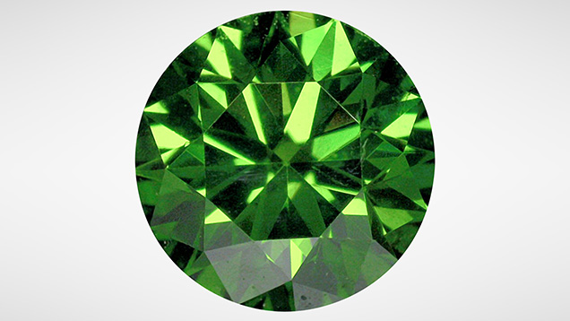
The studied sample was a yellowish green round brilliant cut diamond weighing 0.25 ct (figure 1), provided to us by Mr. Congsen Zhang. The internal and external structural features of the diamond were examined with a standard gemological binocular microscope using a variety of illumination techniques. The UV-Vis-NIR absorption spectrum in the range of 225–1000 nm was recorded at the National Center of Supervision and Inspection of Quality of Gold and Silver Products (NGSTC) using a Spec GEM-3000 fiber-optic instrument equipped with an Ocean Optics QE65Pro spectrometer (1 nm spectral resolution). The sample was immersed in liquid nitrogen for a few seconds and then taken out for immediate UV-Vis-NIR measurement. Room-temperature FTIR absorption measurements in the spectral range 400–6000 cm–1 with a resolution of 4 cm–1 were performed at NGSTC using a Nicolet iN10 spectrometer. Photoluminescence (PL) spectra were collected at liquid nitrogen temperature in the laboratory of the National Gemstone Testing Center (NGTC) using a Renishaw InVia Raman confocal spectrometer equipped with 473 and 532 nm lasers.
RESULTS
Microscopic Features. Examination with the microscope revealed that the green color was evenly distributed over the stone. No green-brown stains characteristic of natural irradiation were observed. Patterned clouds and several fractures under the table were observed. A black graphitized surface-reaching fracture was seen near the girdle (figure 2).

Spectra. FTIR analysis provided important information about the impurity-related defects from nitrogen and hydrogen (Zaitsev et al., 1996). The spectra revealed that the diamond was type IaAB and had high nitrogen content. A minor line detected at 1362 cm–1 (figure 3) was probably an auxiliary peak to isolated nitrogen, given its sharp shape. These features unambiguously pointed to a naturally grown diamond. Hydrogen is a common impurity in diamonds, giving rise to a number of sharp absorption peaks in the IR spectral range from 2700 to 3500 cm–1 (Woods and Collins, 1983; Ardon, 2014; Zaitsev et al., 2016). Many of these hydrogen-related peaks were present in the FTIR spectrum of the studied diamond: strong absorption at 1405 cm–1 and several sharp peaks between 3300 and 2700 cm–1, with the three strongest ones at 3237, 3107, and 2785 cm–1. Weak lines at 4496 and 4168 cm–1 were also detected (figure 3). Besides these features, a strong absorption at 1450 cm–1 (H1a center, an interstitial nitrogen defect; Woods, 1984; Zaitsev, 2001) and a relatively weak absorption at 4930 cm–1 (H1b center) were recorded.

UV-Vis-NIR absorption spectra revealed the strong absorption band of the GR1 center in the spectral range from about 550 to 750 nm with a zero-phonon line (ZPL) at 741 nm, and a broad structureless band spreading from 410 to 510 nm that was presumably due to the H3 center (Dobrinets et al., 2013; Breeding et al., 2018) (figure 4). The H2 center (negatively charged N-V-N defect) with its ZPL at 985 nm was also clearly seen.

PL spectra recorded at 473 nm laser excitation displayed a strong H3 center with a ZPL at 503.2 nm and characteristic phonon side bands at 512 and 520 nm (Zaitsev, 2001) (figure 5A). The emission related to the H4 center was recorded at a wavelength of 496 nm. Lines at wavelengths 484, 489, 613 (612.5 nm in some publications), and 647 nm were also detected. All these features with the exception of the 647 line are common for type Ia diamonds (Zaitsev, 2001). Brown and pink type Ia diamonds frequently reveal a strong 613 nm center in their PL spectra (Tretiakova and Tretyakova, 2008; Gaillou et al., 2010). A ZPL at 613 nm center with its vibrational feature at 625 nm (a weak hillock in spectrum shown in figure 5A) is also a common feature of purple diamonds (Titkov et al., 2008). The 647 nm center has been reported in irradiation-treated diamonds (Wang et al., 2018) and is believed to be an irradiation-related defect (Zaitsev et al., 2018).

Photoluminescence spectra taken with 532 nm laser excitation revealed pronounced emissions of NV0 and NV– centers. Intensity of the NV– center (ZPL at 637 nm, or 638 nm in some publications) was much stronger than that of the NV0 center (ZPL at 575 nm) (figure 5B). Pronounced broad bands at 659 and 680 nm in this spectrum are due to electron-phonon transitions at NV– and 647 nm centers. A doublet line feature at wavelengths 741 and 745 nm is a strongly suppressed and distorted ZPL of the GR1 center. This distortion is a result of strong self-absorption in the ZPL. Self-absorption in the ZPL is a common effect observed in PL spectra of optical centers with strong absorption. For instance, self-absorption can be very pronounced for NV– and H3 centers (Dobrinets et al., 2013). Also detected were lines at 694, 700, and 723 nm of presumably nickel-related centers (Lang et al., 2004; Fritsch et al., 2007; Dobrinets et al., 2013).
DISCUSSION
Characterization of the Sample and Identification of Features. Although graphitization may occur naturally, it is not frequently observed in untreated cut diamonds. If present, the graphitized features are usually considered “suspicious” features, possibly the result of high-temperature treatment. Typical graphitized features in HPHT-treated diamonds have been described in detail by numerous researchers (e.g., Reinitz et al., 2000; Wang et al., 2005). However, the presence of graphitization alone is not reliable proof of HPHT treatment (Overton and Shigley, 2008).
The absence of strong hydrogen-related absorption bands (centered at 730 and 835 nm) and/or a Ni-related band (from approximately 620 to 710 nm), which could contribute to the green color of the diamond, suggests that other optical centers are responsible for the observed color. As shown in figure 4, the strong absorptions in the H3 and GR1 bands result in a transmission window at wavelengths from about 500 to 600 nm. The dominating transmission in this spectral range is the main cause of the yellowish green color. Natural-color green diamonds in which GR1 and H3 centers are the substantial chromophores are relatively common. According to Breeding et al. (2018), they may account for about 8% of the natural-color green diamonds submitted to GIA labs over the last decade. However, the simultaneous presence of these two centers in treated stones is a rare event (Buerki et al., 1999; Reinitz et al., 2000) and thus gives little indication of color treatment.
Indicators of color treatment are seen in IR absorption spectra. It is known that the H1a center, a radiation-related defect, may occur as a weak peak in FTIR spectra of some type Ia diamonds with intense green natural color (Breeding et al., 2018). Consequently, the presence of H1a alone is not a reliable indicator of the origin of green coloration. However, strong H1a absorption is unusual in untreated diamonds and always interpreted as a feature of possible treatment. For instance, a strong H1a center is always observed in multi-step treated “Imperial Red diamonds” (Wang et al., 2005). The H1b center, which is believed to be formed by trapping the 595 nm center on the A-aggregate of nitrogen during annealing at temperatures above 650°C (Woods, 1984; Zaitsev, 2001), is also generally deemed a strong indication of multi-step treatment processes involving radiation and subsequent annealing (Woods and Collins, 1986; Fritsch et al., 1988; Collins, 2003).
Substantial absorption strength of the H2 center contrasts with the vast majority of untreated diamonds, which do not exhibit an H2 center strong enough to be detected in their absorption spectra (Shigley and Fritsch, 1990; Buerki et al., 1999; Dobrinets et al., 2013). Yet the H2 center does occur naturally in type Ia diamonds exposed to natural irradiation and natural annealing at temperatures of about 1000 to 1200°C during geological processes and is, on very rare occasions, seen in absorption. Consequently, pronounced H2 center absorption generally serves as a valuable diagnostic feature for the determination of treatment in diamonds with a green color component (Reinitz and Moses, 1997; Buerki et al., 1999). There are two explanations for the elevated intensity of the H2 center. One is the formation of the negatively charged N-V-N defects (H2 center) in the course of the collapse of H1a and/or H1b defects in irradiated diamonds during their annealing at temperatures above 1300°C (Collins et al., 2005). The other involves HPHT treatment of type Ia diamonds when the dissociation of some nitrogen aggregates produces a measurable amount of single substitutional nitrogen atoms (C defects). C defects are donors in the diamond lattice and negatively charge the naturally occurring N-V-N defects (Collins et al., 2000). This change in the charge state of N-V-N defects is seen as the formation of H2 centers at the expense of H3 centers. The latter model is commonly used to explain the conversion from brown color to green color in type Ia diamonds by HPHT treatment. In the case of brown diamonds, the role of HPHT annealing is to produce C defects and to increase the concentration of N-V-N defects (Dobrinets et al., 2013).
Much stronger intensity of the NV– center relative to the NV0 center also raises suspicion of treatment (Hainschwang et al., 2005; Sriprasert et al., 2007). Those authors regarded the intensity ratio I637/I575>1 as a manifestation of HPHT treatment for type Ia diamonds. For untreated diamonds, the 637 nm center is generally minor with respect to that of the 575 nm center (Dobrinets et al., 2013). It should be noted that the luminescence efficiency of the differently charged NV defects can vary depending on the intensity of the laser excitation (higher-power laser excitation may decrease the I637/I575 ratio). Because of this effect, some natural-color diamonds could reveal an I637/I575 intensity ratio similar to that of HPHT-treated diamonds (e.g., figure 3 in Lim et al., 2006). Unfortunately, the intensity ratio I637/I575>1 as an indicator of treatment does not work well for type Ia diamonds that have remnants of natural C defects sufficient for the enhancement of NV– center luminescence. Thus, this criterion alone cannot be used as a reliable proof of treatment (Dobrinets et al., 2013).
While none of the above features alone provides conclusive proof of treatment, taken together they leave no doubt that the studied diamond is color-treated. Although these features of color treatment are not new, their combination in a single stone does not appear to have been explicitly documented in the literature and as such is worthy of reporting.
Possible Treatment Process. On the basis of the visual microscopic examination and spectroscopic analyses, it is evident that this sample was treated by a complex, multi-step procedure. Taking into account the stability and the transformation pathways of nitrogen-related and radiation defects in diamonds exposed to high-temperature annealing, the treatment processes involved and their sequence could be:
(I) HPHT + Irradiation + Annealing + Irradiation, or
(II) Irradiation + High-Temperature Annealing (about 1400°C) + Irradiation
Detailed analysis of these two treatment procedures is given below.
Treatment Procedure I. Since graphitization is a characteristic feature of HPHT treatment, it offers a hint of HPHT annealing. A much stronger NV– center than the NV0 center is the next clue. The detection of H2 absorption also points to possible HPHT annealing. Evidence of the irradiation-annealing procedure is provided by the presence of H1a and/or H1b centers. It should be noted that the H1a center is destroyed by HPHT annealing (H1a anneals out at a temperature above 1500°C; Zaitsev, 2001; Zaitsev et al., 2018). Thus, assuming the initial HPHT treatment, we also have to assume that after HPHT annealing the diamond was irradiated and annealed again. The very strong GR1 center presented in the absorption spectrum leaves no doubt that there was one more exposure to irradiation. Due to low thermal stability, vacancies cannot survive the temperature required to produce the H1b center. The presence of the GR1 center therefore provides strong evidence that irradiation was the final step of treatment.
Treatment Procedure II. Graphitization of diamond is indicative of high-temperature annealing (usually above 1400°C). It should be noted that the presence of graphitized features does not necessarily imply that annealing was performed under high pressure. After irradiation, therefore, the studied diamond could have been subjected to conventional annealing at high temperature without high pressure. As a result of the annealing, the graphitization and the centers H3, H2, H1a, and H1b appeared. During this course, the newly formed H1a and H1b centers could have been partially annealed out and contributed to the growth of the H2 center (Collins et al., 2005). Taking into account the temperature stability of the H1a and H1b centers, we speculate that the annealing at this temperature was performed for a relatively short time to allow a partial survival of the H1a and H1b centers (prolonged heating at temperatures over 1400°C is needed to completely eliminate H1a or H1b absorption; e.g., Woods, 1984; Collins et al., 2005; Eaton-Magaña et al., 2017; Zaitsev et al., 2018) and the H4 center (Woods and Collins, 1986; Buerki et al., 1999). Besides, type Ia diamonds colored by strong H3 and H2 centers that were produced via irradiation and subsequent prolonged annealing at 1400°C may have an unappealing appearance (a drab, unattractive green color) (Woods and Collins, 1986). A shorter annealing results in a moderate H2 absorption, and diamonds treated in this way tend to have more attractive yellow color, or to show greenish yellow color when H2 absorption is relatively strong (Buerki et al., 1999). If a stronger green color component is needed, the diamond can be re-irradiated. Strong GR1 center in the absorption spectrum offers unambiguous proof that the studied diamond was re-irradiated after the first two steps of treatment with irradiation and annealing.
The treatment process with irradiation, subsequent annealing, and re-irradiation described above resembles the process reported for two green diamonds in Fritsch et al. (1988). However, the substantial presence of H2 absorption suggests that in our case the annealing temperature was higher than that used for the treatment reported by Fritsch et al. (1988).
CONCLUSIONS
We performed detailed studies of a yellowish green diamond that exhibited a range of features indicative of color treatment. Numerous spectroscopic and structural indicators of treatment were identified using microscopy, low-temperature PL spectroscopy, and absorption spectroscopy in a broad spectral range. While the color of this diamond has been unambiguously determined as artificially enhanced, the exact treatment procedure could not be established with certainty. Analyzing the available data, however, we came up with two possible treatment procedures that could explain the results of our measurements. One is a sequence of HPHT annealing, irradiation, conventional annealing, and re-irradiation. The other involves irradiation followed by brief high-temperature annealing at a temperature of about 1400°C and the final re-irradiation.
The major difference between these two procedures is the initial HPHT treatment. HPHT annealing is commonly used for reduction (and eventual elimination) of brown color in type IIa and type IaB diamonds. Applied to type Ia diamonds, HPHT annealing renders conversion of the brown color to a more attractive yellow-green color. Furthermore, HPHT annealing is an effective technique to enhance pink color in some initially brown type IIa diamonds, and to enhance blue color in originally grayish type IIb diamonds. Application of HPHT treatment for a colorless diamond is essentially useless.
Color treatment of colorless diamonds makes economic sense only for low-clarity stones, when it is desirable to conceal their highly included interior. Low-clarity stones have low value, so they can have a higher value as a colored diamond, where clarity is a lesser consideration. Color treatment of pale yellowish diamonds of low color grade is also used as a way to improve their yellow color, or to convert them to green diamonds. These pale yellowish diamonds can be of relatively high clarity grade. Since the studied diamond was not highly included, we assume that the choice of the treatment procedure was dictated by its initial color. If the color was brown, initial HPHT annealing was a likely step. If the color was light brown or yellowish brown, HPHT annealing was not needed and the initial treatment was irradiation. Unfortunately, based on the available spectroscopic data we cannot give a meaningful assumption about the original color of the diamond.


