Published online Jan 25, 2022. doi: 10.5527/wjn.v11.i1.30
Peer-review started: March 26, 2021
First decision: July 31, 2021
Revised: August 7, 2021
Accepted: December 21, 2021
Article in press: December 21, 2021
Published online: January 25, 2022
Unilateral small-sized kidney is a radiological term referring to both the congenital and acquired causes of reduced kidney volume. However, the hypoplastic kidney may have peculiar clinical and radiological characterizations.
To evaluate the clinical presentations, complications, and management approaches of the radiologically diagnosed unilateral hypoplastic kidney.
A retrospective review of the records of patients with a radiological diagnosis of unilateral hypoplastic kidney between July 2015 and June 2020 was done at Assiut Urology and Nephrology Hospital, Assiut University, Egypt.
A total of 33 cases were diagnosed to have unilateral hypoplastic kidney with a mean (range) age of 39.5 ± 11.2 (19-73) years. The main clinical presentation was loin pain (51.5%), stone passer (9.1%), anuria (12.1%), accidental discovery (15.2%), or manifestations of urinary tract infections (12.1%). Computed tomography was the most useful tool for radiological diagnosis. However, radioisotope scanning could be requested for verification of surgical interventions and nephrectomy decisions. Urolithiasis occurred in 23 (69.7%) cases and pyuria was detected in 22 (66.7%) cases where the infection was documented by culture and sensitivity test in 19 cases. While the non-complicated cases were managed by assurance only (12.1%), nephrectomy (15.2%) was performed for persistent complications. However, symptomatic (27.3%) and endoscopic (45.6%) approaches were used for the management of correctable complications.
Unilateral hypoplastic kidney in adults has various complications that range from urinary tract infections to death from septicemia. Diagnosis is mainly radiological and management is usually conservative or minimally invasive.
Core Tip: The study reviewed the clinical characteristics, complications, and management of the unilateral hypoplastic kidney in adults. The various clinical presentations are due to the different complications including urolithiasis, obstruction, urinary tract infections (UTIs), and life-threatening morbidities such as anuria and septicemia. Renal radioisotope scanning is indicated for cases with sizable kidneys, verification of the decision of surgical intervention, and patient preference. Conservative and endoscopic approaches should be tried first for the management of complications. However, laparoscopic nephrectomy is recommended for the treatment of persistent complications such as hypertension and recurrent UTIs or urolithiasis.
- Citation: Gadelkareem RA, Mohammed N. Unilateral hypoplastic kidney in adults: An experience of a tertiary-level urology center . World J Nephrol 2022; 11(1): 30-38
- URL: https://www.wjgnet.com/2220-6124/full/v11/i1/30.htm
- DOI: https://dx.doi.org/10.5527/wjn.v11.i1.30
The term “small-sized kidney” is an imaging-based description that defines the reduction of the kidney mass or volume[1]. It could be unilateral or bilateral, where the latter form is associated with the progression of chronic kidney disease through its different stages[2,3]. However, the unilateral small-sized kidney usually presents clinically with normal total renal functions due to the normal and, in many instances, compensating contralateral kidney[4,5]. It results from many contributing pathological entities such as congenital hypoplasia, chronic pyelonephritis, renovascular ischemia, and urological interventions and surgeries[6]. The hypoplastic kidney is a main contributing factor for this entity and is predominantly unilateral with acquired contralateral compensatory hypertrophy. Although the secreted urine in these kidneys may have normal constituents, its amount is low with subsequent urinary stasis. So, the hypoplastic kidney predisposes to urinary tract infections (UTIs) and urolithiasis. Its share in the total renal function is, definitely, lower than the other kidney down to warrant surgical removal, when indicated, without significant effect on the patient’s total renal function. Hypoplastic dysplastic kidney could be confused with the chronic pyelonephritic kidney which results from repeated attacks of ascending infections. However, the etiology of the hypoplastic kidney is mostly attributed to developmental arrest due to ischemia during embryogenesis[7,8]. Our aim was to study the clinical presentations, radiological differences between the congenital and acquired causes, indications and lines of surgical intervention, and patient’s perception of treatment.
A retrospective search of the manual and electronic patients’ records in our hospital was done for the patients who had a diagnosis of unilateral congenital small-sized kidney or hypoplasia between July 2015 and June 2020. Demographic variables including age and gender were studied. Also, clinical variables including the clinical presentations, laboratory and imaging investigations, complications, and management were studied. Patients’ perception of the diagnosis that they had low function kidneys was traced in the records of their counseling and subsequent follow-up compliance according to the decision of management.
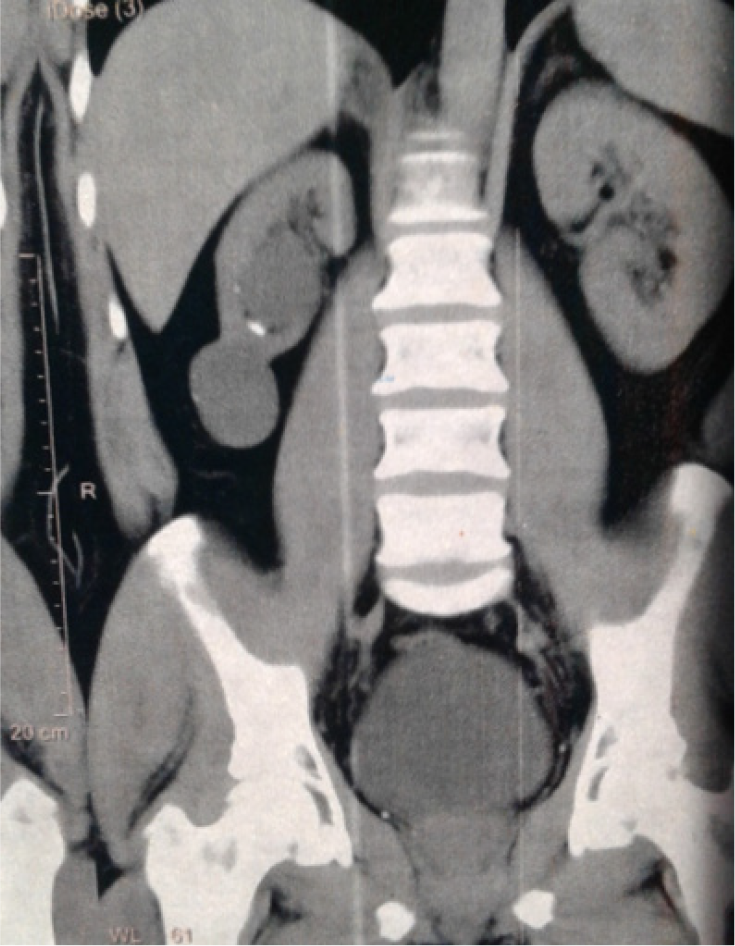
Owing to the difficult differentiation between the hypoplastic kidney and atrophic causes of the unilateral small-sized kidney which could be accurately done only by histopathological studying, we employed the radiological features for the definition of the hypoplastic kidney as a kidney with smooth outline contour without strands in the surrounding fat (Figure 1), a length less than 9 cm or 3-vertebra height, or a glomerular filtration rate less than 40% of a total function that is not less than 60 mL/min/1.73 m2. Patients who had documented acquired causes for the unilateral small-sized kidney including a previous treatment of urolithiasis by surgeries or extracorporeal shock wave lithotripsy, evidence of previous normal kidney size, previous partial nephrectomy, and vesicoureteral reflux disease were excluded from the study.
The data were descriptive and were presented as numbers and percentages or mean ± standard deviation. No biostatistician revision was warranted.
Thirty-three patients were included in the study. The demographic and clinical characteristics are summarized in Table 1.
| Variable | Value |
| Age (yr) | |
| Mean ± SD | 39.5 ± 11.2 |
| Median (range) | 40 (19-73) |
| Gender | |
| Male | 19 (57.6) |
| Female | 14 (42.4) |
| Main clinical presentations | |
| Ipsilateral loin pain | 8 (24.2) |
| Contralateral loin pain | 4 (12.1) |
| Bilateral or vague abdominal pain | 5 (15.2) |
| UTI manifestations | 4 (12.1) |
| Stone passer ± LUTS or colic | 3 (9.1) |
| Anuria/oliguria | 4 (12.1) |
| Accidental discovery F1 | 5 (15.2) |
| Anatomical side | |
| Right | 23 (69.7) |
| Left | 10 (30.3) |
| Laboratory investigations | |
| Serum creatinine mean ± SD; median (range) (mg/dL) | 1.2 ± 0.68; 0.9 (0.66-3.6) |
| Positive for protein in urine | 5 (15.2) |
| Patients with WBCs > 10/HPF in urine F2 | 22 (66.7) |
| Patients with RBCs > 3/HPF in urine | 15 (45.5) |
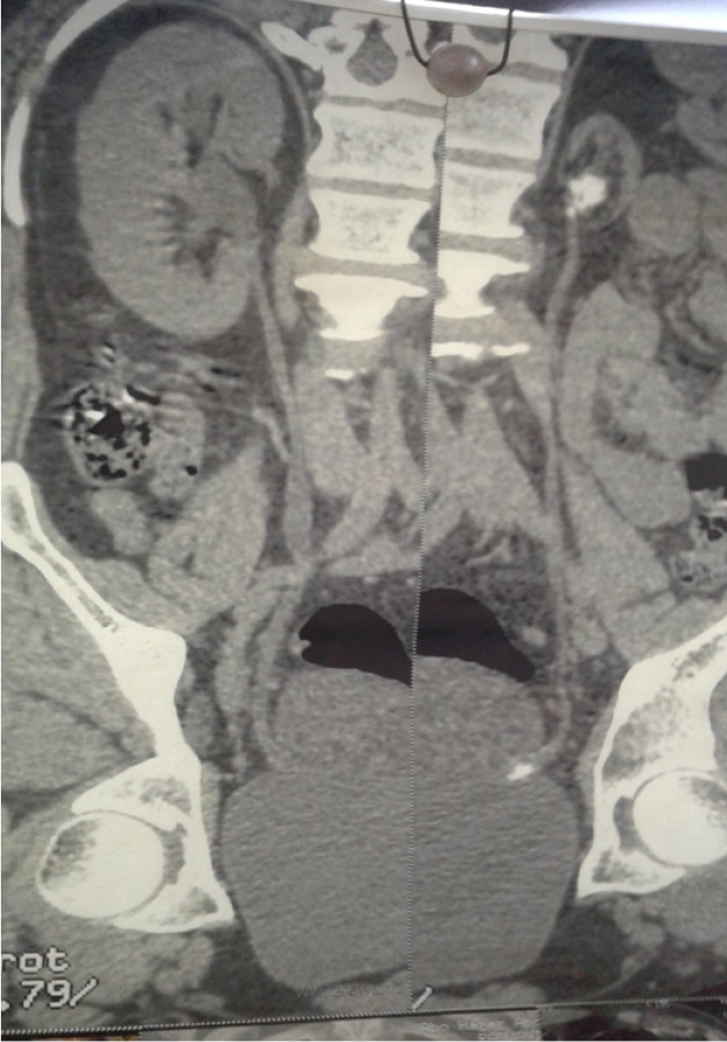
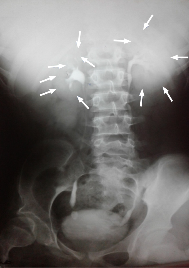
Ultrasonography and plain radiography were routine imaging tools. However, computed tomography (CT) was the best tool for the characterization of the morphological features and complications (Table 2) (Figures 1 and 2). Intravenous urography was performed in two patient (Figure 3). Radioisotope scanning was performed for a limited number of cases (Table 3).
| Imaging modality | Number of patients who had this imaging | Abnormal findings | n (%) |
| US | 33 (100) | Stones | 19 (57.6) |
| Cysts | 3 (9.1) | ||
| Hydronephrosis | 7 (21.2) | ||
| KUB | 33 (100) | Stones | 18 (54.6) |
| IVU | 2 (6.1) | Hydronephrosis | 1 (3) |
| MSCT | 27 (81.8) | Stones | 23 (69.7) |
| Cysts | 3 (9.1) | ||
| Hydronephrosis | 8 (24.2) |
| Case No. | Age (yr) | Gender | GFR (mL/min/1.73 m2) | Indication for isotope scanning | ||
| Total | Right | Left | ||||
| Case 1 | 25 | Male | 92.4 | 61.9 (67) | 30.5 (33) | Kidney donation |
| Case 2 | 42 | Female | 83.2 | 53.4 (64.2) | 29.8 (35.8) | Kidney donation |
| Case 3 | 47 | Female | 88.5 | 67.8 (76.6) | 20.7 (23.4) | To verify decision |
| Case 4 | 45 | Female | 69.7 | 61.2 (87.8) | 8.5 (12.2) | Patient request |
| Case 5 | 21 | Female | 86 | 16.8 (19.5) | 69.2 (80.5) | Patient request |
| Case 6 | 37 | Male | 77.6 | 58.2 (75) | 19.4 (25) | To verify decision |
| Case 7 | 28 | Male | 83.4 | 17.5 (21) | 65.9 (79) | To verify decision |
| Case 8 | 26 | Male | 66.8 | 7.5 (11.2) | 59.3 (88.8) | To verify decision |
Urolithiasis was the most common complication of the hypoplastic kidney (Table 4). One patient died from septicemia due to obstructive pyelonephritis of the contralateral kidney after 2 years from the original diagnosis.
| Complication | Number of patients | Involvement/localization | ||
| Ipsilateral | Contralateral | Bilateral/systemic | ||
| Urolithiasis | 23 (69.7) | 12 (36.4) | 3 (9.1) | 8 (24.2) |
| Renal cysts | 3 (9.1) | 2 (6.1) | 1 (3) | 0 (0) |
| Hydronephrosis | 8 (24.2) | 3 (9.1) | 4 (12.1) | 1 (3) |
| Recurrent UTI | 10 (30.3) | 1 (3) | 2 (6.1) | 7 (21.2) |
| Hypertension | 3 (9.1) | NA | NA | 3 (9.1) |
| Septicemia | 1 (3) | 0 (0) | 1 (3) | 1 (3) |
Different treatment approaches were used, including nephrectomy, endoscopic treatment of stones, conservative and symptomatic treatment, and assurance only for the cases without complications. Laparoscopic nephrectomy was performed in five cases for treatment of uncontrolled hypertension or persistent UTIs (Table 5).
| Approach of management | Category/variety | n (%) |
| Assurance only | 4 (12.1) | |
| Conservative/symptomatic treatment F1 | Total number of patients who received the treatment F1 | 9 (27.3) |
| For hypertension | 2 (6.1) | |
| For UTI | 3 (9.1) | |
| For stones | 5 (15.2) | |
| Hydronephrosis | 1 (3) | |
| For cysts | 1 (3) | |
| Shock wave lithotripsy | 8 (24.2) | |
| Ipsilateral | 2 (6.1) | |
| Contralateral | 5 (15.2) | |
| Bilateral | 1 (3) | |
| Endoscopic procedures | ||
| Ipsilateral ureteroscopy | 3 (9.1) | |
| Contralateral ureteroscopy | 3 (9.1) | |
| Contralateral JJ placement | 5 (15.2) | |
| Laparoscopic nephrectomy | 5 (15.2) | |
| For recurrent UTI | 2 (6.1) | |
| For hypertension | 3 (9.1) | |
| Open nephrectomy | 1 (3) | |
| For stones | 1 (3) |
All patients expressed concerns about the effect on the total kidney function. They had been educated that the lesion was unilateral and should not lead to end-stage renal disease. Four patients without complications preferred to have objective confirmation of the condition by renal radioisotope scanning including two potential kidney donors who were excluded from the donation (Table 3).
Follow-up duration varied between 7-56 mo. Three cases suffered from recurrent UTIs after stone removal and were managed conservatively.
The incidence of the small-sized kidney is variable in clinical settings[9]. Common causes of the unilateral small-sized kidney include chronic pyelonephritis, reflux or obstructive renal atrophy, and renovascular ischemia followed by the uncommon causes represented as congenital renal hypoplasia, tuberculosis, and partial nephrectomy[6]. The unilateral small-sized kidney which results from chronic pyelonephritis, congenital hypoplasia, or both represents a clinical difficulty[9].
Congenital anomalies of the urinary system are usually detected during childhood. However, when the lesion is commonly unilateral such as the hypoplastic kidney, it can pass unnoticed until the accidental discovery or development of complications in adulthood[10]. The common clinical presentations are related to the underlying complications of the hypoplastic kidney such as urolithiasis, recurrent UTIs, and hypertension[6,8]. Other rare presentations include vaginal dribbling due to ectopic ureteral insertion in females[11]. In the current study, loin pain was a cardinal presentation that refers either to the high incidence of complications including urolithiasis, hydronephrosis, and UTIs or the compensatory effect of the contralateral kidney[4-6,8].
Imaging represents a fundamental role in the urological practice with prompt advances through the last decades. Kidney size is a significant predictor of its function. Also, it is a cardinal item in urinary imaging and evaluation of the total renal functions. Bilateral reduction of renal size is imperatively associated with chronic renal impairment, especially with glomerulonephritis and other systemic parenchymal medical disorders[2,3].
Kidney size or volume and length are significant indicators for its function and affecting diseases. Measurement of the size of the kidney according to the old imaging modalities was two-dimensional and expressed relative to the corresponding vertebral heights such as in the plain and excretory radiographs[12]. However, many imaging modalities have been evolved and used recently for the measurement of three-dimensional kidney size. Among these modalities, ultrasonography has been the most practically used one, because it is available, simple, non-invasive, and repeatable. The length and size of the kidney correlate and are usually expressed relative to the whole body anthropometric measures. Size is more accurately expressed as volume by three dimensions which are length, width, and thickness with approximate mean values of 12 cm, 6 cm, and 3 cm, respectively. In spite of the absence of consensus about the definite normal values of renal dimensions among the different populations, renal length is a reproducible, accurate, and more valuable tool for studying renal diseases in adults[12-14]. Accordingly, and in parallel to these established findings, the imaging-based definition was considered in the current study. The need for documentation of the reduction of renal function was warranted only in patients with a relatively minimal size reduction, verification of the interventional management including nephrectomy, and patient insistence on numerical documentation of function. Otherwise, the severe reduction in kidney size and signs of compensation of the contralateral kidney were enough to settle the management decision in most of the cases.
Radiographic features of the uncomplicated hypoplastic kidney include a smooth outer contour of the kidney with a reduced number of calyces without caliceal clubbing or dilatation. However, these features, especially the caliceal morphology, could be disturbed in complicated cases such as urolithiasis and UTIs. These changes may concern its morphological differentiation from the atrophied kidney due to chronic pyelonephritis with an irregular contour and clubbed or dilated calyces due to scarring of the parenchyma which exerts traction forces between the renal surface and the caliceal cavity[6,8]. Renal radioisotope scanning is a tool for accurate and numerical evaluation of renal function[15]. Also, the resistive index by Doppler ultrasound showed a favorable sensitivity in the differentiation of the atrophied and hypoplastic kidneys[16]. In the current study, the indicators of the acquired affection were used to exclude patients with those findings from the study.
In cases of uncomplicated congenital hypoplastic kidney, no symptoms or therapeutic interventions are warranted. However, the management of hypoplastic kidneys is usually directed to the complications rather than the anomaly itself[10]. Indications for nephrectomy include hypertension, recurrent infections, and urolithiasis. In our series, nephrectomy was mainly done for hypertension in relatively young patients, whatever was the degree of hypertension. In the old patients, nephrectomy was preserved for those patients who had uncontrolled hypertension or those who received multiple drugs of more than one antihypertensive drug group for control. Stone passer and UTIs were other indications for surgical removal of the hypoplastic kidney.
Advantages of this series include its presentation in the time that the clinical studying of the clinical entity of hypoplastic kidney in adults has become scarce in the literature[10]. Also, it presented the classic clinical setting of the hypoplastic kidney with the patients’ perception of the potential implications of the disease. Moreover, it provided the clinical experience of a high-volume center and a tertiary level urology hospital with wide geographical drainage of urological disorders. Retrospective studying may not allow an ideal design for studying. However, it is the most suitable form for rare conditions.
In conclusion, unilateral small-sized kidney in adults is a radiological diagnosis. The hypoplastic kidney is a contributing pathology with various clinical presentations due to the development of complications. Although routine imaging by abdominal ultrasonography and radiography is available, abdominal CT is commonly indicated due to complications. In the current study, renal radioisotope scanning was indicated for relatively sizable kidneys, verification of the decision of surgical intervention, and patient request for confirmation of the lesion. The unilateral small-sized kidney is commonly being complicated by urolithiasis, obstruction, or UTIs resulting in more aggressive and life-threatening morbidities such as anuria and septicemia. Endoscopic interventions are mainly for the management of urolithiasis. While conservative management is commonly planned for this lesion, interventional management approaches including nephrectomy are mainly performed for treatment of the complications such as hypertension and recurrent UTIs or urolithiasis.
Unilateral small-sized kidney is a radiological term referring to both the congenital and acquired causes of reduced kidney volume. However, the hypoplastic kidney may have peculiar clinical and radiological characteristics. Its symptomatic clinical presentations are mostly attributed to the occurrence of underlying complications warranting early and proper management.
There is a noticeable lack of research on the clinical aspects of the unilateral hypoplastic kidney in the updated literature. Presentation of the current series may help enrich the literature and enhance the practice.
To study the clinical characteristics, complications, and management approaches of the unilateral radiologically diagnosed hypoplastic kidney in adults.
A retrospective study was carried out on patients with a radiological diagnosis of unilateral hypoplastic kidney between July 2015 and June 2020 at a tertiary-level urology center in Egypt. The demographic, clinical, and radiological characteristics and management approaches were reviewed.
The study included 33 cases with unilateral hypoplastic kidney with a mean (range) age of 39.5 ± 11.2 (19-73) years. Loin pain (51.5%) was the main clinical presentation followed by the accidental discovery (15.2%), anuria (12.1%), manifestations of urinary tract infections (UTIs; 12.1%), and stone passer (9.1%). Radiological diagnosis was commonly done by CT showing the main features including the small volume and the preserved smooth outline and structures. Urolithiasis occurred in 23 (69.7%) cases and pyuria was detected in 22 (66.7%) cases where UTIs were documented by culture and sensitivity test in 19 cases. The non-complicated cases were managed by assurance only (12.1%), symptomatic (27.3%) and endoscopic (45.6%) approaches were used for the management of simple and correctable complications, and nephrectomy (15.2%) was performed for persistent complications.
There are various presentations for the unilateral hypoplastic kidney ranging from accidental discovery to UTIs that may lead to death by septicemia. The diagnosis is mainly radiological and management is usually conservative or minimally invasive relative to the underlying findings.
Presentation of the clinical characteristics and outcomes may enhance the relevant urological practice of this disease. Urologists can provide the proper management including the conservative approaches for the simple complications and laparoscopic nephrectomy for the persistent complications.
Provenance and peer review: Invited article; Externally peer reviewed.
Peer-review model: Single blind
Corresponding Author's Membership in Professional Societies: Egyptian Urological Association; Egyptian Society of Organ Transplantation.
Specialty type: Urology and nephrology
Country/Territory of origin: Egypt
Peer-review report’s scientific quality classification
Grade A (Excellent): 0
Grade B (Very good): 0
Grade C (Good): C
Grade D (Fair): 0
Grade E (Poor): 0
P-Reviewer: Arguelles E S-Editor: Liu M L-Editor: Wang TQ P-Editor: Liu M
| 1. | Yazici R, Guney İ, Altintepe L, Yazici M. Does the serum uric acid level have any relation to arterial stiffness or blood pressure in adults with congenital renal agenesis and/or hypoplasia? Clin Exp Hypertens. 2017;39:145-149. [PubMed] [DOI] [Cited in This Article: ] |
| 2. | Matsell DG, Cojocaru D, Matsell EW, Eddy AA. The impact of small kidneys. Pediatr Nephrol. 2015;30:1501-1509. [PubMed] [DOI] [Cited in This Article: ] |
| 3. | El-Reshaid K, El-Reshaid W, Al-Bader D, Varro J, Madda J, Sallam HT. Biopsy of small kidneys: A safe and a useful guide to potentially treatable kidney disease. Saudi J Kidney Dis Transpl. 2017;28:298-306. [PubMed] [DOI] [Cited in This Article: ] |
| 4. | Rojas-Canales DM, Li JY, Makuei L, Gleadle JM. Compensatory renal hypertrophy following nephrectomy: When and how? Nephrology (Carlton). 2019;24:1225-1232. [PubMed] [DOI] [Cited in This Article: ] |
| 5. | Wang MK, Gaither T, Phelps A, Cohen R, Baskin L. The Incidence and Durability of Compensatory Hypertrophy in Pediatric Patients with Solitary Kidneys. Urology. 2019;129:188-193. [PubMed] [DOI] [Cited in This Article: ] |
| 6. | Neiman HL, Korsower JM, Reeder MM. Unilateral small kidney. JAMA. 1977;238:971-972. [PubMed] [Cited in This Article: ] |
| 7. | MATHE CP. The diminutive kidney; congenital hypoplasia and atrophic pyelonephritis. Calif Med. 1956;84:110-114. [PubMed] [Cited in This Article: ] |
| 8. | Ramanathan S, Kumar D, Khanna M, Al Heidous M, Sheikh A, Virmani V, Palaniappan Y. Multi-modality imaging review of congenital abnormalities of kidney and upper urinary tract. World J Radiol. 2016;8:132-141. [PubMed] [DOI] [Cited in This Article: ] |
| 9. | Bengtsson C, Hood B. The unilateral small kidney with special reference to the hypoplastic kidney. Review of the literature and authors' points of view. Int Urol Nephrol. 1971;3:337-351. [PubMed] [DOI] [Cited in This Article: ] |
| 10. | Xiao GQ, Jerome JG, Wu G. Unilateral hypoplastic kidney and ureter associated with diverse mesonephric remnant hyperplasia. Am J Clin Exp Urol. 2015;3:107-111. [PubMed] [Cited in This Article: ] |
| 11. | Bozorgi F, Connolly LP, Bauer SB, Neish AS, Tan PE, Schofield D, Treves ST. Hypoplastic dysplastic kidney with a vaginal ectopic ureter identified by technetium-99m-DMSA scintigraphy. J Nucl Med. 1998;39:113-115. [PubMed] [Cited in This Article: ] |
| 12. | Emamian SA, Nielsen MB, Pedersen JF, Ytte L. Kidney dimensions at sonography: correlation with age, sex, and habitus in 665 adult volunteers. AJR Am J Roentgenol. 1993;160:83-86. [PubMed] [DOI] [Cited in This Article: ] |
| 13. | Glodny B, Unterholzner V, Taferner B, Hofmann KJ, Rehder P, Strasak A, Petersen J. Normal kidney size and its influencing factors - a 64-slice MDCT study of 1.040 asymptomatic patients. BMC Urol. 2009;9:19. [PubMed] [DOI] [Cited in This Article: ] |
| 14. | Musa MJ, Abukonna A. Sonographic measurement of renal size in normal high altitude Populations. J Radiat Res Appl Sci. 2017;10:178-182. [DOI] [Cited in This Article: ] |
| 15. | Ul Hassan M, Khan SH, Ashraf M, Najar S, Shaheen F. (99m)Tc Diethylenetriaminepentacetic Acid Angiotension-coverting Enzyme Inhibitor Renography as Screening Test for Renovascular Hypertension in Unilateral Small Kidney: A Prospective Study. World J Nucl Med. 2014;13:159-162. [PubMed] [DOI] [Cited in This Article: ] |


