Gout is a rheumatic disease caused by the deposition of monosodium urate crystals. Studies have shown that monosodium urate crystals are more likely to be deposited in areas with relatively low temperatures and pH, so gout nodules are mostly found in the olecranon bursa, peroneal tendon, Achilles tendon, the helix of the ear, peripheral joints, especially the first metatarsophalangeal joint, and some rare sites of gout such as the spine, nose, eyes, and heart have also been reported [1]. Anterior chest pain is common in the population and its causes usually include heart disease, muscle strain, rib fracture, pneumothorax, costochondritis, intercostal neuralgia, lobar pneumonia, aortic dissection, malignancy of the lungs, reflux esophagitis, and autoimmune diseases. However, anterior chest pain caused by gout has rarely been reported and is very easily overlooked. There is no summary in this regard. The purpose of this article is to analyze the clinical features of gout presenting as anterior chest pain, a rare entity, using a database of published literature and to suggest a comprehensive clinical and diagnostic evaluation of patients with joint pain and swelling.
We conducted a systematic literature review in PubMed up to and including October 1, 2022. We used a combination of the following terms: “sternoclavicular joint”, “sternum-clavicle joint”, “manubriosternal joint”, “sternocostal joint, “rib”, “costal cartilage”, “costochondral”, “sternum”, “sternal”, “manubrium”, “clavicle”, “xiphoid process”, “gout”, and “gouty arthritis” and excluded articles for which the full text was not available and the lesion was not located in the anterior chest wall region. Articles involving the acromioclavicular joint that does not present as anterior chest pain and subcutaneous gout are also excluded. The literature search yielded 26 articles after eliminating duplicates. After screening the titles and abstracts and the references of the included papers, eight articles were found to be relevant. The eligible articles included 8 case reports [2–9]. A total of 8 patients were identified, with a male/female ratio of 7/1 and a mean age of 52 years (range: 44–61), with a median age of 52 years (Table I). Seven patients had peripheral joint gout or risk factors for monosodium urate crystal deposits such as hyperuricemia, poorly controlled type 2 diabetes, obesity, alcohol intake, overweight, renal insufficiency, and cardiovascular disease before being diagnosed with gout presenting with anterior chest pain. Only 1 patient had no risk factors or family history and later presented with a painful, increasing bony mass in the sternal notch that resembled a malignant tumor [3]. Of all the sites of gout attacks, the sternoclavicular joint was the most common (n = 4), the others being the costal cartilage (n = 2) and manubriosternal joint (n = 2). Almost all patients presented with anterior chest pain accompanied by localized joint swelling, and in some patients with pain that mimicked myocardial infarction and pleurisy [2, 6]. In addition, some patients presented with night sweats, fever, and other symptoms similar to sternal tuberculosis and infection [4–6]. The main tools for confirming the diagnosis include joint aspiration for polarizing microscopy, mass excision biopsy, and dual-energy CT (DECT). The vast majority of patients had abnormal serum uric acid levels. The differential diagnosis of this disease mainly includes malignant tumors, acute chest pain, costochondral chest wall syndrome, and pyogenic arthritis. Pharmacological treatment included NSAIDs, steroid hormones, allopurinol, febuxostat, colchicine, antibiotics, and the IL-1 receptor antagonist anakinra. In the articles that mentioned treatment outcomes, all patients had a good prognosis.
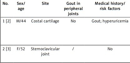
Table I
General clinical characteristics of 8 patients with gout presenting with anterior chest pain
| No. | Sex/age | Site | Gout in peripheral joints | Medical history/risk factors | Clinical manifestations | Confirmation examinations | Uric acid level | Differential diagnosis | Treatment | Prognosis |
|---|---|---|---|---|---|---|---|---|---|---|
| 1 [2] | M/44 | Costal cartilage | No | Gout; hyperuricemia | Anterior chest pain | Dual-energy CT | 10.1 (reference range 2.6–7.0) mg/dl | Heart disease | Febuxostat; colchicine; diclofenac | Recovered |
| 2 [3] | F/52 | Sternoclavicular joint | / | No | A painful and increasing bony mass in the sternal notch | Mass excision biopsy | / | Malignancy | Mass excision | Recovered |
| 3 [4] | M/50 | Sternoclavicular joint | Yes | Chronic gouty arthritis; type 2 diabetes; lumbosacral radicular syndrome; obesity | Widespread pain, especially at the chest level; fever; night sweats | Dual-energy CT | Normal limit (263 mmol/l) | Neoplasia | NSAIDs; febuxostat; corticosteroids; anakinra | Recovered |
| 4 [5] | M/54 | Sternoclavicular joint | Yes | Gouty arthritis; alcoholism | Arthralgia; fever | Joint fluid aspiration analysis reveals monosodium urate crystals | 8 mg/dl | / | Antibiotic therapy; colchicine | Recovered |
| 5 [6] | M/49 | First costochondral junction | Yes | Gout; viral cardiomyopathy; heart transplantation | Pleuritic chest pain; fever | CT-guided core biopsy | 8.6 mg/dl | Infection | Prednisone; colchicine; allopurinol | Recovered |
| 6 [7] | M/52 | Manubriosternal joint | Yes | Gout; alcohol intake | Intense, acute, anterior thoracic pain located on the sternum | Joint fluid aspiration analysis reveals monosodium urate crystals | 9.1 mg/dl | / | Prednisone; NSAIDs; allopurinol | Recovered |
| 7 [8] | M/61 | Sternoclavicular joint | Yes | Carcinoma of the tongue | A progressive tumor over the left sternoclavicular joint. | Tissue biopsy | Slightly elevated | Osteomyelitis; malignancy | / | Recovered |
| 8 [9] | M/54 | Manubriosternal joint | No | Type 2 diabetes; hypertension; renal insufficiency | Tenderness and mild swelling over the upper sternum | Arthrocentesis and polarizing microscopy | 9.6 mg/dl | Ischemic heart disease | Colchicine | Recovered |
Our study indicates that the mean age of onset of gout mimicking anterior chest pain is older (52 years) and that this disease does not occur in younger populations, which increases the differential range of this disease such as acute myocardial infarction, aortic dissection, etc. In addition, the gender ratio of 7 : 1 in this disease is much higher than the 2 : 1 to 4 : 1 gender ratio in epidemiological studies on gout in European and North American population-based studies, indicating that males are the most prevalent group of this rare entity [10]. In terms of laboratory tests, most patients have abnormal serum uric acid levels, but normal uric acid levels do not exclude gout. Anterior chest pain is the most common clinical characteristic of this rare entity in this article. Gout involving the anterior chest (especially at the joints) can mimic painful disorders of the anterior chest region such as acute myocardial infarction, pulmonary embolism, costochondritis, septic arthritis, neoplastic lesions, and rheumatic diseases (e.g., SAPHO syndrome, rheumatoid arthritis, osteoarthritis, relapsing polychondritis, axial spondyloarthritis, fibromyalgia). Notably, there is a strong link between hyperuricemia, gout and cardiovascular risk. Studies have shown that hyperuricemia and gout negatively impact vascular function by exerting pro-oxidant effects and reducing nitric oxide bioavailability, thereby inducing inflammation and endothelial dysfunction, which may contribute to hypertension, metabolic syndrome and cardiovascular disease [11]. Gout itself is a factor in the acceleration of atherosclerosis and subsequent cardiovascular events, including chest pain [11]. In addition, there may be an overlap between the articular-skeletal gout manifestation and the “cardiovascular disease presentation: chest pain”, and there are some suggestions for differential diagnosis, including that patients can be investigated for these episodes of chest pain by blood tests (including troponin), electrocardiogram, echocardiography, and chest CT. In the sample summarized in this article, some patients have completely normal tests mentioned above and even no abnormalities were seen at the chest wall and peripheral joints. Synovial fluid of joints showing monosodium urate crystals under polarized light microscopy is the gold standard for the diagnosis of gout [10]. For some atypical sites such as the spine, heart, lungs, and other organs, the biopsy is difficult, but for sites such as sternoclavicular joints, sternocostal joints, etc., tissue biopsy can accurately diagnose gout [12]. Given the high specificity of dual-energy CT for the detection of urate crystals and its advantages of non-invasiveness, reproducibility, and accurate assessment of atypical sites, it is considered a promising clinical screening tool for gout [13]. Treatment is based on drugs that relieve symptoms and lower blood uric acid, including glucocorticoids, colchicine, NSAIDs, and interleukin-1 receptor antagonists if necessary [1]. All patients have a positive prognosis after treatment. This article explores the general basic features of gout mimicking anterior chest pain using a database of published literature and suggests that chest pain can be a rare manifestation of gout and should be considered as one of the “clinical masks” of the disease; however, the cardiovascular causes of the pain should always be considered and may be a co-existing phenomenon. However, the limitation of this article is that 8 cases are not sufficient to elucidate the clinical features of the disease, and publication bias needs to be considered, so there may be more cases in reality. Further research is needed regarding the more detailed clinical characteristics of the rare entity described in this article.