Published online Mar 26, 2011. doi: 10.4330/wjc.v3.i3.98
Revised: August 12, 2010
Accepted: August 19, 2010
Published online: March 26, 2011
When performing echocardiography in a 74-year-old woman admitted with non ST elevation myocardial infarction and concomitant colorectal cancer (CC), a dense calcification of the mitral annulus was detected. Differential diagnosis between secondary metastasis and other etiologies of cardiac masses was essential for staging and therapeutic decision-making. Multimodality imaging with echocardiography alongside a computed tomography scan and cardiac magnetic resonance was crucial for the final diagnosis of caseous calcification of the mitral annulus (CCMA). CCMA is briefly reviewed and some possible explanations for the previously undescribed association of CC with CCMA are suggested.
- Citation: Providência R, Botelho A, Mota P, Catarino R, Leitão-Marques A. Cardiac mass in a patient with colorectal cancer: “Not all that glitters is gold!”. World J Cardiol 2011; 3(3): 98-100
- URL: https://www.wjgnet.com/1949-8462/full/v3/i3/98.htm
- DOI: https://dx.doi.org/10.4330/wjc.v3.i3.98
Mitral valve calcification (MVC) is a degenerative abnormality of the cardiac fibrous skeleton that occurs mainly in elderly individuals[1]. Caseous calcification of the mitral annulus (CCMA) is a less well known and rarely described entity representing a variant of MVC, typically located in the posterior mitral annulus[2] that should be included in the differential diagnosis of intracardiac masses. It is found in 0.6% of patients with MVC and 0.06% of all echocardiographic studies[3]. In necropsy studies, its prevalence raises to 2.7%[1]. It is more prevalent in the elderly, in women and in patients undergoing dialysis. Histopathologic analysis of this entity reveals an amorphous basophilic fluid, surrounded by macrophages (mainly) and lymphocytes. This condition carries a benign prognosis. Surgery may be rarely needed due to hemodynamic compromise[4,5]. The association of colorectal cancer (CC) with CCMA has not been described previously.
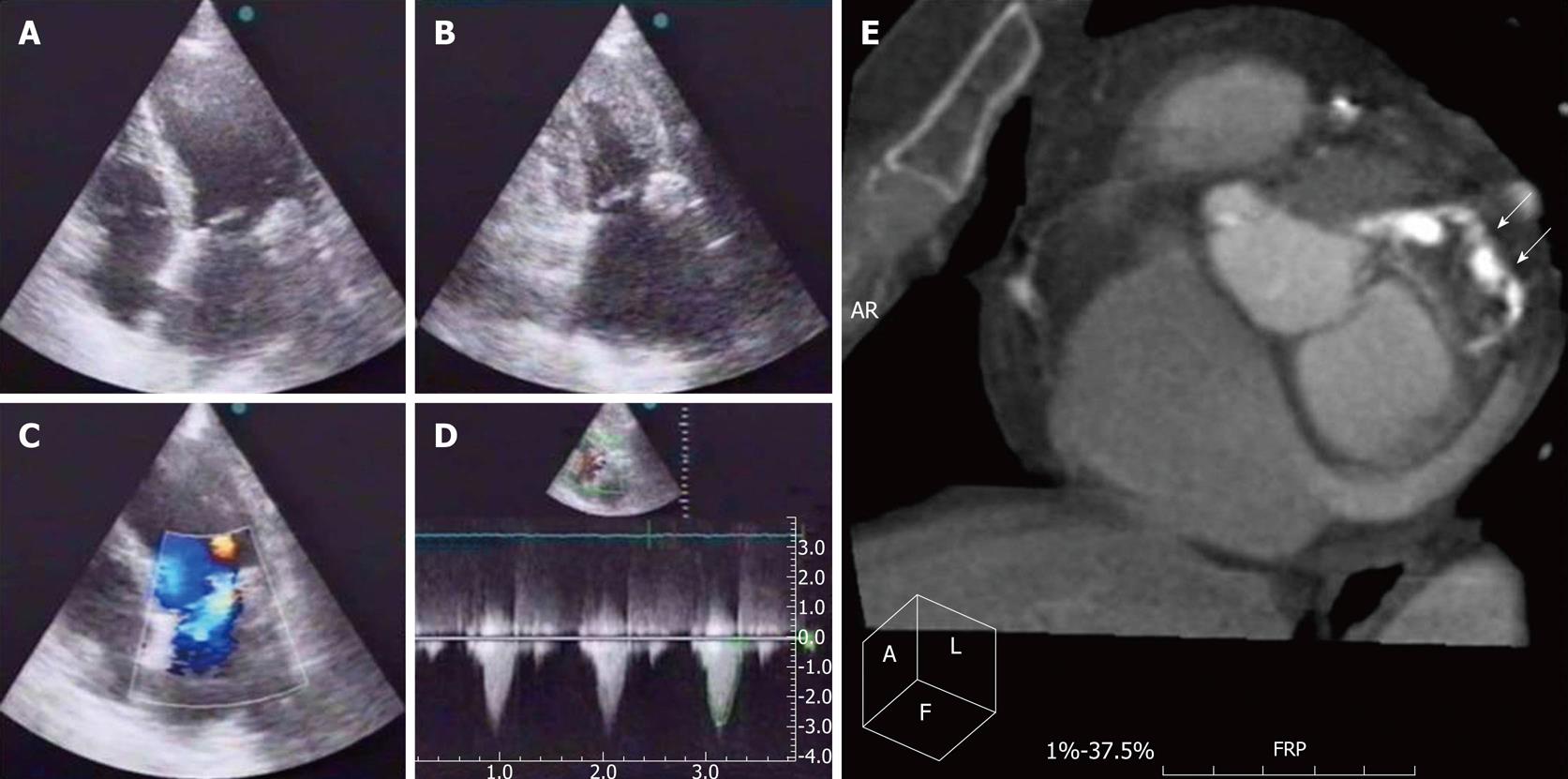
A 74-year-old woman with a history of hypertension was admitted to our department with non ST-elevation myocardial infarction. Standard medical treatment was promptly initiated and a coronary angiogram was performed at 48 h of evolution. Two-vessel coronary artery disease was diagnosed and complete revascularization with 3 bare-metal stents was accomplished by staged percutaneous procedures. A heavily calcified mitral annulus, moderate mitral regurgitation and ejection fraction of 77% were found on the left ventriculogram. The patient had a favorable outcome (Killip class 1). A pre-discharge transthoracic echocardiogram (TTE) (Figure 1A-D) showed moderate enlargement of the left atrium, grade II mitral regurgitation and good left ventricular systolic function. A 2.3 cm × 2.0 cm round echodense mass was found at the posterior side of the mitral annulus adjacent to the atrial side of the posterior leaflet. Fibrocalcification of the aortic valve with moderate stenosis (peak gradient 33 and medium 21 mmHg) was also described. Blood calcium levels and renal function were normal.
Meanwhile, in order to better clarify the persistent microcytic hypochromic anemia (8.6 g/dL hemoglobin, mean globular volume 79 fL) diagnosed during her initial admission, an endoscopic study of the gastrointestinal tract was conducted. An invasive, well differentiated mucinous colorectal adenocarcinoma was detected 20 cm away from the anal margin.
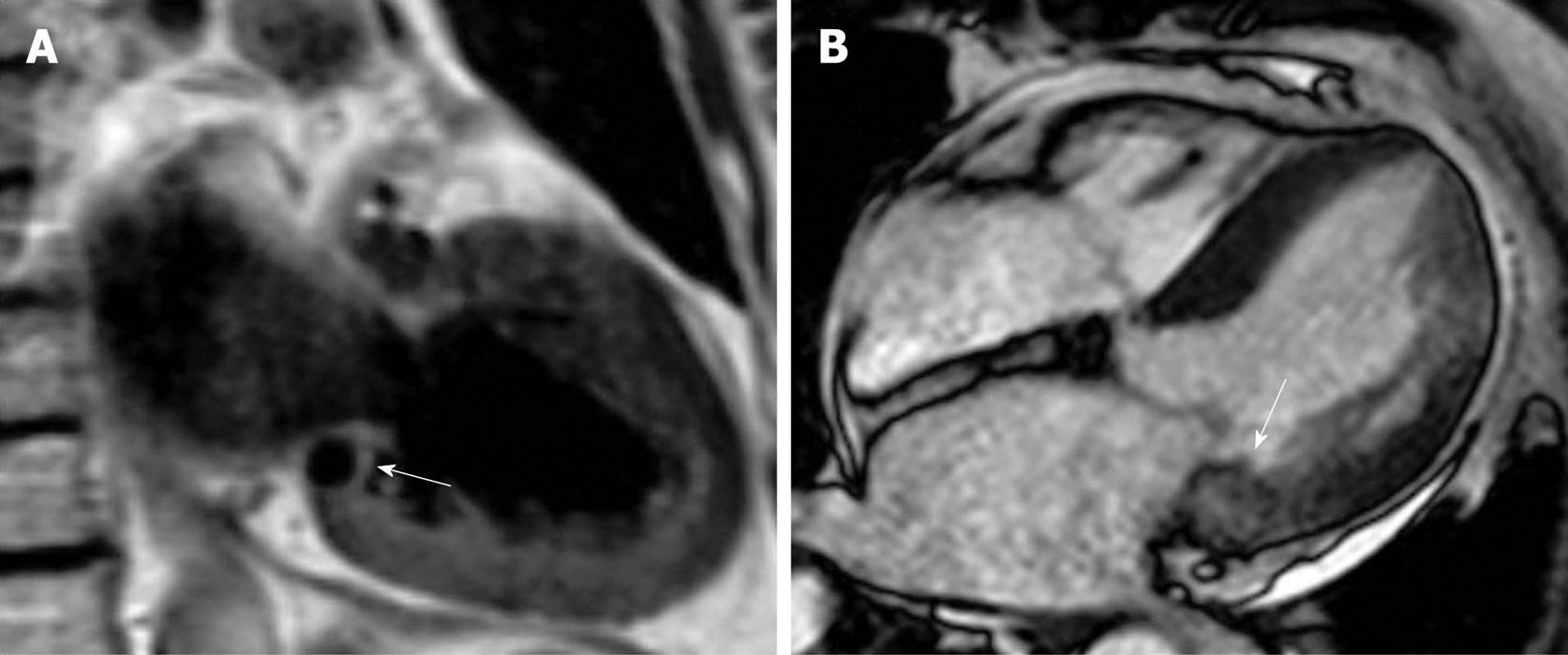
In order to perform cancer staging, the nature of the cardiac mass had to be elucidated. During a routine chest computed tomography (CT) scan for cancer staging, a cardiac CT evaluation was performed (Figure 1E) (suggestive of CCMA). Further evaluation by cardiac magnetic resonance revealing absence of vascular flow within the mass, and the presence of a dark signal in T1 weighted sequences was also consistent with calcification (Figure 2A). In steady state free precession images, the mass appeared only slightly darker than the normal myocardium, with a well defined intramyocardial border (Figure 2B). Along with the TTE, these data suggested a diagnosis in favor of a CCMA. Therefore, as no hepatic, pulmonary or other type of metastasis was found in cancer staging (T4N0M0 - Stage II), left hemicolectomy was successfully performed and the patient was discharged 9 d later.
After 24 mo, the patient is currently stable, with no relapse of cancer nor progression of mitral valve disease (NYHA I and similar findings in follow-up TTE).
Although there are only 9 reports of the association of CC with cardiac metastasis[6], when faced with a patient with CC and a cardiac mass, this is a possible diagnosis, that may be frequently overlooked (metastasis to the heart occurred in 10.7% out of 1,029 autopsy cases in which a malignant neoplasm had been diagnosed[7]). Other possibilities to consider would be a myxoma[8], other types of tumor[9,10], an organized thrombus or abscess. An abscess would be clinically unlikely and the other possibilities could be excluded by the typical presentation of the CCMA that we found. Even though TTE was almost diagnostic by itself, this patient had to undergo chest CT for cancer staging, and cardiac CT along with magnetic resonance imaging were helpful for mass characterization and exclusion of other potential etiologies, and for diagnosis confirmation[11,12].
An accurate diagnosis was of utmost importance in this patient, since it changed both treatment and prognosis. Unlike cardiac metastasis, CCMA has a good prognosis and does not require cardiac surgery, unless it causes mitral stenosis or symptomatic and severe mitral regurgitation. Additionally, the absence of cardiac metastasis turned this into a stage II colorectal cancer, eligible for curative surgical treatment, instead of a stage IV (metastatic).
We may wonder if this association of CC and CCMA is a mere coincidence or if there is any kind of circulating substance produced by the CC that led to increased calcium deposition in the heart. Triple simultaneous involvement of the heart (calcification of coronary vessels, mitral and aortic valves) is unusual in this context. The possibility of a common link between aortic stenosis and coronary heart disease has been described before[13], but nothing has been said about CCMA as a part of this continuum.
Peer reviewers: Jesus Peteiro, MD, PhD, Unit of Echocardiography and Department of Cardiology, Juan Canalejo Hospital, A Coruna University, A Coruna, P/ Ronda, 5-4º izda, 15011, A Coruña, Spain; Yoko Miyasaka, MD, PhD, FACC, Cardiovascular Division, Department of Medicine II, Kansai Medical University, 2-3-1, Shin-machi, Hirakata, Osaka, 573-1191, Japan
S- Editor Cheng JX L- Editor Cant MR E- Editor Zheng XM
| 1. | Pomerance A. Pathological and clinical study of calcification of the mitral valve ring. J Clin Pathol. 1970;23:354-361. [Cited in This Article: ] |
| 2. | Harpaz D, Auerbach I, Vered Z, Motro M, Tobar A, Rosenblatt S. Caseous calcification of the mitral annulus: a neglected, unrecognized diagnosis. J Am Soc Echocardiogr. 2001;14:825-831. [Cited in This Article: ] |
| 3. | Novaro GM, Griffin BP, Hammer DF. Caseous calcification of the mitral annulus: an underappreciated variant. Heart. 2004;90:388. [Cited in This Article: ] |
| 4. | Minardi G, Manzara C, Pulignano G, Pino PG, Pavaci H, Sordi M, Musumeci F. Caseous calcification of the mitral annulus with mitral regurgitation and impairment of functional capacity: a case report. J Med Case Reports. 2008;2:205. [Cited in This Article: ] |
| 5. | Fernandes RM, Branco LM, Galrinho A, Timóteo AT, Tavares A, Feliciano J, Oliveira R, Fiarresga A, Mamede A, Banazol N. Caseous calcification of the mitral annulus. A review of six cases. Rev Port Cardiol. 2007;26:1059-1070. [Cited in This Article: ] |
| 6. | Choi PW, Kim CN, Chang SH, Chang WI, Kim CY, Choi HM. Cardiac metastasis from colorectal cancer: a case report. World J Gastroenterol. 2009;15:2675-2678. [Cited in This Article: ] |
| 8. | Nuño IN, Kang TY 4th, Arroyo H, Starnes VA. Synchronous cardiac myxoma and colorectal cancer: a case report. Tex Heart Inst J. 2001;28:215-217. [Cited in This Article: ] |
| 9. | Kato M, Nakatani S, Okazaki H, Tagusari O, Kitakaze M. Unusual appearance of mitral annular calcification mimicking intracardiac tumor prompting early surgery. Cardiology. 2006;106:164-166. [Cited in This Article: ] |
| 10. | de Vrey EA, Scholte AJ, Krauss XH, Dion RA, Poldermans D, van der Wall EE, Bax JJ. Intracardiac pseudotumor caused by mitral annular calcification. Eur J Echocardiogr. 2006;7:62-66. [Cited in This Article: ] |
| 11. | Monti L, Renifilo E, Profili M, Balzarini L. Cardiovascular magnetic resonance features of caseous calcification of the mitral annulus. J Cardiovasc Magn Reson. 2008;10:25. [Cited in This Article: ] |
| 12. | Blankstein R, Durst R, Picard MH, Cury RC. Progression of mitral annulus calcification to caseous necrosis of the mitral valve: complementary role of multi-modality imaging. Eur Heart J. 2009;30:304. [Cited in This Article: ] |


