Published online Jun 27, 2010. doi: 10.4240/wjgs.v2.i6.210
Revised: February 18, 2010
Accepted: February 25, 2010
Published online: June 27, 2010
Natural orifice translumenal surgery (NOTES) has garnished significant attention from surgeons and gastroenterologists, due to the fusion of flexible endoscopy and operative technique. Preliminary efforts suggest that NOTES holds potential for a less invasive approach with certain surgical conditions. Many of the hurdles encountered during the shift from open to laparoscopic surgery are now being revisited in the development of NOTES. Physician directed efforts, coupled with industry support, have brought about several NOTES specific devices and platforms to help address limitations with current instrumentation. This review addresses current flexible platforms and their attributes, advantages, disadvantages and limitations.
- Citation: Shaikh SN, Thompson CC. Natural orifice translumenal surgery: Flexible platform review. World J Gastrointest Surg 2010; 2(6): 210-216
- URL: https://www.wjgnet.com/1948-9366/full/v2/i6/210.htm
- DOI: https://dx.doi.org/10.4240/wjgs.v2.i6.210
Advances in the fields of laparoscopy and interventional endoscopy have ushered in a new era of minimally invasive surgery. Natural orifice translumenal endoscopic surgery (NOTES) is a new technique that permits flexible endoscopic “scarless” trans-visceral peritoneal access and may be the next evolutionary stride towards progressively less invasive procedures. To date, several important steps have been taken, from simple abdominal exploration in animal models to trans-visceral cholecystectomy in humans[1,2]. As this nascent field matures, technology has produced several platforms that address basic needs and strive to match their surgical counterparts. This article reviews currently available flexible platforms, their advantages, disadvantages and comparisons with currently available tools.
The first published NOTES procedure used a standard forward viewing endoscope, biliary sphincterotome, guide wire, and an esophageal dilatation balloon for transgastric access and liver biopsy[3]. This re-purposed equipment, although rudimentary for these purposes, has been used by gastroenterologists and surgeons alike. More complex trans-gastric procedures were performed in the pre-clinical setting with standard endoscopic equipment including: tubal ligation, hysterectomy/simulated appendectomy[4], cholecystectomy[5] and splenectomy[6]. In this early work, several methodological and technical limitations were identified. An alternative approach from the inferior abdomen was explored for cholecystectomy[7] and pancreatic work[8], and proved useful in addressing certain limitations of upper abdominal surgery, however, other unique problems were revealed.
In 2005, leaders of the ASGE and SAGES (NOSCAR-Natural Orifice Surgery Consortium for Assessment and Research) reviewed the early work and identified several fundamental challenges to NOTES in a manuscript known as the White Paper[9]. Peritoneal access and closure, infection, spatial orientation, management of complications and multi-tasking platforms were identified as critical areas of focus. Further NOTES experience subsequently confirmed the limitations of current instrumentation and identified new problems, which were not initially experienced with the transition from open to laparoscopic surgery.
Laparoscopy introduced several challenges related to visualization and tissue manipulation, however, unlike NOTES, laparoscopy mimics the surgical perspective, maintaining remote visualization using rigid, short length instruments optimally positioned for triangulation, traction, dissection, target mobilization and tissue approximation. Additionally, ports allow for 5, 10, 12 mm and larger access to the peritoneum. Since the initial cholecystectomy by Muhe in 1985[10], laparoscopy has become the gold standard for minimally invasive surgery and is the benchmark to which NOTES is compared.
Purpose-specific equipment and technology is sorely lacking compared to specialized laparoscopic paraphernalia. Current endoscopic technology and design have several shortcomings when applied to more complex surgical procedures and can be framed in the context of basic platform elements, including guide tube attributes, spatial orientation and imaging characteristics.
Shaft: Originally purposed for gastrointestinal procedures, flexibility is desirable for atraumatic endolumenal movement; although this may aide in intra-abdominal navigation it also poses a challenge for maneuvering in open space or when attempting to achieve traction or counter-traction. Conversely, laparoscopic tools are rigid, and the combination of spaced ports of entry with transabdominal fulcrum points allow for a stable platform. Alternative means of target fixation, apart from the physical platform, may aide in stability and organ manipulation and help address current limitations of traction and angles of tissue engagement[11]. Additionally, the increased distance between operator and end-effectors, which is typical with flexible endoscopy, limits haptic feedback.
Working channels: Maximal size on commercially available endoscopes is 3.7 mm, limiting the size of available equipment and contrasts greatly to variable size laparoscopic ports. The proximity and parallel orientation of channels limit triangulation, robust tissue manipulation and traction. Furthermore, current endoscopes are restricted to two working channels that are inadequate for some procedures[4].
Ancillary channels: Channels dedicated for insufflation, irrigation and suction are sub-optimal for routine needs of intra-abdominal surgery and may be inadequate in the event of an emergency.
User interface: Current endoscopes are designed for the endoscopist to control field of view and positioning/orientation. The working channels and navigational/field control elements are in close proximity and lead to complicated team interactions.
Site of access: NOTES entry point (transgastric, transcolonic, transurethral or transvaginal) is often selected for proximity or best en-face view, however, this does not ensure direct or adequate visualization of the desired site. Additionally, trans visceral peritoneoscopy may have limited reach and stability in certain orientations; for example targeting the spleen via the transgastric approach. Considerable maneuvering of the endoscope is often required to achieve acceptable positioning and stability. A “bounce-off” technique, utilizing internal structures to redirect scope vectors and trajectory is often necessary, yet technically challenging. This may be due to several reasons, including inter-patient variability and inherent mobility of internal structures. Conversely, procedure specific transabdominal port placement allows optimal laparoscopic site selection and stability.
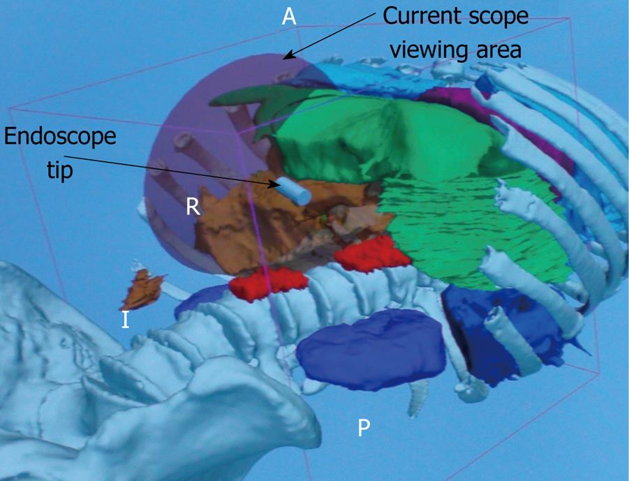
Visual orientation: While endoscopists accustomed to the confines of the gastrointestinal tract are typically comfortable with inverted positioning, surgeons prefer a fixed horizon. With the camera married to the endoscope shaft, horizon is at the mercy of the endoscope’s final position. Laparoscopic visualization is divorced from the effectors allowing remote imaging with maintenance of the horizon. This may diminish work load and prove beneficial when dealing with complex surgeries[12]. Vantage point is yet another issue; the close proximity and magnified endoscopic views may prove advantageous for meticulous dissection, while a remote view of the operative field may be essential for other tasks and harder to achieve with current flexible platforms. Ancillary visualization technologies with computer tomography and 3D image registration may help mitigate these limitations in the future[13] (Figure 1).
Imaging: Current endoscopic depth of field, although excellent for near vision, lacks the needed distant visualization that would aid in complex procedures and abdominal exploration. Additionally, there may be insufficient light for certain procedures, including exploration and cancer staging. A 10-mm laparoscope provides 380 lumens while a typical endoscope with a 3-mm light bundle provides only 25 lumens. Conversely, the magnified endoscopic images may be superior to laparoscopic images for certain procedures.
There is a fundamental difference in work load between laparoscopic and endoscopic paradigms. Laparoscopically, the field of view is maintained by an assistant while instruments are maneuvered and executed by the surgeon. Endoscopically, positioning and field of view are maintained by a complex coordination of positioning-wheels, torque, placement, and locking mechanisms. The endoscopic tools are also positioned by the endoscopist (in unison with the endoscope) while actuated by an assistant. Additionally, mental workload will likely be increased by fluctuating visual frames of reference and angles of approach associated with NOTES procedures.
While many of these differences pose disadvantages compared to the current laparoscopic paradigm, flexible endoscopy’s added reach and close visualization of an operative field may confer advantages still unknown, for example, inspection of the lesser sac and the supra-hepatic/infra diaphragmatic space in cancer staging, which are not readily attainable with current minimally invasive techniques.
NOSCAR’s second meeting included industry participation and several platform solutions were proposed. These have evolved into two broad systems: rigid, mimicking single incision laparoscopic surgery (SILS) and flexible, emulating endoscopy. Flexible platforms may be further divided into two groups: A traditional endoscopic model where the endoscopist controls navigation and instrument position while the assistant exchanges and actuates instruments, and a flexible-laparoscopic paradigm where the interventionalist has complete control of the instruments and the assistant provides visualization and maintains the operative field.
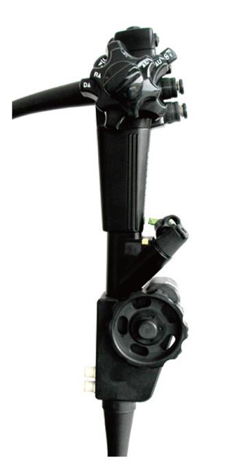
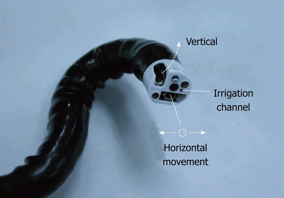
One of the first platforms to be used in NOTES animal models was the “R-scope” (XGIF-2TQ160R; Olympus, Tokyo, Japan) later modified to the NOTES scope. It has been used to perform several procedures in the pre-clinical setting including cholecystectomy[14,15]. This device falls under the traditional endoscopic paradigm. It is a modified dual channel endoscope (DCE) with additional elevator toggles and a larger wheel further down the handle to control a second bending segment (Figure 2). The primary segment is lockable, allowing for a better angle of approach and more precise tissue manipulation with maneuvering of the second segment. Additionally, the two working channels have lifting gates that are orthogonally positioned allowing for simultaneous lifting (vertical motion) and dissection (horizontal motion) (Figure 3). This configuration allows more accurate tissue manipulation off-axis to the visual plane. See Table 1 for device specifics. This endoscope addresses several of the DCE shortcomings, including positioning and to a small degree triangulation. The second bending segment, although useful, can be technically demanding. These features may lead to increased physical and mental work load. Furthermore, the image is still married to the effectors and, as such, has a limited field of view. Essentially the NOTES scope further refined what the current standard endoscope is capable of while partially tackling some tasks germane to complex surgery.
| Prototype | Paradigm | Working Length (cm) | Channels/Size (mm) | Diameter (mm) | Visualization | Positioning mechanism | Specializations | Procedures performed |
| DCE (EGD/Colonoscope) | Endoscopic | 103/168 | Two: 3.7, 2.8/Two: 3.7, 3.2 | 12.6/13.7 | Standard endoscopic | Standard scope shaft | Two small parallel channels | Animal and human NOTES (Hybrid procedures) |
| NOTES scope | Endoscopic | 133 | Two: 2.8, 2.8 | 14.3 | Standard endoscopic | Two bending segments, one lockable | Dual bending segments, orthogonal lifting gates | Animal hybrid NOTES |
| IOP | Endoscopic/Flexible-laparoscopic | 110 | Four: 7, 6, 4, 4 | 18 | N-scope | Built in shaft-stiffening system | 2.5cm gasping forceps with tissue anchors | Human NOTES, endoluminal bariatrics, anti-reflux procedures |
| EndoSAMURAI | Flexible-laparoscopic | 103 | Three: 2.8, 2.8, 2.8 | 15.7 | Endoscopic | Uses a stiffening overtube system | Bimanual control enables 5 degrees of freedom for 2 end effectors | Animal NOTES |
| DDES | Flexible-laparoscopic | 55 | Three: 7, 4.2, 4.2 | 16 × 22 | N-scope | Articulating guide sheath | Bimanual control with 7 degrees of freedom for 2 end effectors | Bench top EMR, ESD and skills assessment models |
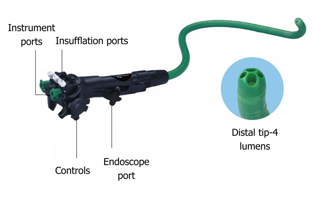
The incisionless operating platform (IOP; USGI Medical, San Capistrano, CA) was initially designed to function within the traditional endoscopic paradigm, with recent modifications bridging to the flexible-laparoscopic model. This device was developed through collaboration of physicians and industry and addresses several NOTES requirements. It has been successfully used in a variety of procedures, and is the first specialized platform to be used in clinical NOTES cases, including human transgastric cholecystectomy[16]. In appearance, the IOP is similar to, but larger than a standard endoscope, with multiple ports and directional wheels at the user interface (Figure 4). Based on endoscopic ergonomics, this platform consists of a 110 cm × 18 mm overtube-like design with a steerable shaft and several channels (Table 1). In some models, the overtube-shaft is capable of stiffening, providing enhanced stability. Its four channels (7, 6, 4 and 4 mm in size) allow for an N-scope (Olympus, Tokyo, Japan) and specialized equipment[17]. The N-scope is independently rotatable within the channel allowing for an adjustable horizon while maintaining instrument position. The other channels can be used for instruments as well as high flow carbon dioxide insufflation[14]. Several specialized tools have been designed for this system including a 2.5-cm grasping jaw, capable of performing tissue plications with unique anchors and several accessories for tissue manipulation. Compared to the DCE, the IOP has enhanced deflection and improved triangulation due to the large channels and effectors’ ability to enter the operative field. However, the IOP’s in-line channel orientation is still subject to parallelism. This may be overcome with instrument modifications. This device presents a new paradigm for the endoscopist as visualization is divorced from the primary operator. Work load for the IOP is high and requires skilled assistants as the primary operator interchanges responsibilities for instrument exchange, device orientation and scope positioning. This is increasingly challenging when the device is in an unstable position. Newer versions of this device allow for bimanual instrument control and are more consistent with the flexible-laparoscopic model.
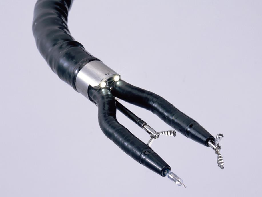
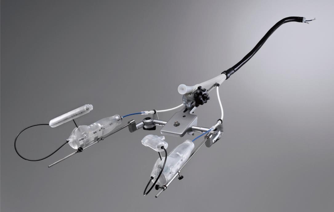
The EndoSAMURAI (Olympus Corp., Tokyo, Japan) was designed to operate within the flexible-laparoscopic paradigm. It has been tested in animal models for cholecystectomy[18]. It consists of a specialized endoscope with a remote working station and a locking overtube. The distal end of the scope has two short modified independent arms, which upon entry remain parallel with the scope shaft, however, open in an elbow-like fashion when in position (Figure 5). These serve as conduits for different effectors, including standard endoscopic accessories, and are manipulated from a control unit apart from the traditional endoscopic user interface (EVIS EXERA II Universal Platform-Olympus Corp; Figure 6). With 5 degrees of freedom and triangulation capabilities, the arms can tie sutures as well as provide traction and counter traction. In addition to the two conduits, it has a third working channel that may be used for ancillary equipment or suction/irrigation. Stiffened by a locking overtube, the scope articulates in the same manner as a standard endoscope with identical visualization. Although essentially a modified DCE, the EndoSAMURAI overcomes parallelism by the angle at which its effectors are positioned. Additionally, it has better stability compared to the DCE because of the locking overtube. As the arms are married to the camera/scope, it still bears the same image-perspective limitations as the standard endoscope. Interestingly, this system employs a “drive, park and move” methodology. Where the user navigates to the target with the endoscope, locks the overtube system and scope in position and then proceeds to the user interface. This effectively allows one operator to perform most of the work load as the image is theoretically kept in place with the locking system with subsequent maintenance of the image by the assistant, which is somewhat similar to traditional laparoscopy.
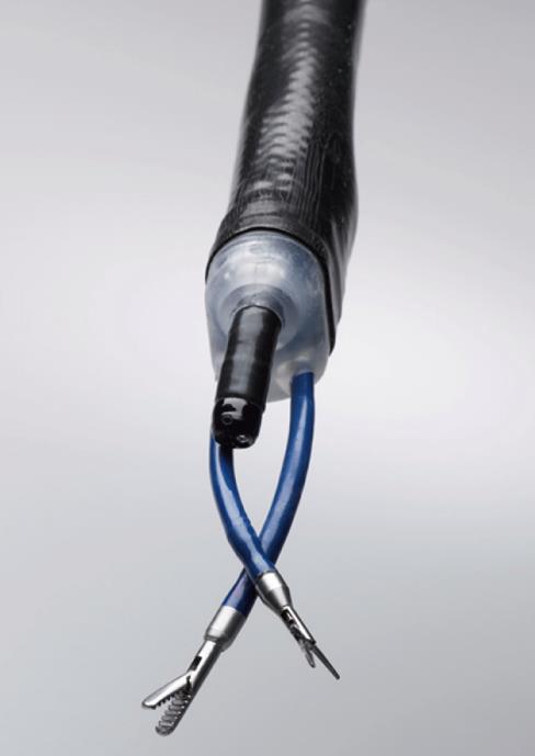
The direct drive endoscopic system (DDES; Boston Scientific, Natick, MA) is a flexible-laparoscopic multitasking platform that consists of a 55-cm steerable guide sheath that houses 3 lumens extending from a rail-based platform with interchangeable 4 mm instruments (Figure 7, Table 1). The user interface consists of ergonomic rail-guided drive handles situated above the surgeon’s waist level. A unique system in the handle allows for seven degrees of freedom: surge, pitch, yaw, roll, tool action, heave and sway. Equipment currently available consists of graspers, scissors, needle pushers and cautery devices. These effectors can traverse a distance from the sheath tip independent of the image (Figure 8). While specialized tools are passed through channels in the guide tube, an N-scope is used for visualization. The N-Scope is freely rotatable and positioned independent of the DDES end effectors. The sheath serves as a guide that can be “docked” once in position. Maintenance of visualization may require adjustment of the endoscope as well as the sheath while tissue is manipulated. This system accomplishes much of what is desired to mimic a laparoscopic approach, including cutting, grasping, suturing, triangulation and knot tying. Of note, current iterations of the DDES do not have a dedicated channel for irrigation and suction and rely on the endoscope’s capabilities, which may not be adequate for intra-abdominal procedures. This system has been tested ex-vivo and in-vivo with suturing tasks accomplished commensurate with laparoscopy, as well as endoscopic mucosal resection and sub-mucosal dissection[15,19].
Parallel to these technical developments, the human NOTES experience has continued to broaden. Early human work out of Ohio State University used standard endoscopic equipment for diagnostic human peritoneal exploration[20]. This study confirmed that the initial steps of NOTES procedures were safe and feasible in humans. A variety of other human NOTES procedures have been performed to date including: transvaginal and transgastric cholecystectomy[21]; transgastric appendectomy[22]; sleeve gastrectomy[23]; and several others. Many of these procedures have been hybrid in nature with laparoscopic components. As tools are further enhanced, current hybrid procedures may evolve to pure NOTES. However, the hybrid approach may be the best course for the near term to maximize patient safety. In addition to the above prototypes, many others are under development and are in various stages of testing. Currently, the best training tools to acquire skills necessary for NOTES, and develop a comfort level with novel instrumentation, are a combination of animal models and human cadavers, which can only approximate the human surgical experience. NOTES simulators are currently under development and may in the future offer a better means of training.
More sophisticated tools are needed to better equip NOTES interventionalists to accomplish tasks that currently fall under the purview of laparoscopic surgery. Acknowledging the inadequacies of current endoscopic equipment in 2006, NOSCAR outlined the ideal attributes of a NOTES platform. Although no current platform meets all of these desired attributes, much progress has been made. It may also be true that we are asking too much of a NOTES platform. It may be more realistic to have specialized platforms that optimally address specific access sites, organs or individual procedures. Endeavors to improve NOTES equipment will likely continue to improve the endolumenal and SILS armamentarium. Additionally, as imaging and proprioception obstacles are encountered, it may become necessary to employ alternative technologies for navigation and orientation. As issues addressed in the original White Paper are investigated, including new platform technology, a word of caution is warranted as the zeal of performing pure NOTES procedures must be tempered with patient safety. Many advances have been made to date. As time has allowed the laparoscopic equipment to evolve, becoming more mature and task-specific, the many NOTES advances over just 5 years are encouraging. With continued oversight and participation of NOSCAR and similar organizations, future progress in flexible platforms holds great potential.
Peer reviewer: Tang Chung Ngai, MB, BS, Professor, Department of Surgery, Pamela Youde Nethersole Eastern Hospital, 3 Lok Man Road, Chai Wan, Hong Kong, China
S- Editor Li LF L- Editor Lutze M E- Editor Yang C
| 1. | Swanstrom L. USGI announces first NOTES Transgastric Cholecystectomy procedures, using the USGI Endosurgical Operating System, performed by Dr. Lee Swanstrom at Legacy Hospital in Portland, OR. USGI Medical, Inc. 2007; Available from: http://www.usgimedical.com/news/releases/062507.htm. [Cited in This Article: ] |
| 2. | Dallemagne B, Perretta S, Allemann P, Asakuma M, Marescaux J. Transgastric hybrid cholecystectomy. Br J Surg. 2009;96:1162-1166. [Cited in This Article: ] |
| 3. | Kalloo AN, Singh VK, Jagannath SB, Niiyama H, Hill SL, Vaughn CA, Magee CA, Kantsevoy SV. Flexible transgastric peritoneoscopy: a novel approach to diagnostic and therapeutic interventions in the peritoneal cavity. Gastrointest Endosc. 2004;60:114-117. [Cited in This Article: ] |
| 4. | Merrifield BF, Wagh MS, Thompson CC. Peroral transgastric organ resection: a feasibility study in pigs. Gastrointest Endosc. 2006;63:693-697. [Cited in This Article: ] |
| 5. | Wagh MS, Thompson CC. Surgery insight: natural orifice transluminal endoscopic surgery--an analysis of work to date. Nat Clin Pract Gastroenterol Hepatol. 2007;4:386-392. [Cited in This Article: ] |
| 6. | Tagaya N, Kubota K. NOTES: approach to the liver and spleen. J Hepatobiliary Pancreat Surg. 2009;16:283-287. [Cited in This Article: ] |
| 7. | Pai RD, Fong DG, Bundga ME, Odze RD, Rattner DW, Thompson CC. Transcolonic endoscopic cholecystectomy: a NOTES survival study in a porcine model (with video). Gastrointest Endosc. 2006;64:428-434. [Cited in This Article: ] |
| 8. | Ryou M, Fong DG, Pai RD, Tavakkolizadeh A, Rattner DW, Thompson CC. Dual-port distal pancreatectomy using a prototype endoscope and endoscopic stapler: a natural orifice transluminal endoscopic surgery (NOTES) survival study in a porcine model. Endoscopy. 2007;39:881-887. [Cited in This Article: ] |
| 9. | Rattner D, Kalloo A. ASGE/SAGES Working Group on Natural Orifice Translumenal Endoscopic Surgery. October 2005. Surg Endosc. 2006;20:329-333. [Cited in This Article: ] |
| 10. | Litynski GS. Erich Mühe and the rejection of laparoscopic cholecystectomy (1985): a surgeon ahead of his time. JSLS. 1998;2:341-346. [Cited in This Article: ] |
| 11. | Ryou M, Thompson CC. Magnetic retraction in natural-orifice transluminal endoscopic surgery (NOTES): addressing the problem of traction and countertraction. Endoscopy. 2009;41:143-148. [Cited in This Article: ] |
| 12. | Swanstrom L, Zheng B. Spatial orientation and off-axis challenges for NOTES. Gastrointest Endosc Clin N Am. 2008;18:315-324; ix. [Cited in This Article: ] |
| 13. | Vosburgh KG, Stylopoulos N, Estepar RS, Ellis RE, Samset E, Thompson CC. EUS with CT improves efficiency and structure identification over conventional EUS. Gastrointest Endosc. 2007;65:866-870. [Cited in This Article: ] |
| 14. | Bardaro SJ, Swanström L. Development of advanced endoscopes for Natural Orifice Transluminal Endoscopic Surgery (NOTES). Minim Invasive Ther Allied Technol. 2006;15:378-383. [Cited in This Article: ] |
| 15. | Astudillo JA, Sporn E, Bachman S, Miedema B, Thaler K. Transgastric cholecystectomy using a prototype endoscope with 2 deflecting working channels (with video). Gastrointest Endosc. 2009;69:297-302. [Cited in This Article: ] |
| 16. | Swanström L, Swain P, Denk P. Development and validation of a new generation of flexible endoscope for NOTES. Surg Innov. 2009;16:104-110. [Cited in This Article: ] |
| 17. | Swanstrom LL, Whiteford M, Khajanchee Y. Developing essential tools to enable transgastric surgery. Surg Endosc. 2008;22:600-604. [Cited in This Article: ] |
| 18. | Spaun GO, Zheng B, Swanström LL. A multitasking platform for natural orifice translumenal endoscopic surgery (NOTES): a benchtop comparison of a new device for flexible endoscopic surgery and a standard dual-channel endoscope. Surg Endosc. 2009;Epub ahead of print. [Cited in This Article: ] |
| 19. | Thompson CC, Ryou M, Soper NJ, Hungess ES, Rothstein RI, Swanstrom LL. Evaluation of a manually driven, multitasking platform for complex endoluminal and natural orifice transluminal endoscopic surgery applications (with video). Gastrointest Endosc. 2009;70:121-125. [Cited in This Article: ] |
| 20. | Hazey JW, Narula VK, Renton DB, Reavis KM, Paul CM, Hinshaw KE, Muscarella P, Ellison EC, Melvin WS. Natural-orifice transgastric endoscopic peritoneoscopy in humans: Initial clinical trial. Surg Endosc. 2008;22:16-20. [Cited in This Article: ] |
| 21. | Salinas G, Saavedra L, Agurto H, Quispe R, Ramírez E, Grande J, Tamayo J, Sánchez V, Málaga D, Marks JM. Early experience in human hybrid transgastric and transvaginal endoscopic cholecystectomy. Surg Endosc. 2010;24:1092-1098. [Cited in This Article: ] |
| 22. | Palanivelu C, Rajan PS, Rangarajan M, Parthasarathi R, Senthilnathan P, Prasad M. Transvaginal endoscopic appendectomy in humans: a unique approach to NOTES--world's first report. Surg Endosc. 2008;22:1343-1347. [Cited in This Article: ] |
| 23. | Ramos AC, Zundel N, Neto MG, Maalouf M. Human hybrid NOTES transvaginal sleeve gastrectomy: initial experience. Surg Obes Relat Dis. 2008;4:660-663. [Cited in This Article: ] |


