Published online Sep 28, 2014. doi: 10.3748/wjg.v20.i36.13191
Revised: March 16, 2014
Accepted: April 15, 2014
Published online: September 28, 2014
Ileal intussusception is the invagination of the small intestine within itself and accounts for 1% of cases of acute obstruction. However, physicians do not initially consider intussusception as a possible diagnosis of obstruction due to its rarity in adults. Herein, we report the case of a 22-year-old male who was admitted to the Emergency Department with continuous abdominal pain. Ultrasonography and computed tomography revealed an ileal intussusception. The patient underwent surgical removal of the segment of the small bowel. Unexpectedly, pathology revealed that the invagination occurred due to a parasite egg, with features suggestive of Schistosoma species. Schistosomiasis, although considered a parasitic disease in tropical countries, is not absent from Europe and though it is highly improbable, it may be responsible for cases of intussusception in adults.
Core tip: This manuscript reports a rare case of intestinal invagination in an adult, which was later found to be caused by a very rare event in non-tropical countries - an egg of a tropical parasite belonging to the Schistosoma species. This diagnosis was highly improbable in a non tropical country and this is the first report of such an event.
- Citation: Pinto JP, Cordeiro A, Ferreira AM, Antunes C, Botelho P, Rodrigues AJ, Leão P. Ileal intussusception due to a parasite egg: A case report. World J Gastroenterol 2014; 20(36): 13191-13194
- URL: https://www.wjgnet.com/1007-9327/full/v20/i36/13191.htm
- DOI: https://dx.doi.org/10.3748/wjg.v20.i36.13191
Ileal intussusception is the invagination of the small intestine within itself and is an exceptional cause of acute obstruction in about 1% of cases[1].
Abdominal pain is a very common complaint in adults seeking help in the Emergency Department. Even when an obstruction is diagnosed, doctors do not initially consider intussusception in adults, unlike in children, since this is a rare cause of obstruction in this age group[2,3]. There are several benign or malignant predisposing causes that account for 90% of cases of small bowel intussusception in adults, such as foreign bodies, polyps or Meckel’s diverticulum[1]. The surgical approach, laparoscopic or laparotomic, invariably turns out to be necessary, before the etiological definition, given the fact that obstruction does not resolve with medical treatment in almost all cases of intussusception. In fact, in nearly 50% of cases, diagnosis of invagination in adults is only made during surgery[4,5].
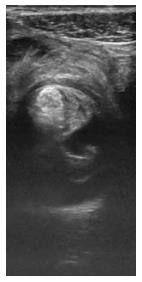
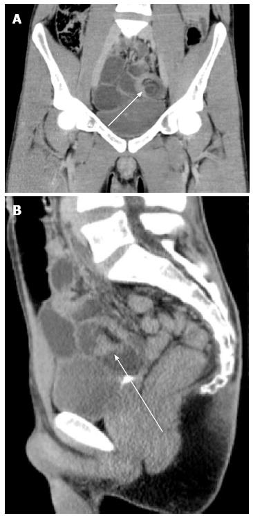
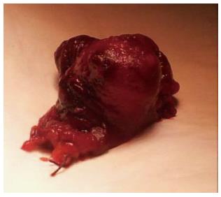
A male 22-year-old patient, with no previous history of medication or known allergies, was admitted to the Emergency Department of the Hospital de Braga with continuous abdominal pain of 24-h onset worsening with time, associated with nausea and vomiting. He reported constipation with two days of evolution. Analytical parameters (WBC, HGB, PCR, metabolic panel and liver function), showed no significant alterations. Ultrasonography and computed tomography revealed requisite findings consistent with ileal intussusception (Figures 1 and 2). The patient was proposed for surgical treatment. At exploratory laparoscopy, no visible alterations were found; hence the team opted for conversion to laparotomy. This allowed the identification of an ileo-ileal intussusception, located approximately 30 cm from the ileocecal valve. During the procedure of desinvagination, we found a sub-mucosal lesion of approximately 2 cm × 1 cm. Posteriorly, enterectomy was performed with lateral anastomosis using a linear stapler; the ileum segment was sent to the pathologist (Figure 3).
The intraoperative and postoperative periods were uneventful, and the patient was discharged on the 6th day of hospitalization when apyretic, presenting no hemodynamic changes, and tolerating an oral diet, with normalization of intestinal transit.
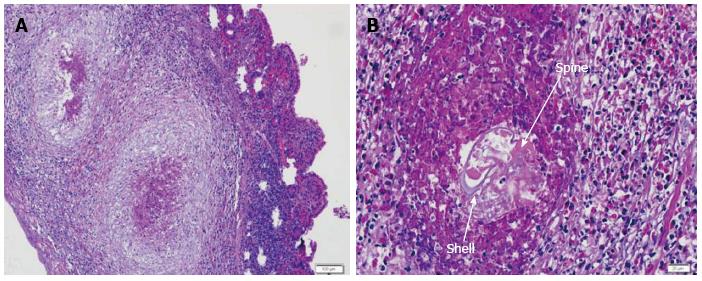
Surprisingly, the pathological anatomy revealed that there was evidence consistent with granulomatous inflammation due to the presence of a parasite egg, with features suggestive of Schistosoma species (Figures 4). Parasite eggs are oval or round reproductive bodies that can be seen on the bowel wall with granuloma formation. They usually have a shell with variable thickness and can be seen on HE preparations. There are three main Schistosoma species causing intestinal infection: S. mansoni (114-175 μm × 45-70 μm wide with a lateral spine), S. japonicum (70-100 μm × 55-65 μm wide, a thin shell with a lateral and small spine) and S. haematobium (112-170 μm × 40-70 μm wide with a terminal and prominent spine). However, there are no special stains or techniques to identify eggs so the diagnosis rests on morphological grounds. We found a 75 μm-wide ovoid structure with a thin and basophilic shell with some distortion so we could not ascertain the species.
The present clinical case illustrates some of the generalities described in the literature on the subject of intussusception. We demonstrate the importance of early surgical approach, even without definitive diagnosis of obstruction. The pathological analysis of the resected specimen turned out to reveal an interesting finding that, to our knowledge, has not been described in the literature to date: an egg of a Schistosoma species. There are three species of Schistosoma with different morphological features; however, in this case the egg was distorted which meant we were unable to make an identification of the organism. These findings were even more interesting and rare in the light of evidence suggesting that this parasite is typical of tropical countries, uncommon in Europe. This patient had not visited any tropical country, which makes this case even more improbable.
We believe that there may be two mechanisms that can potentially explain the observed invagination. First, it may occur due to an inflammatory reaction to a foreign body, with the growth of granuloma, eventually leading to fibrosis, and to a process of retraction of the ileal wall and consequent invagination. On the other hand, this may be a simple physical process, i.e., the size of the granuloma may itself cause the invagination.
Intussusception in adults, although a rare cause of intestinal obstruction, can never be forgotten in the approach to a patient with occlusive or sub-occlusive diagnosis. The most common causes of intussusception are diverticulum or tumors (benign or malignant). However, we must not forget that any foreign body or even a change in endoluminal extrinsic compression may induce intussusception. Schistosomiasis, although considered a parasitic disease in tropical countries, is not absent from Europe and may indeed be responsible for cases of intussusception in adults.
A 22-year-old male patient with no previous relevant history was admitted with continuous abdominal pain, nausea and vomiting.
Intestinal obstruction.
Adhesions, internal hernia, diverticulitis of Meckel.
Analytical parameters were within normal limits.
Ultrasonography and computed tomography revealed anatomic characteristics consistent with ileal intussusception.
Pathological anatomy revealed evidence consistent with granulomatous inflammation due to the presence of a parasite egg, with features suggestive of Schistosoma species.
Exploratory laparoscopy, where no visible alterations were found, hence the team opted for conversion to laparotomy. This identified an ileo-ileal intussusception located approximately 30 cm from the ileocecal valve. During the procedure of desinvagination, we found a sub-mucosal lesion. Posteriorly, enterectomy was performed with lateral anastomosis using a linear stapler.
Despite the fact that this is an extremely rare case, it shows that all the options should be considered when making a diagnosis of intestinal obstruction, even the most improbable ones.
The strength of this article was the finding that the invagination occurred due to the presence of a tropical parasite egg, an exceptionally rare case in Europe, considering the fact that the patient did not visit any tropical country. The limitation was the difficulty in the identification of the Schistosoma species that the egg belonged to due to the lack of specific techniques to identify the different parasites.
P- Reviewer: Berg T S- Editor: Qi Y L- Editor: Logan S E- Editor: Liu XM
| 1. | Albright MT, Grief SN, Carroll RE, Xu J. Ileoileal intussusception in an adult patient. Can Fam Physician. 2007;53:241-243. [PubMed] [Cited in This Article: ] |
| 2. | Oyen TL, Wolthuis AM, Tollens T, Aelvoet C, Vanrijkel JP. Ileo-ileal intussusception secondary to a lipoma: a literature review. Acta Chir Belg. 2007;107:60-63. [PubMed] [Cited in This Article: ] |
| 3. | Eisen LK, Cunningham JD, Aufses AH. Intussusception in adults: institutional review. J Am Coll Surg. 1999;188:390-395. [PubMed] [DOI] [Cited in This Article: ] [Cited by in Crossref: 225] [Cited by in F6Publishing: 224] [Article Influence: 9.0] [Reference Citation Analysis (0)] |
| 4. | Erkan N, Haciyanli M, Yildirim M, Sayhan H, Vardar E, Polat AF. Intussusception in adults: an unusual and challenging condition for surgeons. Int J Colorectal Dis. 2005;20:452-456. [PubMed] [DOI] [Cited in This Article: ] [Cited by in Crossref: 106] [Cited by in F6Publishing: 108] [Article Influence: 5.7] [Reference Citation Analysis (0)] |
| 5. | Gordon RS, O’Dell KB, Namon AJ, Becker LB. Intussusception in the adult--a rare disease. J Emerg Med. 1991;9:337-342. [PubMed] [DOI] [Cited in This Article: ] [Cited by in Crossref: 17] [Cited by in F6Publishing: 19] [Article Influence: 0.6] [Reference Citation Analysis (0)] |


