
Biceps Brachii and Brachioradialis Excitation in Biceps Curl Exercise: Different Handgrips, Different Synergy

 ,
,  ,
,  , , ,
, , ,  and
and 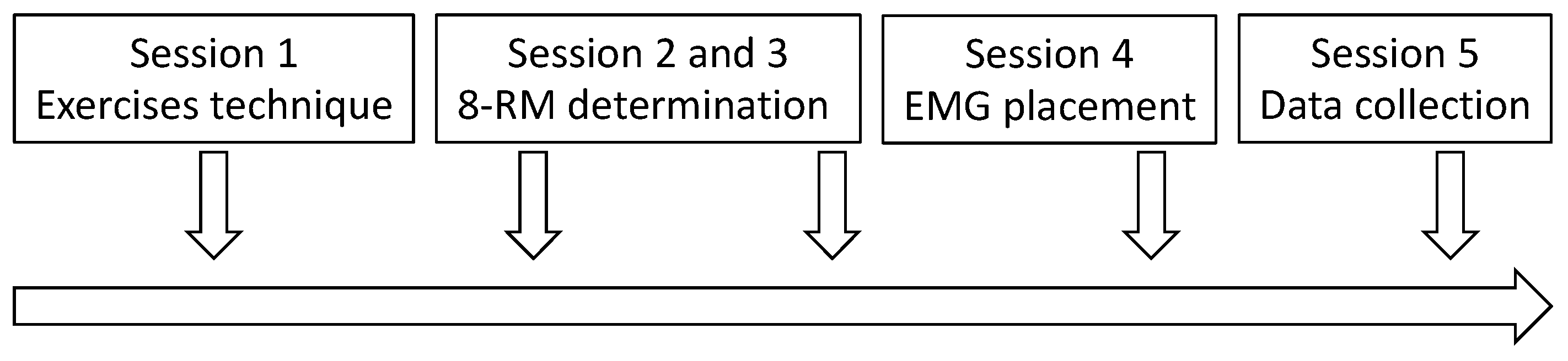
Abstract
:1. Introduction
2. Materials and Methods
2.1. Study Design
2.2. Participants
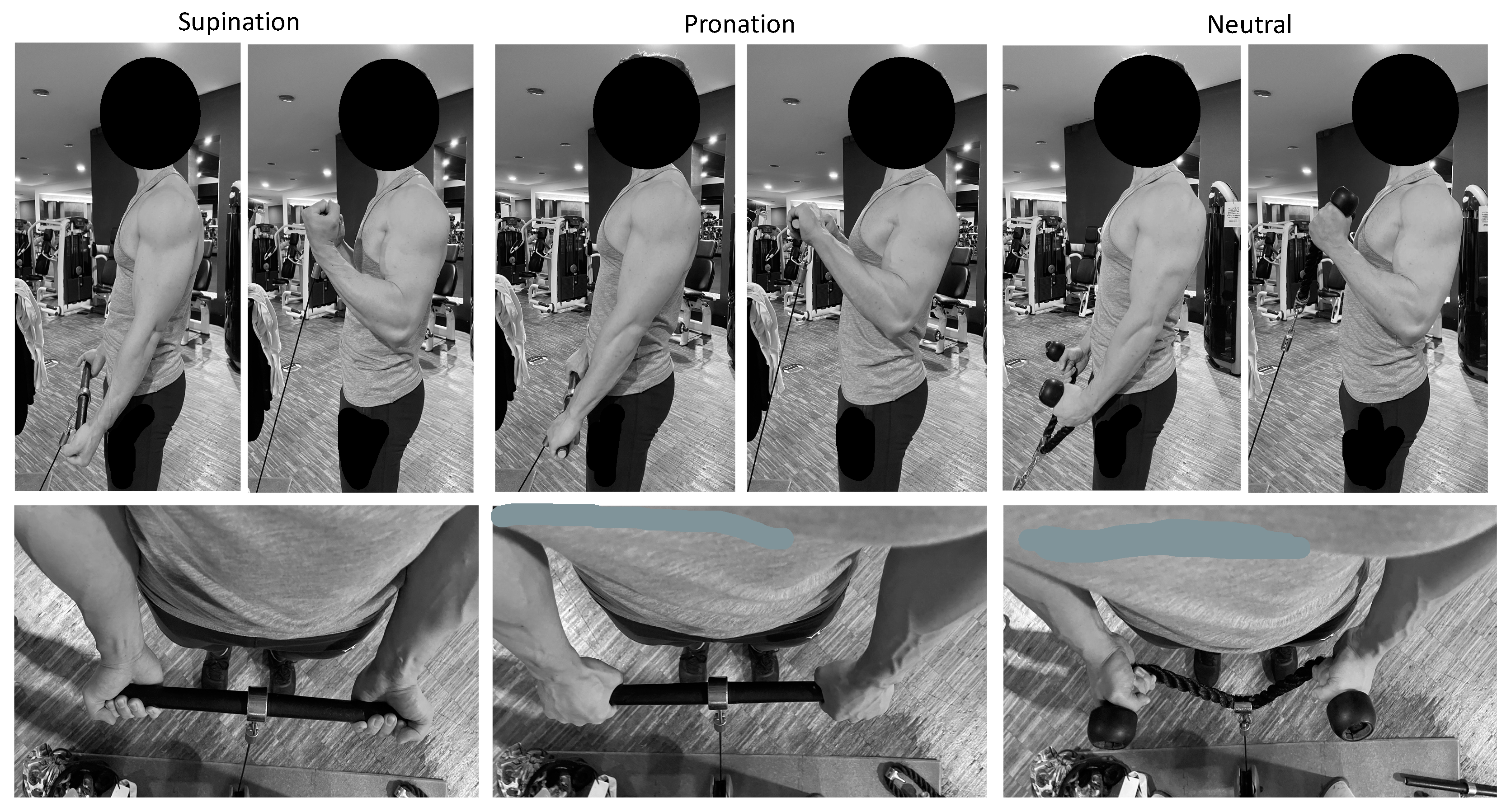
2.3. Exercises Technique
2.4. 8-RM Procedure
2.5. Maximum Voluntary Isometric Excitation
2.6. Data Analysis
2.7. Statistical Analysis
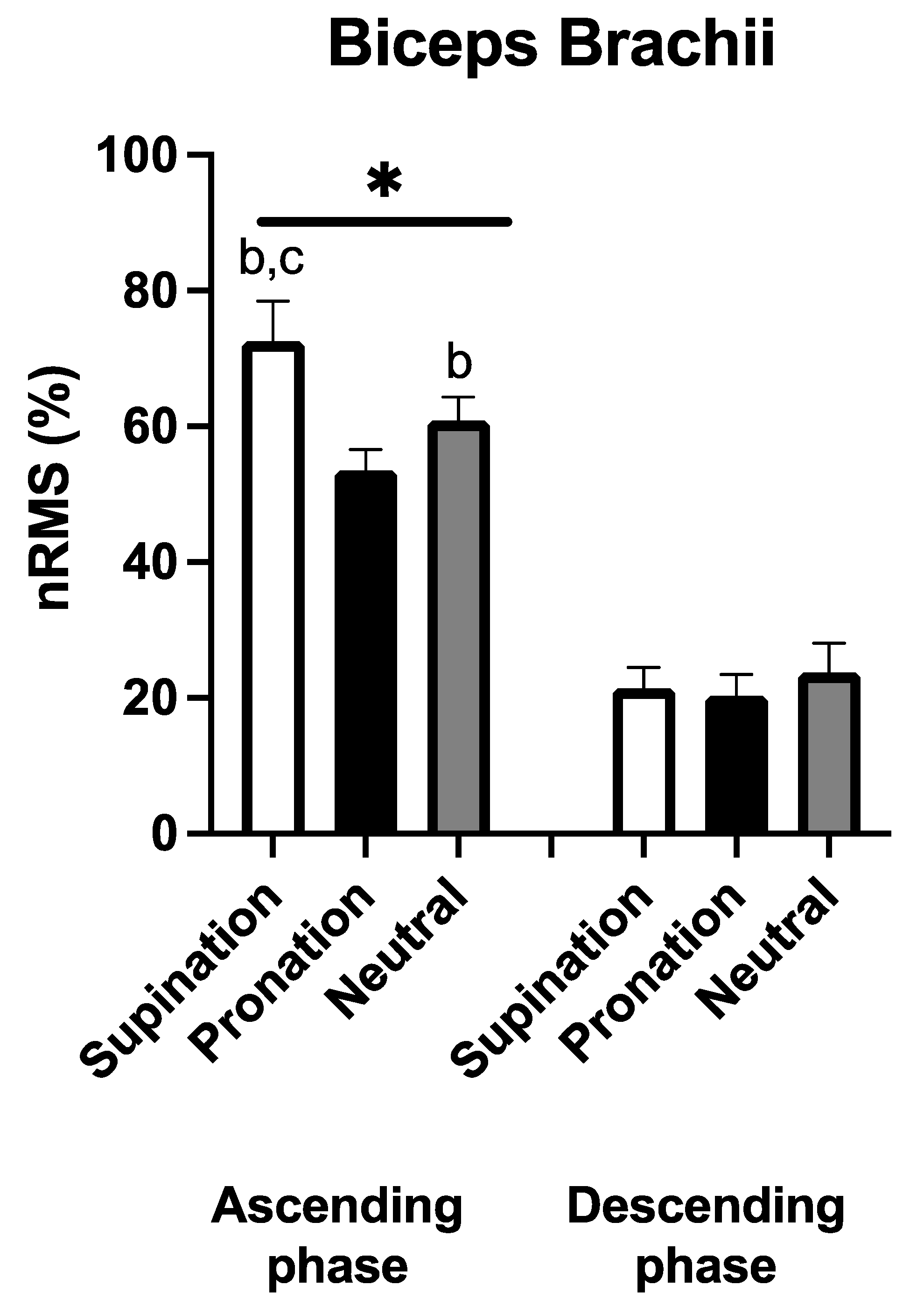
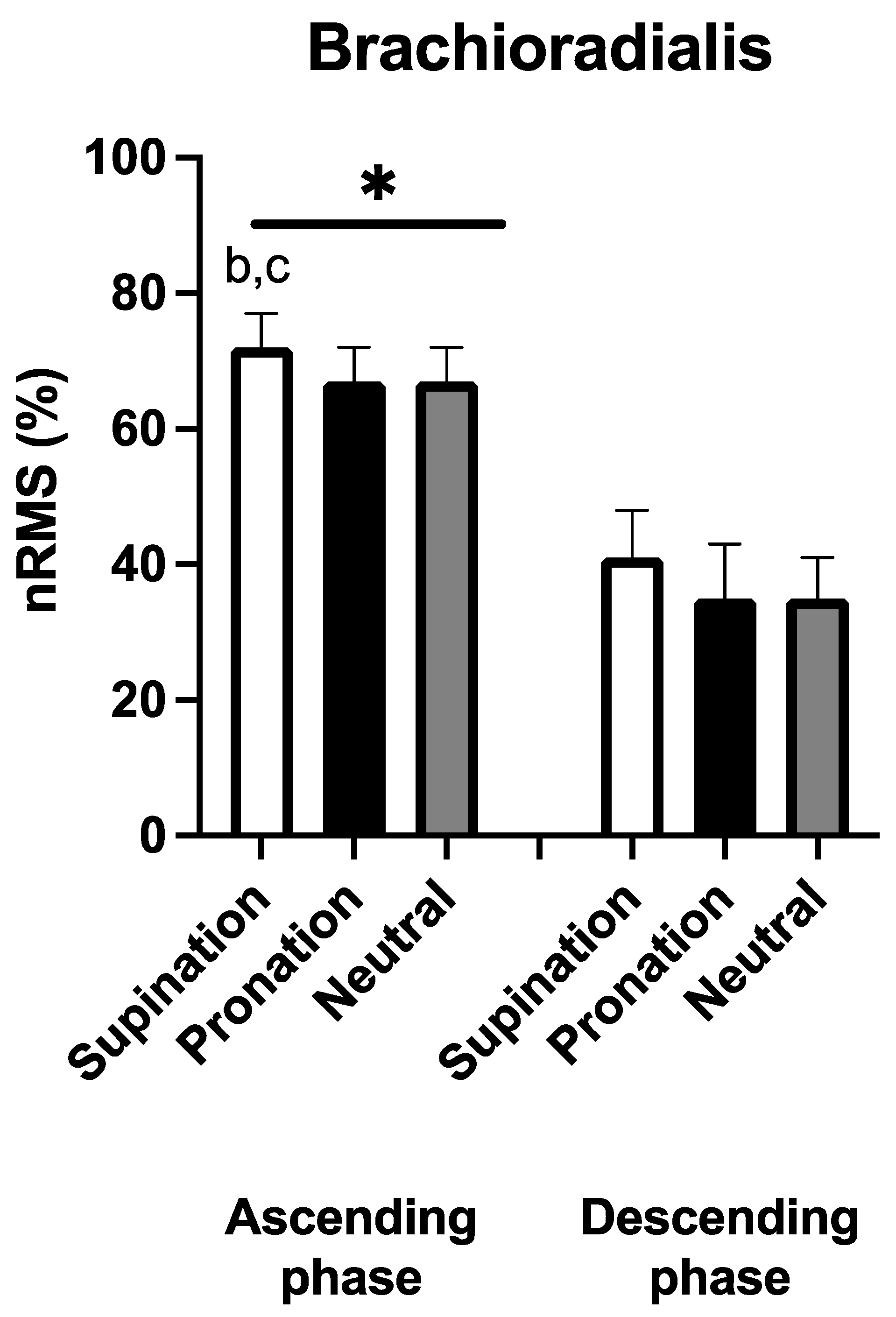
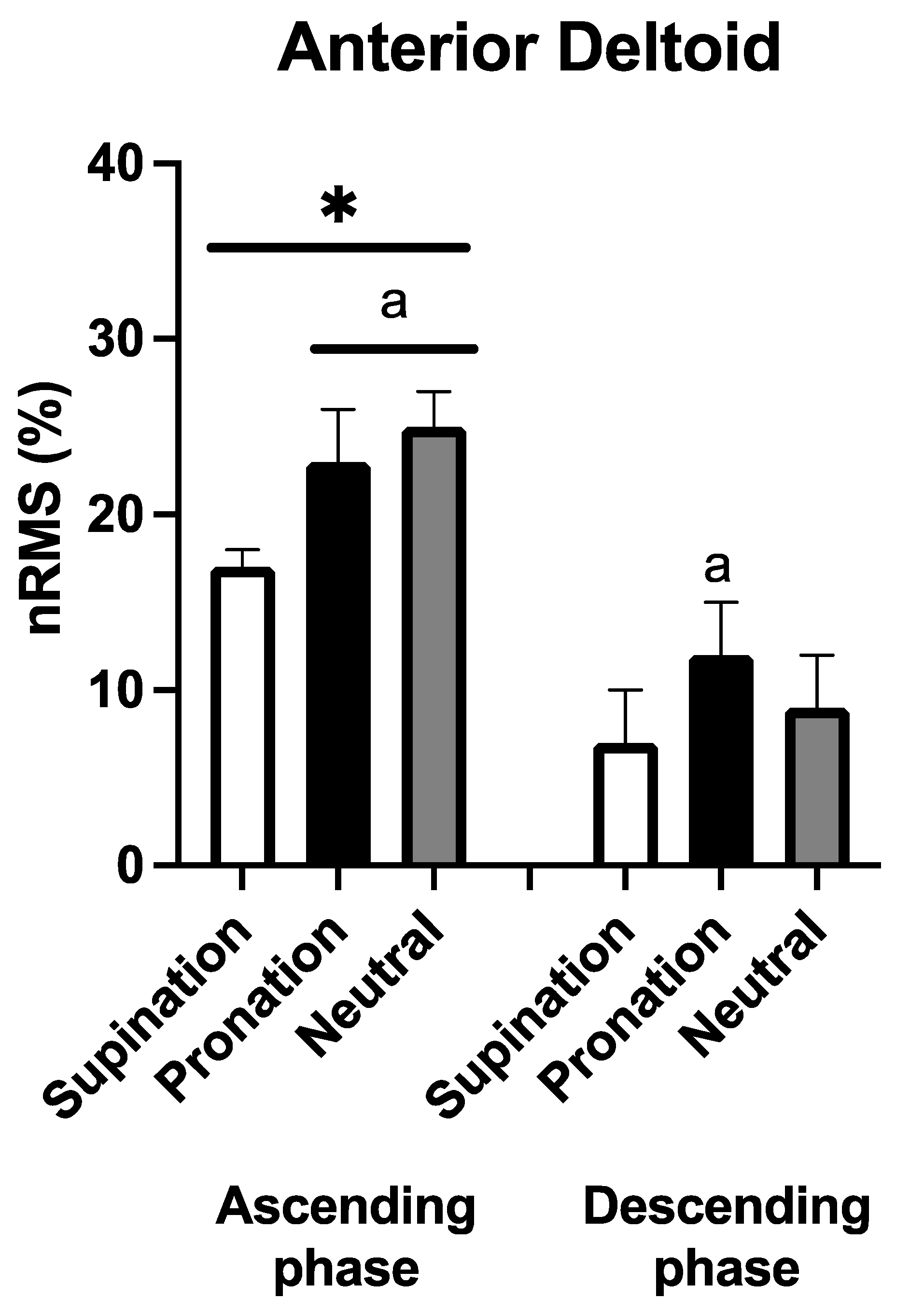
3. Results
4. Discussion
5. Conclusions
Author Contributions
Funding
Institutional Review Board Statement
Informed Consent Statement
Data Availability Statement
Acknowledgments
Conflicts of Interest
References
- Suchomel, T.J.; Nimphius, S.; Bellon, C.R.; Stone, M.H. The Importance of Muscular Strength: Training Considerations. Sports Med. 2018, 48, 765–785. [Google Scholar] [CrossRef]
- Duchateau, J.; Stragier, S.; Baudry, S.; Carpentier, A. Strength Training: In Search of Optimal Strategies to Maximize Neuromuscular Performance. Exerc. Sport Sci. Rev. 2021, 49, 2–14. [Google Scholar] [CrossRef]
- Morton, R.W.; Colenso-Semple, L.; Phillips, S.M. Training for strength and hypertrophy: An evidence-based approach. Curr. Opin. Physiol. 2019, 10, 90–95. [Google Scholar] [CrossRef]
- Nunes, J.P.; Jacinto, J.L.; Ribeiro, A.S.; Mayhew, J.L.; Nakamura, M.; Capel, D.M.G.; Santos, L.R.; Santos, L.; Cyrino, E.S.; Aguiar, A.F. Placing greater torque at shorter or longer muscle lengths? Effects of cable vs. barbell preacher curl training on muscular strength and hypertrophy in young adults. Int. J. Environ. Res. Public Health 2020, 17, 5859. [Google Scholar] [CrossRef]
- Moon, J.; Shin, I.; Kang, M.; Kim, Y.; Lee, K.; Park, J.; Kim, K.; Hong, D.; Koo, D.; O’Sullivan, D. The effect of shoulder flexion angles on the recruitment of upper-extremity muscles during isometric contraction. J. Phys. Ther. Sci. 2013, 25, 1299–1301. [Google Scholar] [CrossRef] [Green Version]
- Cools, A.M.; Borms, D.; Cottens, S.; Himpe, M.; Meersdom, S.; Cagnie, B. Rehabilitation exercises for athletes with biceps disorders and SLAP lesions: A continuum of exercises with increasing loads on the biceps. Am. J. Sports Med. 2014, 42, 1315–1322. [Google Scholar] [CrossRef]
- Yasuda, T.; Loenneke, J.; Ogasawara, R.; Abe, T. Influence of continuous or intermittent blood flow restriction on muscle activation during low-intensity multiple sets of resistance exercise. Acta Physiol. Hung. 2013, 100, 419–426. [Google Scholar] [CrossRef]
- Oliveira, L.F.; Matta, T.T.; Alves, D.S.; Garcia, M.A.C.; Vieira, T.M.M. Effect of the shoulder position on the biceps brachii EMG in different dumbbell curls. J. Sports Sci. Med. 2009, 8, 24–29. [Google Scholar]
- Ha, S.-Y.; Shin, D. The effects of curl-up exercise in terms of posture and muscle contraction direction on muscle activity and thickness of trunk muscles. J. Back Musculoskelet. Rehabil. 2020, 33, 857–863. [Google Scholar] [CrossRef]
- Marcolin, G.; Panizzolo, F.A.; Petrone, N.; Moro, T.; Grigoletto, D.; Piccolo, D.; Paoli, A. Differences in electromyographic activity of biceps brachii and brachioradialis while performing three variants of curl. PeerJ 2018, 6, e5165. [Google Scholar] [CrossRef]
- Naito, A. Electrophysiological studies of muscles in the human upper limb: The biceps brachii. Anat. Sci. Int. 2004, 79, 11–20. [Google Scholar] [CrossRef]
- Oliveira, A.S.; Gonçalves, M. Positioning during resistance elbow flexor exercise affects electromyographic activity, heart rate, and perceived exertion. J. Strength Cond. Res. 2009, 23, 854–862. [Google Scholar] [CrossRef]
- Coratella, G.; Tornatore, G.; Longo, S.; Esposito, F. Bilateral Biceps Curl Shows Distinct Biceps Brachii and Anterior Deltoid Excitation Comparing Straight vs. EZ Barbell Coupled with Arms Flexion/No-Flexion. J. Funct. Morphol. Kinesiol. 2023, 8, 13. [Google Scholar] [CrossRef]
- Bagchi, A.; Raizada, S. A comparative electromyographical analysis of biceps brachii and brachioradialis during eight different types of biceps curl. Indian J. Public Health Res. Dev. 2019, 10, 730–735. [Google Scholar] [CrossRef]
- Enoka, R.M. Eccentric contraction require unique activation strategies by the nervous system. J. Appl. Physiol. 1996, 81, 2339–2346. [Google Scholar] [CrossRef] [Green Version]
- Duchateau, J.; Enoka, R.M. Neural control of shortening and lengthening contractions: Influence of task constraints. J. Physiol. 2008, 586, 5853–5864. [Google Scholar] [CrossRef]
- Duchateau, J.; Enoka, R.M. Neural control of lengthening contractions. J. Exp. Biol. 2016, 219, 197–204. [Google Scholar] [CrossRef] [Green Version]
- Coratella, G.; Chemello, A.; Schena, F. Muscle damage and repeated bout effect induced by enhanced eccentric squats. J. Sports Med. Phys. Fit. 2016, 56, 1540–1546. [Google Scholar]
- Coratella, G.; Bertinato, L. Isoload vs isokinetic eccentric exercise: A direct comparison of exercise-induced muscle damage and repeated bout effect. Sport Sci. Health 2015, 11, 87–96. [Google Scholar] [CrossRef]
- Gibala, M.J.; MacDougall, J.D.; Tarnopolsky, M.A.; Stauber, W.T.; Elorriaga, A. Changes in human skeletal muscle ultrastructure and force production after acute resistance exercise. J. Appl. Physiol. 1995, 78, 702–708. [Google Scholar] [CrossRef]
- Coratella, G.; Beato, M.; Bertinato, L.; Milanese, C.; Venturelli, M.; Schena, F. Including the Eccentric Phase in Resistance Training to Counteract the Effects of Detraining in Women: A Randomized Controlled Trial. J. Strength Cond. Res. 2022, 36, 3023–3031. [Google Scholar] [CrossRef]
- Coratella, G.; Milanese, C.; Schena, F. Unilateral eccentric resistance training: A direct comparison between isokinetic and dynamic constant external resistance modalities. Eur. J. Sport Sci. 2015, 15, 720–726. [Google Scholar] [CrossRef]
- Maeo, S.; Takahashi, T.; Takai, Y.; Kanehisa, H. Trainability of Muscular Activity Level during Maximal Voluntary Co-Contraction: Comparison between Bodybuilders and Nonathletes. PLoS ONE 2013, 8, e79486. [Google Scholar] [CrossRef] [Green Version]
- Coratella, G.; Tornatore, G.; Longo, S.; Esposito, F.; Cè, E. An electromyographic analysis of lateral raise variations and frontal raise in competitive bodybuilders. Int. J. Environ. Res. Public Health 2020, 17, 6015. [Google Scholar] [CrossRef]
- Coratella, G.; Tornatore, G.; Caccavale, F.; Longo, S.; Esposito, F.; Cè, E. The Activation of Gluteal, Thigh, and Lower Back Muscles in Different Squat Variations Performed by Competitive Bodybuilders: Implications for Resistance Training. Int. J. Environ. Res. Public Health 2021, 18, 772. [Google Scholar] [CrossRef]
- Coratella, G.; Tornatore, G.; Longo, S.; Esposito, F.; Cè, E. An electromyographic analysis of Romanian, step-Romanian, and stiff-leg deadlift: Implication for resistance training. Int. J. Environ. Res. Public Health 2022, 19, 1903. [Google Scholar] [CrossRef]
- Coratella, G.; Tornatore, G.; Longo, S.; Esposito, F. Front vs Back and Barbell vs Machine Overhead Press : An Electromyographic Analysis and Implications For Resistance Training. Front. Physiol. 2022, 13, 825880. [Google Scholar] [CrossRef]
- Coratella, G.; Tornatore, G.; Longo, S.; Esposito, F.; Cè, E. Specific prime movers’ excitation during free-weight bench press variations and chest press machine in competitive bodybuilders. Eur. J. Sport Sci. 2020, 20, 571–579. [Google Scholar] [CrossRef]
- Coratella, G. Appropriate reporting of exercise variables in resistance training protocols: Much more than load and number of repetitions. Sports Med.-Open 2022, 8, 99. [Google Scholar] [CrossRef]
- Coratella, G.; Tornatore, G.; Longo, S.; Borrelli, M.; Doria, C.; Esposito, F.; Cè, E. The Effects of Verbal Instructions on Lower Limb Muscles’ Excitation in Back-Squat. Res. Q. Exerc. Sport 2020, 93, 429–435. [Google Scholar] [CrossRef]
- Kompf, J.; Arandjelović, O. Understanding and Overcoming the Sticking Point in Resistance Exercise. Sports Med. 2016, 46, 751–762. [Google Scholar] [CrossRef] [Green Version]
- Hermens, H.J.; Freriks, B.; Disselhorst-Klug, C.; Rau, G. Development of recommendations for SEMG sensors and sensor placement procedures. J. Electromyogr. Kinesiol. 2000, 10, 361–374. [Google Scholar] [CrossRef]
- Marri, K.; Swaminathan, R. Analyzing the influence of curl speed in fatiguing biceps brachii muscles using sEMG signals and multifractal detrended moving average algorithm. In Proceedings of the 2016 38th Annual International Conference of the IEEE Engineering in Medicine and Biology Society (EMBC), Orlando, FL, USA, 16–20 August 2016; Volume 2016, pp. 3658–3661. [Google Scholar] [CrossRef]
- Cohen, J. Statistical Power Analysis for the Behavioral Sciences, 2nd ed.; Lawrence Erlbaum Associates: Mahwah, NJ, USA, 1988. [Google Scholar]
- Hopkins, W.G.; Marshall, S.W.; Batterham, A.M.; Hanin, J. Progressive Statistics for Studies in Sports Medicine and Exercise Science. Med. Sci. Sports Exerc. 2009, 41, 3–13. [Google Scholar] [CrossRef] [Green Version]
- Borms, D.; Ackerman, I.; Smets, P.; Van Den Berge, G.; Cools, A.M. Biceps Disorder Rehabilitation for the Athlete. Am. J. Sports Med. 2017, 45, 642–650. [Google Scholar] [CrossRef]
- Hermens, H.J.; Freriks, B.; Merletti, R.; Stegeman, D.; Blok, J.; Rau, G.; Disselhorst-Klug, C.; Hägg, G. European Recommendations for Surface ElectroMyoGraphy Results of the SENIAM project. Roessingh Res. Dev. 2000, 10, 361–374. [Google Scholar]
- Escamilla, R.F.; Yamashiro, K.; Paulos, L.; Andrews, J.R. Shoulder muscle activity and function in common shoulder rehabilitation exercises. Sports Med. 2009, 39, 663–685. [Google Scholar] [CrossRef]
- Alves, D.; Matta, T.; Oliveira, L. Effect of shoulder position on triceps brachii heads activity in dumbbell elbow extension exercises. J. Sports Med. Phys. Fit. 2018, 58, 1247–1252. [Google Scholar] [CrossRef]
- Coratella, G.; Schena, F. Eccentric resistance training increases and retains maximal strength, muscle endurance, and hypertrophy in trained men. Appl. Physiol. Nutr. Metab. 2016, 41, 1184–1189. [Google Scholar] [CrossRef]
- Coratella, G.; Galas, A.; Campa, F.; Pedrinolla, A.; Schena, F.; Venturelli, M. The Eccentric Phase in Unilateral Resistance Training Enhances and Preserves the Contralateral Knee Extensors Strength Gains After Detraining in Women: A Randomized Controlled Trial. Front. Physiol. 2022, 13, 788473. [Google Scholar] [CrossRef]
- Herzog, W. Mechanisms of enhanced force production in lengthening (eccentric) muscle contractions. J. Appl. Physiol. 2014, 116, 1407–1417. [Google Scholar] [CrossRef] [Green Version]
- Herzog, W. Why are muscles strong, and why do they require little energy in eccentric action? J. Sport Health Sci. 2018, 7, 255–264. [Google Scholar] [CrossRef]
- Herzog, W.; Powers, K.; Johnston, K.; Duvall, M. A new paradigm for muscle contraction. Front. Physiol. 2015, 6, 174. [Google Scholar] [CrossRef] [Green Version]
- Hill, E.; Housh, T.; Smith, C.; Schmidt, R.; Johnson, G. Muscle-and mode-specific responses of the forearm flexors to fatiguing, concentric muscle actions. Sports 2016, 4, 47. [Google Scholar] [CrossRef] [Green Version]


Disclaimer/Publisher’s Note: The statements, opinions and data contained in all publications are solely those of the individual author(s) and contributor(s) and not of MDPI and/or the editor(s). MDPI and/or the editor(s) disclaim responsibility for any injury to people or property resulting from any ideas, methods, instructions or products referred to in the content. |
© 2023 by the authors. Licensee MDPI, Basel, Switzerland. This article is an open access article distributed under the terms and conditions of the Creative Commons Attribution (CC BY) license (https://creativecommons.org/licenses/by/4.0/).
Share and Cite
Coratella, G.; Tornatore, G.; Longo, S.; Toninelli, N.; Padovan, R.; Esposito, F.; Cè, E. Biceps Brachii and Brachioradialis Excitation in Biceps Curl Exercise: Different Handgrips, Different Synergy. Sports 2023, 11, 64. https://doi.org/10.3390/sports11030064
Coratella G, Tornatore G, Longo S, Toninelli N, Padovan R, Esposito F, Cè E. Biceps Brachii and Brachioradialis Excitation in Biceps Curl Exercise: Different Handgrips, Different Synergy. Sports. 2023; 11(3):64. https://doi.org/10.3390/sports11030064
Chicago/Turabian StyleCoratella, Giuseppe, Gianpaolo Tornatore, Stefano Longo, Nicholas Toninelli, Riccardo Padovan, Fabio Esposito, and Emiliano Cè. 2023. "Biceps Brachii and Brachioradialis Excitation in Biceps Curl Exercise: Different Handgrips, Different Synergy" Sports 11, no. 3: 64. https://doi.org/10.3390/sports11030064