Article

Clinical Evaluation of Respiratory Rate Measurements on COPD (Male) Patients Using Wearable Inkjet-Printed Sensor

1
Mechatronics Engineering Department/NanoLab, School of Applied Technical Sciences, German Jordanian University, P.O. Box 35247, Amman 11180, Jordan
2
Institute of Microtechnology, Technische Universität Braunschweig, 38124 Braunschweig, Germany
3
Faculty of Engineering, Middle East University, Amman 11831, Jordan
4
Mechanical Engineering Department, University of Kentucky, Lexington, KY 40506, USA
5
Department of Internal Medicine, Faculty of Medicine, University of Jordan, Amman 11942, Jordan
6
Research Centre for Intelligent Healthcare, Faculty of Health and Life Sciences, Coventry University, Coventry CV1 5FB, UK
7
Department of Electrical and Electronic Engineering, Southern University of Science and Technology, Shenzhen 518055, China
*
Author to whom correspondence should be addressed.
Sensors 2021, 21(2), 468; https://doi.org/10.3390/s21020468
Submission received: 22 November 2020 / Revised: 6 January 2021 / Accepted: 8 January 2021 / Published: 11 January 2021

Abstract

:
Introduction: Chronic Obstructive Pulmonary Disease (COPD) is a progressive disease that causes long-term breathing problems. The reliable monitoring of respiratory rate (RR) is very important for the treatment and management of COPD. Based on inkjet printing technology, we have developed a stretchable and wearable sensor that can accurately measure RR on normal subjects. Currently, there is a lack of comprehensive evaluation of stretchable sensors in the monitoring of RR on COPD patients. We aimed to investigate the measurement accuracy of our sensor on COPD patients. Methodology: Thirty-five patients (Mean ± SD of age: 55.25 ± 13.76 years) in different stages of COPD were recruited. The measurement accuracy of our inkjet-printed (IJPT) sensor was evaluated at different body postures (i.e., standing, sitting at 90°, and lying at 45°) on COPD patients. The RR recorded by the IJPT sensor was compared with that recorded by the reference e-Health sensor using paired T-test and Wilcoxon signed-rank test. Analysis of variation (ANOVA) was performed to investigate if there was any significant effect of individual difference or posture on the measurement error. Statistical significance was defined as p-value less than 0.05. Results: There was no significant difference between the RR measurements collected by the IJPT sensor and the e-Health reference sensor overall and in three postures (p > 0.05 in paired T-tests and Wilcoxon signed-rank tests). The sitting posture had the least measurement error of −0.0542 ± 1.451 bpm. There was no significant effect of posture or individual difference on the measurement error or relative measurement error (p > 0.05 in ANOVA). Conclusion: The IJPT sensor can accurately measure the RR of COPD patients at different body postures, which provides the possibility for reliable monitoring of RR on COPD patients.

1. Introduction

Respiratory rate (RR) is a vital sign that is related to, therefore regulated by, multiple physiological and neural activities [1,2,3]. RR plays an important role in the detection of various cardiovascular and respiratory diseases, as well as relevant clinical events [4,5]. The variation of RR reflects the deterioration of respiratory diseases including asthma, chronic obstructive pulmonary disease (COPD), and other clinical conditions including fever, infection, and drug overdose [6]. Despite the development of RR measurement technologies, RR is still often neglected or inaccurately estimated in clinical practice [7].
Recently, wearable and flexible sensors (WFS) have been widely applied in medical and healthcare areas [8,9,10,11,12,13]. Especially, WFS can be used in the non-invasive monitoring of vital signs such as blood pressure [14], cardiac [15], and respiratory activities [15,16,17]. More specifically, several studies have investigated the use of WFS extensively in RR monitoring [18,19,20,21,22,23,24]. For instance, Jeong et al. [25] developed a fabric piezo-resistive sensor of RR and tested it in different physiological conditions, including resting, walking at two different speeds, and running activities. It was found that the averaged relative error of the sensor in RR measurement was about 3%. Lei et al. [26] proposed a PVDF-based (Piezoelectric-film) sensor patch and tested it in static (e.g., sitting or sleeping) and dynamic (e.g., walking) conditions. No significant measurement error was found when compared to the reference sensor (p > 0.05). Moreover, WFS can be used in the monitoring of vital signs on patients who have special medical conditions such as Chronic Obstructive Pulmonary Disease (COPD) [27].
COPD is a term to describe lung diseases such as chronic bronchitis, emphysema, and refractory asthma, and it is usually characterized by increasing breathlessness. The symptoms of COPD may differ between individuals, but commonly include shortness of breath, wheezing, and tightness in the chest [28]. The link between dyspnea and respiratory rate is of particular interest in COPD patients [3]. Moreover, it was suggested that hypnosis could contribute to the improvement of anxiety levels and breathing mechanics in severe COPD patients [29]. Several studies in the literature highlighted the importance of reliable RR monitoring in the early detection of COPD [30,31,32], where a RR over 25 breaths per min (bpm) was considered as a sign of COPD exacerbation [30,33]. Hence, the comprehensive evaluation of the measurement accuracy and the suitability of WFS in RR monitoring on COPD patients is important for clinical application.
Regarding RR measurement, the clinical evaluation of WFS was mainly performed on healthy subjects [34,35,36,37,38,39]. Few studies have investigated the accuracy of WFS in monitoring COPD patients’ vital signs [31,33,40]. Bellos et al. [31] investigated the accuracy of a wearable vest in monitoring different vital signs including RR on COPD patients. The system consisted of a wearable platform and some external devices connected to a smart device and a home patient monitor. The wearable platform along with the external devices extracted some useful information about the patient’s activities, living environment, and lifestyle. The collected data were then processed to evaluate the patient’s health status. The system achieved about 94% accuracy in RR monitoring. Moreover, Rubio et al. [33] evaluated the accuracy of five home-based RR measurement devices on 21 stable COPD patients during daily activities. The authors also investigated the acceptability (comfort) of the patients regarding wearing of the sensors. They concluded that the chest-band sensor was the most acceptable sensor for patients with good measurement accuracy with a bias of −1.60 bpm.
Al-Halhouli et al. [16] have presented the development of stretchable and wearable strain gauge sensor using inkjet printing technology for RR monitoring. The accuracy and performance of the developed sensor have been validated on healthy subjects at different body locations [41] and at different body postures [17]. They concluded that the inkjet-printed (IJPT) sensor was accurate and had a good potential for monitoring RR on non-healthy patients such as COPD patients. To the best of our knowledge, there is a lack of studies that investigated the accuracy of WFS for RR measurements on COPD patients at different postures. Hence, this study aimed to comprehensively evaluate the accuracy of the IJPT RR sensor- developed in [16] on COPD patients at different body postures at rest. Despite that, this study did not aim to investigate the ability of the developed sensor to diagnose COPD types, however the use of this sensor could help in reducing the frequency and severity of COPD exacerbation symptoms by early detection of abnormal physiological measurements including the respiratory rate [42] especially with the use of wireless sensors that are capable of monitoring the RR continuously. The preliminary version evaluated in this study is a wired one for the sake of sensor’s clinical evaluation.

2. Methodology

The extraction of the RR using the IJPT on COPD patients consists of several stages, including the fabrication of the IJPT sensor and the signal processing of the RR signal. Figure 1 shows a brief summary of the fabrication process of the IJPT sensor as well as the RR extraction procedure on COPD patients. The following sections elaborate more on these stages.

2.1. Ethical Approval

The protocol adhered to the tenets of the Declaration of Helsinki [43]. Each patient had the freedom of choice to participate in this work. The procedure was explained in details with questions answered. Then, the participants signed the consent form, which was reviewed and approved by Jordan University Hospital Ethics Committee (REF 67/2019/6480).

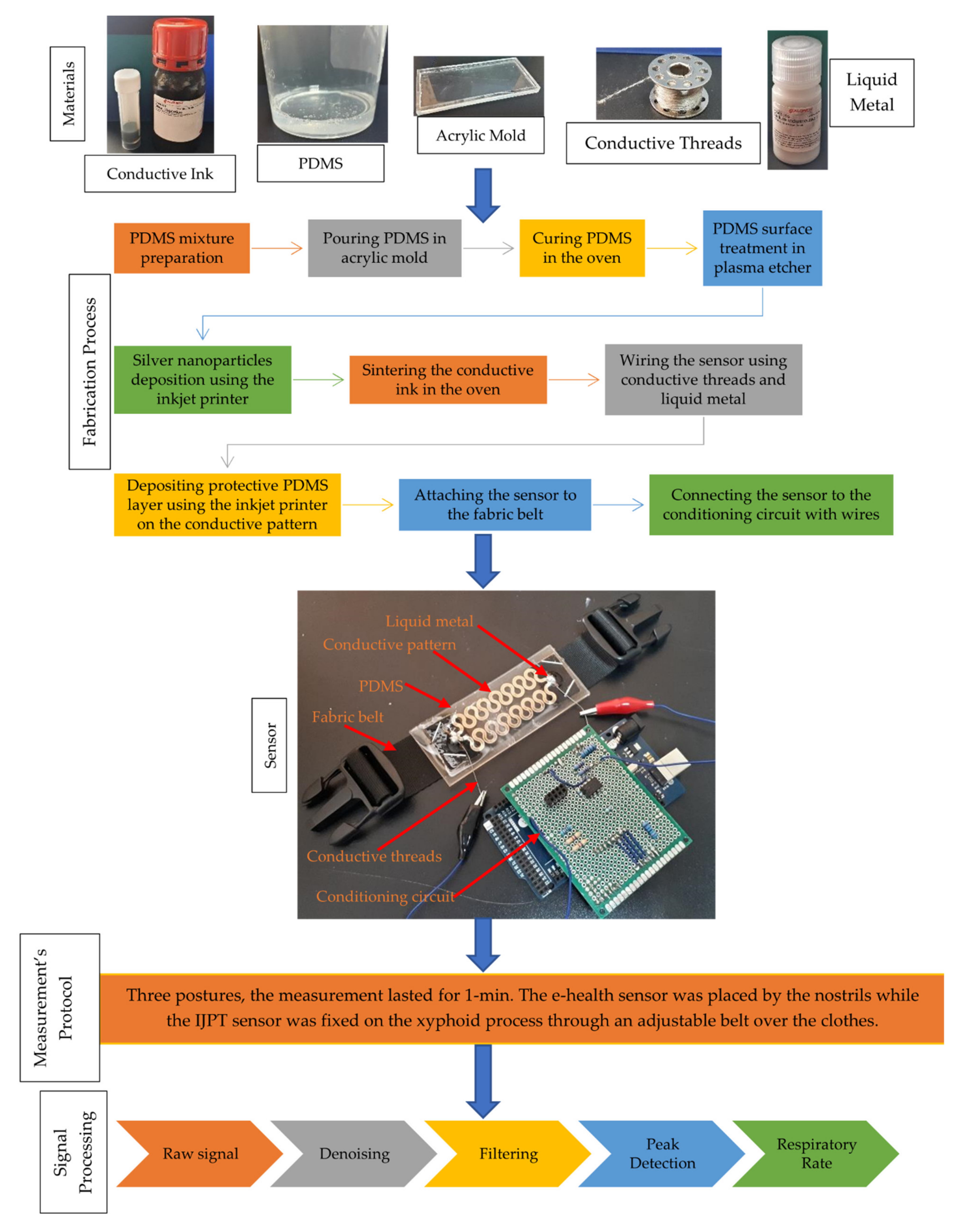
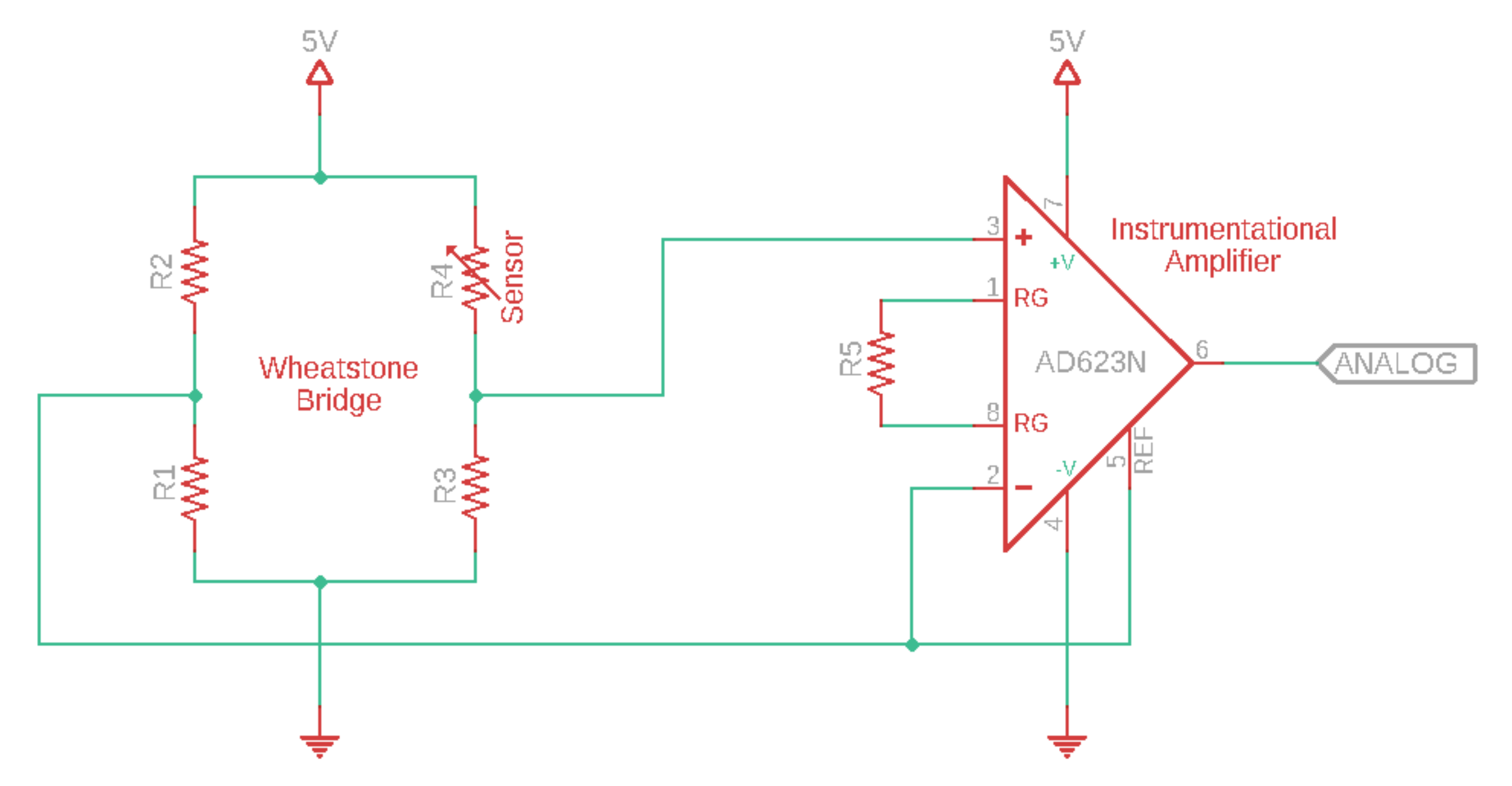
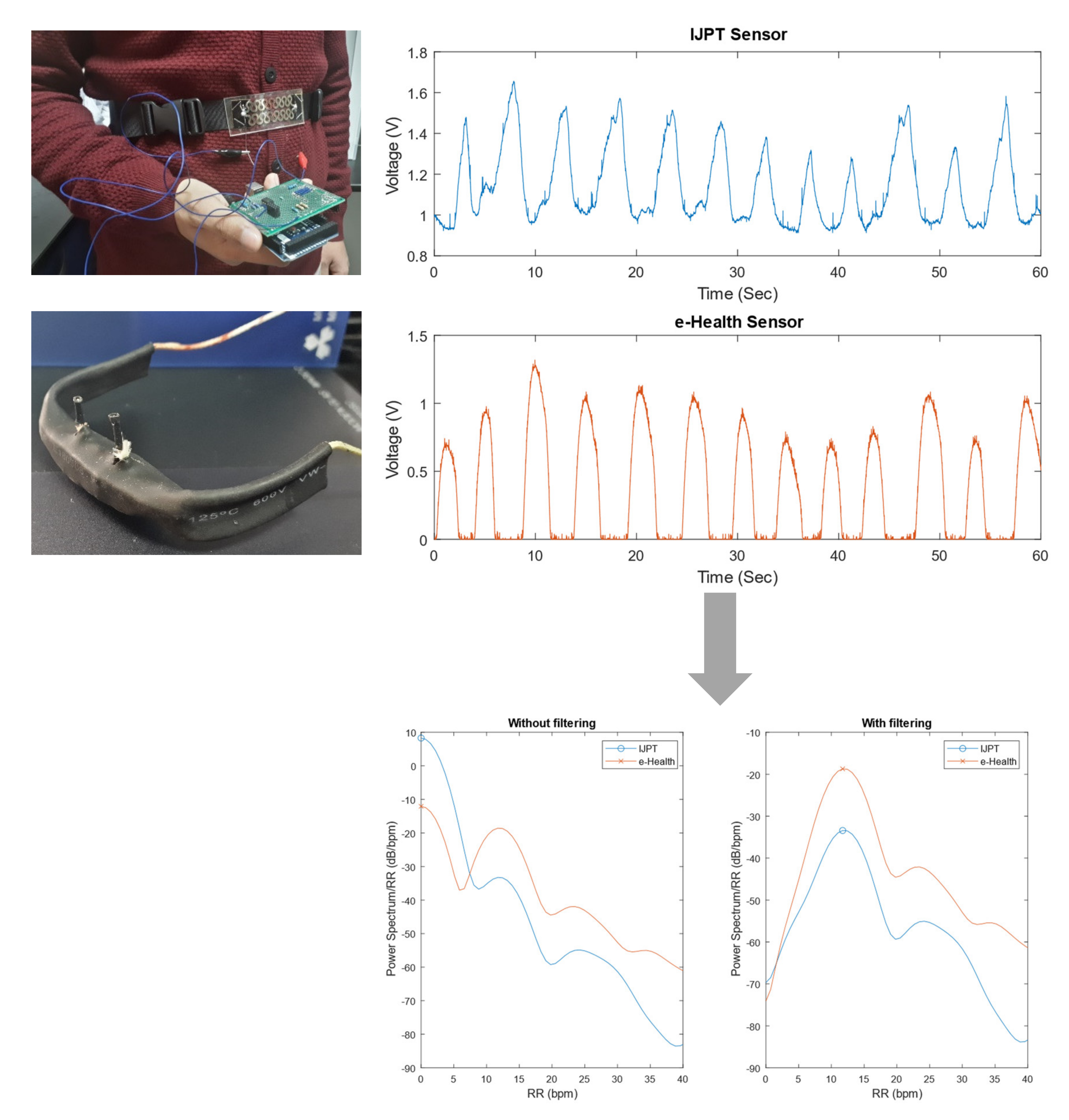
2.2. Inkjet-Printed RR Sensor

The fabrication of flexible electronics using inkjet printing technology has gained significant interest in recent years. The RR sensor evaluated in this study was fabricated using this technology where silver nanoparticle ink was deposited on polydimethylsiloxane (PDMS) substrates. The RR was detected by the IJPT sensor via the variations of the resistance of the conductive traces caused by the volumetric change in the ribcage or abdomen areas during the respiration process. In other words, the IJPT sensor acted as a strain gauge sensor. The details of the fabrication process of the IJPT sensor can be found in [16].

2.3. Measurements Protocol

Thirty-five patients were included in the study after taking their permissions using the informed consent form. All of them were diagnosed with COPD, and managed with adequate medication and on regular follow-up. Firstly, patients were asked to rest for five minutes while the questionnaire was filled with the health records in patient’s file validated. Then, the respiration rate was measured with the reference e-Health nasal flow sensor and the IJPT sensor simultaneously. The measurement was repeated in 3 different positions: Lying position 45°, sitting upright, and standing. In each position, the measurement lasted for one minute. The e-Health sensor was placed in the nostrils while the IJPT sensor was fixed on the xiphoid process through an adjustable belt over the clothes. It should be noted that the patients were asked not to move while taking the measurements.
The patients included in the study had different GOLD stages of COPD (1, 2, 3 or 4), different age groups (mean ± standard deviation (SD) of age: 55.25 ± 13.76 years) and different smoking status (ex-smoker, quit smoking, or smoker). Some of the limitations faced prevented the measurement of the RR at some postures, such as the inability to take readings from patients on continuous oxygen support as the e-Health sensor was placed by the nostrils. Another limitation was the endurance of some patients to stay in a certain position as it caused fatigue and irritability. In addition, some patients had chest tubes, which caused improper mounting of the IJPT sensor. Despite these limitations, the respiratory signals were successfully recorded in most measurements. A summary of patients’ information, as well as CAGE (Cut-Annoyed-Guilty-Eye) questionnaire summary, can be found in Table A1 and Table A2 in Appendix A, respectively.

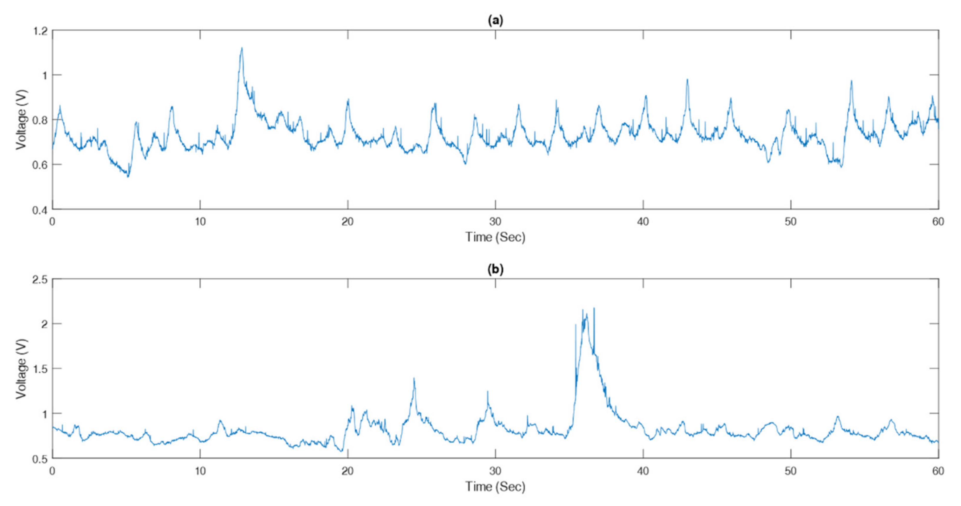
2.4. Respiration Rate Derivation

The strain gauge sensor, whose resistance value varied during respiration, was connected to a Wheatstone Bridge with suitable values for the resistors. The output was connected to an Instrumentational Amplifier with a suitable value for the gain resistor, as shown in Figure 2. The output of the amplifier was connected to an analog input of the Arduino board. The airflow sensor was connected to e-Health Sensor Shield V2.0 for Arduino [44] and connected to the same Arduino board as the strain gauge circuitry. Arduino Mega2560 with an ATMega2560 chip as its microcontroller was used with maximum sampling rate of 15 kSPS at maximum resolution [45,46].
The signals of the strain gauge sensor and the airflow sensor were sampled at 100 Hz using the Arduino board, which was connected to a MATLAB/Simulink file to save the data. Afterward, a MATLAB-based bandpass filter was used to filter both signals with cut-off frequencies of 3 and 90 bpm, or 0.05 and 1.5 Hz, respectively, with the function configured to an infinite impulse response. The calculation of the respiratory rate was done by finding the frequency at which the power spectral density of the filtered data had its maximum value. Figure 3 shows the raw RR signals from the IJPT and e-Health sensors as well as power spectral densities of both signals before and after the filtering. The circle and cross indicate the maximum value of the power spectral density of each signal at which the respiratory rate was found.

2.5. Statistical Analysis

2.5.1. Data Cleaning

For the data of each measurement, the quality of reference (airflow) respiratory signal was double-checked. If the quality of reference respiratory signal was low (e.g., blurred waveform, missing period longer than 5 s, or missing more than two consecutive respiratory cycles), the data of that measurement were discarded. The considered RR data can be found in Table A3 in Appendix A.

2.5.2. Comparison of RR Values

The RR values derived from inkjet-printed strain gauge (RRSG) and airflow (RRAF) were compared to investigate if there was any significant difference. Firstly, the Shapiro–Wilk test was performed on RR values to investigate if they followed normal distribution. If both RRSG and RRAF groups followed normal distribution (defined as p > 0.05 in Shapiro-Wilk test), the paired T-test was performed to investigate if there was any significant difference (defined as p < 0.05) between RRSG and RRAF. If normal distribution was not followed in any one group, the Wilcoxon signed-rank test (significant difference defined as p < 0.05) was performed as the non-parametric substitute of paired T-test. Considering the missing data in some subjects, the test was performed globally and for each posture, respectively.

2.5.3. Analysis of Errors of RR

The estimation error of RR was calculated as in Equation. (1). The analysis of variance (ANOVA) was performed to investigate if there was any significant effect of individual difference or posture on the results. It has been proven that ANOVA is applicable even if the data deviate from normal distribution [47]. For reliable estimation, the Shapiro–Wilk test was performed to investigate if the error followed normal distribution in three postures respectively. The paired T-test was performed on the data of error derived in different postures if both groups followed normal distribution. Otherwise, the Kruskal–Wallis test was used to investigate if the effect of posture on error was significant (defined as p < 0.05), while the Wilcoxon signed-rank test was performed as the non-parametric substitute of paired T-test.
E = RR SG RR AF

2.5.4. Analysis of Relative Errors of RR

The relative error of RR estimation was calculated as in Equation (2). The analysis method is the same as that for error of RR.
E r = RR SG RR AF RR AF

2.5.5. Bland-Altman Analysis

To illustrate the difference between RRSG and RRAF in different postures, the Bland–Altman analysis was performed on RRSG and RRAF for each posture, respectively [48,49]. In signal processing, the resolution of RR was 0.001 bpm. To accurately show and compare the biases, the results of Bland-Altman analysis were rounded to the third significant digit after the decimal point.

2.5.6. Regression Analysis

Linear regression analysis was used to inspect whether the correlation between the RRSG and the RRAF followed a linear correlation or not. The consistency in the RRSG and RRAF was evaluated using the regression coefficient of the linear correlation.

3. Results

3.1. Comparison of RR Values

The overall distribution of RRSG and RRAF followed normal distribution (p = 0.689 and p = 0.066 in Shapiro–Wilk test). The paired T-test showed no significant difference between RRSG and RRAF (p = 0.572). In sitting posture, normal distribution was followed by RRAF but not RRSG (p = 0.104 and p = 0.045 in Shapiro–Wilk test, respectively). The Wilcoxon signed-rank test showed no significant difference between RRSG and RRAF (p = 0.968). In standing posture, both RRSG and RRAF followed normal distribution (p = 0.977 and p = 0.652 in Shapiro–Wilk test, respectively). The paired T-test showed no significant difference between RRSG and RRAF (p = 0.873). In lying45° posture, both RRSG and RRAF (p = 0.629 and p = 0.242 in Shapiro–Wilk test, respectively) followed normal distribution. The paired T-test showed no significant difference between RRSG and RRAF (p = 0.619).

3.2. Analysis of Errors of RR

The ANOVA showed no significant effect of posture (p = 0.318) or individual difference (p = 0.857) on the measurement error. The normal distribution was followed by the data of standing posture (p = 0.516 in Shapiro-Wilk test) but not those of sitting (p = 0.007) or lying45° posture (p < 0.001). The Wilcoxon signed-rank test showed no significant difference between the data of sitting and standing (p = 0.754), sitting and lying45° (p = 0.107), or standing and lying45° posture (p = 0.944).

3.3. Analysis of Relative Errors of RR

The ANOVA showed no significant effect of posture (p = 0.418) or individual difference (p = 0.959) on the relative measurement error. The normal distribution was followed by the data of standing posture (p = 0.278 in Shapiro-Wilk test) but not those of sitting (p = 0.031) or lying45° posture (p < 0.001). The Wilcoxon signed-rank test showed no significant difference between the data of sitting and standing (p = 0.496), sitting and lying45° (p = 0.176), or standing and lying45° posture (p = 0.889).

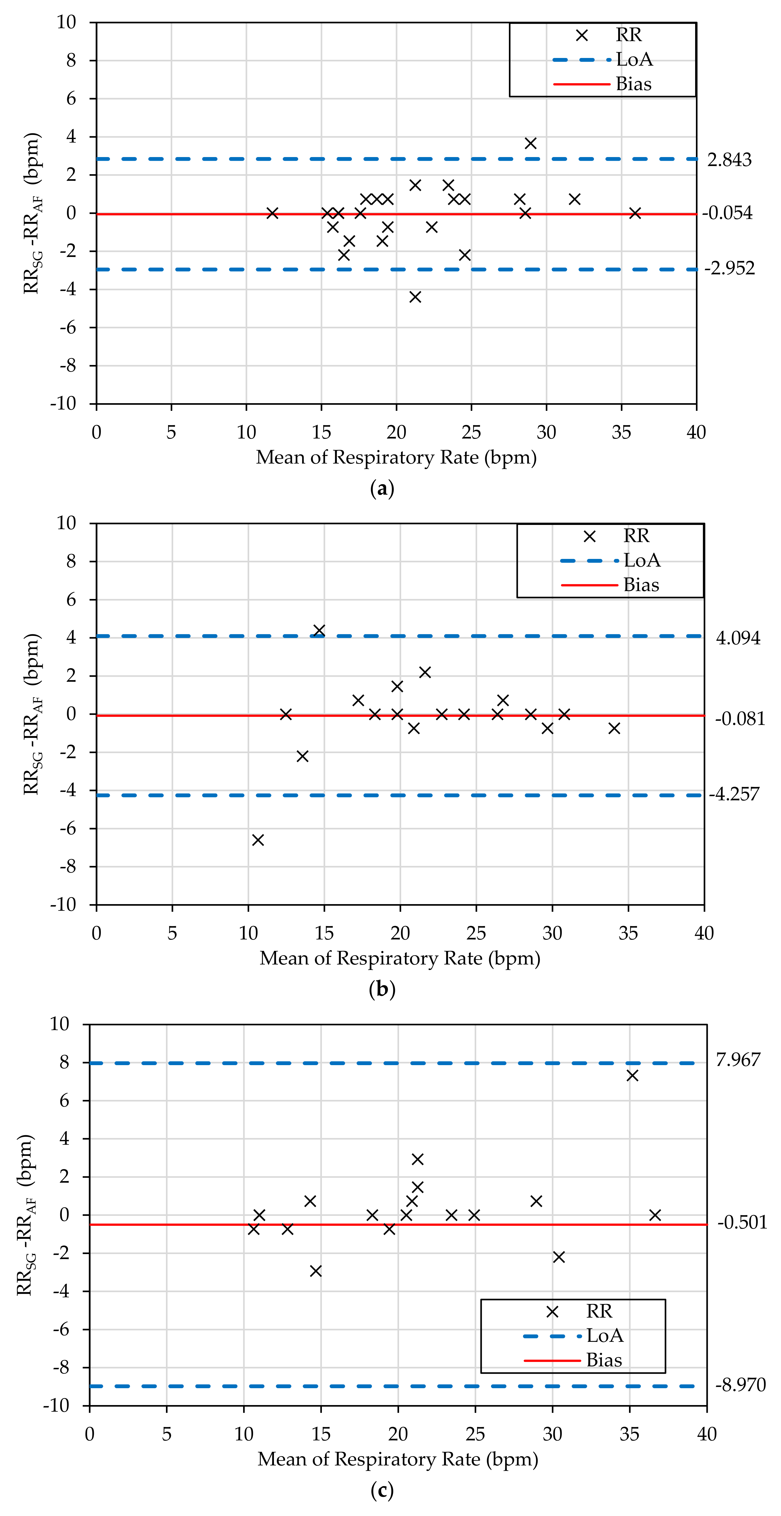
3.4. Bland-Altman Analysis

As shown in Figure 4, the smallest bias between the SG-derived and reference RRs was from the measurement in sitting posture (bias: −0.0543 bpm, 95% limits of agreement (LoA): −2.952 to 2.843 bpm). The measurement in standing posture had a similar bias but a wider LoA. The measurement in lying45° posture has the largest bias (−0.501 bpm) and the widest LoA (−8.970 to 7.967 bpm). Thus, the SG measurement was most accurate in sitting posture and least accurate in the lying45° posture.

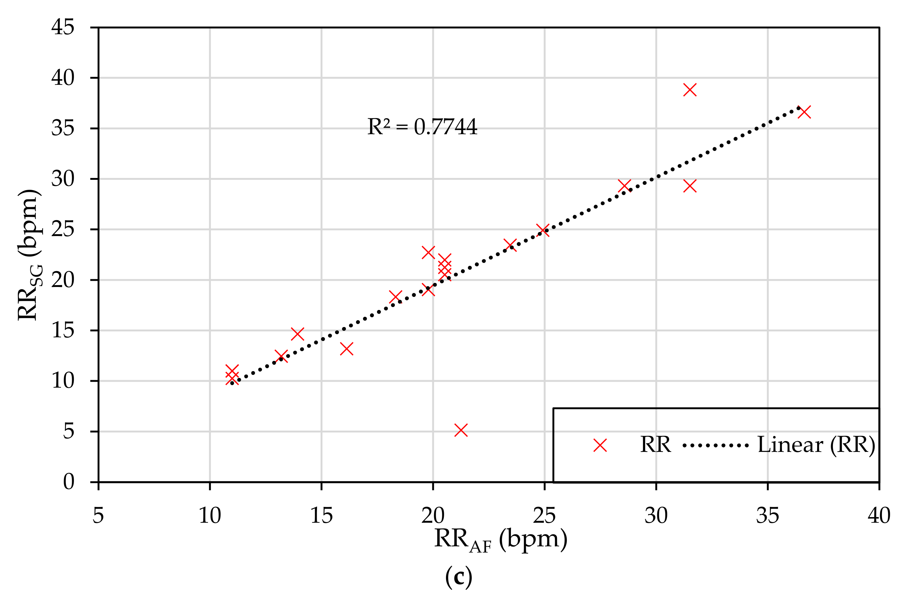
3.5. Regression Analysis

The linear regression coefficient between RRSG and RRAF measurements was high for the sitting and standing postures (0.9394 and 0.9067, respectively) while the value was lower in the lying45° posture (0.7744), as shown in Figure 5. The small regression coefficient in the lying45° posture was largely related to the result from an outlier (patient #2) due to losing mounting of the IJPT sensor.

4. Discussion

In this work, the IJPT sensor was comprehensively evaluated on 35 COPD patients to measure the RR at different body postures namely standing, sitting at 90°, and lying at 45° positions. The results in this work indicated the high accuracy of the IJPT sensor in RR measurement, with minor differences between different postures.

4.1. Measurement of RR on COPD Patients: Difficulties and Approaches

The RR value is one of the most important indicators of several chronic diseases including COPD [50]. RR over 25 bpm is considered as one of the COPD exacerbation signs while the normal range of RR in adults is about 12–20 bpm [40]. RR monitoring of COPD patients is usually carried out in hospitals and clinics via manual counting of breaths, using nasal sensors, or extracted from the electrocardiogram (ECG) signals. The measurement with manual counting is inaccurate [16] while the use of nasal sensors is difficult especially when patients are on continuous oxygen support. Additionally, ECG and nasal sensors are cumbersome and uncomfortable for long-term monitoring. On the other hand, significant progress in the development of WFS has provided the possibility for the convenient, long-term, and low-cost monitoring of vital signs including RR for COPD patients [16,33]. However, there is a lack of validation and clinical evaluation of WFS on COPD patients in different postures, daily activities, and other physiological conditions where the noises could affect the signal quality [31,33,40]. Our results provided a reference for the development of WFS for clinical use.
Another challenge is to get accurate estimation of RR in varying breathing patterns. COPD patients usually have an unstable breathing pattern, which causes the variations of RR during measurement. It was observed that RR changed from 15.2 ± 4.3 bpm to 19.1 ± 5.9 bpm during the exacerbation of COPD [24]. COPD patients show characteristic symptoms like breathlessness and cough [51,52]. These symptoms in addition to body posture are associated with the respiratory volume change. Resultantly, the deformed respiratory waveform and amplitude of peaks can lead to the inaccuracy in RR estimation, as observed in this study. Figure 6 shows a comparison between good and deformed respiratory signals measured by the IJPT. It should be noted that the signal quality depends on many factors including the stability of the sensor attachment on the patients. Another factor that affects the signal is the amount of clothing on the patients. Furthermore, the amount of volume change in the abdominal area varies significantly among individuals and also among postures as reported in the literature [17]. These changes in the volume could significantly affect the ability of the IJPT sensor to measure the RR accurately, as seen in the lying45° posture, which is the most challenging posture for RR measurement and yields the results consistent with what was reported in the literature [17]. Furthermore, some COPD patients cannot endure standing or sitting for long periods. Therefore, the comprehensive evaluation of RR monitoring in different circumstances is important for the clinical application of the IJPT sensor [30]. The high measurement accuracy of the IJPT sensor at different postures and among different patients highlighted its potential for reliable monitoring of RR in COPD patients.

4.2. Accuracy of IJPT Sensor: Comparison with Other Sensors

The statistical analysis demonstrated a high accuracy of the IJPT sensor in RR monitoring on COPD patients with good stability at different posture positions. In addition, there was insignificant effect of posture or individual difference on the estimation error. Moreover, the measurement accuracy of the IJPT sensor was comparable with other sensors that have been clinically evaluated on healthy human subjects reported in [35,36,37,38,39]. As shown in Table 1, the IJPT sensor showed higher accuracy in RR monitoring on COPD patients (except in the lying posture, which could be related to losing mounting of the IJPT sensor as aforementioned) than most of the existing sensors.

4.3. Applications of IJPT Sensor

Currently, the RR monitoring is often neglected or inaccurately recorded even on patients with respiratory diseases due to the lack of appropriate devices for clinical use [7]. The low fabrication cost, biocompatible substrate, skin-friendly attachment on body surface, and the ease of movement without restrictions in daily activities, as well as the high measurement accuracy of the IJPT sensor make it a promising technology for remote and continuous monitoring of RR for patients with COPD and other respiratory diseases [13]. Reliable RR monitoring for COPD patients using the IJPT sensor could reduce the healthcare cost and the pressure on healthcare facilities especially in low-resource settings such as refugee camps [54]. A recent systematic review disclosed that the majority of physiological monitoring methods for COPD are intermittent with no more than twice a day measurements’ frequency [55]. Acute exacerbations of COPD require intensive care treatment immediately. The evaluation of vital signs is necessary to detect physiological abnormalities (micro events), but patients may deteriorate between measurements [56]. The continuous monitoring of RR on COPD patients is an unmet clinical need that has attracted increasing research focus. Especially, the RR monitoring based on WFS on chest band has been proven with the highest reliability compared with other sensors [57], which makes it possible to provide reliable daily monitoring of COPD based on WFS. Furthermore, the frequency and severity of COPD exacerbation symptoms would be reduced by early detection of abnormal physiological measurements including the respiratory rate [42]. To fight the ongoing pandemic of COVID-19, the combination of internet-of-things (IoT) technology and the IJPT sensor can generate a low-cost, safe, and convenient approach for the remote monitoring, management, and early intervention of the patients where recent studies [58,59] have mentioned that RR could serve as a leading indicator of COVID-19.

4.4. Limitations and Future Work

There were some limitations associated with this study. Firstly, the IJPT sensor was tested on 35 COPD patients with only one female and also not all of the measurements at the three postures were available due to the limitations aforementioned in Section 2.3. Therefore, further investigation with more diverse COPD patients is required. Secondly, further clinical evaluation of the IJPT sensor on COPD patients during walking, running, sleeping, and other daily activities can be carried out to expand its application scenarios. Finally, longer time series data should be recorded and clinically evaluated on healthy and non-healthy subjects to further investigate the applicability of the IJPT sensor for continuous monitoring of RR. For future work, an innovative mounting mechanism can be developed as the substitute for the fabric belt to achieve more convenient and reliable attachment in different postures. A wireless platform can be designed to achieve more comfortable measurement for the end user, which will ease the remote and continuous monitoring of RR.

5. Conclusions

The results of clinical evaluation on 35 COPD patients in the present work indicated that the IJPT sensor was able to accurately measure the RR at different postures. It can be concluded that the IJPT sensor showed comparable accuracy with other wearable sensors on COPD patients evaluated in the literature with the absolute relative error of 4.49%, 7.29%, and 9.47% at sitting, standing, and lying45° postures, respectively. The IJPT sensor is promising in achieving reliable RR monitoring for COPD patients where the use of this sensor would contribute to mitigating the frequency and severity of COPD exacerbation symptoms by early detection of abnormal physiological measurements including the respiratory rate.

Author Contributions

Conceptualization, A.A.-H., L.A.-G., H.L., and D.Z.; data curation, O.K. and H.L.; formal analysis, L.A.-G. and H.L.; funding acquisition, A.A.-H., D.Z. and F.C.; investigation, L.A.-G., O.K., A.R., J.A., and H.L.; methodology, L.A.-G. and H.L.; project administration, A.A.-H., L.A.-G., and D.Z.; resources, A.A.-H. and D.Z.; software, O.K.; supervision, A.A.-H., L.A.-G., J.A., H.L., K.A.O., F.C., and D.Z.; validation, A.R., H.L., and K.A.O.; visualization, L.A.-G., O.K., and H.L.; writing—original draft, L.A.-G., O.K., A.R., J.A., and H.L.; writing—review and editing, A.A.-H., L.A.-G., H.L., and F.C. All authors have read and agreed to the published version of the manuscript.

Funding

This research was funded by the Royal Academy of Engineering under grant Ref. IAPP1R2\100204. The work of Fei Chen was supported by High-level University Fund G02236002 of Southern University of Science and Technology.

Institutional Review Board Statement

The study was conducted according to the guidelines of the Declaration of Helsinki, and approved by Jordan University Hospital Ethics Committee (REF 67/2019/6480).

Informed Consent Statement

Informed consent was obtained from all subjects involved in the study.

Data Availability Statement

The data presented in this study are available in Appendix A.

Acknowledgments

The authors would like to thank the Royal Academy of Engineering for Funding this project under grant Ref. IAPP1R2\100204. Moreover, the authors would like to thank the Industrial Research & Development Fund in the Higher Council for Science & Technology for their support of the project. In addition, the authors would like to thank Eng. Ali Al-Ghussein from the GJU Innovation and Entrepreneurship laboratory for providing the e-Health sensor. Finally, the authors would like to thank Eng. Ahmed Albagdady for providing some of the photos used in the manuscript.

Conflicts of Interest

The authors declare no conflict of interest.

Ethical Statements

All subjects gave their informed consent for inclusion before they participated in the study. The study was conducted in accordance with the Declaration of Helsinki, and the protocol was approved by the Ethics Committee of REF 67/2019/6480.

Appendix A

Table A1. Summary of patients’ information collected.
PatientAgeGenderHeight (m)Weight (kg)Smoking StatusGOLD DiagnosisFirst Diagnosed
150M16962Ex-Smoker312/2018
251M17071Smoker111/2001
346M17483Ex-Smoker35/2010
430M18062Smoker1N/A
579M17598Ex-Smoker24/2008
658M16278Smoker34/2019
739M17085Quit smoking211/2017
856M17590Smoker18/2013
958M17380Ex-Smoker22/2009
1026M17384Ex-Smoker18/2016
1152M17663Ex-Smoker22009
1256M17070SmokerN/A2014
1347M17580Ex-smoking17/2018
1452M16880Smoker25/2007
1524M17363Smoker11/2020
1642M16485Smoker110/2014
1760M17884Smoker18/2019
1842M16575Smoker28/2014
1949M16764Smoker31/2020
2037M17298Smoker31/2020
2157M16760Smoker32/2020
2270M16050Smoker22/2020
2366M17977Smoker19/2013
2467M17570Ex-Smoker35/2001
2555M16758Quit smoking211/2019
2669M17588Ex-Smoker2N/A
2753F16790Smoker12/2012
2858M16374Ex-Smoker11/2019
2979M17472Smoker38/2017
3067M16762Smoker28/2017
3166M17585Ex-Smoker16/2002
3267M17469Smoker36/2014
3358M17273Ex-Smoker33/2012
3475M17468Smoker111/2009
3573M17065Smoker17/2013
N/A: not available. quit smoking: stopped within the last 6 months of interview. Ex-Smoker: stopped smoking after 6 months of interview. A/V fistula: arteriovenous fistula.
Table A2. Summary of CAGE (cut-annoyed-guilty-eye) questionnaire.
PatientCoughPhlegm (mucus)Tightness of ChestNot Able to Climb a Flight of StairsCannot Perform Home ActivitiesCan Go Out AnytimeGood SleepHaving Energy
1frequentlyrareneveralwaysrarealwaysfrequentlyalways
2rareraresometimesalwaysneveralwaysfrequentlyrare
3alwayssometimessometimesalwaysrareneversometimesrare
4rarerarefrequentlyfrequentlyneveralwaysfrequentlyalways
5alwayssometimesrarefrequentlyfrequentlyraresometimesrare
6alwaysalwayssometimessometimessometimesalwayssometimesnever
7rareraresometimesrarerarerarefrequentlysometimes
8frequentlysometimesraresometimesneveralwaysalwaysalways
9sometimesrareneverneverneveralwaysfrequentlysometimes
10neverneverneverneverneveralwaysfrequentlyalways
11raresometimesneverrarerarefrequentlyfrequentlysometimes
12sometimessometimesneverneverneverfrequentlyalwaysalways
13raresometimesneverneverneveralwaysalwaysalways
14sometimesneverneverrareneverfrequentlyfrequentlyfrequently
15rareneverneverneverneveralwaysalwaysalways
16sometimesfrequentlyfrequentlyrarerareneversometimesfrequently
17rarerarerareneverneveralwaysfrequentlyfrequently
18frequentlyraresometimesneverneveralwaysalwaysfrequently
19frequentlyfrequentlysometimessometimesrarefrequentlysometimesfrequently
20frequentlyfrequentlyrarealwaysalwayssometimesrarefrequently
21frequentlyrarealwaysalwaysalwaysalwayssometimesrare
22alwayssometimessometimessometimessometimesalwaysfrequentlynever
23rareneverneverneverneveralwaysfrequentlyfrequently
24sometimesneverneverneverfrequentlyfrequentlysometimesalways
25sometimessometimesfrequentlyalwaysalwayssometimesrarenever
26sometimesalwayssometimesfrequentlyfrequentlysometimessometimesfrequently
27frequentlyrareneversometimessometimesalwaysfrequentlyfrequently
28alwaysalwaysrarerareneveralwaysalwaysfrequently
29alwaysfrequentlyneveralwaysrarefrequentlyrarefrequently
30alwaysalwaysalwaysalwaysfrequentlyrarerarenever
31sometimesneversometimesfrequentlyalwaysrarefrequentlyrare
32sometimesneverneverrarerarefrequentlyalwaysalways
33frequentlyalwaysneversometimesfrequentlyalwaysraresometimes
34frequentlyrareraresometimesneveralwayssometimesfrequently
35sometimesraresometimessometimesraresometimesfrequentlysometimes
Table A3. RRAF and RRSG of the COPD measured in addition to the relative error at different body postures.
PatientPostureRRSG (bpm)RRAF (bpm)Error (bpm)
1Sitting28.5728.570
Standing30.7730.770
2Sitting19.0518.320.73
Standing20.5119.051.46
Lying45°5.1321.25−16.12
3Sitting32.2331.500.73
Standing29.3030.04−0.74
Lying45°29.3031.50−2.2
4Sitting18.3217.580.74
Standing7.3313.92−6.59
Lying45°12.4513.19−0.74
5Sitting21.9822.71−0.73
Standing22.7122.710
6Sitting19.0523.44−4.39
7Sitting15.3817.58−2.2
Standing22.7120.512.2
8Sitting30.7727.113.66
Standing18.3218.320
9Sitting11.7211.720
Standing12.4512.450
Lying45°10.9910.990
10Sitting16.1217.58−1.46
Standing19.7819.780
11Sitting19.0519.78−0.73
12Sitting18.3219.78−1.46
Standing24.1824.180
Lying45°14.6513.920.73
13Sitting16.1216.120
Lying45°10.9910.990
14Sitting23.4425.64−2.2
Standing20.5121.25−0.74
15Sitting21.9820.511.47
Lying45°19.0519.78−0.73
16Standing28.5728.570
Lying45°21.2520.510.74
17Lying45°13.1916.12−2.93
18Sitting17.5817.580
19Sitting16.1216.120
Standing16.8512.454.4
Lying45°18.3218.320
20Sitting35.9035.900
Standing33.7034.43−0.73
Lying45°36.6336.630
21Lying45°22.7119.782.93
22Sitting28.5727.840.73
Lying45°24.9124.910
23Sitting24.9124.180.73
24Sitting15.3816.12−0.74
Standing17.5816.850.73
25Lying45°23.4423.440
26Lying45°21.9820.511.47
27Sitting19.7819.050.73
Standing12.4514.65−2.2
Lying45°10.2610.99−0.73
28Lying45°38.8331.507.33
29Sitting15.3815.380
30Sitting24.1822.711.47
31Lying45°20.5120.510
32Sitting19.7819.050.73
33Sitting24.1823.440.74
Standing26.3726.370
34Lying45°29.3028.570.73
35Sitting16.1216.120
Standing27.1126.370.74

References

  1. Lucchini, M.; Pini, N.; Burtchen, N.; Signorini, M.G.; Fifer, W.P. Transfer Entropy Modeling of Newborn Cardiorespiratory Regulation. Front. Physiol. 2020, 11, 1095. [Google Scholar] [CrossRef] [PubMed]
  2. Liu, H.; Chen, F.; Hartmann, V.; Khalid, S.G.; Hughes, S.; Zheng, D. Comparison of different modulations of photoplethysmography in extracting respiratory rate: From a physiological perspective. Physiol. Meas. 2020, 41, 094001. [Google Scholar] [CrossRef]
  3. Nicolò, A.; Massaroni, C.; Schena, E.; Sacchetti, M. The Importance of Respiratory Rate Monitoring: From Healthcare to Sport and Exercise. Sensors 2020, 20, 6396. [Google Scholar] [CrossRef]
  4. Flenady, T.; Dwyer, T.; Applegarth, J. Accurate respiratory rates count: So should you! Australas. Emerg. Nurs. J. 2017, 20, 45–47. [Google Scholar] [CrossRef] [PubMed] [Green Version]
  5. Nicolò, A.; Marcora, S.M.; Sacchetti, M. Time to reconsider how ventilation is regulated above the respiratory compensation point during incremental exercise. J. Appl. Physiol. 2020, 128, 1447–1449. [Google Scholar] [CrossRef] [PubMed]
  6. Hill, B.; Annesley, S.H. Monitoring respiratory rate in adults. Br. J. Nurs. 2020, 29, 12–16. [Google Scholar] [CrossRef]
  7. Liu, H.; Allen, J.; Zheng, D.; Chen, F. Recent development of respiratory rate measurement technologies. Physiol. Meas. 2019, 40, 07TR01. [Google Scholar] [CrossRef] [Green Version]
  8. Nag, A.; Mukhopadhyay, S.C.; Kosel, J. Wearable Flexible Sensors: A Review. IEEE Sens. J. 2017, 17, 3949–3960. [Google Scholar] [CrossRef] [Green Version]
  9. Rodgers, M.M.; Pai, V.M.; Conroy, R.S. Recent advances in wearable sensors for health monitoring. IEEE Sens. J. 2015, 15, 3119–3126. [Google Scholar] [CrossRef]
  10. Yetisen, A.K.; Martinez-Hurtado, J.L.; Ünal, B.; Khademhosseini, A.; Butt, H. Wearables in Medicine. Adv. Mater. 2018, 30, e1706910. [Google Scholar] [CrossRef]
  11. Dittmar, A.; Meffre, R.; De Oliveira, F.; Gehin, C.; Delhomme, G. Wearable Medical Devices Using Textile and Flexible Technologies for Ambulatory Monitoring. In Proceedings of the 2005 IEEE Engineering in Medicine and Biology 27th Annual Conference, Shanghai, China, 1–4 September 2005; pp. 7161–7164. [Google Scholar]
  12. Khan, Y.; Ostfeld, A.E.; Lochner, C.M.; Pierre, A.; Arias, A.C. Monitoring of Vital Signs with Flexible and Wearable Medical Devices. Adv. Mater. 2016, 28, 4373–4395. [Google Scholar] [CrossRef] [PubMed]
  13. Liu, Y.; Wang, H.; Zhao, W.; Zhang, M.; Qin, H.; Xie, Y. Flexible, stretchable sensors for wearable health monitoring: Sensing mechanisms, materials, fabrication strategies and features. Sensors 2018, 18, 645. [Google Scholar] [CrossRef] [PubMed] [Green Version]
  14. Abu-khalaf, J.M.; Saraireh, R.; Eisa, S.M.; Al-halhouli, A. Experimental Characterization of Inkjet-Printed Stretchable Circuits for Wearable Sensor Applications. Sensors 2018, 18, 3476. [Google Scholar] [CrossRef] [PubMed] [Green Version]
  15. Yilmaz, T.; Foster, R.; Hao, Y. Detecting vital signs with wearablewireless sensors. Sensors 2010, 10, 10837–10862. [Google Scholar] [CrossRef] [PubMed]
  16. Al-Halhouli, A.; Al-Ghussain, L.; El Bouri, S.; Liu, H.; Zheng, D. Fabrication and Evaluation of a Novel Non-Invasive Stretchable and Wearable Respiratory Rate Sensor Based on Silver Nanoparticles Using Inkjet Printing Technology. Polymers 2019, 11, 1518. [Google Scholar] [CrossRef] [Green Version]
  17. Al-Halhouli, A.; Al-Ghussain, L.; El Bouri, S.; Habash, F.; Liu, H.; Zheng, D. Clinical Evaluation of Stretchable and Wearable Inkjet-Printed Strain Gauge Sensor for Respiratory Rate Monitoring at Different Body Postures. Appl. Sci. 2020, 10, 480. [Google Scholar] [CrossRef] [Green Version]
  18. Nicolò, A.; Massaroni, C.; Passfield, L. Respiratory frequency during exercise: The neglected physiological measure. Front. Physiol. 2017, 8, 922. [Google Scholar] [CrossRef]
  19. Massaroni, C.; Nicolò, A.; Presti, D.L.; Sacchetti, M.; Silvestri, S.; Schena, E. Contact-based methods for measuring respiratory rate. Sensors 2019, 19, 908. [Google Scholar] [CrossRef] [Green Version]
  20. Naranjo-Hernández, D.; Talaminos-Barroso, A.; Reina-Tosina, J.; Roa, L.M.; Barbarov-Rostan, G.; Cejudo-Ramos, P.; Márquez-Martín, E.; Ortega-Ruiz, F. Smart vest for respiratory rate monitoring of copd patients based on non-contact capacitive sensing. Sensors 2018, 18, 2144. [Google Scholar] [CrossRef] [Green Version]
  21. Hesse, M.; Christ, P.; Hormann, T.; Ruckert, U. A respiration sensor for a chest-strap based wireless body sensor. In Proceedings of the IEEE SENSORS 2014 Proceedings, Valencia, Spain, 2–5 November 2014; IEEE: Piscataway, NJ, USA, 2014; Volume 2014, pp. 490–493. [Google Scholar]
  22. Chiarugi, F.; Karatzanis, I.; Zacharioudakis, G.; Meriggi, P.; Rizzo, F.; Stratakis, M.; Louloudakis, S.; Biniaris, C.; Valentini, M.; Di Rienzo, M.; et al. Measurement of heart rate and respiratory rate using a textile-based wearable device in heart failure patients. Comput. Cardiol. 2008, 35, 901–904. [Google Scholar]
  23. Furtak, N.T.; Skrzetuska, E.; Krucińska, I. Development of Screen-Printed Breathing Rate Sensors. Fibres Text. East. Eur. 2013, 21, 84–88. [Google Scholar]
  24. Wu, D.; Wang, L.; Zhang, Y.; Huang, B.; Wang, B.; Lin, S.; Xu, X. A wearable respiration monitoring system based on digital respiratory inductive plethysmography. In Proceedings of the 2009 Annual international conference of the IEEE Engineering in Medicine and Biology Society, Minneapolis, MN, USA, 3–6 September 2009; pp. 4844–4847. [Google Scholar]
  25. Jeong, J.W.; Jang, Y.W.; Lee, I.; Shin, S.; Kim, S. Wearable Respiratory Rate Monitoring using Piezo-resistive Fabric Sensor. In Proceedings of the World Congress on Medical Physics and Biomedical Engineering, Munich, Germany, 7–12 September 2009; Dössel, O., Schlegel, W.C., Eds.; Springer: Berlin/Heidelberg, Germany, 2009; pp. 282–284, ISBN 978-3-642-03904-1. [Google Scholar]
  26. Lei, K.F.; Hsieh, Y.Z.; Chiu, Y.Y.; Wu, M.H. The structure design of piezoelectric poly(vinylidene fluoride) (PVDF) polymer-based sensor patch for the respiration monitoring under dynamic walking conditions. Sensors 2015, 15, 18801–18812. [Google Scholar] [CrossRef] [Green Version]
  27. Bonato, P. Clinical applications of wearable technology. In Proceedings of the 2009 Annual International Conference of the IEEE Engineering in Medicine and Biology Society, Minneapolis, MN, USA, 2–6 September 2009; pp. 6580–6583. [Google Scholar]
  28. Berry, C.E.; Wise, R.A. Mortality in COPD: Causes, Risk Factors, and Prevention. COPD J. Chronic Obstr. Pulm. Dis. 2010, 7, 375–382. [Google Scholar] [CrossRef] [PubMed]
  29. Anlló, H.; Herer, B.; Delignières, A.; Bocahu, Y.; Segundo, I.; Mach Alingrin, V.; Gilbert, M.; Larue, F. Hypnosis for the management of anxiety and dyspnea in COPD: A randomized, sham-controlled crossover trial. Int. J. Chronic Obstr. Pulm. Dis. 2020, 15, 2609–2620. [Google Scholar]
  30. Yañez, A.M.; Guerrero, D.; Pérez De Alejo, R.; Garcia-Rio, F.; Alvarez-Sala, J.L.; Calle-Rubio, M.; Malo De Molina, R.; Valle Falcones, M.; Ussetti, P.; Sauleda, J.; et al. Monitoring breathing rate at home allows early identification of COPD exacerbations. Chest 2012, 142, 1524–1529. [Google Scholar] [CrossRef] [PubMed]
  31. Bellos, C.C.; Papadopoulos, A.; Rosso, R.; Fotiadis, D.I. Identification of COPD patients’ health status using an intelligent system in the CHRONIOUS wearable platform. IEEE J. Biomed. Heal. Inform. 2014, 18, 731–738. [Google Scholar] [CrossRef] [PubMed]
  32. Rolfe, S. The importance of respiratory rate monitoring. Br. J. Nurs. 2019, 28, 504–508. [Google Scholar] [CrossRef] [PubMed]
  33. Rubio, N.; Parker, R.A.; Drost, E.M.; Pinnock, H.; Weir, C.J.; Hanley, J.; Mantoani, L.C.; Macnee, W.; McKinstry, B.; Rabinovich, R.A. Home monitoring of breathing rate in people with chronic obstructive pulmonary disease: Observational study of feasibility, acceptability, and change after exacerbation. Int. J. Chronic Obstr. Pulm. Dis. 2017, 12, 1221–1231. [Google Scholar] [CrossRef] [PubMed] [Green Version]
  34. Dias, D.; Cunha, J.P.S. Wearable health devices—Vital sign monitoring, systems and technologies. Sensors 2018, 18, 2414. [Google Scholar] [CrossRef] [Green Version]
  35. Ciocchetti, M.; Massaroni, C.; Saccomandi, P.; Caponero, M.; Polimadei, A.; Formica, D.; Schena, E. Smart Textile Based on Fiber Bragg Grating Sensors for Respiratory Monitoring: Design and Preliminary Trials. Biosensors 2015, 5, 602–615. [Google Scholar] [CrossRef] [Green Version]
  36. Sharma, P.; Hui, X.; Zhou, J.; Conroy, T.B.; Kan, E.C. Wearable radio-frequency sensing of respiratory rate, respiratory volume, and heart rate. NPJ Digit. Med. 2020, 3, 1–10. [Google Scholar] [CrossRef] [PubMed]
  37. Yamamoto, A.; Nakamoto, H.; Bessho, Y.; Watanabe, Y.; Oki, Y.; Ono, K.; Fujimoto, Y.; Terada, T.; Ishikawa, A. Monitoring respiratory rates with a wearable system using a stretchable strain sensor during moderate exercise. Med. Biol. Eng. Comput. 2019, 57, 2741–2756. [Google Scholar] [CrossRef] [PubMed]
  38. Chu, M.; Nguyen, T.; Pandey, V.; Zhou, Y.; Pham, H.N.; Bar-Yoseph, R.; Radom-Aizik, S.; Jain, R.; Cooper, D.M.; Khine, M. Respiration rate and volume measurements using wearable strain sensors. NPJ Digit. Med. 2019, 2, 8. [Google Scholar] [CrossRef] [PubMed]
  39. Massaroni, C.; Lopes, D.S.; Lo Presti, D.; Schena, E.; Silvestri, S. Contactless monitoring of breathing patterns and respiratory rate at the pit of the neck: A single camera approach. J. Sens. 2018, 2018, 1–13. [Google Scholar] [CrossRef]
  40. Tomasic, I.; Tomasic, N.; Trobec, R.; Krpan, M.; Kelava, T. Continuous remote monitoring of COPD patients—justification and explanation of the requirements and a survey of the available technologies. Med. Biol. Eng. Comput. 2018, 56, 547–569. [Google Scholar] [CrossRef] [PubMed] [Green Version]
  41. Al-Halhouli, A.; Al-Ghussain, L.; El Bouri, S.; Habash, F.; Liu, H.; Zheng, D. Clinical evaluation of stretchable and wearable inkjet-printed strain gauge sensor for respiratory rate monitoring at different measurements locations. J. Clin. Monit. Comput. 2020, 10, 480. [Google Scholar] [CrossRef]
  42. Cordova, F.C.; Ciccolella, D.; Grabianowski, C.; Gaughan, J.; Brennan, K.; Goldstein, F.; Jacobs, M.R.; Criner, G.J. A Telemedicine-Based Intervention Reduces the Frequency and Severity of COPD Exacerbation Symptoms: A Randomized, Controlled Trial. Telemed. Health 2016, 22, 114–122. [Google Scholar] [CrossRef] [Green Version]
  43. Carlson, R.V.; Boyd, K.M.; Webb, D.J. The revision of the Declaration of Helsinki: Past, present and future. Br. J. Clin. Pharmacol. 2004, 57, 695–713. [Google Scholar] [CrossRef]
  44. Hacks, C. e-Health Sensor Platform V2.0 for Arduino and Raspberry Pi [Biometric/Medical Applications]. Available online: https://www.cooking-hacks.com/documentation/tutorials/ehealth-biometric-sensor-platform-arduino-raspberry-pi-medical (accessed on 20 February 2019).
  45. Arduino Mega 2560 Datasheet. Available online: http://eprints.polsri.ac.id/4598/8/File%20VIII%20%28Lampiran%29.pdf (accessed on 10 January 2021).
  46. ATmega640/V-1280/V-1281/V-2560/V-2561/V. Available online: https://ww1.microchip.com/downloads/en/DeviceDoc/ATmega640-1280-1281-2560-2561-Datasheet-DS40002211A.pdf (accessed on 10 January 2021).
  47. Schmider, E.; Ziegler, M.; Danay, E.; Beyer, L.; Bühner, M. Is It Really Robust? Methodology 2010, 6, 147–151. [Google Scholar] [CrossRef]
  48. Bland, J.M.; Altman, D.G. Statistical methods for assessing agreement between two methods of clinical measurement. Lancet 1986, 1, 307–310. [Google Scholar] [CrossRef]
  49. Giavarina, D. Understanding Bland Altman analysis. Biochem. Med. 2015, 25, 141–151. [Google Scholar] [CrossRef] [PubMed] [Green Version]
  50. Siafakas, N.M.; Vermeire, P.; Pride, N.B.; Paoletti, P.; Gibson, J.; Howard, P.; Yernault, J.C.; Decramer, M.; Higenbottam, T.; Postma, D.S.; et al. Optimal assessment and management of chronic obstructive pulmonary disease (COPD). Eur. Respir. J. 1995, 8, 1398–1420. [Google Scholar] [CrossRef] [PubMed]
  51. Roche, N.; Chavannes, N.H.; Miravitlles, M. COPD symptoms in the morning: Impact, evaluation and management. Respir. Res. 2013, 14, 112. [Google Scholar] [CrossRef] [PubMed] [Green Version]
  52. Miravitlles, M.; Worth, H.; Soler Cataluña, J.J.; Price, D.; De Benedetto, F.; Roche, N.; Godtfredsen, N.S.; van der Molen, T.; Löfdahl, C.G.; Padullés, L.; et al. Observational study to characterise 24-hour COPD symptoms and their relationship with patient-reported outcomes: Results from the ASSESS study. Respir. Res. 2014, 15, 122. [Google Scholar] [CrossRef] [PubMed]
  53. Soler, J.; Alves Pegoraro, J.; Le, X.L.; Nguyen, D.P.Q.; Grassion, L.; Antoine, R.; Guerder, A.; Gonzalez-Bermejo, J. Validation of respiratory rate measurements from remote monitoring device in COPD patients. Respir. Med. Res. 2019, 76, 1–3. [Google Scholar] [CrossRef]
  54. Klersy, C.; De Silvestri, A.; Gabutti, G.; Raisaro, A.; Curti, M.; Regoli, F.; Auricchio, A. Economic impact of remote patient monitoring: An integrated economic model derived from a meta-analysis of randomized controlled trials in heart failure. Eur. J. Heart Fail. 2011, 13, 450–459. [Google Scholar] [CrossRef] [Green Version]
  55. Al Rajeh, A.; Hurst, J. Monitoring of Physiological Parameters to Predict Exacerbations of Chronic Obstructive Pulmonary Disease (COPD): A Systematic Review. J. Clin. Med. 2016, 5, 108. [Google Scholar] [CrossRef]
  56. Elvekjaer, M.; Aasvang, E.K.; Olsen, R.M.; Sørensen, H.B.D.; Porsbjerg, C.M.; Jensen, J.-U.; Haahr-Raunkjær, C.; Meyhoff, C.S. Physiological abnormalities in patients admitted with acute exacerbation of COPD: An observational study with continuous monitoring. J. Clin. Monit. Comput. 2020, 34, 1051–1060. [Google Scholar] [CrossRef]
  57. Parker, R.A.; Weir, C.J.; Rubio, N.; Rabinovich, R.; Pinnock, H.; Hanley, J.; McCloughan, L.; Drost, E.M.; Mantoani, L.C.; MacNee, W.; et al. Application of Mixed Effects Limits of Agreement in the Presence of Multiple Sources of Variability: Exemplar from the Comparison of Several Devices to Measure Respiratory Rate in COPD Patients. PLoS ONE 2016, 11, e0168321. [Google Scholar] [CrossRef]
  58. Miller, D.; Capodilupo, J.; Lastella, M.; Sargent, C.; Roach, G.; Lee, V.; Capodilupo, E. Analyzing changes in respiratory rate to predict the risk of COVID-19 infection. PLoS ONE 2020, 15, e0243693. [Google Scholar] [CrossRef]
  59. Nakano, H.; Kadowaki, M.; Furukawa, T.; Yoshida, M. Rise in nocturnal respiratory rate during CPAP may be an early sign of COVID-19 in patients with obstructive sleep apnea. J. Clin. Sleep Med. 2020, 6, 1811–1813. [Google Scholar] [CrossRef] [PubMed]
Figure 1. Schematic diagram of the inkjet-printed (IJPT) sensor fabrication process and respiratory rate (RR) extraction on Chronic Obstructive Pulmonary Disease (COPD) patients.
Sensors 21 00468 g001
Figure 2. Electrical circuit configuration of the strain gauge sensor.
Sensors 21 00468 g002
Figure 3. Sample of RR signal processing and RR derivation obtained from patient #9 while sitting using the strain gauge sensor and the e-Health airflow sensor.
Sensors 21 00468 g003
Figure 4. Bland-Altman analysis of the measured respiratory rate of the COPD patients at different postures, namely at (a) sitting, (b) standing, and (c) lying45° posture. Each cross represents the data point of RR measurement. The continuous red line represents the bias, which is the average difference between the RRAF and the RRSG while the dashed blue lines are the limits of agreement where 95% of the data lies in between.
Sensors 21 00468 g004
Figure 5. Linear regression analysis of the measured respiratory rate of the COPD patients at different postures namely at: (a) Sitting, (b) standing, and (c) lying45° posture.
Sensors 21 00468 g005aSensors 21 00468 g005b
Figure 6. Samples of raw respiratory signals acquired by the IJPT on COPD patient while standing: (a) Good signal (test subject #8) and (b) deformed signal (test subject #10).
Sensors 21 00468 g006
Table 1. Comparison between the accuracy and performance of the IJPT sensor and other sensors reported in the literature.
Ref.MethodPostureAccuracy ParameterProtocolNumber of COPD Patients
[33]ImpedanceActivities of daily living protocolBias (bpm)−1.18Attached to the chest and upper abdomen44
LoA (bpm)−20.07 to 17.72
Photoplethysmography (PPG)Bias (bpm)3.01Worn on the wrist with a finger probe
LoA (bpm)−11.17 to 17.19
CameraBias (bpm)−3.21Participant was videoed while in sitting position
LoA (bpm)−12.71 to 6.30
AccelerometerBias (bpm)−2.18Attached to the upper abdomen just below the ribs and taped to the skin
LoA (bpm)−8.63 to 4.27
Chest-Band (strain gauge)Bias (bpm)−1.60Chest strap and an electronics module that attaches to the strap62
LoA (bpm)−9.99 to 6.80
[20]CapacitiveRest (lying)Bias (bpm)−0.14 bpmRest (after exercises)9
SD (bpm)0.28
[31]Respiration band (strain gauge)-Relative Error (%)17.43Attached to the wearable Jacket30
[53]Airflow pressure sensor-Bias (bpm)0.046Hoses attached to the nose14
LoA (bpm)3.865 to 3.957
This studyStrain gauge SittingBias (bpm)−0.0542 bpm135
LoA (bpm)−2.951 to 2.842
SD (bpm)1.451
Absolute relative error (%) 4.49
StandingBias (bpm)−0.0814
LoA (bpm)−4.257 to 4.094
SD (bpm)2.071
Absolute relative error (%) 7.29
Lying45°Bias (bpm)−0.501
LoA (bpm)−8.969 to 6.807.967
SD (bpm)4.227
Absolute relative error (%) 9.47
Publisher’s Note: MDPI stays neutral with regard to jurisdictional claims in published maps and institutional affiliations.

