Multi-Sensors Integration in a Human Gut-On-Chip Platform †
by
 , ,
, ,
Lucia Giampetruzzi
1,* ,
,
Amilcare Barca
2,
Flavio Casino
1,
Simonetta Capone
1,
Tiziano Verri
2,
Pietro Siciliano
1 and
Luca Francioso
1 1
Institute for Microelectronics and Microsystems IMM-CNR, Via per Monteroni “Campus Ecotekne”, 73100 Lecce, Italy
2
Department of Biological and Environmental Sciences and Technologies (DiSTeBA), University of Salento, Via per Monteroni “Campus Ecotekne”, 73100 Lecce, Italy
*
Author to whom correspondence should be addressed.
†
Presented at the Eurosensors 2018 Conference, Graz, Austria, 9–12 September 2018.
Proceedings 2018, 2(13), 1022; https://doi.org/10.3390/proceedings2131022
Published: 13 November 2018
(This article belongs to the Proceedings of EUROSENSORS 2018)
Abstract
:In the conventional culture systems in vitro, the challenging organoid approach have recently been overcome by the development of microfluidic Organ Chip models of human intestine. The potential future applications of Intestine-on-Chips in disease modelling, drug development and personalized medicine are leading research to identify and investigate limitations of modern chip-based systems and to focus the attention on the gut epithelium and its specific barrier function playing a significant role in many human disorders and diseases. In this paper, we propose and discuss the importance to implement a multi-parameter analysis on an engineered platform for developing an Epithelial Gut On Chip model.
1. Introduction
In the biomedical research, the gastrointestinal tract (gut, or GI) is gaining interest for its numerous key roles, not only in nutrient and drug digestion/absorption but also in the pathways of immune response, as well as in those processes and mechanisms triggered by drug-based therapies and pharmacokinetics [1]. Also, the GI is a protective interface in the interaction of the body with agents from the external environment, and recent advances in research are progressively describing the specific importance of gut microbiota in influencing the bidirectional communication between the central and the enteric nervous system, i.e., the gut-brain axis (GBA) [2]. Although its increasing importance, some of small intestine’s functional mechanisms and pathologies still remain indefinite, due to the difficulties of traditional planar culture models, to take into account more than a few crucial aspects of the intestinal tracts. The failure of traditional cell-based models and in vivo tests in being predictive for the analyses of drug efficacy and toxicity, has led to the need of testing pharmaceutical compounds for a long period of time in conditions that mimic the human in vivo situation [3] (Figure 1).
In this respect, the development of Organ-On-Chip technologies (OOC) allows a better reproduction of a human gut, implementing microfluidic techniques which permit the handling of small amount of liquids and automated and controlled fluid flows, in order to deliver nutrients to the biological matrices. Also, for better reproducing the physiological human gut conditions, Gut-On-Chip (GOC) systems have been implemented with multiple factors/components, such as the presence of commensal microbes/pathogenic bacteria, the triggering of inflammatory responses in the presence of immune cells, or the induction of physiological movements (e.g., by applying vacuum). However, current intestine-on-a-chip systems show some disadvantages such as the inaccurate intestine’s peristalsis simulation, and the insufficient in situ sensing network to analyze procedures and effects of nutrients/stimuli on growth and adhesion of cell models in culture [4]. Considering the idea that a multiparameter analysis could be a crucial tool for diagnostics, therapy and therapy control under the light of personalized medicine, this ambition could be addressed only integrating multiple microfluidic actuators and sensors within a single platform interacting with a an automated instrumentation [5].
In this view, we propose a GOC platform that will be potentially integrated with a large number of analytical tools to apply and integrate many methods for detecting the fluctuation of different parameters: the membrane tightness and the passage of nutrient/drugs fluxes through it; the extracellular microenvironment parameters as the pH gradients, O2 concentration/availability, solute gradients and the plasma membrane potentials. These intertwined parameters are the basis of various physiological processes and of numerous responses to drug-based treatments. Here, to perform an in situ continuous automated sensing of microenvironment parameters, a multifunctional real-time detection device is projected. A previous evaluation has been conducted on the physiological parameters that guide cellular and gut tissue formation. Many of them still remain indefinite but are crucial for understanding the processes elicited in the response to a normal stimulus, or under an inflammatory state, or a chronic disease. The automated microfluidic control, the application and the detection of physiologically mechanical/chemical stimuli are preliminarily investigated. In addition, processes of cell adhesion, growth and polarization of a functional monolayer are described using two different human-derived epithelial cell lines.
2. Materials and Methods
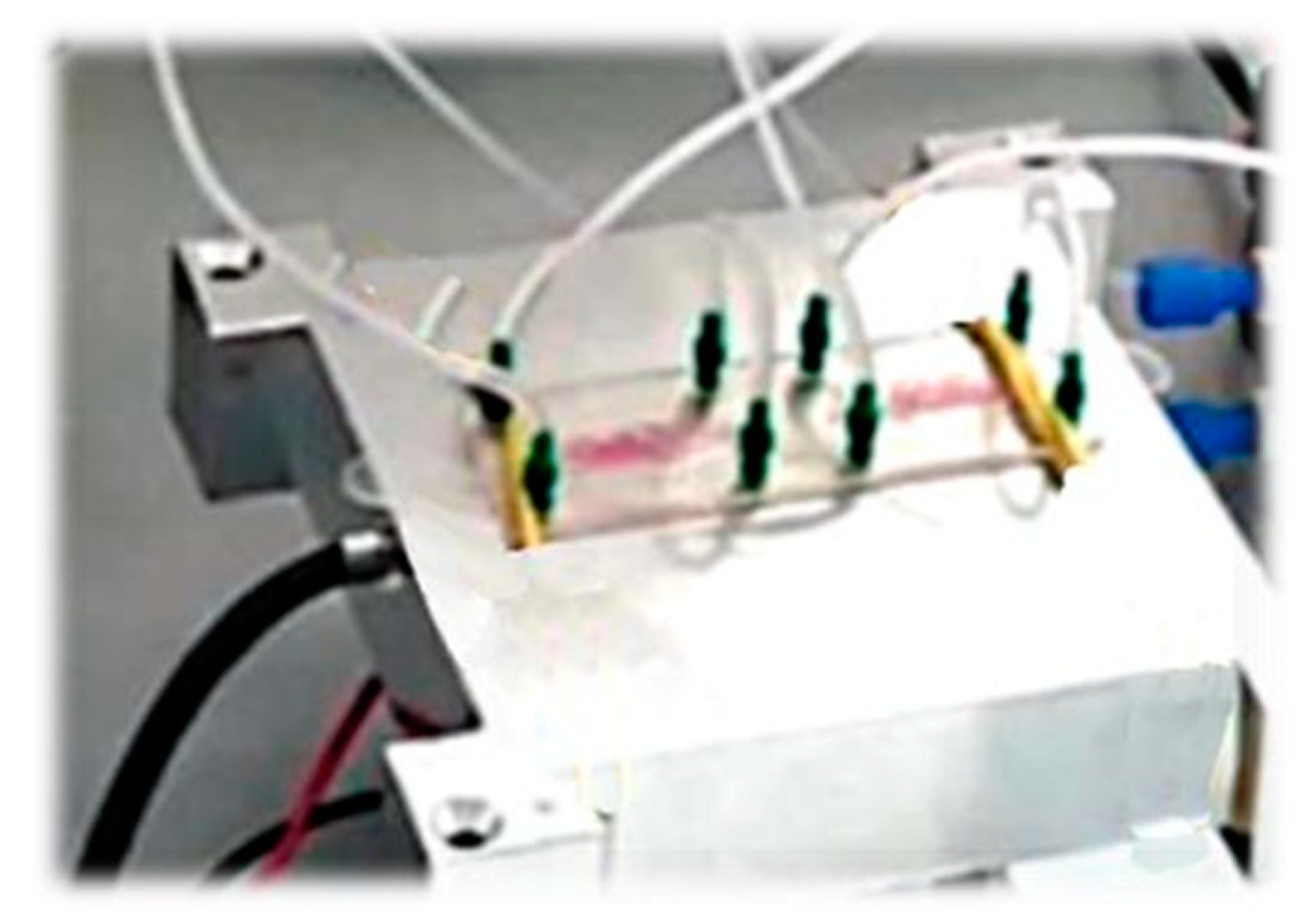
In this work, the proposed Gut-on-Chip system is a modified and customized commercial Topas polymer-made platform, with two (lumen vs. blood) aligned fluidic chambers separated by a transparent and porous PET (Polyethylene terephthalate) membrane for cell culture (Figure 2). The Membrane size is 11 × 8.5 mm2. Membrane thickness is 23 µm, the pore dimension is about 200 nm, with a pores density of 5 × 10⁸ pores/cm2.
This microfluidic platform is equipped with transparent ITO (Indium tin Oxide) electrodes for TEER (Transepithelial Electrical Resistance) monitoring during the cell culture experiments, because TEER values are indicators of cellular barriers integrity and of tight junction dynamics in cultured cells monolayers. The measure of pH, Zn and Cu passage through the apical and basolateral chamber, will be realized by implementing miniaturized Ion Selective Electrodes (ISE). The simultaneous embedded sensors measurements and a software-controlled mechanical deflection also help in reproducing local mechanical stretches/bends/etc. To preliminarily investigate cell-to-chip interactions, we assayed cell adhesion on our PET porous membrane, and consequent proliferation, by adopting the human-derived, epithelial-like HeLa and Caco-2 cell lines. Cells were seeded on the membrane exposed to medium with no cover lid; then, the chip chamber was sealed on top and bottom sides with Corning slides equipped with electrodes, and the system was incubated for 24 h (HeLa) or 72 h (Caco-2) at 37 °C before assessing cell viability by a standard MTT assay to detect metabolic activity of cells. (Figure 3).
3. Results
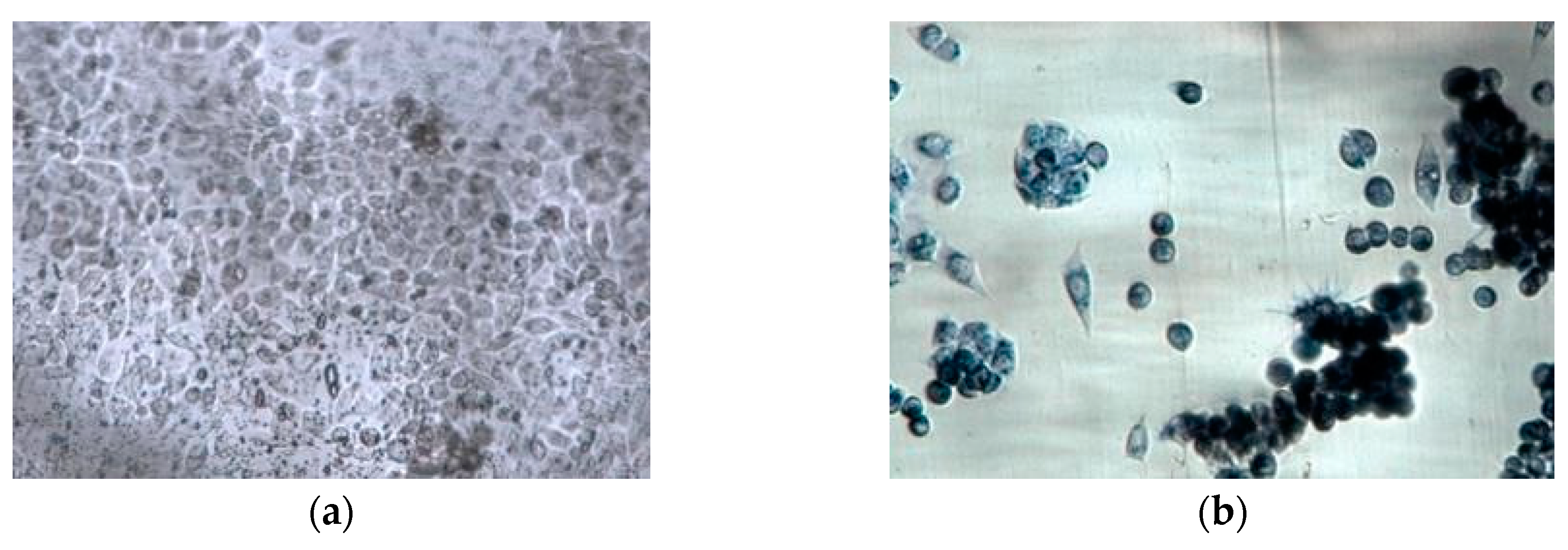
A complete, microfluidic-based and multiparameter Epithelial Gut on Chip model has been developed that could enable human intestinal epithelium growth. We basically evaluated the cell interaction, using the HeLa cell line seeded on the PET transparent porous membrane (a non-biological matrix) which expose cells to a microfluidic flow. The cell behavior on an early prototype has been observed to reduce critical aspects deriving from biological components. The sterility maintenance, the monitoring of cell viability and growth during the epithelial barriers formation and the presence of air bubbles on cell culture are challenging aspects that were evaluated using HeLa cells as preliminary study before the evaluation of a more specific cell lines such as the intestinal Caco-2 cells. Indeed, the multifunctional microsystem for monitoring epithelia parameters has been set up with proper material and microfluidics tubes and it has been primarily investigated in its ability to support cell adhesion and growth, for several days, before electrical and mechanical test on the epithelium. In our experimental setup, viable HeLa (and then Caco-2) cells have been detected by metabolization of the MTT compound producing the dark blue-to-black staining (i.e., formazan crystals in viable cells) (Figure 3). The MTT assay is a preliminary evaluation to demonstrate a considerable efficiency of adhesion, viability and migration of human-derived intestinal cells on microfluidic devices. The optimization of in vitro conditions provides the implementation of some enabling technologies to monitor in real time important parameters that are crucial in several physiological pathways. An example is characterized by the measure of pH, or the translocation of Zn and Cu ions across the apical and basolateral chambers, that will be implemented by using miniaturized Ion Selective Electrodes (ISE). The Impedance measurements will be regularly acquired, to detect and evaluate the TEER behavior in situ. Additionally, the actuation of mechanotransduction could permit the understanding of important mechanisms of GI tissue organization; also, concomitant pharmacological assays based on nutrient/drugs intestinal interactions are under analysis. Our cell-to-device combination may represent a useful study of the epithelial tightness and the mechanotransduction in the regulatory pathways at cellular and epithelial level in the GI tissue districts, in GI drug toxicity and drug screening.
Author Contributions
L.G., A.B., L.F. and F.C. performed the experiments; A.B., L.F., P.S. and T.V. analyzed the data; S.C., P.S. and T.V. contributed reagents/materials/analysis tools; L.G. wrote the paper.
References
- Bein, A.; Shin, W.; Jalili-Firoozinezhad, S.; Park, M.H.; Sontheimer-Phelps, A.; Tovaglieri, A.; Chalkiadaki, A.; Kim, H.J.; Ingber, D.E. Microfluidic Organ-on-a-Chip Models of Human Intestine. Cell. Mol. Gastroenterol. Hepatol. 2018, 5, 659–668. [Google Scholar] [CrossRef] [PubMed]
- Carabotti, M.; Scirocco, A.; Maselli, M.A.; Severi, C. The gut-brain axis: Interactions between enteric microbiota, central and enteric nervous systems. Ann. Gastroenterol. 2014, 28, 203–209. [Google Scholar]
- Andersen, M.E.; Betts, K.; Dragan, Y.; Fitzpatrick, S.; Goodman, J.L.; Hartung, T.; Himmelfarb, J.; Ingber, D.E.; Jacobs, A.; Kavlock, R. Developing microphysiological systems for use as regulatory tools–challenges and opportunities. ALTEX 2014, 31, 364–367. [Google Scholar] [CrossRef] [PubMed]
- Lee, J.; Choi, J.H.; Kim, H.J. Human gut-on-a-chip technology: Will this revolutionize our understanding of IBD and future treatments? Expert Rev. Gastroenterol. Hepatol. 2016, 10, 883–885. [Google Scholar] [CrossRef] [PubMed]
- Schumacher, S.; Nestler, J.; Otto, T.; Wegener, M.; Ehrentreich-Förster, E.; Michel, D.; Wunderlich, K.; Palzer, S.; Sohn, K.; Weber, A. Highly-integrated lab-on-chip system for point-of-care multiparameter analysis. Lab Chip 2012, 12, 464–473. [Google Scholar] [CrossRef] [PubMed]
- Qian, X.; Nguyen, H.N.; Song, M.M.; Hadiono, C.; Ogden, S.C.; Hammack, C.; Yao, B.; Hamersky, G.R.; Jacob, F.; Zhong, C.; et al. Brain-region-specific organoids using mini-bioreactors for modeling ZIKV exposure. Cell 2016, 165, 1238–1254. [Google Scholar] [CrossRef] [PubMed]
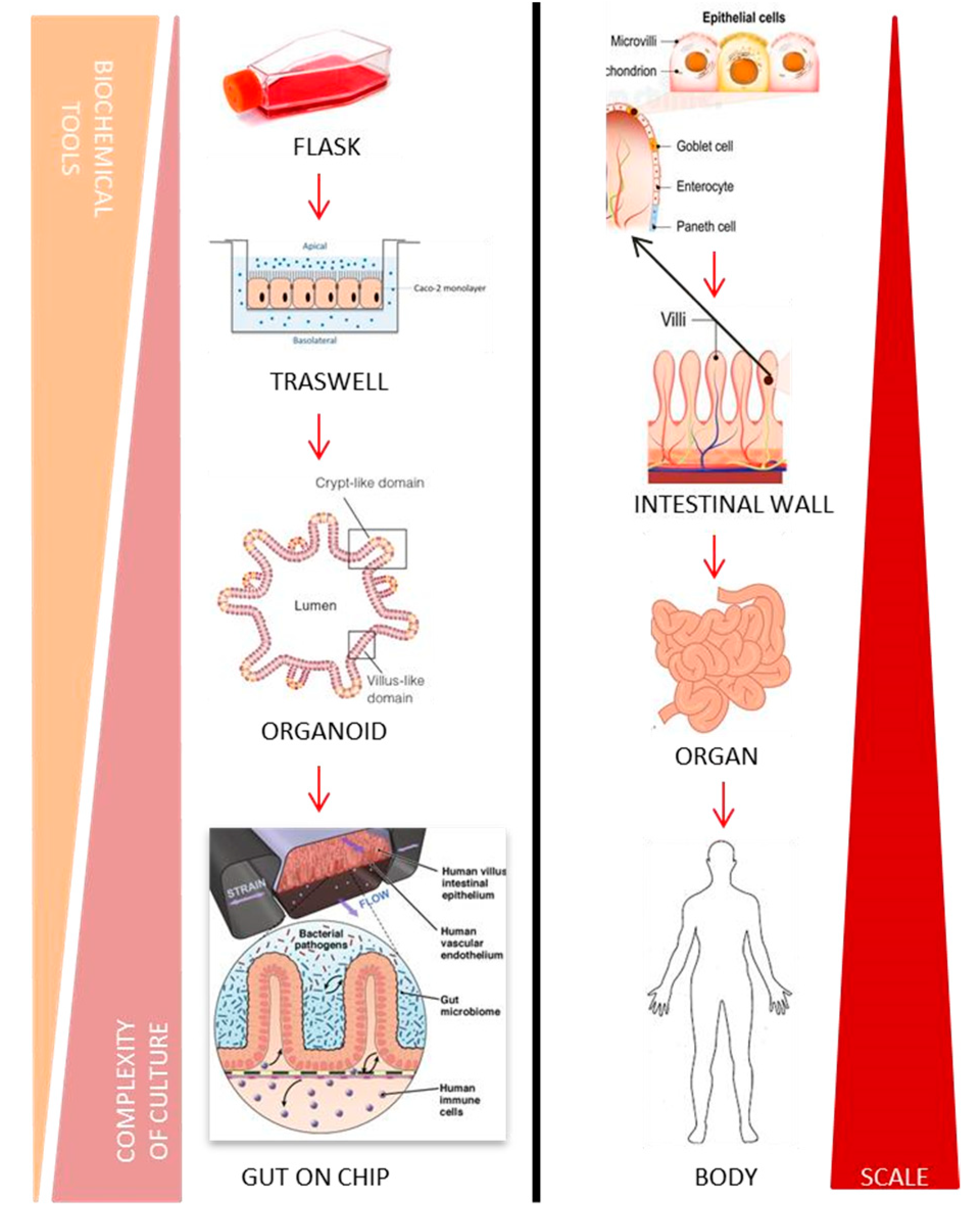
Figure 1.
On the left many models are presented of witch that have been developed across this organismal hierarchy, to address specific challenges in medicine. On the right there is a life system hierarchy: from the subcellular level to the whole body. Inspired from Cell Press Copyright© 2016 Elsevier Inc. Published by Elsevier Inc. (Amsterdam, The Netherlands) [6].

Figure 2.
Modified commercial multifunctional platform for Gut-On-Chip Caco-2 cultures with embedded ITO electrodes.
Figure 2.
Modified commercial multifunctional platform for Gut-On-Chip Caco-2 cultures with embedded ITO electrodes.

Figure 3.
In-Device Seeding through Microfluidic Path of 1 × 10⁵ cells/mL (representative images of HeLa cells): (a) Detection 24 h after seeding of adhered cells in proliferating culture; (b) In-Device MTT Delivery by controlled flux (7 µL/min) for 2.5 h. The MTT salts are metabolized by functional enzymes of viable cells only, which produce the precipitation of dark-blue formazan crystals.

Publisher’s Note: MDPI stays neutral with regard to jurisdictional claims in published maps and institutional affiliations. |
© 2018 by the authors. Licensee MDPI, Basel, Switzerland. This article is an open access article distributed under the terms and conditions of the Creative Commons Attribution (CC BY) license (https://creativecommons.org/licenses/by/4.0/).
Share and Cite
MDPI and ACS Style
Giampetruzzi, L.; Barca, A.; Casino, F.; Capone, S.; Verri, T.; Siciliano, P.; Francioso, L. Multi-Sensors Integration in a Human Gut-On-Chip Platform. Proceedings 2018, 2, 1022. https://doi.org/10.3390/proceedings2131022
AMA Style
Giampetruzzi L, Barca A, Casino F, Capone S, Verri T, Siciliano P, Francioso L. Multi-Sensors Integration in a Human Gut-On-Chip Platform. Proceedings. 2018; 2(13):1022. https://doi.org/10.3390/proceedings2131022
Chicago/Turabian StyleGiampetruzzi, Lucia, Amilcare Barca, Flavio Casino, Simonetta Capone, Tiziano Verri, Pietro Siciliano, and Luca Francioso. 2018. "Multi-Sensors Integration in a Human Gut-On-Chip Platform" Proceedings 2, no. 13: 1022. https://doi.org/10.3390/proceedings2131022