Exploring Enzymatic Hydrolysis of Urine Samples for Investigation of Drugs Associated with Drug-Facilitated Sexual Assault

 , , , and
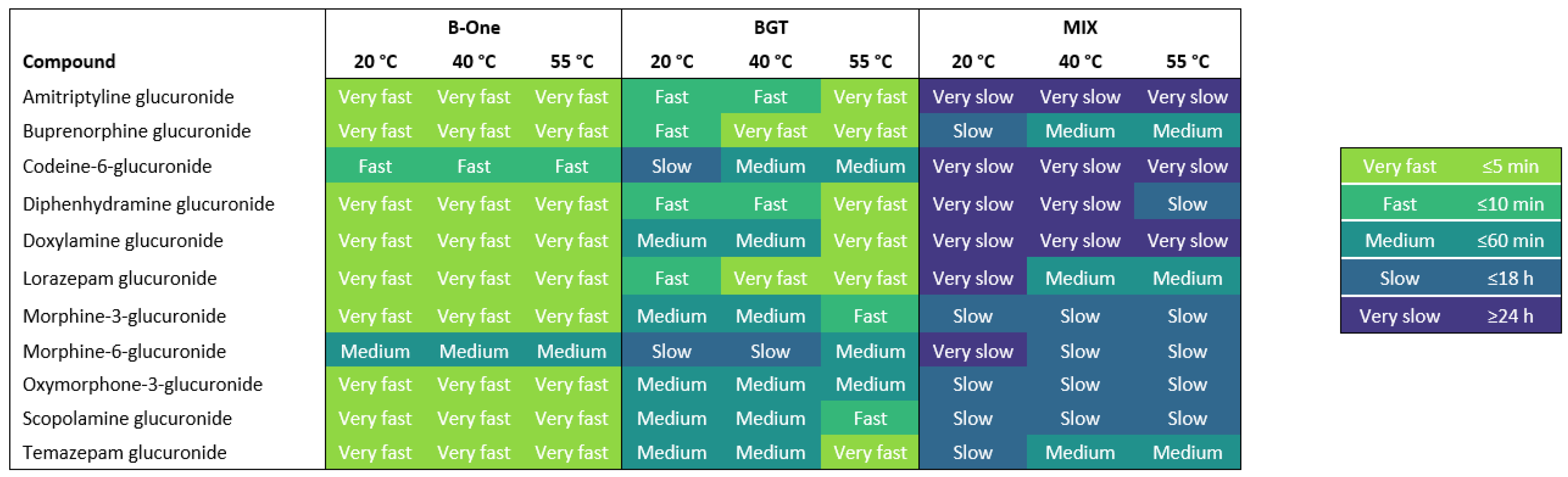
Abstract
:1. Introduction
2. Results and Discussion
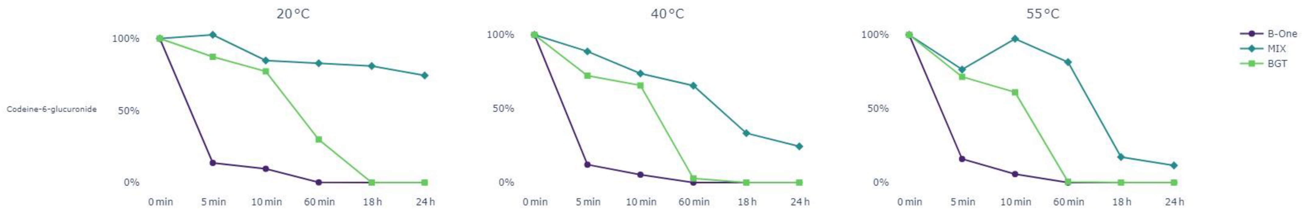
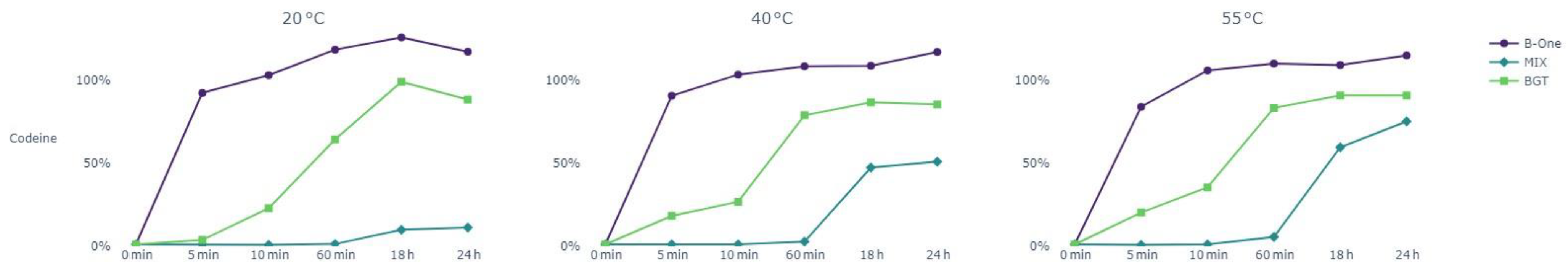
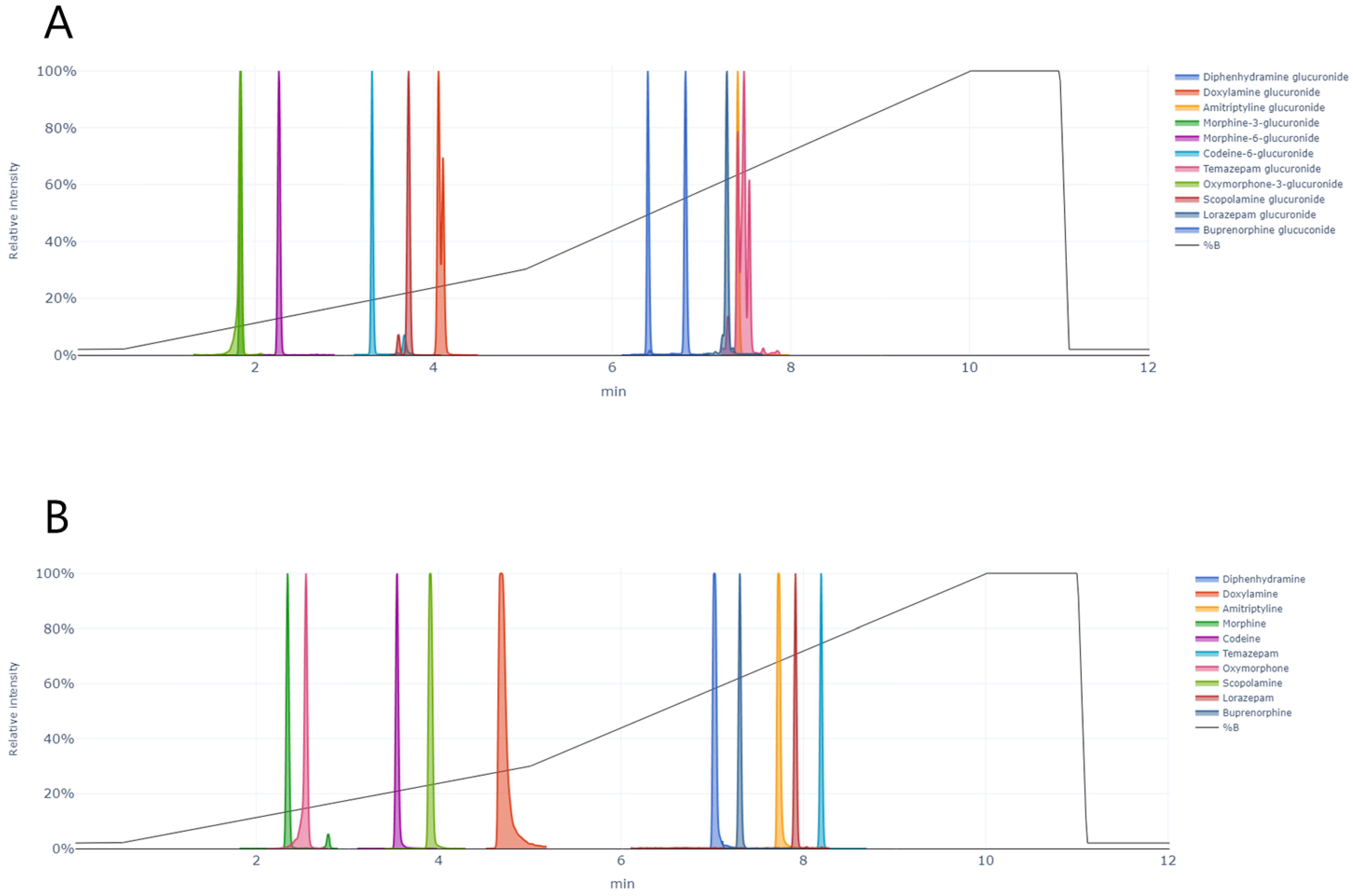
2.1. Hydrolysis Efficiency
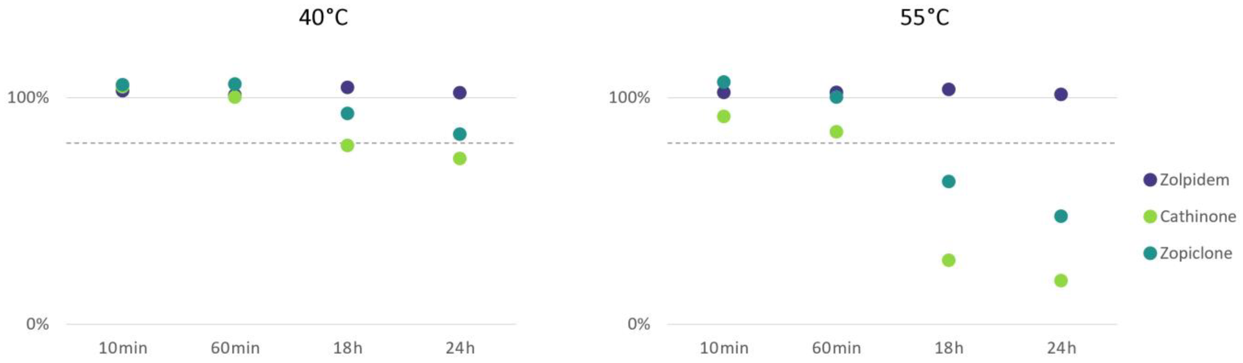
2.2. Stability of Drugs during Incubation
2.3. Considerations for Choice of Enzyme
3. Materials and Methods
3.1. Chemicals and Reagents
3.2. Enzymes
3.3. Preparation of Standard Solutions
3.4. Fortified Urine Pools and Calibrators
3.5. Hydrolysis of Urine Samples
3.6. Testing Stability during Incubation
3.7. Quantification by UHPLC–MS/MS
3.8. Estimation of Hydrolysis Efficiency
3.9. Ethical Considerations
4. Conclusions
Supplementary Materials
Author Contributions
Funding
Institutional Review Board Statement
Informed Consent Statement
Data Availability Statement
Conflicts of Interest
References
- Anderson, L.J.; Flynn, A.; Pilgrim, J.L. A global epidemiological perspective on the toxicology of drug-facilitated sexual assault: A systematic review. J. Forensic Leg. Med. 2017, 47, 46–54. [Google Scholar] [CrossRef] [PubMed]
- García, M.G.; Pérez-Cárceles, M.D.; Osuna, E.; Legaz, I. Drug-facilitated sexual assault and other crimes: A systematic review by countries. J. Forensic Leg. Med. 2021, 79, 102151. [Google Scholar] [CrossRef] [PubMed]
- Bertol, E.; Di Milia, M.G.; Fioravanti, A.; Mari, F.; Palumbo, D.; Pascali, J.P.; Vaiano, F. Proactive drugs in DFSA cases: Toxicological findings in an eight-years study. Forensic Sci. Int. 2018, 291, 207–215. [Google Scholar] [CrossRef] [PubMed]
- Tiemensma, M.; Davies, B. Investigating drug-facilitated sexual assault at a dedicated forensic centre in Cape Town, South Africa. Forensic Sci. Int. 2018, 288, 115–122. [Google Scholar] [CrossRef] [PubMed]
- Caballero, C.G.; Jorge, Ó.Q.; Landeira, A.C. Alleged drug-facilitated sexual assault in a Spanish population sample. Forensic Chem. 2017, 4, 61–66. [Google Scholar] [CrossRef]
- Hall, J.; Goodall, E.A.; Moore, T. Alleged drug facilitated sexual assault (DFSA) in Northern Ireland from 1999 to 2005. A study of blood alcohol levels. J. Forensic Leg. Med. 2008, 15, 497–504. [Google Scholar] [CrossRef] [PubMed]
- Bosman, I.J.; Verschraagen, M.; Lusthof, K.J. Toxicological Findings in Cases of Sexual Assault in the Netherlands. J. Forensic Sci. 2011, 56, 1562–1568. [Google Scholar] [CrossRef]
- Birkler, R.I.D.; Telving, R.; Ingemann-Hansen, O.; Charles, A.V.; Johannsen, M.; Andreasen, M.F. Screening analysis for medicinal drugs and drugs of abuse in whole blood using ultra-performance liquid chromatography time-of-flight mass spectrometry (UPLC-TOF-MS)-Toxicological findings in cases of alleged sexual assault. Forensic Sci. Int. 2012, 222, 154–161. [Google Scholar] [CrossRef]
- Skov, K.; Johansen, S.S.; Linnet, K.; Nielsen, M.K.K. A review on the forensic toxicology of global drug-facilitated sexual assaults. Eur. Rev. Med. Pharmacol. Sci. 2022, 26, 183–197. [Google Scholar] [CrossRef]
- Jones, A.W.; Holmgren, A.; Ahlner, J. Toxicological analysis of blood and urine samples from female victims of alleged sexual assault. Clin. Toxicol. 2012, 50, 555–561. [Google Scholar] [CrossRef]
- Poulsen, H.; McCarthy, M.-J.; Baker, J.; Verma, A.; Moir, H.J.; Brodie, T.; Thatti, B.; Trotter, G.; Rooney, B. Toxicological Assessment of the Role of Alcohol and Drugs in Drug-Facilitated Sexual Assault Cases in New Zealand. J. Anal. Toxicol. 2021, 45, 44–52. [Google Scholar] [CrossRef] [PubMed]
- Madea, B.; Mußhoff, F. Knock-Out Drugs. Dtsch. Aerzteblatt Online 2009, 106, 341–347. [Google Scholar] [CrossRef] [PubMed]
- Kaeferstein, H. Forensic relevance of glucuronidation in phase-II-metabolism of alcohols and drugs. Leg. Med. 2009, 11, S22–S26. [Google Scholar] [CrossRef] [PubMed]
- Ding, Y.; Peng, M.; Zhang, T.; Tao, J.S.; Cai, Z.Z.; Zhang, Y. Quantification of conjugated metabolites of drugs in biological matrices after the hydrolysis with β-glucuronidase and sufatase: A review of bio-analytical methods. Biomed. Chromatogr. 2013, 27, 1280–1295. [Google Scholar] [CrossRef] [PubMed]
- Kapur, B.M.; Aleksa, K. What the lab can and cannot do: Clinical interpretation of drug testing results. Crit. Rev. Clin. Lab. Sci. 2020, 57, 548–585. [Google Scholar] [CrossRef]
- Johnson-Davis, K.L. Opiate & Benzodiazepine Confirmations: To Hydrolyze or Not to Hydrolyze is the Question. J. Appl. Lab. Med. 2018, 2, 564–572. [Google Scholar] [CrossRef]
- Morris, A.A.; Chester, S.A.; Strickland, E.C.; McIntire, G.L. Rapid enzymatic hydrolysis using a novel recombinant Β-glucuronidase in benzodiazepine urinalysis. J. Anal. Toxicol. 2014, 38, 610–614. [Google Scholar] [CrossRef]
- Dwivedi, P.; Zhou, X.; Powell, T.G.; Calafat, A.M.; Ye, X. Impact of enzymatic hydrolysis on the quantification of total urinary concentrations of chemical biomarkers. Chemosphere 2018, 199, 256–262. [Google Scholar] [CrossRef]
- Lee, L.A.; McGee, A.C.; Sitasuwan, P.; Tomashek, J.J.; Riley, C.; Muñoz-Muñoz, A.C.; Andrade, L. Factors Compromising Glucuronidase Performance in Urine Drug Testing Potentially Resulting in False Negatives. J. Anal. Toxicol. 2021, 46, 689–696. [Google Scholar] [CrossRef]
- Zenser, T.V.; Lakshmi, V.M.; Davis, B.B. Human and Escherichia coli β-glucuronidase hydrolysis of glucuronide conjugates of benzidine and 4-aminobiphenyl, and their hydroxy metabolites. Drug Metab. Dispos. 1999, 27, 1064–1067. [Google Scholar]
- Babu, S.R.; Lakshmi, V.M.; Huang, G.P.W.; Zenser, T.V.; Davis, B.B. Glucuronide conjugates of 4-aminobiphenyl and its N-hydroxy metabolites pH stability and synthesis by human and dog liver. Biochem. Pharmacol. 1996, 51, 1679–1685. [Google Scholar] [CrossRef] [PubMed]
- Hackett, L.P.; Dusci, L.J.; Ilett, K.F.; Chiswell, G.M. Optimizing the hydrolysis of codeine and morphine glucuronides in urine. Ther. Drug Monit. 2002, 24, 652–657. [Google Scholar] [CrossRef] [PubMed]
- Glicksberg, L.; Kerrigan, S. Stability of synthetic cathinones in Urine. J. Anal. Toxicol. 2018, 42, 77–87. [Google Scholar] [CrossRef] [PubMed]
- Adamowicz, P.; Malczyk, A. Stability of synthetic cathinones in blood and urine. Forensic Sci. Int. 2019, 295, 36–45. [Google Scholar] [CrossRef] [PubMed]
- Glicksberg, L.; Rana, S.; Kerrigan, S. Cathinone stability in authentic urine specimens. Forensic Sci. Int. 2018, 286, 54–60. [Google Scholar] [CrossRef] [PubMed]
- Nilsson, G.H.; Kugelberg, F.C.; Kronstrand, R.; Ahlner, J. Stability tests of zopiclone in whole blood. Forensic Sci. Int. 2010, 200, 130–135. [Google Scholar] [CrossRef] [PubMed]
- Nilsson, G.H.; Kugelberg, F.C.; Ahlner, J.; Kronstrand, R. Influence of pre-analytical conditions on the interpretation of zopiclone concentrations in whole blood. Forensic Sci. Int. 2011, 207, 35–39. [Google Scholar] [CrossRef]
- Mata, D.C. Stability of 26 sedative hypnotics in six toxicological matrices at different storage conditions. J. Anal. Toxicol. 2016, 40, 663–668. [Google Scholar] [CrossRef]
- Nilsson, G.H.; Kugelberg, F.C.; Ahlner, J.; Kronstrand, R. Quantitative analysis of zopiclone, N-desmethylzopiclone, zopiclone N-oxide and 2-amino-5-chloropyridine in urine using LC-MS-MS. J. Anal. Toxicol. 2014, 38, 327–334. [Google Scholar] [CrossRef]
- Jantos, R.; Vermeeren, A.; Sabljic, D.; Ramaekers, J.G.; Skopp, G. Degradation of zopiclone during storage of spiked and authentic whole blood and matching dried blood spots. Int. J. Leg. Med. 2013, 127, 69–76. [Google Scholar] [CrossRef]
- Briscoe, C.J.; Hage, D.S. Factors affecting the stability of drugs and drug metabolites in biological matrices. Bioanalysis 2009, 1, 205–220. [Google Scholar] [CrossRef] [PubMed]
- Baselt, R.C. Disposition of Toxic Drugs and Chemicals in Man, 12th ed.; Biomedical publications: Seal Beach, CA, USA, 2020; ISBN 978-0-578-57749-4. [Google Scholar]
- Lantz, R.J.; Gillespie, T.A.; Rash, T.J.; Kuo, F.; Skinner, M.; Kuan, H.Y.; Knadler, M.P. Metabolism, excretion, and pharmacokinetics of duloxetine in healthy human subjects. Drug Metab. Dispos. 2003, 31, 1142–1150. [Google Scholar] [CrossRef] [PubMed]


Disclaimer/Publisher’s Note: The statements, opinions and data contained in all publications are solely those of the individual author(s) and contributor(s) and not of MDPI and/or the editor(s). MDPI and/or the editor(s) disclaim responsibility for any injury to people or property resulting from any ideas, methods, instructions or products referred to in the content. |
© 2023 by the authors. Licensee MDPI, Basel, Switzerland. This article is an open access article distributed under the terms and conditions of the Creative Commons Attribution (CC BY) license (https://creativecommons.org/licenses/by/4.0/).
Share and Cite
Skov, K.; Johansen, S.S.; Linnet, K.; Rasmussen, B.S.; Nielsen, M.K.K. Exploring Enzymatic Hydrolysis of Urine Samples for Investigation of Drugs Associated with Drug-Facilitated Sexual Assault. Pharmaceuticals 2024, 17, 13. https://doi.org/10.3390/ph17010013
Skov K, Johansen SS, Linnet K, Rasmussen BS, Nielsen MKK. Exploring Enzymatic Hydrolysis of Urine Samples for Investigation of Drugs Associated with Drug-Facilitated Sexual Assault. Pharmaceuticals. 2024; 17(1):13. https://doi.org/10.3390/ph17010013
Chicago/Turabian StyleSkov, Kathrine, Sys Stybe Johansen, Kristian Linnet, Brian Schou Rasmussen, and Marie Katrine Klose Nielsen. 2024. "Exploring Enzymatic Hydrolysis of Urine Samples for Investigation of Drugs Associated with Drug-Facilitated Sexual Assault" Pharmaceuticals 17, no. 1: 13. https://doi.org/10.3390/ph17010013