Review

The Potential Properties of Natural Compounds in Cardiac Stem Cell Activation: Their Role in Myocardial Regeneration

1
Institute of Research for Food Safety & Health IRC-FSH, University Magna Graecia, 88100 Catanzaro, Italy
2
Nutramed S.c.a.r.l., Complesso Ninì Barbieri, Roccelletta di Borgia, 88100 Catanzaro, Italy
*
Authors to whom correspondence should be addressed.
These authors contributed equally to this work.
Nutrients 2021, 13(1), 275; https://doi.org/10.3390/nu13010275
Submission received: 19 December 2020 / Revised: 7 January 2021 / Accepted: 12 January 2021 / Published: 19 January 2021
(This article belongs to the Special Issue Nutraceutical Supplements in Heart Failure)

Abstract

:
Cardiovascular diseases (CVDs), which include congenital heart disease, rhythm disorders, subclinical atherosclerosis, coronary heart disease, and many other cardiac disorders, cause about 30% of deaths globally; representing one of the main health problems worldwide. Among CVDs, ischemic heart diseases (IHDs) are one of the major causes of morbidity and mortality in the world. The onset of IHDs is essentially due to an unbalance between the metabolic demands of the myocardium and its supply of oxygen and nutrients, coupled with a low regenerative capacity of the heart, which leads to great cardiomyocyte (CM) loss; promoting heart failure (HF) and myocardial infarction (MI). To date, the first strategy recommended to avoid IHDs is prevention in order to reduce the underlying risk factors. In the management of IHDs, traditional therapeutic options are widely used to improve symptoms, attenuate adverse cardiac remodeling, and reduce early mortality rate. However, there are no available treatments that aim to improve cardiac performance by replacing the irreversible damaged cardiomyocytes (CMs). Currently, heart transplantation is the only treatment being carried out for irreversibly damaged CMs. Hence, the discovery of new therapeutic options seems to be necessary. Interestingly, recent experimental evidence suggests that regenerative stem cell medicine could be a useful therapeutic approach to counteract cardiac damage and promote tissue regeneration. To this end, researchers are tasked with answering one main question: how can myocardial regeneration be stimulated? In this regard, natural compounds from plant extracts seem to play a particularly promising role. The present review will summarize the recent advances in our knowledge of stem cell therapy in the management of CVDs; focusing on the main properties and potential mechanisms of natural compounds in stimulating and activating stem cells for myocardial regeneration.

Graphical Abstract

1. Introduction

Cardiovascular diseases (CVDs) are the leading cause of death in the developed world; accounting for approximately 30% of deaths globally, according to the World Health Organization, [1], thus, representing one of the main health problems worldwide [2,3]. The increase in life expectancy, together with several risk factors such as smoking, poor nutritional habits, sedentary lifestyle, obesity, high blood pressure, and diabetes is closely linked to the onset of CVDs [2,3,4]. To date, the number of people with CVDs in most European countries and various other countries around the world, moving a volume of billions of dollars in healthcare [5,6,7,8], is expected to further increase according to the American Heart Association reports [7].
Among CVDs, ischemic heart disease (IHD), such as myocardial infarction (MI), is one of the major causes of morbidity and mortality throughout Europe, America and Asia [5,9,10]. When myocardial ischemia occurs, the main consequence is represented by irreversible cell death, which is accompanied by the loss of viable myocardial tissue, pathological remodeling and scar tissue formation; dramatically predisposing to heart failure (HF) [11]. In this condition, “time” represents the main discriminant factor on which current pharmacological and surgical therapies focus, in order to reduce serious adverse events and ultimately the mortality rate.
Although traditional therapeutic options are highly efficient in improving symptoms, decelerating adverse cardiac remodeling and reducing early mortality rate, there is no available treatment that aims to improve cardiac performance by replacing the irreversible damaged CMs [12].
It is now clear that despite the adult heart being composed of mainly terminally differentiated cells, unable to re-enter the cell cycle, it cannot be considered a terminally differentiated organ because it also contains stem cells that support its regeneration [13]. However, the loss of CMs due to ischemic heart events is currently counteracted only by heart transplantation [11].
Over the last two decades, new alternative therapies have been studied to address the underlying problem of CM loss and improve the clinical condition of patients. Among these, stem cell therapy has proven to be a promising therapeutic approach for the treatment of IHDs [14]. Stem cells possess specific features of self-renewing, clonogenicity, and differentiation into specialized cell types under appropriate conditions [15]. Stem cells are commonly divided into various major subgroups such as embryonic stem cells (ESCs), induced pluripotent stem cells (iPSCs), and adult or somatic stem cells. The latter include bone marrow stem cells (BM-SCs), (which in turn consist of hematopoietic stem cells -HSCs, bone marrow derived mesenchymal stem cells -BM-MSCs, multipotent adult progenitor cells -MAPCs and endothelial progenitor cells -EPCs), mesenchymal stem cells (MSCs), cardiac stem cells (CSCs), and skeletal myoblasts [15]. During the last twenty years, several pre-clinical studies have revealed the great potential and also the serious limitations in the use of different type of stem cells, such as ESCs [16,17,18], iPSCs [19,20,21], or adult/somatic stem cells [22,23] in the treatment of CVDs. Moreover, many advances have been made to enable the application of stem cell therapy in patients with CVDs through the implementation of different clinical trials [22,24,25,26,27,28].
In short, today knowledge on the use of stem cells in regenerative therapy is manifold. Several animal studies and early clinical trials clearly show the feasibility of the stem cell-based therapies and the beneficial effects of numerous stem cell types in the regeneration of injured myocardium. However, the beneficial effects of stem cell-based treatment on myocardial performance are still quite limited by the low rates of engraftment and cell survival. These limitations are due to the microenvironment of the damaged site of cardiac tissue which is often associated with inflammatory response, oxidative stress, and extracellular matrix (ECM) degradation [11]. Therefore, it is essential to solve the main issue of understanding how to boost the efficacy of stem cell repair to make cardiac regenerative therapy an effective therapeutic tool.
To date, many different methods to improve the efficiency of stem cell therapy and to better understand their way of action have been investigated. Among these, there are several cell-delivery methods which include intra-myocardial, intra-coronary or intra-venous injection of stem cells. However, using these methods, the survival rate of injected cells was below 1% [29,30,31]. Both cell-sheet technology and the injectable hydrogels offer more advantageous cell-delivery systems by reducing the mechanical damage to the host myocardium; thus improving the survival rate of the transplanted cells [32,33]. In addition, continuous advances in genetically engineered stem cell techniques, such us the overexpression of cytoprotective or anti-apoptotic genes, are enhancing the outcomes of the stem cell therapy [34,35]. Before transplantation, cell preconditioning strategies such as culturing the cells with agonists, exposing cells to hypoxia, heat shock, or to oxidative stress have also been used [36,37,38]. The mechanisms of cardiac repair by transplanted stem cells represent another important issue which is not yet fully understood [11]. The scientific discussion about the possible mechanisms of action underlying the ability of stem cells to regenerate damaged heart tissue is highly debated. Suggested mechanisms include the direct cardiomyogenic differentiation of the transplanted stem cells and progenitor cells, after isolation from autologous or allogeneic source tissues, and the paracrine effect of stem cells, which indirectly stimulate the regenerative process through secretion of soluble cytokines and growth factors [39]. Interestingly, the indirect paracrine effect of transplanted stem cells also seems to stimulate the activation and differentiation of resident CSCs [40].
Nowadays, the paracrine effect on stem cells has been identified as a key factor of stem cell-therapy efficacy. Indeed, it has been shown that paracrine signaling enables transplanted stem cells to protect the surrounding cardiovascular tissue due to the release of different growth factors, such as transforming growth factor (TGF)-β, vascular endothelial growth factor (VEGF), fibroblast growth factor (β-FGF), hepatocyte growth factor (HGF), insulin-like growth factor 1 (IGF-1), stromal cell-derived factor (SDF)-1, epidermal growth factor (EGF), thymosin b4, and a variety of cytokines [41].
It is now well-established that the secretion of biologically active molecules from transplanted stem cells leads to the activation of angiogenic, antiapoptotic and antifibrotic paracrine patterning within the damaged heart; these actions, also referred to as “trophic effects”, play a major role in tissue repair [42]. Moreover, the administration of stem cell-conditioned medium has shown beneficial effects, comparable to those of the transplanted cells. These beneficial effects include promoting regenerative processes, protecting the existing CMs from damage and death, reducing matrix remodeling and enhancing the recruitment of circulating progenitor cells, and activation of cell proliferation and CM differentiation [43,44]. A major role in the paracrine effect of stem cells seems to be played by a type of extracellular vesicles called exosomes. Stem cell-secreted exosomes, function as cargo of nucleic acids, proteins, lipids, and several cardio-protective and pro-angiogenic mRNAs and non-coding RNAs; suggesting their critical role in exchange of biological information and clinical usefulness as biomarkers of CVDs and as therapeutic tools [45,46]. Indeed, it was recently shown that direct injection of exosomes derived from stem cells into a damaged heart decreases the systemic inflammation, oxidative stress, and significantly improves the regenerative effects produced by stem cells [47].
Interestingly, pharmacological preconditioning represents a novel and efficient technique for stimulating the secretory activity of stem cells. For example, in vitro and in vivo studies reported the effectiveness of oxytocin in stimulating the release of cytokines [48] and the anti-ischemic effect of trimetazidine and sildenafil, when applied to stem cells prior to transplantation [49,50]. Moreover, drug-mediated activation or inhibition of pathways can modify stem cell physiology; enhancing cell survival and engraftment thereby improving cardiac regeneration [51].
In this regard, the use of natural compounds from plant extracts seems to play a particularly intriguing role. Several studies have provided a number of molecules able to selectively control stem cell growth and differentiation. Natural compounds such as: ascorbic acid, indirubin, resveratrol, icariin, bergamot polyphenols, are emerging as useful tools capable of orchestrating the homing, engraftment, and survival of cells; improving self-renewal and modulating stem cell cardiovascular lineage specification, possibly through regulation of paracrine signaling (Figure 1).
The present review briefly summarizes the knowledge on stem cell therapy in the management of CVDs and focuses on the potential properties of natural compounds in stimulating and activating stem cells for myocardial regeneration.

2. Cardiac Regeneration and Stem Cells Therapy

The presence of damaged and necrotic heart tissue, with stromal connective tissue formation and deposition of fibrotic scar are the main features of IHDs and particularly of MI. These conditions, as well as in non-IHDs, are characterized by a loss of CMs, expansion of the ECM and increase in hypertrophic CMs as an attempt to compensate for the reduced cardiac function. Persistence of pathological stress, cardiac remodeling and development of HF with poor prognosis are treated with limited therapeutic intervention such as ventricular assist devices and cardiac transplantation [52,53,54]. In addition, targeted pharmacological treatments are used to counteract HF development [52,53,54]. In this regard, research on new strategies to prevent cardiac damage and dysfunction has focused on cardiac regeneration by promoting cell-based therapy, mainly based on transplantation, into the damaged heart, of several stem cell types or in vitro stem cell derived- CMs, endorsing the regeneration of resident CSCs [54]. Originally, the heart was considered a terminally differentiated organ, unable to replicate and to be responsive to myocardial detriment [55,56,57,58]. After the discovery of the regenerating adult mammalian heart’s capacity the potential replicative activity of CMs was investigated [55,56,57,58]. This ability was observed in normal and pathological condition, by the presence of cycling ventricular myocytes that rise in case of damage, in contrast with the idea that after birth CMs lost the possibility to divide and regenerate, giving rise to the question about the origin of these cycling myocytes [58,59]. The studies about putative CSCs, showed that they shared certain features with stem cells such as the markers on the cell surface like the stem cell factor receptor (c-kit), the Stem cells antigen-1 (Sca1), the multidrug resistance gene product-1 (MDR-1), the capacity to self-renewing, generate clones and differentiate into myocytes, smooth muscle cells and endothelial cells [58,60]. However, CMs turnover is only 0.5% to 2% per year and was found to be insufficient to supply the necessity of a damaged heart, leading to terminal HF without a proper intervention [61]. Therefore, new alternative therapies were investigated to address the underlying problem of CM loss and improve patients’ clinical condition. Among all, stem cell therapy has proven to be a promising therapeutic approach for the treatment of IHDs [14]. The intriguing capability of stem cells to support cardiac repair has been well investigated in many in-depth studies [62,63,64].
Several preclinical studies aimed to investigate the characteristics of different stem cell types in cardiac damage, including ESCs, iPSCs and adult or somatic stem cells, showing the possibilities of improving cardiac dysfunction and promoting tissue regeneration [65,66,67], translating this knowledge in early clinical trials [68,69]. Despite this evidence and the observation that after cardiac injury it is possible to observe cardiac recovery [70], the data about what type of cells robustly contributes to cardiac regeneration, and which provides the real therapeutic benefit eventually deriving from the use of stem cells, is still under debate [71]. The main results obtained using different stem cell types in in vitro and in vivo studies and in the early clinical trials are summarized below and in Table 1.

2.1. Embryonic Stem Cells

Embryonic stem cells (ESCs) are pluripotent cells derived from the inner cell mass of pre-implantation blastocysts of a murine or human host. ESCs showed the ability to discriminate in cells of all three germ layers, thus, including the generation of cardiac myocytes [40,72,73]. From early studies conducted on different animal models of acute MI and IHDs, such as pig or guinea pig, the injected ESCs have shown the ability to act as a biological pacemaker in electro-physiologically silenced or atrioventricular blocked hearts [74,75]. However, in these experiments, ESCs give rise not only to CMs, but also to other non-cardiac cell types. For this reason, the need arose to achieve a more specific ESC differentiation into CMs [74,76]. Over the last decades, numerous procedures have clearly demonstrated that ESCs, adequately stimulated, express markers, such as Receptor Tyrosine Kinase-Like Orphan Receptor2 (ROR2), CD13, Vascular Endothelial Growth Factor Receptor 2 (VEGFR-2), Platelet-Derived Growth Factor Receptor-α (PDGFR-α) and can differentiate into endothelial cells, vascular smooth muscle cells and CMs in vitro [77,78]. It was observed that the use of different growth factors or cytokines, such as activin, bone morphogenetic proteins (BMPs), β-FGF, TGF-β, or tumor necrosis factor-α (TNF-α) could be employed to efficiently generate CMs from ESCs [75,79]. Moreover, manipulating the specific cardiac signal pathway, such as with the use of a glycogen synthase kinase 3 (GSK3) inhibitor followed by a chemical inhibitor of wnt signaling, makes the differentiation more specific [76,80]. Subsequent several preclinical investigations have been well demonstrated that ESCs when transferred into the infarcted myocardium well-differentiate, produce CM grafts and leads to a significant improvement in left ventricular function [16,17]. The injection of ESC-derived CMs into rat myocardium was able to reduce the severity of HF after MI and ameliorate left ventricular fractional shortening (FS), ejection fraction (EF) and left ventricular regional wall motion, improving cardiac function [81]. Furthermore, a limited number of CMs derived from ESCs were observed to survive after transplantation in animal trials, forming desmosome and gap-junction after several weeks [71]. A further study conducted on non-human primates with MI showed remuscularization of the infarcted heart with the formation of electromechanical junctions and ventricular myocytes able to restore cardiac function [82]. Nevertheless, despite the documented capability of ESCs in cardiac regeneration their clinical application as a treatment modality in patients clashes with some important ethical issues and biological considerations [25]. For this reason, many countries have imposed prohibitions on research funding and on the use of human ESCs (hESCs) derived from an embryo destruction [18]. In addition, some studies have identified the ability of ESCs to generate teratomas (tumors of mixed germ layers) and graft refusal when transplanted undifferentiated [73], together with genetic variability and the risk of immune rejection due to their nature of pluripotency and allogeneic cell type [83]. Many efforts to enhance purifications and differentiations protocols are ongoing [84]. Recently, hESC-derived differentiated cells expressing cardiac transcription factors Insulin gene enhancer protein (Isl-1) and Stage Specific Embryonic Antigen-1 (SSEA-1) when transplanted into the infarct site of 68-year-old patients with impaired left ventricular EF showed significant improvement in left ventricular function without complications like arrhythmias, tumor creation, or detrimental immunosuppression [24]. The feasibility and safety of hESCs were recently evaluated in an early trial of six patients with severe ischemic left ventricular dysfunction. Human ESC-derived cardiovascular progenitors embedded in a fibrin patch were epicardially delivered during a coronary artery bypass procedure leading to an increased systolic motion of the cell-treated segments without the identification of tumor formation or signs of arrhythmias during follow-up [85]. However, ESCs remain more difficult to manage than the more recently discovered iPSCs, which have been suggested as potential substitutes for hESCs.

2.2. Induced Pluripotent Stem Cells

Induced PSCs are produced from adult somatic cells over a genetic reprogramming process [86], capable of obtaining cells with properties similar to those of hESCs, reaching a solution ethically shared by the scientific community [87]. The ease of sampling and propagation of iPSCs has made it possible to draft advanced reprogramming protocols [88] and established models of CVDs [89]. After the discovery of iPSCs in 2006 [90], several studies have successfully differentiated them into many distinct cell types, yielding in vitro model specific for tissue, disease, and type of patient [91]. Due to the overexpression of several transcription factors, such as Kruppel-like factor 4 (Klf4), SRY-box transcription factor 2 (Sox2), cellular myelocytomatosis oncogene (c-Myc), Octamer-binding transcription factor 4 (Oct3/4), iPSCs become similar to ESCs and able to indefinitely proliferate in vitro, form beating embryoid bodies (EBs) and differentiate into all somatic cells including smooth muscle cells, endothelial cells and CMs. In particular, CM derived from iPSCs (iPSC-CMs) express cardiac specific markers GATA-binding protein (GATA-4), GATA-2, NK2 transcription factor related locus 5 (Nkx2.5), showing mature electrophysiological function [92,93]. During the last decade, methods for differentiating iPSCs into beating CMs (iPSC-CMs) have been efficiently optimized [94]. Evidence from preclinical studies has shown the capacity of iPSCs-CMs to restore cardiac contractility. Transplantation of iPSCs-CMs into non-human primate model of MI leads to partial remuscularization of the scar tissue promoted by grafted CMs [95]. Moreover, in a mouse model of acute MI, the injection of iPSCs ameliorates left ventricular function and attenuates cardiac remodeling [96]. Importantly, CMs derived from human iPSCs (hiPSC-CMs) have been shown to replicate the genome of the patient donor [97], contract [98] and successfully engraft into a host organism [19]. Although, the resulting cells remain qualitatively and quantitatively immature in their structure and function, particularly showing fetal gene expression, disorganized morphology, immature calcium processing and ultrastructural and electrophysiological features [99,100]. Recently, many approaches have been successfully developed to induce a more defined maturation of hiPSC-CMs leading to substantial progress in addressing this limitation [101]. These include the use of small molecules combined with hormones [102] electrical and/or mechanical stimulation [103] together with the use of conductive materials [104] and the development of three-dimensional (3D) culture conditions or organoids [105]. Despite the previously discussed gaps in structural and electrophysiological characters of hiPSC-derived and adult CMs, several studies have proven the exceptional perspective that the use of hiPSC offers for cardiovascular disease modelling and in treatment approaches based on personalized medicine [25]. Furthermore, innovative methods, such as transitory expression of the reprogramming factors [106], have been defined to avoid the onset of teratoma, due to the pluripotent feature of iPSCs [20,21].

2.3. Bone Marrow Derived Cells

Nowadays, the existence of stem cell niches, residing in different organs and tissues of the body, is well established. These cells, defined as adult stem cells, are multipotent stem cells (e.g., BMSCs, CSCs), which can differentiate into limited lineages, with a greater regenerative potential in organs/tissues with a high cell turnover and in response to injury. Adult stem cells have been extensively studied in the last 20 years proving to be safe and effective in myocardial regeneration process thanks to their high degree of plasticity [15]. Bone marrow derived stem cells (BMSCs) are multipotent stem cells derived and purified from patients through bone marrow aspiration [107]. These cells are made up of heterogeneous population of different cell types, such as hematopoietic stem cells, endothelial stem/progenitor cells and mesenchymal stem cells [108]. As early as 2001, one of the first pre-clinical studies reported the regenerative effects of BMSCs negatively selected for blood lineage markers (Lin) and positively selected for c-kit (c-kit+) in the repair of infarcted cardiac tissue and in the improvement of ventricular function [109]. Indeed, Linc-kit+ BMSCs locally transplanted into the peri-infarcted left ventricle of a murine model of coronary ligation differentiate into cardiac tissue reducing the infarcted area thanks to the formation of new myocytes, endothelial cells and smooth muscle cells generating de novo myocardium, inclusive of coronary arteries, arterioles and capillaries [109]. Afterwards, other important studies demonstrated the ability of BMSCs to differentiate into CMs and vascular cells [110], leading to a rapid translation of stem cell therapies in the clinical setting. Indeed, in 2002 the first autologous intracoronary mononuclear BMSC transplantation was performed in patients with MI showing a clear therapeutic effect related to myocardial regeneration and neovascularization due to BMSCs [22]. BMSC transplantation was also performed in patients with IHDs improving cardiac remodeling and function and reducing infarct size and the resulting incidence of death [111,112]. However, the benefits obtained in the clinical practice are controversial. Indeed, some other trials have shown that the administration of BMSCs did not improve long term left ventricular function and remodeling in patients with MI or with non-ischemic dilated cardiomyopathy [113,114]. It was also reported that there is no clear in vivo evidence on the ability of BMSCs to differentiate into CMs [115]. Furthermore, the mechanism underlying the benefits observed in treated patients is not well-understood and it has been proposed that the latter cannot be due to a direct effect of the limited number of injected BMCs, but instead relates to the activity of the released growth factors by BMSCs [108].

2.4. Mesenchimal Stem Cells

Mesenchymal stem cells (MSCs) are stem cells capable of forming colonies, in vitro, and of differentiating into different cell types, such as osteocytes, chondrocytes, adipocytes, myocytes, and other lineages [116]. MSCs derive from different sources, and more commonly, research has focused on those of the bone marrow, adipose tissue origin [117]. MSCs have shown beneficial effects in different MI model of pre-clinical and clinical studies, improving systolic and diastolic function through both paracrine effects and trans-differentiation into CMs, vascular endothelium and smooth muscle cells [118,119,120,121,122]. Although the mechanism by which MSCs restore the myocardium has not been established, it has been proposed that MSC homing into damage heart invading the ECM [123], while the paracrine and immunomodulatory effects are the main mechanisms proposed. These two mechanisms are related to the secretion of various cytokines like VEGF, β-FGF, interleukin (IL)-6, HGF, SDF-1, and IGF-1 and to the ability of MSCs to mobilize autologous CSCs or progenitor cells to differentiate into CMs and proliferate [124,125]. However, experiments with the use of MSCs have encountered several side effects: The delivery of MSCs often leads to the formation of calcified or ossified structures after injection and cardiac sarcoma or other tumors formation has been observed after transplantation of unmodified MSCs in the peri-infarct area of the heart, suggesting that more eligible methods of delivery, differentiation and cell survival are needed to promote survival of transplanted MSCs [125].

2.5. Cardiac Stem Cells

More recently, the endogenous regenerative capacity of the heart has been an area of extensive research. The discovery that the adult heart contains a pool of cardiac stem and progenitor cells able to regenerate CM population and coronary vessels has been extensively studied [126,127,128]. Initially, primitive cells that express c-kitSca-1 and MDR-1 were identified in mammalian heart, and importantly, in human control hearts [129,130]. In 2003, for the first time, Beltrami and colleagues identified a pool of endogenous cardiac stem cell in the adult mammalian myocardium as a small cell population that does not express blood lineage markers (Lin), and likewise stem cells, express c-kit [59,60]. This specific cardiac stem cell population, mainly displayed as niches and localized in the atria and ventricular apex, has been shown to be self-renewing, clonogenic and multipotent [59,60]. Moreover, fate mapping studies well-demonstrated that resident cardiac stem and progenitor cells contribute to adult cardiomyogenesis by direct differentiation [13,23]. Several preclinical studies showed that CSCs or cardiac progenitor cells (CPCs) when inoculated into the infarcted myocardium are able to replenish the damaged heart tissue (scar size) due to their proliferative capacity, re-establishing cardiac structure and activity and improving left ventricular function [131]. The regenerative capacity seems to be connected with mechanisms of excretion of cytokines and growth factors by the exogenous CSCs that would lead to the activation of endogenous CSC pool, their increased proliferation and commitment into adult cardiac cells [68,132]. It was detected that treatment with IGF-1 and HGF, in a large animal model of acute myocardial infarction (AMI), was able to activate CSCs promoting cardiac muscle regeneration, ameliorating cardiac function and reducing fibrosis and hypertrophic reaction, probably thanks to the resulting paracrine effects [133]. More recently, an in depth study demonstrated well that clones derived from Lin c-kit+ CSCs showed a full transcriptome and protein expression, as well as the presence of sarcomere structures, spontaneous contraction and electrophysiological properties of differentiated CMs, after an adequate stimulation with TGF-β/wnt molecules [134]. In addition, this specific pool of CSCs, when injected into a murine infarcted myocardium, can replenish damaged CMs and contribute to the development of capillaries, possibly through activation of the SMAD2 pathway, which is involved in the commitment of CM lineage [134].
The experimental evidence obtained in different animal models has stimulated several clinical trials in patients with MI or IHDs. In 2011, phase one of the first clinical trial performed on patients with ischemic cardiomyopathy began [26]. Interesting data emerged from the aforementioned and other clinical studies that followed, highlighting the beneficial effects of CSCs delivery in reducing infarct size, improving global heart function, contractility, systolic wall thickening and cardiac fibrosis [27,135,136,137]. Some studies have also shown that intracoronary injection of autologous CSCs [27] and also of allogeneic CSCs was safe and feasible [28]. However, the applicability of autologous CSCs in human cell therapy would be limited mainly due to patient-specific invasive myocardial biopsy and cell processing time and the resulting delay in therapy administration. Conversely, the use of allogeneic CSCs avoids logistical and economic constraints [28]. Nevertheless, the actual significance of CSCs in cardiac cell repair, particularly in adults after injury, is still widely debated [138]. Some contradictory reports have been reported on a specific subgroup of endogenous CSCs, mainly identified by the expression of c-kit, discussing the extent of CSC regenerative capacity and even the reliability of the methods used to quantify it [139,140].
In conclusion, to date, there is no unanimous opinion about the intrinsic endogenous regenerative capacity of the adult mammalian heart and subsequent studies will be necessary to better understand the underlying mechanism of the regenerative response and to develop clear protocols of myocardial regeneration and protection of cardiac function [141].
Table 1. Stem cell properties in cardiac regeneration.
Stem Cell TypePropertiesIn Vitro/In Vivo ModelsClinical TrialsReferences
Embryonic stem cells (ESCs)
  • Differentiate into CMs
  • Engraftment of ESC-derived CMs:
    ↑ LV functions after MI
  • ESCs-derived CMs:
    ↓ HF post MI
    ↑ LVFS, LVEF, LV
    wall motion
  • ESCs-derived CMs determine:
    deformation of desmosome and gap-junction after MI
  • ESCs-derived CMs:
    ↑ remuscolarization of infarcted heart
    ↑ formation of electromechanical junction
    ↑ formation of ventricular myocytes
    ↑ cardiac functions
  • ↑ hESCs-derived CPCs:
    ↑ systolic motion of the treated segments
  • ESCs treated with: BMP4, β-FGF, TGFβ, TNF-α, wnt inhibitors or GSK3 inhibitors
  • MI in mice and rats
  • MI in guinea pig or pig
  • MI in non-human primates
  • infarct site of a 68-year-old patients with impaired LVEF
  • patients with severe ischemic LV dysfunction.
[24,71,74,75,76,79,82,85]
Induced Pluripotent stem cells (iPSCs)
  • indefinitely proliferate
  • form beating embryoid bodies
  • differentiate into all somatic cells, including CMs
  • iPSC-CMs express cardiac specific markers GATA-4, GATA-2, Nkx2.5, showing mature electrophysiological function
  • iPSCs-CMs:
    ↑cardiac contractility,
    ↑ remuscularisation of the scar tissue
    ↑ cadherins and gap junctions
    ↑ FS
  • injection of iPSCs:
    ↑ LV function
    ↓ cardiac remodelling
  • hiPSC-CMs:
    replicate the genome of the patient donor contract successfully engraft in a host organism
  • iPSCs treated with Klf4, Sox2, c-Myc, Oct3/4
  • I in non-human primate
  • acute MI in mice
  • LAD-ligated mice
  • 3D Cultured iPSCs
  • iPSC-derived CMs transplanted in patients after MI
[19,74,92,93,95,96,97]
Bone marrow derived cells (BMSCs)
  • ↑ ventricular function
  • ↑ myocardial regeneration
  • ↑ neovascularization
  • ↓ cardiac remodelling
  • ↓ infarct size
  • ↓ incidence of death
  • coronary ligation in mice
  • MI in mice
  • MI in patients
  • IHDs in patients
[22,109,110,111,112]
Mesenchimal stem cells (MSCs)
  • ↑cardiac function
  • ↑ systolic and diastolic function through paracrine effect and trans- differentiation into CMs, VECs and SMCs.
  • MSCs homing into damage heart:
    invading ECM
    secrete various cytokines (VEGF, IL-6, β-FGF, HGF, SDF-1, IGF-1)
    mobilize autologous CSCs or progenitor cells to differentiate into CMs
  • cultured CMs from neonatal rats exposed to H/R and treated with MSC medium
  • MI in pigs
  • MSCs
    intracoronary injection in patients with systolic heart failure
[118,119,120,121,122,123,124,125]
Cardiac stem cells (CSCs)
  • proliferate and differentiate into cardiomyogenic lineage under specific differentiation conditions
  • ↑ cardiac muscle regeneration,
  • ↑ cardiac function
  • ↓ fibrosis ↓ hypertrophic reaction
  • CSCs or CPCs when inoculated into the infarcted myocardium:
    ↓ scar size,
    ↑ cardiac structure and activity
    ↑ LV function
  • exogenous CD45c-kit+ CSCs excreted cytokines and growth factors:
    ↑ activation of endogenous CD45
    c-kit+ CSCs ↑ proliferation
    ↑ commitment into adult cardiac cells
  • c-kit+ CSCs delivery:
    ↓ infarct size
    ↑ global heart function
    ↑ contractility
    ↓ systolic wall thickening
    ↓ cardiac fibrosis
  • specific Linc-kit+ cloned progeny injected into the infarcted myocardium:
    ↑ CSC engraftment rate
    ↑ formation of new CMs, arterioles,
    capillaries
    ↓ myocyte apoptosis, hypertrophy
    ↓ scar size, LV dilation
    ↑ FS, EF
  • CD45c-kit+ CSCs treated with IGF-1 and HGF
  • AMI in large animals
  • MI in murine models
  • patients with ischemic cardiomyopathy
  • (SCIPIO trial) patients with ventricular dysfunction after MI (CADUCEUS trial)
  • CPCs infusion in patients with univentricular heart disease (PERSEUS trial)
[13,23,26,27,58,59,60,137]
↑ increase, ↓ decrease. CMs: cardiomyocytes; LV: left ventricle; MI: myocardial infarction; HF: heart failure; LVFS: left ventricular fractional shortening; LVEF: left ventricular ejection fraction; hESC: human embryonic stem cell; CPC: cardiac progenitor cell; BMP4: bone morphogenetic protein-4; β-FGF: fibroblast growth factor-β, TGF-β: transforming growth factor-β; TNF-α: tumor necrosis factor-α; GSK3: glycogen synthase kinase 3; iPSC-CMs: induced pluripotent stem cell-derived cardiomyocytes; GATA-4: GATA-binding protein 4; GATA-2: GATA-binding protein 2; Nkx2.5: NK2 transcription factor related locus 5; hiPSC-CMs: human induced pluripotent stem cell-derived cardiomyocytes; Klf4: Kruppel-like factor 4; Sox2: SRY-box transcription factor 2; c-Myc: cellular myelocytomatosis oncogene; Oct3/4: Octamer-binding transcription factor 3/4; LAD: left coronary artery ligation; IHDs: ischemic heart diseases; VECs: vascular endothelial cells; SMCs: smooth muscle cells; ECM: extracellular matrix; VEGF: vascular endothelial growth factor; IL-6: interleukin-6; HGF: hepatocyte growth factor; SDF-1: stromal cell-derived factor-1; IGF-1: insulin-like growth factor-1; H/R: hypoxia/reoxygenation; c-kit: stem cell factor receptor; AMI: acute myocardial infarction.

2.6. Mechanisms Involved in Cardiac Regeneration

When stem cells where injected into damaged heart, they found a hostile microenvironment, that was characterized by a reduction of blood supply, deprivation of nutrients and hypoxic condition, secretion of cytokines with pro-inflammatory properties, such as TNF-α, IL-1β, and IL-6. In this context, an overproduction of free radicals determines an alteration of myocardial energy and metabolism, contributing to CM loss, pathological remodeling processes, promoting inflammation and cell death and reducing stem cell engraftment [142,143]. Accordingly, as mentioned above, albeit several stem cell types were employed to promote cardiac restoration, it is still necessary to develop new and safety approaches to be able to counteract cardiac cell death, hostile microenvironment, and promote stem cell proliferation and survival. To develop new methods to sustain stem cell therapy, it is important to have a better comprehension about the principal processes behind stem cell induced cardiac repair. On the other hand, heart development represents a dynamic process directed by a complex network of signals and gene transcription [144]. Some major signaling pathways are involved in the process from early cardiac tissue specification of mesoderm progenitors to subsequent differentiation into cardiac progenitors. These are the members of the TGF-β cytokine family, BMPs, Nodal/activin and the wnt/β-catenin pathway [73,90]. Moreover, a key role in heart development is played by paracrine signals responsible for the fine-tuning of those pathways [73].
In particular, a critical and most studied wnt pathway is the canonical signaling by the wnt family of secreted glycolipoproteins (hereafter wnt/β-catenin signaling pathway) which is an essential mechanism that direct cell proliferation and cell fate determination during embryonic development, adult homeostasis, tissue and organ regeneration, and specifically, is an important key factor in heart muscle development [73,74,75,76,77,78,79,80,81,82,83,84,85,86,87,88,89,90,91,92,93,94,95,96,97,98,99,100,101,102,103,104,105,106,107,108,109,110,111,112,113,114,115,116,117,118,119,120,121,122,123,124,125,126,127,128,129,130,131,132,133,134,135,136,137,138,139,140,141,142,143,144,145,146].
Studies in the past decades have identified wnt/β-catenin signaling in different physiological and pathophysiological processes, including angiogenesis, vasculature remodeling, myogenesis, adipogenesis, lipid and glucose metabolism and stem cell renewal and differentiation [147]. Several studies conducted on wnt/β-catenin pathway showed that it has biphasic role in heart muscle development: In the early phase of development, the activation of wnt/β-catenin pathway leads to cardiac specification, while in a late phase it inhibits it. However, its modulation can lead to different effects. For example, the treatment of ESCs with wnt ligands leads to CM formation through mesoderm specification, and a subsequent treatment with Dickkopf1 (Dkk1), a specific inhibitor of the wnt pathway, promotes CM differentiation (Figure 2) [148,149]. It was observed that the wnt pathway is also implicated in the differentiation of BMSCs through the specific regulatory activity of miR1-2 [150]. Indeed, the over-expression of miR1-2 in mouse BM-derived MSCs increases the expression of cardiac-specific genes Nkx2.5, cardiac Troponin I (cTnI) and GATA-4 which drive cells to a cardiac phenotype. In addition, miR1-2 activity leads to increased expression of wnt11, c-Jun N-terminal kinase (JNK), β-catenin and T-cell factor (TCF) in the wnt/β-catenin signaling pathway (Figure 2) [150]. However, this type of cells still lacks the CM morphology and did not beat unlike it has been observed in ESCs [151]. Moreover, in CSCs the presence of wnt ligand promotes clonogenicity and proliferation, while, the inhibition of canonical pathway of wnt, using Dkk1, promotes CM differentiation [152]. Despite some studies have suggested that the engraftment of stem cells leads to cardiac benefits, it was observed that the small number of cells engrafted cannot be the only responsible of this effect [153,154] (Figure 2). As previously introduced, the paracrine effect plays a pivotal role in the reparative action of stem cells after their injection into the damaged heart. Indeed, stem cells are able to release growth factors and/or chemokines that can promote cardiac repair in autocrine or paracrine manner [154]. The release of paracrine factors has been shown to be important for stem cell differentiation into CMs and in apoptosis reduction, thus, promoting the differentiation of resident CSCs and neovascularization. Moreover, anti-fibrotic, anti-oxidant and anti-inflammatory effects have been recognized, able to protect the microenvironment of the transplanted CSCs or resident CSCs [155,156]. Interestingly, it was observed that the use of preconditioned medium rich of VEGF, β-FGF, IL-1β, Platelet-Derived Growth Factor (PDGF), IGF-1, and TGF-β of BM-mononuclear cells, cultured in normoxic condition and amplified under hypoxic condition, improves cardiac function, reduces apoptosis of CMs and promotes angiogenesis [155,156]. Furthermore, Miao et al. clearly showed that the paracrine effects related to MSCs also determine an anti-inflammatory action, with a reduction of TNF-α, IL-6, and IL-1β, while, the release of VEGF and SDF-1 determine pro-angiogenic effects. It was also observed that the treatment with VEGF in combination with transplanted MSCs could increase vascular density, decrease the formation of scar tissue and improve cardiac function [124]. Moreover, the activation of endogenous CSCs through paracrine mechanism seems to be helpful, as it was recently reported. Injection of the growth factors IGF-1 and HGF in an animal model of AMI, evidently shown a regeneration of damaged myocardium, promoting proliferation and reducing fibrosis and CM reactive hypertrophy [140]. These and other evidence strongly support the idea that paracrine effects mainly promote cardioprotective actions and suggest that the use of growth factors can be useful for cardiac repair. In addition to soluble paracrine molecules, exosomes were proposed as mediators of adjacent or distant cells communication [157]. Immune-regulatory, proliferative and differentiation effects are ascribed to these vesicles due to the presence of proteins, lipids, mRNA, miRNAs [154]. They can function as signaling modulators, transferring membrane receptors and other proteins to the target cells, or modifying cell phenotype by the transfer of genetic information [158]. In particular, exosome properties were related to cardiac protection and regeneration [159]. Recently, it was shown that exogenous transplanted stem and progenitor cells can release exosomes, which were taken up by myocardial cells thus carrying out cardioprotective effects improving tissue microenvironment and boosting survival and engraftment of the transplanted cells [160]. Exosomes exert their cardioprotective activity through the delivery of mRNAs, miRNAs, and proteins to the injured heart muscle ameliorating resident cardiac stem cell activity, promoting CM proliferation, beneficial cardiac remodeling and angiogenesis. Several preliminary studies showed that exosomes released from different cell type, such as CPCs, MSCs or hiPSC-derived CMs are able to improve cardiac function through the modulation of specific gene expression, the stimulation of some metabolic processes and the reduction of oxidative stress, activating the Phosphoinositide 3-Kinases (PI3K)/Akt pathway and enhancing CM viability in different experimental model of damaged heart [47,161,162]. Moreover, cell precondition promotes exosome release [163] and increases the content of miRNAs within the exosomes resulting in a greater spread of cardioprotective signals within the myocardium [47]. Therefore, the study of stem cell-derived exosomes and their potential use as genetically engineered exosomes could be advantageous because these are cell-free component, have long-term stability, and low immune response. However, there are some limitations to their use, such as the need of repeated injections, target cell selection, and the random packing of the exosome cargo [164].
Based on the abovementioned mechanisms, researches proposed the use of growth factors, wnt modulators, genetic process or tissue engineering to increase cell viability, cell engraftment and reducing cell death to promote cardiac restoration. However, no safe and efficacy methods is actually available to support stem cells therapy in counteract cardiac remodeling, HF and cardiac dysfunction [75,140,165]. Interestingly, several studies reported below, have suggested that the use of natural compounds could be useful in stimulating stem cell survival, proliferation and differentiation, enhancing cell related paracrine effects and definitively cardiac repair.

3. Plant Extracts and Their Role in Nutraceutical Supplementation

The use of natural compounds in the treatment of several diseases dates back to ancient times around 3900 b.C. The Egyptians, Oakes and Gahlin, Greek, Chinese and Indian employed herbal remedies and wrote about their properties (for example in “The Ebers Papyrus”, “The Materia Medica” or “Pen T’Sao,”) handing down their use. The disclosure of possible therapeutic benefits of natural products traced back to 60,000 years ago. From that moment the knowledge about natural remedies began to pass from person to person, according to the characteristics of the different societies that continued to pass on and enrich it [165]. Traditional use of natural products includes several plants handled by different populations worldwide. For example, in the United States and in Northern Mexico, plant genus Salvia, was burned by Indian tribes to obtain hot ashes to cover the body of expectant mother during childbirth, while, in China the root of Salvia miltiorrhiza was used to dilate coronary arteries or as an antianginal drug, circulatory stimulant or sedative [166,167]. Ayurvedic therapies suggested the use of Rauwolfia serpentine root as a remedy for psychosis and hypertension, while, in the traditional Chinese medicine Stephania tetandra was used to treat hypertension, and the root of Panax notoginseng was employed in patients with angina and coronary artery diseases [166]. Furthermore, plants of the genus Mercurialis spp. were widely used in Spain to different aims depending on the species of Mercurialis (M. annua, M. ambigua, M. perennis, M. tomentosa): It can be used as an anti-hypertensive, laxative, abortive, anti-hyperglycemic, anti-arthrosis, hepatoprotective or anticolagogue [168]. In the 18th century, thanks to “Systema Naturae” (1735) and “Species Planetarium”, from Carolus Linnaeus, the botanical classification and the identification of thousands plants with specific characteristics began. These works represented a fundamental and useful resource for botanists and taxonomists in the drafting of subsequent collections about traditional herbal remedies, described by anthropologies and ethnobotanicals in several cultures [169], and which represent important sources for the discovery of new natural drugs in the field of scientific research [170]. The historical use of plants for health care has come down to the present day thanks to the beneficial effects in the treatment of pain, inflammation, microbial infection or gastrointestinal diseases observed in several experimental studies [171]. In particular, it was found that the beneficial effects of plants include their ability to counteract congestive HF, systolic hypertension, angina pectoris, atherosclerosis, venous insufficiency and arrhythmia, probably due to the presence of several biomolecules acting through different mechanisms [171]. The most common examples are cardioactive glycosides, present in Strophanthus hispidus and Strophanthus kombe, Thevetia peruviana and Urginea maritima, which have positive inotropic actions. Even the biomolecules digitoxin in Digitalis purpurea, or digoxin in Digitalis lanata are glicosides responsible for the beneficial effects in different heart conditions and used in case of congestive HF [166,169]. In addition, flavonoids, oleuropein, ω-3 fatty acids, alkaloids and lycopene are natural compounds found in several plants, which can be useful in the prevention and management of CVDs as has been observed in many studies. The beneficial effects of all these natural compounds are mainly linked to their antioxidant, anti-inflammatory, antiplatelet, anti-hypercholesterolemic, anti-hypertensive actions, and to their ability to modulate High Density Lipoprotein (HDL)/Low Density Lipoprotein (LDL) ratio, protecting the endothelial function [164,165,166,167,168,169,170,171,172,173,174]. All these characteristics suggest that natural compounds could potentially be beneficial in counteracting the main risk factors and early pathological features of CVDs contributing to cardiac recovery. The study on the synergistic modulation of natural compounds and stem cells in counteracting cardiac damage highlights the key role played by some of them.

Natural Compounds and Cardiovascular Protection

In this review, we focus on lupinine, resveratrol, ginseng components, icariin, curcumin and BPF. Recent data interestingly showed how these compounds met the requirements for the activation, proliferation, differentiation and survival of stem cells that promote cardiac repair, suggesting a possible combined use of natural cardioprotective agents with stem cell-based therapy, as will be discussed in the next section.
Concurrently, the safety profile and specific cardioprotective properties of these natural compounds have been extensively described in several experimental models and clinical trials of CVDs such as HF, cardiac ischaemia and myocardial fibrosis.
In brief, lupine belongs to the quinolizide family and is one of the major alkaloids present in the seeds of Lupinus luteus and other various lupine species (Lupinus caudatus L., Lupinus albus L.) [175]. Although there are few studies regarding the possible beneficial effects of lupine against CVDs, some reports have shown specific hypoglycemic properties of lupine extracts leading to improved glucose homeostasis in glucose-resistant experimental models [175,176]. Moreover, interesting cardiovascular health benefits, in terms of insulin sensitivity and blood pressure were observed in a randomized controlled weight loss trial of overweight individuals treated with a lupine-enriched diet [177].
The major bioactive compound of horny goat weed is icariin, also known as Epimedii Herba or Ying Yang Huo [178,179] which represents the principal pharmacologically active constituent in E. brevicornu Maxim, E. koreanum Nakai, E. sagittatim Maxim, E. pubescens Maxim and E. wushanense T. S. Ying [179]. Different studies clearly shown that icariin exerts a strong anti-oxidative, anti-inflammatory, and lipid-modulatory effects, preventing the development of cardiovascular risk factors. Indeed, icariin has protective effects against free-radical-induced peroxidation [180] and has shown anti-oxidant properties after a pre-treatment in cardiac myocytes, reducing cell apoptosis, preventing mitochondria membrane potential and preserving Ca2+ homeostasis, increasing superoxide dismutase (SOD) activity and reducing malondialdehyde (MDA) levels. These effects were related to a direct reactive oxygen species (ROS) scavenge action and to the up-regulation of p-ERK exerted by icariin [181]. Icariin is able to suppress the expression of TNFα, cyclooxygenase-2 (COX2), inducible NOS, and myeloperoxidase (MPO) activity through the inhibition p38/MAPK and NF-κB p65 pathways [182]. Icariin also showed specific atherosclerotic activity, improving the lipid profile in experimental models of atherosclerosis [183]. It also suppresses the oxidized LDL formation, leading to anti-proliferative effects in VSMCs, and mediates nitric oxide (NO) production and vasorelaxation, thus, preventing endothelial dysfunction through the activation of PI3K/pAkt/p-eNOS pathway in several animal models of atherosclerotic diseases [178,184]. Specifically, icariin increases endothelial Nitric Oxide Synthase (eNOS) level and NO production through the activation of PI3K/pAkt/p-eNOS pathway and ERK pathway, reducing eNOS uncoupling thanks to the inhibition of ROS levels [184]. The cardioprotective properties of icariin and its potential involvement in the modulation of the PI3K/Akt signaling pathway were also observed in different animal models of ischemia-reperfusion injury and cardiomyopathies. Indeed, icariin perfusion before ischemia induction and throughout reperfusion, ameliorates cardiac function improving left ventricular diastolic pressure, coronary flow and reducing infarct size, oxidative stress and cardiomyocyte apoptosis [178,185,186,187]. Moreover, icariin preserves the injured heart from pathological cardiac remodeling, characterized by hypertrophy and degeneration of CMs and inflammatory infiltration, down-regulating Matrix Metallopeptidases (MMP)-2 and MMP-9, increasing Bcl2 levels and decreasing Bax and caspase 3 [187].
Ginseng is a perennial umbel plant and belongs to the genus Panax, commonly referred to the dry root and rhizome of Panax ginseng C.A. Meyer of the family Araliaceae [188,189]. Traditionally, Ginseng was considered a panacea in promoting longevity, and its use as a tonic was considered helpful for mind and fatigue, also useful to increase physical strength, to prevent aging, to increase vigor, and was reported in many herbals which led to counteract several illnesses in East Asian countries [188,189]. All the positive actions ascribed to this plant, bring to the study of the active biomolecules of ginseng, leading to the isolation of ginsenosides, alkaloids, phenolics, phytosterol, polypeptides, ginseng oils, nitrogenous substances and vitamins [188]. Ginsenosides are triterpene saponins, which represent the main bioactive metabolites of ginseng are divided into the panaxadiol group (Rb1, Rb2, Rb3, Rc, Rd, Rg3, Rh2, Rs1) and the panaxatriol group, (Re, Rf, Rg1, Rg2, Rh1), and account for ginseng beneficial effects in several pathological conditions, such as CVDs, cancer and immune deficiency [190]. The cardiovascular benefits of ginseng and ginsenoides are mainly attributable to their antioxidant properties but interesting anticoagulant, antihypertensive antihyperglycaemic and hyperlipemic effects are also observed in several studies [189]. The antioxidant activity of ginseng specifically concerns the reduction of MDA levels and the increase in endogenous antioxidant enzymes concentration, such as SOD, CAT, and GPx. The administration of ginseng water extract exhibits a significant reduction in oxidative stress, downregulating ROS-stimulated mitogen-activated protein kinase (MAPK) and Akt pathways in in vitro and in vivo models [188,190,191]. The antioxidant activity of ginseng is also mediated by the activation of Nrf2, a transcription factor of endogenous anti-oxidative defence systems [192]. In addition, other several studies have shown that total ginsenosides activate PI3K, eNOS and Akt pathway, determining an increase in NO levels and a subsequent improvement of vascular function and platelet aggregation rate [193,194]. Moreover, pre-treatment with ginseng was found able to counteract pathological electrocardiogram (ECG) abnormalities and changes in left ventricular systolic pressure in a rat model of isoproterenol-induced cardiac injury through the increase of myocardial antioxidative defense system and the inhibition of neutrophil infiltration in cardiac tissue [195,196]. In particular, ginsenoside Rg3, when administrated in a rat model of myocardial ischemia reperfusion, improves cardiac function and decreases left ventricular diastolic pressure, inhibiting caspase-3, p53 and reducing left ventricle TNF-α and IL-1β levels [196]. Interestingly, the treatment with ginseng extract for 8 months in ST-elevation AMI patients, after coronary stenting, reduces inflammatory cytokines (IL-6, TNFα, VCAM-1) production, increases circulating angiogenic cells ameliorating the recovery of microvascular integrity [197]. Several other randomized clinical trials reported clear pharmacological and medical applications of ginseng and gingenosides, resulting in beneficial effects on cardiac and vascular diseases through the improvement of lipid profiles, the adjustment of blood pressure, the enhancement of cardiac function and the reduction in platelet adhesion [189,198]. Another extensively studied natural antioxidant compound is curcumin, a diferuloylmethane which represents the major natural polyphenol derived from the rizoma of turmeric, Curcuma longa and Curcuma spp. [199,200]. The protective role of curcumin in CVDs is related to its antioxidant and anti-inflammatory properties associated with the chemotherapeutic effects and, in particular, the anti-thrombotic, and cardioprotective action [201,202]. The antioxidant effect of curcumin seems to be mediated by Nrf2, which interacts with antioxidant response elements by inducing the transcription of antioxidant enzymes, while, its anti-inflammatory properties are mediated by the activation of heme oxigenase (HO-1) [203]. Several studies identified curcumin as a remedy to counteract many cardiovascular pathological features such as aortic aneurysm, atherosclerosis, cardiac hypertrophy, drug-induced cardiotoxicity, diabetic complications [201]. Based on its antioxidant activity curcumin inhibits lipid peroxidation, oxidized LDL formation and inflammation. Curcumin also reduces the total cholesterol content into foam cells, preventing smooth muscle cells migration and proliferation and monocyte adhesion [204]. Interestingly, curcumin has shown a suppressive action in several experimental model of cardiac hypertrophy and HF, mainly due to its well-known inhibitory activity on the transcriptional co-activator p-300 involved in the pathological growth of CMs and on the interaction between p300 and the key transcription factor GATA-4 [205]. Curcumin significantly prevents apoptotic cell death, reducing infarct size, inflammatory response and pathological cardiac remodeling in different animal models of MI [206]. The authors suggested that the cardioprotective effect is due to the ability of curcumin to modulate the PI3K/Akt, ERK1/2 and GSK3β pathways [207]. Its antioxidant and anti-apoptotic properties lead to the phosphorylation of JAK2 and STAT3, enhancing Bcl-2/Bax expression and reducing caspase-3 [208]. Also noteworthy is Resveratrol, a proanthocyanidins, specifically hydroxy stilbenes, derived from grapes, plumps, blueberries red wine with powerful antioxidant and anti-inflammatory properties [209]. The influence on NO availability and the antioxidant effect explain the beneficial effects of resveratrol on CVDs. Indeed, resveratrol significantly increases NO levels through the upregulation of eNOS and VEGF levels, preventing endothelial dysfunction in a rat model of myocardial ischaemia [210]. Resveratrol has been shown to increase the endogenous antioxidant protective mechanism in the heart by reducing free radical species through inhibition of pro-apoptotic JNK and c-fos proteins, after myocardial ischaemic/reperfusion injury [210]. Moreover, resveratrol is able to reduce oxidative stress by enhancing MnSOD expression and glutathione levels, decreasing stress markers (MAPK p38, ERk1/2) and increasing prosurvival marker phosphor-Akt and GSK3β [211]. In addition, pre-treatment with resveratrol is able to reduce cardiac hypertrophy and fibrosis, decrease infarct size in several experimental models of HF thus restoring left ventricular function [212]. Several studies reported clearly antihypertensive properties of resveratrol, able to reduce blood pressure in different animal models of myocardial ischaemia with potential regeneration capacity in the affected area, maybe due to the activation of Akt and Bcl2 and of the autophagic pathway [213]. Interestingly, patients with stable coronary artery disease showed an improved diastolic function after resveratrol treatment [214]. Moreover, experimental evidence exists about the ability of resveratrol to decrease LDL-cholesterol, improve systolic function and endothelial function, and decrease platelet aggregation in post-infarction patients [215]. Among all, bergamot polyphenolic fraction (BPF), derived from Citrus bergamia Risso et Poiteau, shows potential protective activities in the management of different features of atherosclerosis, metabolic disorders, and cardiotoxicity due to its pleiotropic anti-oxidative, anti-inflammatory and lipid-lowering effects [216]. In the last decades, several in depth studies, performed on cellular and animal models, and clinical trials demonstrated hypolipemic and anti-atherogenic effects of BPF, associated with the modulation of the activity of some enzymes responsible for cholesterol esterification reactions and lipid trafficking [217,218]. Moreover, some of these polyphenols inhibit the rate-limiting step in cholesterol synthesis due to structural similarity to the HMGCoA reductase substrate [219]. Furthermore, BPF is able to interfere with the autophagic pathway preventing the pathogenic lipid accumulation and strongly induce lipophagy, as shown in a rat model of metabolic syndrome [220]. In particular, it was recently observed that BPF directly induces the modulation of JNK/p38 MAPKs, ameliorating insulin sensitivity in animal models of metabolic syndrome and pathological fatty liver [221]. The powerful antioxidant effects of BPF underlie all the observed protective effects. BPF directly reduces lipid peroxidation biomarkers (TBARS), MDA, strongly inhibits protein tyrosine nitration levels and prevents ROS accumulation in the nucleus of several cell types [222,223,224]. BPF also improves the activity of endogenous antioxidant enzymes, such as SOD, Glutathione Peroxidase (GPx) and Glutathione S Transferase P1 (GSTP1) [225,226]. The additive vaso-protective effect of BPF, related to its antioxidant property, demonstrated its ability to reduce oxidized low-density lipoprotein receptor-1 (LOX-1) expression levels, highly modulated in the development and progression of endothelial dysfunction to atherosclerosis, and increase PKB phosphorylation, providing protection against vascular atherogenic injury in patient with hyperlipemia [222]. Interestingly, BPF exerts cardioprotective effects suppressing ROS production, excessive autophagy activation and apoptosis of CMs and resident endogenous CSC compartment in an experimental model of doxorubicin-induced acute cardiomyopathy, thereby, attenuating pathological cardiac remodeling and left ventricular dysfunction [223]. In the light of these experimental evidence about the beneficial cardioprotective effects of compounds deriving from natural products these may represent an innovative tool in supporting stem cell therapy looking for new safety and efficacy approaches in cardiac restoration.

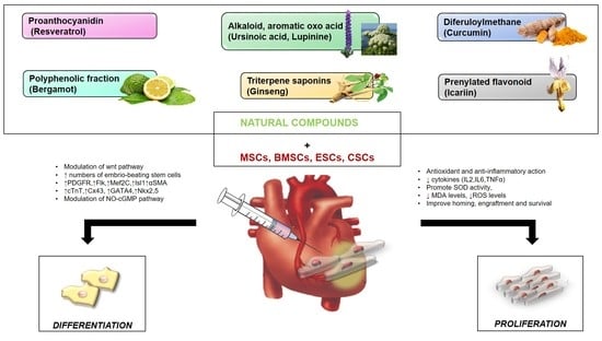
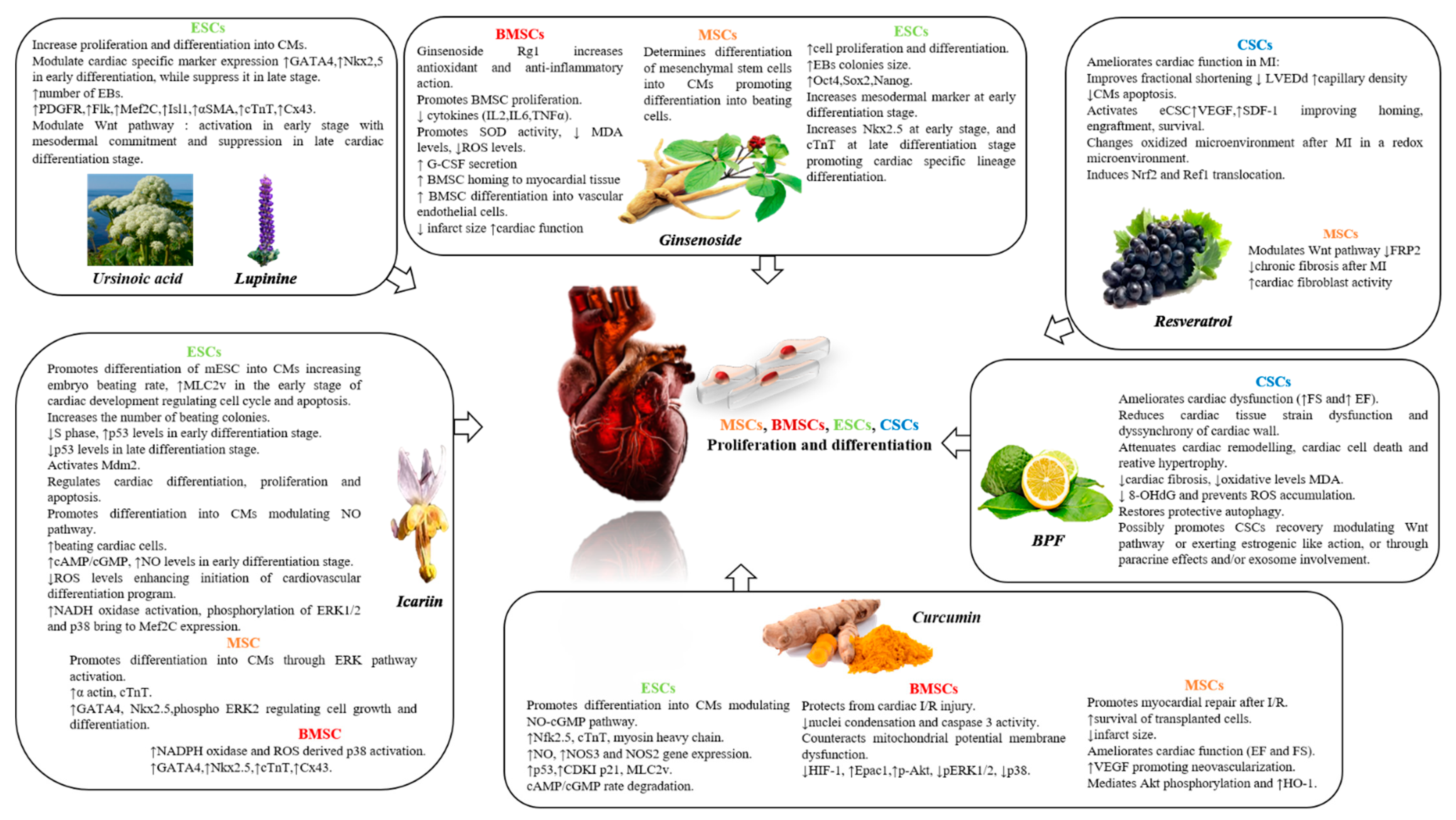
4. Natural Compounds and Stem Cells Activation and Differentiation in CVDs

Although limited data is available on the role of nutraceuticals in the modulation of stem cell properties, it is interesting to note how the targeted use of certain natural occurring compounds can contribute to the activation, proliferation and differentiation of different types of stem cells, intensifying their protective activity against cardiovascular damage. The most interesting and recent results obtained from the analysis of the mainly studied natural compounds are discussed (Figure 3).

4.1. Lupinine and Ursinoic Acid

As mentioned above, lupinine, represents the main alkaloid in the seeds of Lupinus luteus and other various species of lupine (Lupinus caudatus L., Lupinus albus L.) [175], while ursinoic acid is an aromatic oxo acid isolated from the roots of Angelica ursina but it is not pointed out for any therapeutic application [226]. However, an in-depth study recently shown that the concomitant use of these two natural compounds promotes the commitment of pluripotent stem cells to cardiac mesoderm and contribute to CM differentiation [227]. Lupinine and ursinoic acid were used to treat mouse embryonic CSCs and mouse embryo-derived teratocarcinoma cells (P19 cells). In particular, a CM-specific readout reporter system based on the P19 cell line (which has a pluripotent nature) was used expressing the α-myosin heavy chain (α-MHC) promoter-driven fluorescent mCherry protein, which is activated upon cardiac induction. After in vitro formation of EBs from both cell lines, P19 and mouse embryonic CSCs, were treated for three days with or without natural product library containing 800 compounds of various alkaloids, flavonoids, and sterols to initiate differentiation. At the end of the treatment, the cells continued their differentiation under attachment conditions for another three days and then were observed for mCherry expression. Among a number of natural compounds, lupinine and ursinoic acid successfully induced a consistent improvement in the fluorescence of mCherry and 0.5 mM of lupinine and 0.25 mM of ursinoic acid concentrations were chosen for subsequent experiments [227]. Treatment of cells with these two natural compounds showed higher differentiation efficiency when compared to the untreated cells or cells treated with oxytocin, used as a positive control. In particular, the rate of differentiation was observed by the increase in the number and size of the beating colonies in lupinine and ursinoic acid treated cells. Analysis of the transcript levels of early cardiac-specific differentiation markers, GATA-4 and Nkx2.5, recognized an increase in both P19 and mouse embryonic CSCs, which gradually increase up to 10 days. After 10 days the levels of these cardiac markers decrease, suggesting that cells treated with natural compounds proceed to the next stage of maturation where these markers were not relevant. In addition, lupinine and ursinoic acid are involved in cardiogenesis has demonstrated by the upregulation of PDGFR-α and Flk, cardiac progenitor cell markers involved in the middle stage of cardiac differentiation. Furthermore, the combined use of these two natural compounds also increases the Myocyte Enhancer Factor 2c (MEF2C) and the Insulin gene enhancer protein (Isl), markers of middle and early stages of cardiac differentiation. Muscle and cardiac markers of the most differentiated cells (α-Smooth Muscle Actin-α-SMA and cTnT) were also increased after treatment of both lupinine and ursinoic acid proving the efficacy of differentiation. Levels of α-SMA, cTnT and Connexin 43 (Cx43) were also increased in mouse embryonic stem cells (mESC) the levels of [227]. Interestingly, the improvement in cardiogenesis due to ursinoic acid and lupinine treatment was associated with the activation of the wnt pathway. In particular, an upregulation of the wnt pathway in EBs was observed after 24 h of treatment with these two alkaloids, which was suppressed after 72 h. These data suggest that lupinine and ursinoic acid promote the activation of the wnt pathway during the early phase of EBs, contributing to an efficient cardiac mesoderm commitment and subsequently their effects decrease, leading to the transition from mesoderm to the next stage of cardiac commitment [227].

4.2. Resveratrol

The properties of resveratrol in stem cell therapy has been demonstrated in several experimental studies. Among the reported results, it was observed that the administration of 2.5 mg/Kg of resveratrol can improve cardiac regeneration through the activation of endogenous CSCs before and after the induction of AMI, together with the injection of CSCs into the peri-ischemic area [228]. In this experimental model, resveratrol improves cardiac function ameliorating FS and reducing left ventricular-end diastolic diameter. This effect is linked to an increase in capillary density, decreased CM apoptosis and upregulation of VEGF and SDF-1, suggesting that the activation of endogenous CSCs could enhance cardiac recovery [229]. Furthermore, resveratrol improves the homing, engraftment and survival of injected CSCs in an animal model of MI. In particular, the CSCs were preconditioned with 2.5 uM of resveratrol before being injected in the border zone of ischemia [230]. Interestingly, improved cardiac function and a modification of the microenvironment was observed, as demonstrated by the nuclear translocation of Nrf2. It binds to the antioxidant response element and to Ref-1, a redox protein that regulates redox-sensitive transcription factors, leading to an increased engraftment of the implanted stem cells [230]. The ability of resveratrol to improve stem cell survival and engraftment was confirmed by the expression of the cell proliferation marker Ki67, while the expression of SDF-1 and myosin clearly demonstrated CSC homing in the infarcted myocardium and its regeneration which ameliorates cardiac function [230]. These results showed that resveratrol is able to modify the oxidized microenvironment after MI in a redox setting for CSCs, exerting an interesting beneficial effect in cardiac restoration [228,230]. In another in-depth study, intraperitoneal injection of 2.5 mg/kg of resveratrol and a tail vein injection of conditioning MSCs with 0.1 µM resveratrol were performed [231]. Resveratrol has been shown to reduce sFRP2, a key regulator of the wnt pathway. sFRP2 binds to Fz receptors and wnt ligands, playing a key role in chronic fibrosis after MI and may enhance the activity of adult mouse cardiac fibroblasts through wnt/β-catenin signaling. The observed effects are related to an improvement in EF, as well as a reduction of cardiac remodeling and cardiac fibrosis [231].
Moreover, resveratrol is able to promote mesoderm differentiation of human iPSCs under EB condition, up-regulating the expression of the Mesoderm Posterior BHLH Transcription Factor 1 (MESP1), Brachyury, and Mix Paired-Like Homeobox-1 (Mxl1) markers. In addition, the rate of beating EBs, after resveratrol treatment, was higher than in the control group. Resveratrol significantly reduces Oct4 and Nanog expression and substantially enhances the expression of specific cardiac genes Nkx2.5, GATA-4, cTnT, α-Myosin Heavy Chain (α-MHC), thus promoting CM differentiation of human iPSCs [232]. An interesting in vitro study using mouse ESCs identified the optimal concentration of resveratrol (10 umol/L) capable of directing mouse ESC differentiation into CMs and of achieving the beating properties of EBs [233]. Notably, the authors reported significantly higher levels of the five cardiac gene markers analyzed, including Nkx2.5, MEF2C, Tbx5, dHand and α-MHC, in mouse ESC treated with 10 umol/L of resveratrol compared to other concentrations. Additionally, the highest protein levels of Cx43 and Troponin C1, maturity indicative cardiac markers, were observed. These results were also associated with the largest calculated beating areas [233]. The mechanism underlying these properties of resveratrol seems to be related to the inhibition of the canonical wnt pathway, and upregulation of SRF/mir-1 expression, both of which are involved in CM commitment from human cardiovascular progenitors [232,233]. In an animal model of cardiomyopathy resulting from doxorubicin-induced cardiotoxicity the intriguing effect of resveratrol, associated with adipose-derived MSCs was investigated [234]. The data showed that the injection of resveratrol and adipose-derived MSCs significantly prevent the onset and severity of cardiotoxicity induced by concomitant treatment with doxorubicin. Animals treated with both resveratrol and adipose-derived MSCs showed no signs of cardiac necrosis or edema, while myocardial healing consisted of myofibril regeneration was observed. Furthermore, cell cluster of adipose-derived MSCs were observed suggesting that differentiation of adipose-derived MSCs into CMs associated with MSC paracrine capacity contributes to cardiac improvement against doxorubicin detrimental effects [234]. In addition to the reported data, MSCs previously treated with resveratrol and injected into a failed myocardium, induce cardiac differentiation and enhance MSC paracrine effects in an animal model of doxorubicin-induced cardiomyopathy [235].

4.3. Ginseng

Although there is few information about ginseng treatment and stem cell differentiation into CMs, an interesting report has shown that the use of ginsenoside Rg1 improves the antioxidant and anti-inflammatory capacities of BMSCs in the hematopoietic microenvironment of aged rats [236]. Aging is a condition characterized by chronic inflammation, increased levels of ROS and decreased levels of anti-oxidant defense. These conditions were reverted by the use of ginsenoside Rg1, able to counteract the increase in inflammatory cytokines (IL-2, IL-6, TNF-α), MDA content and ROS levels and promote SOD activity, leading to BMSC proliferation [236]. As mentioned above, one of the mechanism through which ginseng determines cardioprotection is the upregulation of NO. Indeed, ginseng is an angiogenesis inducer of eNOS expression thanks to the ginsenoside Rg1. Rg1 was found to decrease miR-214, which appears to be involved in the negative modulation of eNOS expression in human umbilical vein endothelial cells (HUVECs), suggesting a possible angiogenic mechanism induced by ginsenoside [237]. In particular, BMSC-derived exosomes have been observed to strongly suppress apoptosis and ROS production in CSCs after oxidative stress injury, mainly due to the action of mir-214 in silencing Ca2+/calmodulin-dependent protein kinase II (CaMKII) target [238]. However, there are some studies that determined the action of ginseng on stem cells. Ginseng extract has been observed to facilitate proliferation and differentiation of hESCs [239]. In brief, ginseng extract was added to undifferentiated hESCs and differentiating cells at 0.125, 0.25, and 0.5 mg/mL. Human-ESC colonies were cultured in suspension for several days, while undifferentiated hESCs were treated to differentiate them into cardiac progenitor cells. At the end of the experimental period it was shown that ginseng-treated hESCs had larger colony size and increased proliferation rate and expression of pluripotency markers such as Oct4, Sox2, and Nanog. Ginseng also allows differentiation into mesendoderm lineage when added to EBs for 10 days, showing increased expression of the mesodermal marker (Brachyury and winged helix transcription factor hepatocyte nuclear factor 3β (HNF3β or Foxa2). Moreover, ginseng treatment promotes mesodermal lineage in the early stage of hESC-derived cardiac progenitor cell differentiation, while, it promotes cardiac specific lineage differentiation in the later stage of hESC-derived cardiac progenitor cells as showed by increased levels of Nkx2.5 and cTnI [239]. Interestingly, some of the compounds contained in the water soluble fraction of the ginseng fraction (in particular vitamin B12 and methionine) are able to promote the differentiation of MSCs into CMs, enhancing the differentiation rate into beating cells [240]. Finally, ginsenoside-Rg-1 was identified as a potent proangiogenic agent. Indeed, rg-1 has been shown to reduce miR-15b expression, leading to a temporal induction of VEGFR-2 in HUVEC cells. It has also demonstrated that rg1 induces motility and tubulogenesis through the upregulation of VEGFR-2 in treated cells. Confirming this result, in an in vivo model of zebrafish embryos, injection of the pre-miR-15b precursor was found to significantly suppress sub-intestinal vessels formation [241]. Other in vivo experiments also reported that Rg1 stimulates the myocardial tissue secretion of granulocyte colony-stimulating factor (G-CSF), able to promotes BMSC homing to myocardial tissue and differentiation into vascular endothelial cells. Interestingly, a reduction in the size of the myocardial infarction and an improvement in cardiac function were observed in this condition [242]. In a rat model of acute MI it was also observed how the continuous application of Rg1 enhances the number of peripheral blood CD34+ stem cells and stimulates the homing of stem cells to the infarcted area, leading to ventricular remodeling with reduction of infarct size and significant myocardial regeneration [243]. A detailed study recently published in Nature has identified the 20(R)-ginsenoside Rh2 among several ginseng saponins, capable of increasing the proliferation of skeletal myoblasts in vitro [244]. The molecular mechanism underlying this important effect was identified in the activation of Akt signaling and in the inhibition of the cyclin-dependent kinases inhibitors (CDKIs), p27Kip1 and p57Kip2 which negatively regulate cell proliferation. Furthermore, an animal model of MI and an animal model of skeletal muscle degeneration were used to assess the potential beneficial effect of ginsenoside Rh2 in vivo. The results showed that treatment with ginsenoside Rh2 increases cardiac recovery through improved left ventricular function and increased proliferation of CMs in the infarcted area which was indicated by higher levels of cTnI and Ki67. Additionally, in the animal model of barium chloride-induced skeletal muscle degeneration, treatment with ginsenoside Rh2 leads to a reduction in muscle scarring and fewer inflammatory cells. These features are associated with reduced tissue degeneration and a greater proportion of smaller diameter fibers, which increase in diameter as the muscle heals [244].

4.4. Icariin

Interesting data are collected about the effects of icariin in the modulation of cardiomyogenesis and in differentiation into CMs through several specific actions. An an in vitro study showed the ability of icariin to modulate cardiomyogenesis and promote differentiation of mouse ESCs into CMs [245]. Briefly, ESCs were cultured to form EBs and then plated on a gelatin-coated plate and treated with icariin 1×10⁻⁷ mol/L for 23 days. The rate of beating EBs was increased compared to control and cardiac differentiation process was improved. Indeed, an increase in cardiac-specific protein such as mRNA level of cardiac α-MHC and myosin light chain 2v (MLC2v) was observed in EBs in the early stage of cardiac development. Furthermore, the inducible effect of icariin was related to cell cycle regulation and induction of apoptosis as a signal of ESC differentiation [245]. In addition, treatment of ESC derived-embryoid stem cells with 10−7 M icariin for 24–48 h confirmed its ability to promote differentiation into beating colonies increasing the levels of cardiac specific markers. Moreover, in the early phase of differentiation, icariin induces cell cycle arrest and apoptosis, reducing the S phase in ESCs before transitioning to the CM phenotype with an increase in p53 levels. However, during the last phase of differentiation, a reduction in p53 protein expression levels, accompanied by the activation of the E3 ubiquitin-protein ligase (Mdm2), was observed, suggesting a potential mechanism by which icariin could regulate cardiac differentiation, proliferation and apoptosis [246]. Subsequent studies suggested that icariin mediates the differentiation of ESCs into CMs through modulation of the endogenous NO signaling pathway [247]. After EB induction, cells were treated with 1×10⁻⁷ mol/L icariin. The data showed that, in accordance with the study mentioned above, icariin increases beating cardiac cells expressing α-actinin and cardiac transcription factors. In particular, levels of GATA-4 and Nkx2.5 were increased by icariin treatment during early cardiac differentiation, as well as, the levels of α-MHC, MLC2v and β-adrenergic receptor (β-AR) [247]. Cardiomyocytes treated with icariin were shown to be more sensitive to isoproterenol than control cells. Furthermore, after 24–48 h of icariin treatment the ratio of cAMP/cGMP and NO levels was increased, before ESCs became CMs. Instead, the cGMP level was increased within 24 h, suggesting that the NO-cGMP pathway is mainly involved in the early differentiation phase of icariin treated ESCs. Therefore, the upregulation of NO production appears to be a mechanism by which icariin modulates the early differentiation of ESCs into CMs as has been observed using NO inhibitors which leads to a reduction in the differentiation process [247]. Interestingly, modulation of ROS levels was involved in icariin induced ESC-differentiation into CMs. Low levels of ROS during the early phase of differentiation of ESCs have been observed to be helpful in initiating the cardiovascular differentiation program [248]. The in vitro model of mouse ESCs-derived EBs treated with 100 nM icariin, paradoxically, showed that icariin (and other prenylflavonoids such as icaritin and desmetilicaritiin) was able to increase the number of spontaneous beating EBs, cardiac specific genes and cardiac transcription factors by regulating the intracellular concentration of ROS [248]. In particular, the effects of icariin are related to the activation of NADPH oxidase, to the phosphorylation of ERK1/2 and p38, bringing to MEF2C expression and CM differentiation [248]. Other reports support the theory that icariin promotes cardiac differentiation in BM-MSCs enhancing NADPH oxidase and ROS-derived p-38 activation. Co-administration of icariin (0.1 μM) and the BMP-2 (10 μg/L) for 24 h has been observed to exert a synergistic effect in CM differentiation of BM-MSCs in vitro, enhancing the expression of specific markers such as GATA-4, Nkx2.5, cTnT, and Cx43, higher levels of NADPH oxidase, H2O2 and p38MAPK [249]. Icariin was also found to induce CM differentiation in MSCs derived from adipose tissue and this effect was related to the activation of the ERK pathway [250]. Adipose-derived MSCs co-cultured with CMs showed increased protein levels of α-actinin and cTnT at 21 days after treatment with 10−7 mol/l icariin for 24 h, increasing the rate of differentiated cells compared to non-treated cells. In addition, GATA-4, Nkx2.5, MLC-2v markers increased after 3 weeks of treatment. These effects were supported by increased levels of p-ERK-1 and p-ERK-2, a general regulator of cell growth and differentiation in response to mitogenic stimuli [250].

4.5. Curcumin

Several studies reported a powerful effect of curcumin on histone acetylation, which can be useful in counteracting pathological cardiac growth, detrimental remodeling or HF. Indeed, cardiac growth and gene expression are regulated through histone acetylation/deacetylation in response to acute and chronic stress [251]. Recently, curcumin has been found to modulate cardiogenesis through inhibition of histone acetylation. In an experimental model, H9c2 cells were transfected with BMP-2 to induce cardiac specific gene expression of GATA-4 and MEF2C through p300 activation, thereby, increasing the level of H3 histone acetylation. Interestingly, this effect was reversed by curcumin’s inhibition of p300 [252]. An in-depth study investigated the key role of curcumin in epigenetic modifications of embryonic heart development such as in prenatal alcohol exposure model [253]. Indeed, in cardiac progenitor cells, curcumin inhibits alcohol-induced hyperacetylation of hystone H3, leading to a reduction in the expression of GATA-4 and MEF2C and subsequent abnormal heart development and congenital heart disease [253]. In addition to the above data, some studies have shown that pre-treatment of adipose MSCs with curcumin promotes myocardial repair in an animal model of cardiac ischemic-reperfusion injury, performed by left coronary artery ligation (LAD) [254]. One week after ligation, adipose MSCs treated with 10 μM of curcumin for 24 h were injected into the peri-infarcted regions and within the infarcted area. After 28 days, an increase in the number of vital transplanted cells was observed, accompanied by a reduction in the infarct size and an improvement in cardiac function with an increase in the level of VEGF. Furthermore, in vitro data showed that curcumin protects stem cells from H2O2 and reduces apoptosis by mediating Akt phosphorylation, increasing HO-1 expression and inhibiting phosphatase and tensin homolog (PTEN), p53, and caspase-3 expression levels [254]. Curcumin has also been found to modulate ESCs differentiation into cardiac lineage through the modulation of NO-cyclic GMP pathway [255]. Human-ESCs were cultured in suspension to form EBs and then transferred to coated plates with differentiation medium. EBs at day 1 and 2 and partially differentiated cells at day seven, were treated with curcumin (5–20 μmol/L) at day 7, 9, 11 and 13. At the end of the experimental period, increased levels of cardiac specific markers (NKx2.5, cTnI and α-MHC) determined by curcumin were observed. Moreover, an increase in intracellular nitrite, induction of the NOS-3 gene and reduction of NOS-2 was recorded alongside an increase in the expression of the soluble Guanylyl Cyclase α1 (sGC α1) gene, suggesting that NO pathway is involved in curcumin-induced stem cell differentiation [255]. Co-administration of curcumin with NO donors leads to an increase in the protein levels of p53, cyclin-dependent kinase inhibitor (CDKI) p21 and of the specific cardiac proteins MLC2 and cTnI. Analysis of the basal level of cAMP and cGMP, in partially differentiated cells treated with curcumin, showed an increase in the level ofcGMP and a decrease in those of cAMP. The rate of cAMP/cGMP degradation was increased after long term treatment with curcumin, while, direct administration of curcumin to cell extracts results in suppression of cAMP and cGMP, probably related to curcumin induced phosphodiesterase degradation [255]. Finally, an in vitro model of hypoxia and reoxygenation, which mimics ischemia/reperfusion injury, further confirms the protective effects of curcumin against cardiovascular injury [256]. Curcumin was revealed to significantly reduce cell loss, nuclei condensation and caspase 3 activity, induced by hypoxia and reoxygenation, protecting cells from mitochondrial dysfunction. In addition, curcumin inhibits Hypoxia-Inducible Factor-1α (HIF-1α) expression, directly activated by the cAMP-1 (Epac-1)/Akt pathway, increasing Epac1 levels, Akt phosphorylation and downregulating Erk1/2 and p38 phosphorylation. Overall, these effects strongly improve BMSC survival [256].

4.6. BPF

The potential role of bergamot polyphenols in modulating stem cell properties remains to be extensively evaluated. To date, the first interesting results were collected by analyzing the effects of the polyphenolic fraction of bergamot (BPF) on an experimental model of anthracycline-induced HF [223]. For the first time, BPF was observed to prevent eCSCs attrition, improving the number of resident c-kit+CD45negCD31negeCSCs and inhibiting 8-OHdG nuclear accumulation. Interestingly, these effects were associated with CM replenishment, recognized through an increasing number of newly formed BrdUpos small myocytes. These data suggest that the direct ROS-scavenging properties of BPF, already discussed in the previous paragraph, lead to additional beneficial effects on the eCSC compartment by promoting the proliferation and differentiation of the eCSCs. Indeed, the maintenance of low ROS levels could guarantee the constitutive activity of the endogenous antioxidant enzymes thus ensuring the right redox status of the eCSC niches. In support of these findings, recent evidence shown that naringin, one of the most abundant flavonoids contained in BPF, protects human adipose-derived MSCs from the detrimental action of H2O2 which inhibits osteogenic differentiation. The protective effect of naringin is probably exerted through its action in modulating wnt signaling [257]. As extensively reported, wnt pathway is crucial for heart development and is probably inhibited in the adult heart [258], suggesting that the latter event may be beneficially modulated by BPF-derived naringin. Moreover, other hypothetical mechanisms leading to cardioprotective effects of citrus-derived flavonoids against HF were identified, such as an estrogenic like action which might induce progenitor cell mediated recovery [259]. Furthermore, in addition to the anti-oxidant actions, other mechanisms are currently being studied that participate in the mediation of the protective actions of BPF on the CSCs compartment, probably through paracrine effects and/or exosome involvement.
Interestingly, all these studies pointed out that the use of natural compounds could be involved in the cardiogenesis of different stem cell types by directly stimulating endogenous stem cells and their secretory activity and/or using conditioned media containing cytokines and signal modulators, increasing the level of some cardiac specific markers. The data suggest a possible future approach to obtain an optimal condition for efficient cardiac commitment with the use of natural inducers.

5. Conclusions

After many years, the belief that the heart was a terminally differentiated organ has been denied by groundbreaking discoveries about CM turnover and the existence of stem and progenitor cells located in the myocardium, which support its self-renewal potential. In this regard, stem cell therapy has gained particular attention, emerging as a novel approach to restore damaged myocardial tissue. Interesting pre-clinical studies reporting significant stem cell-mediated cardiac regeneration, encouraging subsequent first clinical trials. However, the beneficial effects of stem cell-based treatment on myocardial performance are still quite limited by the low engraftment and survival rate of the cells due to the microenvironment of the damaged site of the cardiac tissue. Therefore, boosting the efficacy of stem cell repair is essential, as well as better understanding the heart endogenous reparative mechanisms and their interactions with stem cell regenerative properties, making cardiac regenerative therapy an effective therapeutic tool. The indirect stimulation of cardiovascular commitment and paracrine regulation in various stem cell populations opens a new scenario for cardiac stem cell therapy.
Interestingly, pharmacological and non-pharmacological preconditioning represents a novel and efficient technique for stimulating the secretory activity of stem cells and drug-mediated activation or inhibition of pathways, which can modify stem cell physiology by improving cell survival, engraftment and regeneration.
Although limited data is available, the targeted use of certain natural compounds from plant extracts is emerging as a useful tool for orchestrating stem cell homing, engraftment and survival in heart diseases.
The upcoming challenges will concern a deeper understanding of the underlying mechanisms by which natural compounds can modulate stem cell cardiovascular lineage specification, and the main role played by stimulating their secretory activity and regulating the paracrine signaling. It will also be necessary to investigate whether the effects observed in experimental models of myocardial diseases are retained in humans with a sufficient degree of therapeutic efficacy without toxic effects.
CVDs represent complex disorders characterized by several disease patterns and pathologic mechanisms that make it difficult and ineffective to provide a uniform therapeutic intervention protocol. Therefore, future strategies should aim at identifying the optimal cell type, the specific stimulation/differentiation protocol, dosage and delivery approach that are effective and safe allowing for personalized therapy. In this perspective, the use of nutraceuticals can contribute significantly to the optimization of stem cell therapy in the clinical practice also thanks to their overall safety profile, feasibility and wide availability.

Author Contributions

V.M. (Vincenzo Mollace), C.C. and M.S. conceptualized and designed the manuscript; C.C. and M.S. wrote the manuscript; V.M. (Vincenzo Mollace) and M.G. revised the manuscript; F.S., R.M. (Roberta Macrì), F.B., S.N., S.R., M.C.Z., R.M. (Rocco Mollace), L.G., A.R.C., V.M. (Vincenzo Musolino), J.M. and E.P. participated in drafting the article and revising it critically. All authors have read and agreed to the published version of the manuscript.

Funding

The work has been supported by the public resources from the Italian Ministry of Research, PON-MIUR 03PE000_78_1, PONMIUR 03PE000_78_2 and POR Calabria FESR FSE 2014–2020 Asse 12-Azioni 10.5.6 e 10.5.12.

Institutional Review Board Statement

Not applicable.

Informed Consent Statement

Not applicable.

Data Availability Statement

Data available in a publicly accessible repository.

Conflicts of Interest

The authors declare no conflict of interest.

References

  1. WHO. Cardiovascular Diseases (CVDs). Available online: http://www.who.int/en/news-room/fact-sheets/detail/cardiovascular-diseases (accessed on 1 June 2019).
  2. Mozaffarian, D.; Benjamin, E.J.; Go, A.S.; Arnett, D.K.; Blaha, M.J.; Cushman, M.; Das, S.R.; de Ferranti, S.; Després, J.-P.; Fullerton, H.J.; et al. Executive Summary: Heart Disease and Stroke Statistics—2015 Update. A Report from the American Heart Association. Circulation 2015, 131, 434–441. [Google Scholar] [CrossRef]
  3. Fuster, V.; Kelly, B.B. (Eds.) Institute of Medicine (US) Committee on Preventing the Global Epidemic of Cardiovascular Disease: Meeting the Challenges in Developing Countries. In Promoting Cardiovascular Health in the Developing World: A Critical Challenge to Achieve Global Health; National Academies Press: Washington, DC, USA, 2010. [Google Scholar]
  4. Cesselli, D.; Aleksova, A.; Mazzega, E.; Caragnano, A.; Beltrami, A.P. Cardiac stem cell aging and heart failure. Pharmacol. Res. 2018, 127, 26–32. [Google Scholar] [CrossRef] [PubMed]
  5. Wilkins, E.; Wilson, L.; Wickramasinghe, K.; Bhatnagar, P.; Leal, J.; Luengo-Fernandez, R.; Burns, R.; Rayner, M.; Townsend, N. European Cardiovascular Disease Statistics 2017; European Heart Network: Brussels, Belgium, 2017. [Google Scholar]
  6. Epidemiology of Cardiovascular Disease. Available online: https://www.nature.com/collections/bedbejdbij?proof=t (accessed on 20 November 2020).
  7. Benjamin, E.J.; Muntner, P.; Alonso, A.; Bittencourt, M.S.; Callaway, C.W.; Carson, A.P.; Chamberlain, A.M.; Chang, A.R.; Cheng, S.; Das, S.R.; et al. Heart Disease and Stroke Statistics-2019 Update: A Report From the American Heart Association. Circulation 2019, 139, e56–e528. [Google Scholar] [CrossRef] [PubMed]
  8. Arnett, D.K.; Blumenthal, R.S.; Albert, M.A.; Buroker, A.B.; Goldberger, Z.D.; Hahn, E.J.; Himmelfarb, C.D.; Khera, A.; Lloyd-Jones, D.; McEvoy, J.W.; et al. 2019 ACC/AHA Guideline on the Primary Prevention of Cardiovascular Disease: A Report of the American College of Cardiology/American Heart Association Task Force on Clinical Practice Guidelines. Circulation 2019, 140, e596–e646. [Google Scholar] [CrossRef]
  9. Sanchis-Gomar, F.; Perez-Quilis, C.; Leischik, R.; Lucia, A. Epidemiology of coronary heart disease and acute coronary syndrome. Ann. Transl. Med. 2016, 4, 256. [Google Scholar] [CrossRef] [PubMed] [Green Version]
  10. Sakboonyarat, B.; Rangsin, R. Prevalence and associated factors of ischemic heart disease (IHD) among patients with diabetes mellitus: A nationwide, cross-sectional survey. BMC Cardiovasc. Disord. 2018, 18, 151. [Google Scholar] [CrossRef] [Green Version]
  11. Yu, H.; Lu, K.; Zhu, J.; Wang, J. Stem cell therapy for ischemic heart diseases. Br. Med. Bull. 2017, 121, 135–154. [Google Scholar] [CrossRef] [Green Version]
  12. Terashvili, M.; Bosnjak, Z.J. Stem Cell Therapies in Cardiovascular Disease. J. Cardiothorac. Vasc. Anesth. 2019, 33, 209–222. [Google Scholar] [CrossRef]
  13. Hsieh, P.C.; Segers, V.F.; Davis, M.E.; MacGillivray, C.; Gannon, J.; Molkentin, J.D.; Robbins, J.; Lee, R.T. Evidence from a genetic fate-mapping study that stem cells refresh adult mammalian cardiomyocytes after injury. Nat. Med. 2007, 13, 970–974. [Google Scholar] [CrossRef]
  14. Sun, R.; Li, X.; Liu, M.; Zeng, Y.; Chen, S.; Zhang, P. Advances in Stem Cell Therapy For Cardiovascular Disease (Review). Int. J. Mol. Med. 2016, 38, 23–29. [Google Scholar] [CrossRef] [Green Version]
  15. Wernly, B.; Mirna, M.; Rezar, R.; Prodinger, C.; Jung, C.; Podesser, B.K.; Kiss, A.; Hoppe, U.C.; Lichtenauer, M. Regenerative Cardiovascular Therapies: Stem Cells and Beyond. Int. J. Mol. Sci. 2019, 20, 1420. [Google Scholar] [CrossRef] [Green Version]
  16. Ménard, C.; Hagège, A.A.; Agbulut, O.; Barro, M.; Morichetti, M.C.; Brasselet, C.; Bel, A.; Messas, E.; Bissery, A.; Bruneval, P.; et al. Transplantation of cardiac-committed mouse embryonic stem cells to infarcted sheep myocardium: A preclinical study. Lancet 2005, 366, 1005–1012. [Google Scholar] [CrossRef]
  17. Sartiani, L.; Bettiol, E.; Stillitano, F.; Mugelli, A.; Cerbai, E.; Jaconi, M.E. Developmental changes in cardiomyocytes differentiated from human embryonic stem cells: A molecular and electrophysiological approach. Stem Cells 2007, 25, 1136–1144. [Google Scholar] [CrossRef] [PubMed]
  18. Jain, K.K. Ethical and regulatory aspects of embryonic stem cell research. Expert Opin. Biol. Ther. 2005, 5, 153–162. [Google Scholar] [CrossRef] [PubMed]
  19. Rojas, S.V.; Kensah, G.; Rotaermel, A.; Baraki, H.; Kutschka, I.; Zweigerdt, R.; Martin, U.; Haverich, A.; Gruh, I.; Martens, A. Transplantation of purified iPSC-derived cardiomyocytes in myocardial infarction. PLoS ONE 2017, 12, e0173222. [Google Scholar] [CrossRef] [PubMed]
  20. Riggs, J.W.; Barrilleaux, B.L.; Varlakhanova, N.; Bush, K.M.; Chan, V.; Knoepfler, P.S. Induced pluripotency and oncogenic transformation are related processes. Stem Cells Dev. 2012, 22, 37–50. [Google Scholar] [CrossRef]
  21. Yoshihara, M.; Hayashizaki, Y.; Murakawa, Y. Genomic instability of iPSCs: Challenges towards their clinical applications. Stem Cell Rev. Rep. 2017, 13, 7–16. [Google Scholar] [CrossRef] [Green Version]
  22. Strauer, B.E.; Brehm, M.; Zeus, T.; Kostering, M.; Hernandez, A.; Sorg, R.V.; Kögler, G.; Wernet, P. Repair of infarcted myocardium by autologous intracoronary mononuclear bone marrow cell transplantation in humans. Circulation 2002, 106, 1913–1918. [Google Scholar] [CrossRef] [Green Version]
  23. Ellison, G.M.; Vicinanza, C.; Smith, A.J.; Aquila, I.; Leone, A.; Waring, C.D.; Henning, B.J.; Stirparo, G.G.; Papait, R.; Scarfò, M.; et al. Adult c-kit(pos) cardiac stem cells are necessary and sufficient for functional cardiac regeneration and repair. Cell 2013, 154, 827–842. [Google Scholar] [CrossRef] [Green Version]
  24. Menasché, P.; Vanneaux, V.; Hagège, A.; Bel, A.; Cholley, B.; Cacciapuoti, I.; Parouchev, A.; Benhamouda, N.; Tachdjian, G.; Tosca, L.; et al. Human embryonic stem cell-derived cardiac progenitors for severe heart failure treatment: First clinical case report. Eur. Heart J. 2015, 36, 2011–2017. [Google Scholar] [CrossRef] [Green Version]
  25. Musunuru, K.; Sheikh, F.; Gupta, R.M.; Houser, S.R.; Maher, K.O.; Milan, D.J.; Terzic, A.; Wu, J.C. Induced pluripotent stem cells for cardiovascular disease modeling and precision medicine: A scientific statement from the American Heart Association. Circ. Genom. Precis Med. 2018, 11, e000043. [Google Scholar] [CrossRef] [Green Version]
  26. Bolli, R.; Chugh, A.R.; D’Amario, D.; Loughran, J.H.; Stoddard, M.F.; Ikram, S.; Beache, G.M.; Wagner, S.G.; Leri, A.; Hosoda, T.; et al. Cardiac stem cells in patients with ischaemic cardiomyopathy (SCIPIO): Initial results of a randomised phase 1 trial. Lancet 2011, 378, 1847–1857. [Google Scholar] [CrossRef] [Green Version]
  27. Malliaras, K.; Makkar, R.R.; Smith, R.R.; Cheng, K.; Wu, E.; Bonow, R.O.; Marban, L.; Mendizabal, A.; Cingolani, E.; Johnston, P.V.; et al. Intracoronary cardiosphere-derived cells after myocardial infarction: Evidence of therapeutic regeneration in the final 1-year results of the CADUCEUS trial (CArdiosphere-Derived aUtologous stem CElls to reverse ventricUlar dySfunction). J. Am. Coll. Cardiol. 2014, 63, 110–122. [Google Scholar] [CrossRef] [PubMed] [Green Version]
  28. Chakravarty, T.; Makkar, R.R.; Ascheim, D.D.; Traverse, J.H.; Schatz, R.; DeMaria, A.; Francis, G.S.; Povsic, T.J.; Smith, R.R.; Lima, J.A.; et al. ALLogeneic Heart STem Cells to Achieve Myocardial Regeneration (ALLSTAR) Trial: Rationale and Design. Cell Transplant. 2017, 26, 205–214. [Google Scholar] [CrossRef] [PubMed]
  29. Ott, H.C.; Kroess, R.; Bonaros, N.; Marksteiner, R.; Margreiter, E.; Schachner, T.; Laufer, G.; Hering, S. Intramyocardial microdepot injection increases the efficacy of skeletal myoblast transplantation. Eur. J. Cardiothorac. Surg. 2005, 27, 1017–1021. [Google Scholar] [CrossRef] [PubMed]
  30. Forest, V.F.; Tirouvanziam, A.M.; Perigaud, C.; Fernandes, S.; Fusellier, M.S.; Desfontis, J.C.; Toquet, C.S.; Heymann, M.F.; Crochet, D.P.; Lemarchand, P.F. Cell distribution after intracoronary bone marrow stem cell delivery in damaged and undamaged myocardium: Implications for clinical trials. Stem Cell Res. Ther. 2010, 1, 4. [Google Scholar] [CrossRef] [Green Version]
  31. Aicher, A.; Brenner, W.; Zuhayra, M.; Badorff, C.; Massoudi, S.; Assmus, B.; Eckey, T.; Henze, E.; Zeiher, A.M.; Dimmeler, S. Assessment of the tissue distribution of transplanted human endothelial progenitor cells by radioactive labeling. Circulation 2003, 107, 2134–2139. [Google Scholar] [CrossRef] [Green Version]
  32. Miyagawa, S.; Saito, A.; Sakaguchi, T.; Yoshikawa, Y.; Yamauchi, T.; Imanishi, Y.; Kawaguchi, N.; Teramoto, N.; Matsuura, N.; Iida, H.; et al. Impaired myocardium regeneration with skeletal cell sheets—A preclinical trial for tissue-engineered regeneration therapy. Transplantation 2010, 90, 364–372. [Google Scholar] [CrossRef]
  33. Zhang, X.; Wang, H.; Ma, X.; Adila, A.; Wang, B.; Liu, F.; Chen, B.; Wang, C.; Ma, Y. Preservation of the cardiac function in infarcted rat hearts by the transplantation of adipose-derived stem cells with injectable fibrin scaffolds. Exp. Biol. Med. (Maywood) 2010, 235, 1505–1515. [Google Scholar] [CrossRef]
  34. Lim, S.Y.; Kim, Y.S.; Ahn, Y.; Jeong, M.H.; Hong, M.H.; Joo, S.Y.; Nam, K.I.; Cho, J.G.; Kang, P.M.; Park, J.C. The effects of mesenchymal stem cells transduced with Akt in a porcine myocardial infarction model. Cardiovasc. Res. 2006, 70, 530–542. [Google Scholar] [CrossRef]
  35. Wang, J.; Yang, H.; Hu, X.; Fu, W.; Xie, J.; Zhou, X.; Xu, W.; Jiang, H. Dobutamine-mediated heme oxygenase-1 induction via PI3K and p38 MAPK inhibits high mobility group box 1 protein release and attenuates rat myocardial ischemia/reperfusion injury in vivo. J. Surg. Res. 2013, 183, 509–516. [Google Scholar] [CrossRef] [PubMed]
  36. Ma, Q.; Xia, X.; Tao, Q.; Lu, K.; Shen, J.; Xu, Q.; Hu, X.; Tang, Y.; Block, N.L.; Webster, K.A.; et al. Profound actions of an agonist of growth hormone-releasing hormone on angiogenic therapy by mesenchymal stem cells. Arterioscler. Thromb. Vasc. Biol. 2016, 36, 663–672. [Google Scholar] [CrossRef] [PubMed] [Green Version]
  37. Hu, X.; Wei, L.; Taylor, T.M.; Wei, J.; Zhou, X.; Wang, J.A.; Yu, S.P. Hypoxic preconditioning enhances bone marrow mesenchymal stem cell migration via kv2.1 channel and FAK activation. Am. J. Physiol. Cell Physiol. 2011, 301, C362–C372. [Google Scholar] [CrossRef] [PubMed]
  38. Wei, H.; Li, Z.; Hu, S.; Chen, H.; Cong, X. Apoptosis of mesenchymal stem cells induced by hydrogen peroxide concerns both endoplasmic reticulum stress and mitochondrial death pathway through regulation of caspases, p38 and JNK. J. Cell Biochem. 2010, 111, 967–978. [Google Scholar] [CrossRef]
  39. Huang, K.; Hu, S.; Cheng, K. A New Era of Cardiac Cell Therapy: Opportunities and Challenges. Adv. Healthc. Mater. 2019, 8, e1801011. [Google Scholar] [CrossRef]
  40. Rikhtegar, R.; Pezeshkian, M.; Dolati, S.; Safaie, N.; Afrasiabi Rad, A.; Mahdipour, M.; Nouri, M.; Jodati, A.R.; Yousefi, M. Stem cells as therapy for heart disease: iPSCs, ESCs, CSCs, and skeletal myoblasts. Biomed. Pharmacother. 2019, 109, 304–313. [Google Scholar] [CrossRef]
  41. Wang, J.A.; Li, C.L.; Fan, Y.Q.; He, H.; Sun, Y. Allograftic bone marrow-derived mesenchymal stem cells transplanted into heart infarcted model of rabbit to renovate infarcted heart. J. Zhejiang. Univ. Sci. 2004, 5, 1279–1285. [Google Scholar] [CrossRef] [Green Version]
  42. Ventura, C.; Cavallini, C.; Bianchi, F.; Cantoni, S. Stem Cells and Cardiovascular Repair: A Role for Natural and Synthetic Molecules Harboring Differentiating and Paracrine Logics. Cardiovasc. Hematol. Agents. Med. Chem. 2008, 6, 60–68. [Google Scholar] [CrossRef]
  43. Gnecchi, M.; He, H.; Noiseux, N.; Liang, O.D.; Zhang, L.; Morello, F.; Mu, H.; Melo, L.G.; Pratt, R.E.; Ingwall, J.S.; et al. Evidence supporting paracrine hypothesis for Akt-modified mesenchymal stem cell-mediated cardiac protection and functional improvement. FASEB J. 2006, 20, 661–669. [Google Scholar] [CrossRef]
  44. Uemura, R.; Xu, M.; Ahmad, N.; Ashraf, M. Bone marrow stem cells prevent left ventricular remodeling of ischemic heart through paracrine signaling. Circ. Res. 2006, 98, 1414–1421. [Google Scholar] [CrossRef] [Green Version]
  45. Zhu, H.; Fan, G.C. Extracellular/circulating microRNAs and their potential role in cardiovascular disease. Am. J. Cardiovasc. Dis. 2011, 1, 138–149. [Google Scholar] [PubMed]
  46. Sluijter, J.P.; Verhage, V.; Deddens, J.C.; van den Akker, F.; Doevendans, P.A. Microvesicles and exosomes for intracardiac communication. Cardiovasc. Res. 2014, 102, 302–311. [Google Scholar] [CrossRef] [PubMed] [Green Version]
  47. Arslan, F.; Lai, R.C.; Smeets, M.B.; Akeroyd, L.; Choo, A.; Aguor, E.N.; Timmers, L.; van Rijen, H.V.; Doevendans, P.A.; Pasterkamp, G.; et al. Mesenchymal stem cell-derived exosomes increase ATP levels, decrease oxidative stress and activate pi3k/Akt pathway to enhance myocardial viability and prevent adverse remodeling after myocardial ischemia/reperfusion injury. Stem Cell Res. 2013, 10, 301–312. [Google Scholar] [CrossRef] [PubMed] [Green Version]
  48. Noiseux, N.; Borie, M.; Desnoyers, A.; Menaouar, A.; Stevens, L.M.; Mansour, S.; Danalache, B.A.; Roy, D.C.; Jankowski, M.; Gutkowska, J. Preconditioning of stem cells by oxytocin to improve their therapeutic potential. Endocrinology 2012, 153, 5361–5372. [Google Scholar] [CrossRef] [PubMed]
  49. Wisel, S.; Khan, M.; Kuppusamy, M.L.; Mohan, I.K.; Chacko, S.M.; Rivera, B.K.; Sun, B.C.; Hideg, K.; Kuppusamy, P. Pharmacological preconditioning of mesenchymal stem cells with trimetazidine (1-2,3,4-trimethoxybenzylpiperazine) protects hypoxic cells against oxidative stress and enhances recovery of myocardial function in infarcted heart through Bcl-2 expression. J. Pharmacol. Exp. Ther. 2009, 329, 543–550. [Google Scholar] [CrossRef] [PubMed]
  50. Hoke, N.N.; Salloum, F.N.; Kass, D.A.; Das, A.; Kukreja, R.C. Preconditioning by phosphodiesterase-5 inhibition improves therapeutic efficacy of adipose-derived stem cells following myocardial infarction in mice. Stem Cells 2012, 30, 326–335. [Google Scholar] [CrossRef]
  51. Müller, P.; Lemcke, H.; David, R. Stem Cell Therapy in Heart Diseases—Cell Types, Mechanisms and improvement Strategies. Cell Physiol. Biochem. 2018, 48, 2607–2655. [Google Scholar] [CrossRef]
  52. Thygesen, K.; Alpert, J.S.; Jaffe, A.S.; Chaitman, B.R.; Bax, J.J.; Morrow, D.A.; White, H.D. Executive Group on behalf of the Joint European Society of Cardiology (ESC)/American College of Cardiology (ACC)/American Heart Association (AHA)/World Heart Federation (WHF) Task Force for the Universal Definition of Myocardial Infarction. Fourth universal definition of myocardial infarction (2018). J. Am. Coll. Cardiol. 2018, 72, 2231–2264. [Google Scholar]
  53. Talman, V.; Ruskoaho, H. Cardiac fibrosis in myocardial infarction—From repair and remodeling to regeneration. Cell Tissue Res. 2016, 365, 563–581. [Google Scholar] [CrossRef] [Green Version]
  54. Hashimoto, H.; Olson, E.N.; Bassel-Duby, R. Therapeutic approaches for cardiac regeneration and repair. Nat. Rev. Cardiol. 2018, 15, 585–600. [Google Scholar] [CrossRef]
  55. Turan, R.D.; Aslan, G.S.; Yücel, D.; Döğer, R.; Kocabaş, F. Evolving approaches to heart regeneration by therapeutic stimulation of resident cardiomyocyte cell cycle. Anatol. J. Cardiol. 2016, 16, 881–886. [Google Scholar] [PubMed]
  56. Zhao, X.; Huang, I. Cardiac stem cells in patients with heart disease. Exp. Ther. Med. 2013, 5, 1273–1276. [Google Scholar] [CrossRef] [PubMed] [Green Version]
  57. Laflamme, M.A.; Murry, C.E. Heart Regeneration. Nature 2011, 473, 326–335. [Google Scholar] [CrossRef] [PubMed] [Green Version]
  58. Anversa, P.; Leri, A.; Kajstura, J.; Nadal-Ginard, B. Myocyte Growth and Cardiac Repair. J. Mol. Cell Cardiol. 2002, 34, 91–105. [Google Scholar] [CrossRef]
  59. Beltrami, A.P.; Barlucchi, L.; Torella, D.; Baker, M.; Limana, F.; Chimenti, S.; Kasahara, H.; Rota, M.; Musso, E.; Urbanek, K.; et al. Adult Cardiac Stem Cells Are Multipotent and Support Myocardial Regeneration. Cell 2003, 114, 763–776. [Google Scholar] [CrossRef] [Green Version]
  60. Barile, L.; Messina, E.; Giacomello, A.; Marbán, E. Endogenous Cardiac Stem Cells. Progr. Cardiovasc. Dis. 2007, 50, 31–48. [Google Scholar] [CrossRef]
  61. Buja, L.M. Cardiac repair and the putative role of stem cells. J. Mol. Cell. Cardiol. 2019, 128, 96–104. [Google Scholar] [CrossRef]
  62. Biehl, J.K.; Russell, B. Introduction to Stem Cell Therapy. J. Cardiovasc. Nurs. 2009, 24, 98–105. [Google Scholar] [CrossRef] [Green Version]
  63. Leong, Y.Y.; Ng, W.H.; Ellison- Hughes, G.M.; Tan, J.J. Cardiac Stem Cells for Myocardial Regeneration: They Are Not Alone. Front. Cardiovasc. Med. 2017, 4, 47. [Google Scholar] [CrossRef] [Green Version]
  64. Ge, Z.; Lal, S.; Le, T.Y.L.; dos Remedios, C.; Chong, J.J.H. Cardiac stem cells: Translation to human studies. Biophys. Rev. 2015, 7, 127–139. [Google Scholar] [CrossRef] [Green Version]
  65. Rota, M.; Padin-Iruegas, M.E.; Misao, Y.; De Angelis, A.; Maestroni, S.; Ferreira-Martins, J.; Fiumana, E.; Rastaldo, R.; Arcarese, M.L.; Mitchell, T.S.; et al. Local activation or implantation of cardiac progenitor cells rescues scarred infarcted myocardium improving cardiac function. Circ. Res. 2008, 103, 107–116. [Google Scholar] [CrossRef] [PubMed] [Green Version]
  66. Bearzi, C.; Rota, M.; Hosoda, T.; Tillmanns, J.; Nascimbene, A.; De Angelis, A.; Yasuzawa-Amano, S.; Trofimova, I.; Siggins, R.W.; Lecapitaine, N.; et al. Human cardiac stem cells. Proc. Natl. Acad. Sci. USA 2007, 104, 14068–14073. [Google Scholar] [CrossRef] [PubMed] [Green Version]
  67. Oettgen, P.; Boyle, A.; Schulman, S.P.; Hare, J.M. Cardiac Stem Cell Therapy. Need for Optimization of Efficacy and Safety Monitoring. Circulation 2006, 114, 353–358. [Google Scholar] [CrossRef] [PubMed] [Green Version]
  68. Tang, X.L.; Rokosh, G.; Sanganalmath, S.K.; Yuan, F.; Sato, H.; Mu, J.; Dai, S.; Li, C.; Chen, N.; Peng, Y.; et al. Intracoronary administration of cardiac progenitor cells alleviates left ventricular dysfunction in rats with a 30-day-old infarction. Circulation 2010, 121, 293–305. [Google Scholar] [CrossRef] [PubMed] [Green Version]
  69. Goradel, N.H.; Hour, F.G.; Negahdari, B.; Malekshahi, Z.V.; Hashemzehi, M.; Masoudifar, A.; Mirzaei, H. Stem Cell Therapy: A New Therapeutic Option for Cardiovascular Diseases. J. Cell. Biochem. 2017, 119, 95–104. [Google Scholar] [CrossRef] [PubMed]
  70. Laflamme, M.A.; Myerson, D.; Saffitz, J.E.; Murry, C.E. Evidence for Cardiomyocyte Repopulation by Extracardiac Progenitors in Transplanted Human Hearts. Circ. Res. 2002, 90, 634–640. [Google Scholar] [CrossRef] [Green Version]
  71. du Pré, B.C.; Doevendans, P.A.; van Laake, L.W. Stem cells for cardiac repair: An introduction. J. Geriatr. Cardiol. 2013, 10, 186−197. [Google Scholar]
  72. Thomson, J.A.; Itskovitz-Eldor, J.; Shapiro, S.S.; Waknitz, M.A.; Swiergiel, J.J.; Marshall, V.S.; Jones, J.M. Embryonic stem cell lines derived from human blastocysts. Science 1998, 282, 1145–1147. [Google Scholar] [CrossRef] [Green Version]
  73. Samak, M.; Hinkel, R. Stem Cells in Cardiovascular Medicine: Historical Overview and Future Prospects. Cells 2019, 8, 1530. [Google Scholar] [CrossRef] [Green Version]
  74. Duelen, R.; Sampaolesi, M. Stem Cell Technology in Cardiac Regeneration: A Pluripotent Stem Cell Promise. EBioMedicine 2017, 16, 30–40. [Google Scholar] [CrossRef] [Green Version]
  75. Leri, A.; Kajstura, J.; Anversa, P. Cardiac Stem Cells and Mechanisms of Myocardial Regeneration. Physiol. Rev. 2005, 85, 1373–1416. [Google Scholar] [CrossRef] [PubMed] [Green Version]
  76. Henning, R.J. Current status of stem cells in cardiac repair. Future Cardiol. 2018, 14, 181–192. [Google Scholar] [CrossRef] [PubMed]
  77. Kehat, I.; Kenyagin-Karsenti, D.; Snir, M.; Segev, H.; Amit, M.; Gepstein, A.; Livne, E.; Binah, O.; Itskovitz-Eldor, J.; Gepstein, L. Human embryonic stem cells can differentiate into myocytes with structural and functional properties of cardiomyocytes. J. Clin. Investig. 2001, 108, 407–414. [Google Scholar] [CrossRef] [PubMed]
  78. Ardehali, R.; Ali, S.R.; Inlay, M.A.; Abilez, O.J.; Chen, M.Q.; Blauwkamp, T.A.; Yazawa, M.; Gong, Y.; Nusse, R.; Drukker, M.; et al. Prospective isolation of human embryonic stem cell-derived cardiovascular progenitors that integrate into human fetal heart tissue. Proc. Natl. Acad. Sci. USA 2013, 110, 3405–3410. [Google Scholar] [CrossRef] [PubMed] [Green Version]
  79. Singla, D.K. Embryonic Stem Cells in Cardiac Repair and Regeneration. Antioxid. Redox Signal 2009, 11, 1857–1863. [Google Scholar] [CrossRef] [PubMed]
  80. Jiang, Y.; Lian, X.L. Heart regeneration with human pluripotent stem cells: Prospects and challenges. Bioact. Mater. 2020, 5, 74–81. [Google Scholar] [CrossRef]
  81. Laflamme, M.A.; Chen, K.Y.; Naumova, A.V.; Muskheli, V.; Fugate, J.A.; Dupras, S.K.; Reinecke, H.; Xu, C.; Hassanipour, M.; Police, S.; et al. Cardiomyocytes derived from human embryonic stem cells in pro-survival factors enhance function of infarcted rat hearts. Nat. Biotechnol. 2007, 25, 1015–1024. [Google Scholar] [CrossRef]
  82. Liu, Y.W.; Chen, B.; Yang, X.; Fugate, J.A.; Kalucki, F.A.; Futakuchi-Tsuchida, A.; Couture, L.; Vogel, K.W.; Astley, C.A.; Baldessari, A.; et al. Human ESC-Derived Cardiomyocytes Restore Function in Infarcted Hearts of Non-Human Primates. Nat. Biotechnol. 2018, 36, 597–605. [Google Scholar] [CrossRef]
  83. Nouspikel, T. Genetic instability in human embryonic stem cells: Prospects and caveats. Future Oncol. 2013, 9, 867–877. [Google Scholar] [CrossRef]
  84. Cohen, I.G.; Adashi, E.Y. Human embryonic stem-cell research under siege—Battle won but not the war. N. Engl. J. Med. 2011, 364, e48. [Google Scholar] [CrossRef]
  85. Menasché, P.; Vanneaux, V.; Hagège, A.; Bel, A.; Cholley, B.; Parouchev, A.; Cacciapuoti, I.; Al-Daccak, R.; Benhamouda, N.; Blons, H.; et al. Transplantation of human embryonic stem cell–derived cardiovascular progenitors for severe ischemic left ventricular dysfunction. J. Am. Coll. Cardiol. 2018, 71, 429–438. [Google Scholar]
  86. Yu, J.; Vodyanik, M.A.; Smuga-Otto, K.; Antosiewicz-Bourget, J.; Frane, J.L.; Tian, S.; Nie, J.; Jonsdottir, G.A.; Ruotti, V.; Stewart, R.; et al. Induced pluripotent stem cell lines derived from human somatic cells. Science 2007, 318, 1917–1920. [Google Scholar] [CrossRef] [PubMed]
  87. Choi, J.; Lee, S.; Clement, K.; Mallard, W.; Tagliazucchi, G.M.; Lim, H.; Choi, I.Y.; Ferrari, F.; Tsankov, A.; Pop, R.; et al. A comparison of genetically matched cell lines reveals the equivalence of human iPSCs and ESCs. Nat. Biotechnol. 2015, 33, 1173–1181. [Google Scholar] [CrossRef] [PubMed]
  88. Gonzalez, F.; Boue, S.; Izpisua Belmonte, J.C. Methods for making induced pluripotent stem cells: Reprogramming a la carte. Nat. Rev. Genet. 2011, 12, 231–242. [Google Scholar] [CrossRef] [PubMed]
  89. Moretti, A.; Laugwitz, K.L.; Dorn, T.; Sinnecker, D.; Mummery, C. Pluripotent stem cell models of human heart disease. Cold Spring Harb. Perspect. Med. 2013, 3, a014027. [Google Scholar] [CrossRef] [PubMed]
  90. Takahashi, K.; Yamanaka, S. Induction of pluripotent stem cells from mouse embryonic and adult fibroblast cultures by defined factors. Cell 2006, 126, 663–676. [Google Scholar] [CrossRef] [PubMed] [Green Version]
  91. Hamazaki, T.; El Rouby, N.; Fredette, N.C.; Santostefano, K.E.; Terada, N. Concise Review: Induced Pluripotent Stem Cell Research in the Era of Precision Medicine. Stem Cells. 2017, 35, 545–550.3de. [Google Scholar] [CrossRef] [Green Version]
  92. Mauritz, C.; Schwanke, K.; Reppel, M. Generation of Functional Murine Cardiac Myocytes from Induced Pluripotent Stem Cells. Circulation 2008, 118, 507–517. [Google Scholar] [CrossRef] [Green Version]
  93. Wang, H.; Xi, Y.; Zheng, Y.; Wang, X.; Cooney, A.J. Generation of Electrophysiologically Functional Cardiomyocytes from Mouse Induced Pluripotent Stem Cells. Stem Cell Res. 2016, 16, 522–530. [Google Scholar] [CrossRef] [Green Version]
  94. Kattman, S.J.; Witty, A.D.; Gagliardi, M.; Dubois, N.C.; Niapour, M.; Hotta, A.; Ellis, J.; Keller, G. Stage-specific optimization of activin/nodal and BMP signaling promotes cardiac differentiation of mouse and human pluripotent stem cell lines. Cell Stem Cell 2011, 8, 228–240. [Google Scholar] [CrossRef] [Green Version]
  95. Shiba, Y.; Gomibuchi, T.; Seto, T.; Wada, Y.; Ichimura, H.; Tanaka, Y.; Ogasawara, T.; Okada, K.; Shiba, N.; Sakamoto, K.; et al. Allogeneic transplantation of iPS cell-derived cardiomyocytes regenerates primate hearts. Nature 2016, 538, 388–391. [Google Scholar] [CrossRef] [PubMed]
  96. Ong, S.G.; Huber, B.C.; Lee, W.H.; Kodo, K.; Ebert, A.D.; Ma, Y.; Nguyen, P.K.; Diecke, S.; Chen, W.Y.; Wu, J.C. Microfluidic Single-Cell Analysis of Transplanted Human Induced Pluripotent Stem Cell-Derived Cardiomyocytes After Acute Myocardial Infarction. Circulation 2015, 132, 762–771. [Google Scholar] [CrossRef] [PubMed] [Green Version]
  97. Karakikes, I.; Ameen, M.; Termglinchan, V.; Wu, J.C. Human induced pluripotent stem cell-derived cardiomyocytes: Insights into molecular, cellular, and functional phenotypes. Circ. Res. 2015, 117, 80–88. [Google Scholar] [CrossRef] [PubMed] [Green Version]
  98. Matsa, E.; Burridge, P.W.; Wu, J.C. Human stem cells for modeling heart disease and for drug discovery. Sci. Transl. Med. 2014, 6, 239ps6. [Google Scholar] [CrossRef] [Green Version]
  99. Tu, C.; Chao, B.S.; Wu, J.C. Strategies for Improving the Maturity of Human Induced Pluripotent Stem Cell Derived Cardiomyocytes. Circ. Res. 2018, 123, 512–514. [Google Scholar] [CrossRef]
  100. Goversen, B.; van der Heyden, M.A.G.; van Veen, T.A.B.; de Boer, T.P. The immature electrophysiological phenotype of iPSC-CMs still hampers in vitro drug screening: Special focus on IK1. Pharmacol. Ther. 2018, 183, 127–136. [Google Scholar] [CrossRef]
  101. Machiraju, P.; Greenway, S.C. Current methods for the maturation of induced pluripotent stem cell-derived cardiomyocytes. World J. Stem Cells 2019, 11, 33–43. [Google Scholar] [CrossRef]
  102. Parikh, S.S.; Blackwell, D.J.; Gomez-Hurtado, N.; Frisk, M.; Wang, L.; Kim, K.; Dahl, C.P.; Fiane, A.; Tønnessen, T.; Kryshtal, D.O.; et al. Thyroid and Glucocorticoid Hormones Promote Functional T-Tubule Development in Human-Induced Pluripotent Stem Cell-Derived Cardiomyocytes. Circ. Res. 2017, 121, 1323–1330. [Google Scholar] [CrossRef]
  103. Ruan, J.L.; Tulloch, N.L.; Razumova, M.V.; Saiget, M.; Muskheli, V.; Pabon, L.; Reinecke, H.; Regnier, M.; Murry, C.E. Mechanical Stress Conditioning and Electrical Stimulation Promote Contractility and Force Maturation of Induced Pluripotent Stem Cell-Derived Human Cardiac Tissue. Circulation 2016, 134, 1557–1567. [Google Scholar] [CrossRef]
  104. Cui, Z.; Ni, N.C.; Wu, J.; Du, G.Q.; He, S.; Yau, T.M.; Weisel, R.D.; Sung, H.W.; Li, R.K. Polypyrrole-chitosan conductive biomaterial synchronizes cardiomyocyte contraction and improves myocardial electrical impulse propagation. Theranostics 2018, 8, 2752–2764. [Google Scholar] [CrossRef]
  105. Correia, C.; Koshkin, A.; Duarte, P.; Hu, D.; Carido, M.; Sebastião, M.J.; Gomes-Alves, P.; Elliott, D.A.; Domian, I.J.; Teixeira, A.P.; et al. 3D aggregate culture improves metabolic maturation of human pluripotent stem cell derived cardiomyocytes. Biotechnol. Bioeng. 2018, 115, 630–644. [Google Scholar] [CrossRef] [PubMed]
  106. Soldner, F.; Hockemeyer, D.; Beard, C.; Gao, Q.; Bell, G.W.; Cook, E.G.; Hargus, G.; Blak, A.; Cooper, O.; Mitalipova, M.; et al. Parkinson’s disease patient-derived induced pluripotent stem cells free of viral reprogramming factors. Cell 2009, 136, 964–977. [Google Scholar] [CrossRef] [PubMed] [Green Version]
  107. Behfar, A.; Crespo-Diaz, R.; Terzic, A.; Gersh, B.J. Cell therapy for cardiac repair--lessons from clinical trials. Nat. Rev. Cardiol. 2014, 11, 232–246. [Google Scholar] [CrossRef] [PubMed]
  108. Dawn, B.; Bolli, R. Adult bone marrow-derived cells: Regenerative potential, plasticity, and tissue commitment. Basic Res. Cardiol. 2005, 100, 494–503. [Google Scholar] [CrossRef] [PubMed] [Green Version]
  109. Orlic, D.; Kajstura, J.; Chimenti, S.; Jakoniuk, I.; Anderson, S.M.; Li, B.; Pickel, J.; McKay, R.; Nadal-Ginard, B.; Bodine, D.M.; et al. Bone marrow cells regenerate infarcted myocardium. Nature 2001, 410, 701–705. [Google Scholar] [CrossRef]
  110. Czarna, A.; Sanada, F.; Matsuda, A.; Kim, J.; Signore, S.; Pereira, J.D.; Sorrentino, A.; Kannappan, R.; Cannatà, A.; Hosoda, T.; et al. Single-cell analysis of the fate of c-kit-positive bone marrow cells. NPJ Regen. Med. 2017, 2, 27. [Google Scholar] [CrossRef] [Green Version]
  111. Jeevanantham, V.; Butler, M.; Saad, A.; Abdel-Latif, A.; Zuba-Surma, E.K.; Dawn, B. Adult Bone Marrow Cell Therapy Improves Survival and Induces Long-Term Improvement in Cardiac Parameters: A Systematic Review and Meta-Analysis. Circulation 2012, 126, 551–568. [Google Scholar] [CrossRef] [Green Version]
  112. Afzal, M.R.; Samanta, A.; Shah, Z.I.; Jeevanantham, V.; Abdel-Latif, A.; Zuba-Surma, E.K.; Dawn, B. Adult Bone Marrow Cell Therapy for Ischemic Heart Disease: Evidence and Insights from Randomized Controlled Trials. Circ. Res. 2015, 117, 558–575. [Google Scholar] [CrossRef] [Green Version]
  113. Traverse, J.H.; Henry, T.D.; Pepine, C.J.; Willerson, J.T.; Chugh, A.; Yang, P.C.; Zhao, D.X.M.; Ellis, S.G.; Forder, J.R.; Perin, E.C.; et al. TIME Trial-Effect of Timing of Stem Cell Delivery Following ST-Elevation Myocardial Infarction on the Recovery of Global and Regional Left Ventricular Function: Final 2-Year Analysis. Circ. Res. 2018, 122, 479–488. [Google Scholar] [CrossRef]
  114. Martino, H.; Brofman, P.; Greco, O.; Bueno, R.; Bodanese, L.; Clausell, N.; Maldonado, J.A.; Mill, J.; Braile, D.; Moraes, J., Jr.; et al. Multicentre, randomized, double-blind trial of intracoronary autologous mononuclear bone marrow cell injection in non-ischaemic dilated cardiomyopathy (the dilated cardiomyopathy arm of the MiHeart study). Eur. Heart J. 2015, 36, 2898–2904. [Google Scholar] [CrossRef] [Green Version]
  115. Balsam, L.B.; Wagers, A.J.; Christensen, J.L.; Kofidis, T.; Weissman, I.L.; Robbins, R.C. Haematopoietic stem cells adopt mature haematopoietic fates in ischaemic myocardium. Nature 2004, 428, 668–673. [Google Scholar] [CrossRef] [PubMed]
  116. Friedenstein, A.J.; Chailakhjan, R.K.; Lalykina, K.S. The development of fibroblast colonies in monolayer cultures of guinea-pig bone marrow and spleen cells. Cell Tissue Kinet. 1970, 3, 393–403. [Google Scholar] [CrossRef] [PubMed]
  117. Hass, R.; Kasper, C.; Böhm, S.; Jacobs, R. Different populations and sources of human mesenchymal stem cells (MSC): A comparison of adult and neonatal tissue-derived MSC. Cell Commun. Signal 2011, 9, 12. [Google Scholar] [CrossRef] [PubMed] [Green Version]
  118. Amado, L.C.; Saliaris, A.P.; Schuleri, K.H.; St John, M.; Xie, J.S.; Cattaneo, S.; Durand, D.J.; Fitton, T.; Kuang, J.Q.; Stewart, G.; et al. Cardiac repair with intramyocardial injection of allogeneic mesenchymal stem cells after myocardial infarction. Proc. Natl. Acad. Sci. USA 2005, 102, 11474–11479. [Google Scholar] [CrossRef] [PubMed] [Green Version]
  119. Xiang, M.X.; He, A.N.; Wang, J.A.; Gui, C. Protective paracrine effect of mesenchymal stem cells on cardiomyocytes. J. Zhejiang Univ. Sci. B 2009, 10, 619–624. [Google Scholar] [CrossRef]
  120. Toma, C.; Pittenger, M.F.; Cahill, K.S.; Byrne, B.J.; Kessler, P.D. Human mesenchymal stem cells differentiate to a cardiomyocyte phenotype in the adult murine heart. Circulation 2002, 105, 93–98. [Google Scholar] [CrossRef]
  121. Thakker, R.; Yang, P. Mesenchymal Stem Cell Therapy for Cardiac Repair. Curr Treat. Options Cardiovasc. Med. 2014, 16, 323. [Google Scholar] [CrossRef] [Green Version]
  122. Fan, M.; Huang, Y.; Chen, Z.; Xia, Y.; Chen, A.; Lu, D.; Wu, Y.; Zhang, N.; Qian, J. Efficacy of mesenchymal stem cell therapy in systolic heart failure: A systematic review and meta-analysis. Stem Cell Res. Ther. 2019, 10, 150. [Google Scholar] [CrossRef] [Green Version]
  123. Van Linthout, S.; Stamm, C.; Schultheiss, H.P.; Tschöpe, C. Mesenchymal Stem Cells and Inflammatory Cardiomyopathy: Cardiac Homing and Beyond. Cardiol. Res. Pract. 2011, 2011, 757154. [Google Scholar] [CrossRef] [Green Version]
  124. Miao, C.; Lei, M.; Hu, W.; Han, S.; Wang, Q. A brief review: The therapeutic potential of bone marrow mesenchymal stem cells in myocardial infarction. Stem Cell Res. Ther. 2017, 8, 242. [Google Scholar] [CrossRef] [Green Version]
  125. Singh, A.; Singh, A.; Sen, D. Mesenchymal stem cells in cardiac regeneration: A detailed progress report of the last 6 years (2010–2015). Stem Cell Res. Ther. 2016, 7, 82. [Google Scholar] [CrossRef] [PubMed] [Green Version]
  126. Beltrami, A.; Urbanek, K.; Kajstura, J.; Yan, S.M.; Finato, N.; Bussani, R.; Nadal-Ginard, B.; Silvestri, F.; Leri, A.; Beltrami, C.A.; et al. Evidence that human cardiac myocytes divide after myocardial infarction. N. Engl. J. Med. 2001, 344, 1750–1757. [Google Scholar] [CrossRef] [PubMed]
  127. Urbanek, K.; Quaini, F.; Tasca, G.; Torella, D.; Castaldo, C.; Nadal-Ginard, B.; Leri, A.; Kajstura, J.; Quaini, E.; Anversa, P. Intense myocyte formation from cardiac stem cells in human cardiac hypertrophy. Proc. Natl. Acad. Sci. USA 2003, 100, 10440–10445. [Google Scholar] [CrossRef] [PubMed] [Green Version]
  128. Oh, H.; Bradfute, S.B.; Gallardo, T.D.; Nakamura, T.; Gaussin, V.; Mishina, Y.; Pocius, J.; Michael, L.H.; Behringer, R.R.; Garry, D.J.; et al. Cardiac progenitor cells from adult myocardium: Homing, differentiation, and fusion after infarction. Proc. Natl. Acad. Sci. USA 2003, 100, 12313–12318. [Google Scholar] [CrossRef] [Green Version]
  129. Anversa, P.; Nadal-Ginard, B. Myocyte renewal and ventricular remodeling. Nature 2002, 415, 240–243. [Google Scholar] [CrossRef]
  130. Quaini, F.; Urbanek, K.; Beltrami, A.P.; Finato, N.; Beltrami, C.A.; Nadal-Ginard, B.; Kajstura, J.; Leri, A.; Anversa, P. Chimerism of the transplanted heart. N. Engl. J. Med. 2002, 346, 5–15. [Google Scholar] [CrossRef]
  131. Hong, K.U.; Bolli, R. Cardiac stem cell therapy for cardiac repair. Curr. Treat. Options Cardiovasc. Med. 2014, 16, 1–19. [Google Scholar] [CrossRef] [Green Version]
  132. Bolli, R.; Tang, X.L.; Sanganalmath, S.K.; Rimoldi, O.; Mosna, F.; Abdel-Latif, A.; Jneid, H.; Rota, M.; Leri, A.; Kajstura, J. Intracoronary delivery of autologous cardiac stem cells improves cardiac function in a porcine model of chronic ischemic cardiomyopathy. Circulation 2013, 128, 122–131. [Google Scholar] [CrossRef] [Green Version]
  133. Ellison, G.M.; Torella, D.; Dellegrottaglie, S.; Perez-Martinez, C.; Perez de Prado, A.; Vicinanza, C.; Purushothaman, S.; Galuppo, V.; Iaconetti, C.; Waring, C.D.; et al. Endogenous cardiac stem cell activation by insulin-like growth factor-1/hepatocyte growth factor intracoronary injection fosters survival and regeneration of the infarcted pig heart. J. Am. Coll Cardiol. 2011, 58, 977–986. [Google Scholar] [CrossRef] [Green Version]
  134. Vicinanza, C.; Aquila, I.; Scalise, M.; Cristiano, F.; Marino, F.; Cianflone, E.; Mancuso, T.; Marotta, P.; Sacco, W.; Lewis, F.C.; et al. Adult cardiac stem cells are multipotent and robustly myogenic: C-kit expression is necessary but not sufficient for their identification. Cell Death Differ. 2017, 24, 2101–2116. [Google Scholar] [CrossRef] [Green Version]
  135. Chugh, A.R.; Beache, G.M.; Loughran, J.H.; Mewton, N.; Elmore, J.B.; Kajstura, J.; Pappas, P.; Tatooles, A.; Stoddard, M.F.; Lima, J.A.; et al. Administration of cardiac stem cells in patients with ischemic cardiomyopathy: The SCIPIO trial: Surgical aspects and interim analysis of myocardial function and viability by magnetic resonance. Circulation 2012, 126, S54–S64. [Google Scholar] [CrossRef] [PubMed] [Green Version]
  136. Makkar, R.R.; Smith, R.R.; Cheng, K.; Malliaras, K.; Thomson, L.E.; Berman, D.; Czer, L.S.; Marbán, L.; Mendizabal, A.; Johnston, P.V.; et al. Intracoronary cardiosphere derived cells for heart regeneration after myocardial infarction (CADUCEUS): A prospective, randomised phase 1 trial. Lancet 2012, 379, 895–904. [Google Scholar] [CrossRef] [Green Version]
  137. Ishigami, S.; Ohtsuki, S.; Eitoku, T.; Ousaka, D.; Kondo, M.; Kurita, Y.; Hirai, K.; Fukushima, Y.; Baba, K.; Goto, T.; et al. Intracoronary Cardiac Progenitor Cells in Single Ventricle Physiology: The PERSEUS (Cardiac Progenitor Cell Infusion to Treat Univentricular Heart Disease) Randomized Phase 2 Trial. Circ. Res. 2017, 120, 1162–1173. [Google Scholar] [CrossRef] [PubMed]
  138. Hesse, M.; Fleischmann, B.K.; Kotlikoff, M.I. Concise review: The role of C-kit expressing cells in heart repair at the neonatal and adult stage. Stem Cells 2014, 32, 1701–1712. [Google Scholar] [CrossRef] [PubMed]
  139. Jesty, S.A.; Steffey, M.A.; Lee, F.K.; Breitbach, M.; Hesse, M.; Reining, S.; Lee, J.C.; Doran, R.M.; Nikitin, A.Y.; Fleischmann, B.K.; et al. c-kit+ precursors support postinfarction myogenesis in the neonatal, but not adult, heart. Proc. Natl. Acad. Sci. USA 2012, 109, 13380–13385. [Google Scholar] [CrossRef] [Green Version]
  140. Nadal-Ginard, B.; Ellison, G.M.; Torella, D. The cardiac stem cell compartment is indispensable for myocardial cell homeostasis, repair and regeneration in the adult. Stem Cell Res. 2014, 13, 615–630. [Google Scholar] [CrossRef] [Green Version]
  141. Nadal-Ginard, B.; Torella, D.; De Angelis, A.; Rossi, F. Monographic issue of pharmacological research on adult myocardial repair/regeneration. Pharmacol. Res. 2018, 127, 1–3. [Google Scholar] [CrossRef]
  142. Khodayari, S.; Khodayari, H.; Amiri, A.Z.; Eslami, M.; Farhud, D.; Hescheler, J.; Nayernia, K. Inflammatory Microenvironment of +Acute Myocardial Infarction Prevents Regeneration of Heart with Stem Cells Therapy. Cell Physiol. Biochem. 2019, 53, 887–909. [Google Scholar]
  143. Abdelwahid, E.; Kalvelyte, A.; Stulpinas, A.; de Carvalho, K.A.; Guarita-Souza, L.C.; Foldes, G. Stem cell death and survival in heart regeneration and repair. Apoptosis 2016, 21, 252–268. [Google Scholar] [CrossRef] [Green Version]
  144. De Bakker, B.S.; de Jong, K.H.; Hagoort, J.; de Bree, K.; Besselink, C.T.; de Kanter, F.E.; Veldhuis, T.; Bais, B.; Schildmeijer, R.; Ruijter, J.M.; et al. An interactive three-dimensional digital atlas and quantitative database of human development. Science 2016, 354, aag0053. [Google Scholar] [CrossRef]
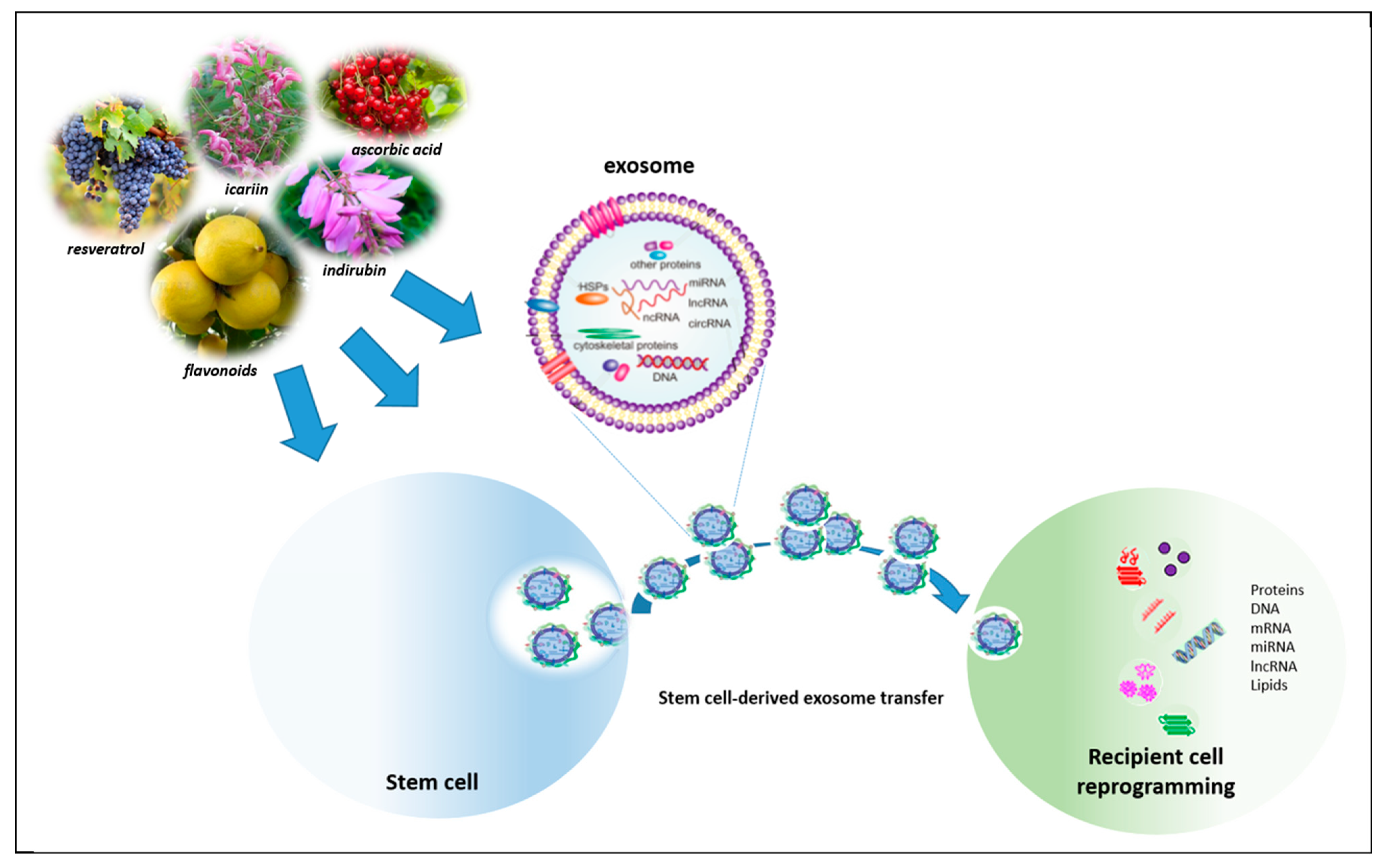
  145. Fan, Y.; Ho, B.X.; Pang, J.K.S.; Pek, N.M.Q.; Hor, J.H.; Ng, S.Y.; Soh, B.S. Wnt/β-catenin-mediated signaling re-activates proliferation of matured cardiomyocytes. Stem Cell Res. Ther. 2018, 9, 338. [Google Scholar] [CrossRef] [PubMed] [Green Version]
  146. MacDonald, B.T.; Tamai, K.; He, X. Wnt/β-catenin signaling: Components, mechanisms, and diseases. Dev. Cell 2009, 17, 9–26. [Google Scholar] [CrossRef] [PubMed] [Green Version]
  147. Wu, D.; Pan, W. GSK3: A multifaceted kinase in Wnt signaling. Trends Biochem. Sci. 2010, 35, 161–168. [Google Scholar] [CrossRef] [PubMed] [Green Version]
  148. Ozhan, G.; Weidinger, G. Wnt/β-catenin signaling in heart regeneration. Cell Regen. 2015, 4, 3. [Google Scholar] [CrossRef] [PubMed] [Green Version]
  149. Hua, Y.; Yang, Y.; Li, Q.; He, X.; Zhu, W.; Wang, J.; Gan, X. Oligomerization of Frizzled and LRP5/6 protein initiates intracellular signaling for the canonical WNT/β-catenin pathway. J. Biol. Chem. 2018, 293, 19710–19724. [Google Scholar] [CrossRef] [Green Version]
  150. Shen, X.; Pan, B.; Zhou, H.; Liu, L.; Lv, T.; Zhu, J.; Huang, X.; Tian, J. Differentiation of mesenchymal stem cells into cardiomyocytes is regulated by miRNA-1-2 via WNT signaling pathway. J. Biomed. Sci. 2017, 24, 29. [Google Scholar] [CrossRef]
  151. Fu, J.D.; Rushing, S.N.; Lieu, D.K.; Chan, C.W.; Kong, C.W.; Geng, L.; Wilson, K.D.; Chiamvimonvat, N.; Boheler, K.R.; Wu, J.C.; et al. Distinct roles of microRNA-1and -499 in ventricular specification and functional maturation of human embryonic stem cells-derived cardiomyocytes. PLoS ONE 2011, 6, e27417. [Google Scholar] [CrossRef] [Green Version]
  152. Marino, F.; Scalise, M.; Cianflone, E.; Mancuso, T.; Aquila, I.; Agosti, V.; Torella, M.; Paolino, D.; Mollace, V.; Nadal-Ginard, B.; et al. Role of c-Kit in Myocardial Regeneration and Aging. Front. Endocrinol. 2019, 10, 371. [Google Scholar] [CrossRef]
  153. Der Sarkissian, S.; Lévesque, T.; Noiseux, N. Optimizing stem cells for cardiac repair: Current status and new frontiers in regenerative cardiology. World J. Stem Cells 2017, 9, 9–25. [Google Scholar] [CrossRef]
  154. Hodgkinson, C.P.; Bareja, A.; Gomez, J.A.; Dzau, V.J. Emerging Concepts in Paracrine Mechanisms in Regenerative Cardiovascular Medicine and Biology. Circ. Res. 2016, 118, 95–107. [Google Scholar] [CrossRef] [Green Version]
  155. Takahashi, M.; Li, T.S.; Suzuki, R.; Kobayashi, T.; Ito, H.; Ikeda, Y.; Matsuzaki, M.; Hamano, K. Cytokines produced by bone marrow cells can contribute to functional improvement of the infarcted heart by protecting cardiomyocytes from ischemic injury. Am. J. Physiol. Heart Circ. Physiol. 2006, 291, H886–H893. [Google Scholar] [CrossRef] [PubMed]
  156. Song, Y.S.; Joo, H.W.; Park, I.H.; Shen, G.Y.; Lee, Y.; Shin, J.H.; Kim, H.; Kim, K.S. Bone marrow mesenchymal stem cell-derived vascular endothelial growth factor attenuates cardiac apoptosis via regulation of cardiac miRNA-23a and miRNA-92a in a rat model of myocardial infarction. PLoS ONE 2017, 12, e0179972. [Google Scholar] [CrossRef] [PubMed] [Green Version]
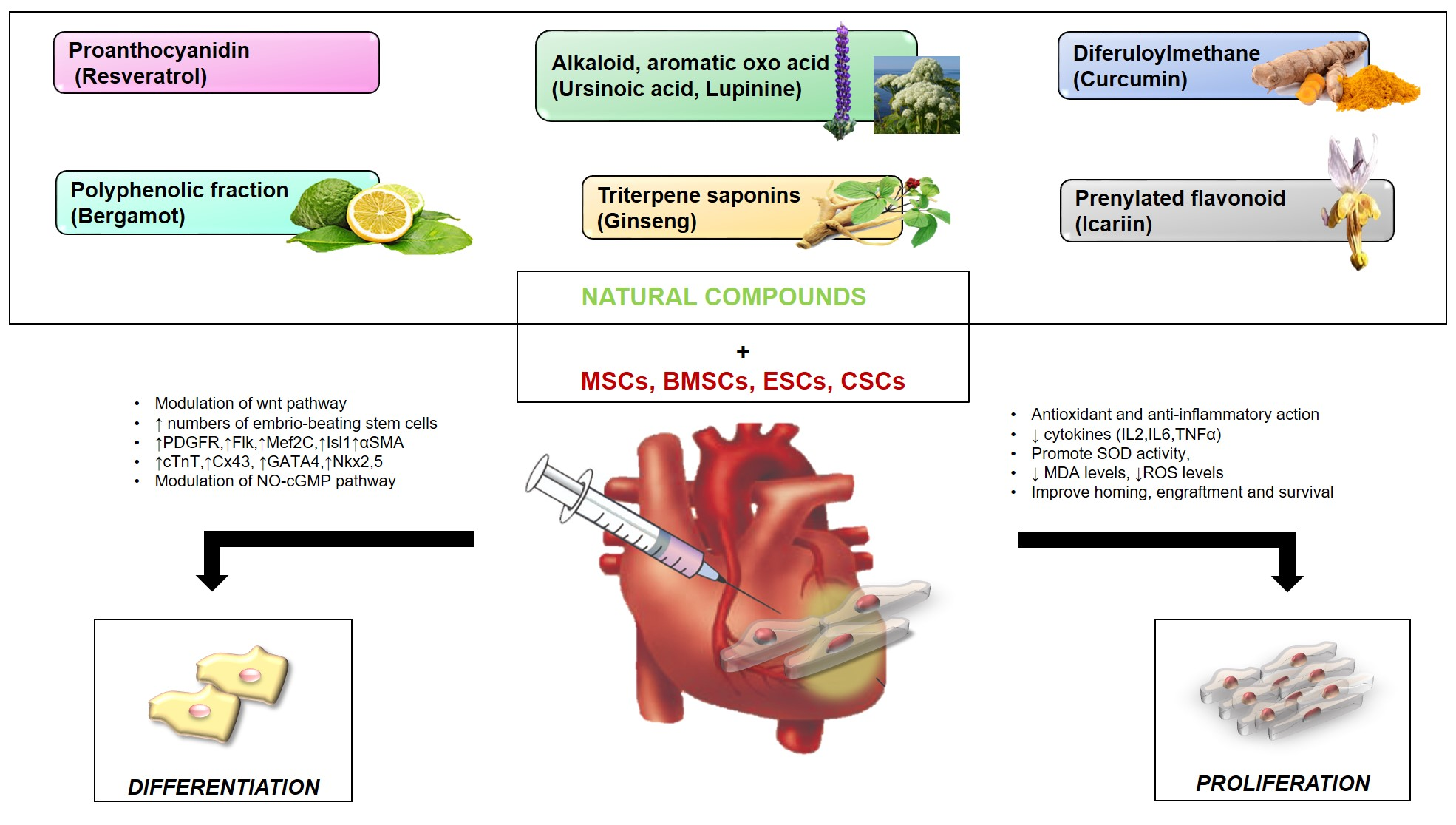
  157. Yuan, Y.; Du, W.; Liu, J.; Ma, W.; Zhang, L.; Du, Z.; Cai, B. Stem Cell-Derived Exosome in Cardiovascular Diseases: Macro Roles of Micro Particles. Front. Pharmacol. 2018, 9, 547. [Google Scholar] [CrossRef] [PubMed] [Green Version]
  158. Mathieu, M.; Martin-Jaular, L.; Lavieu, G.; Théry, C. Specificities of secretion and uptake of exosomes and other extracellular vesicles for cell-to-cell communication. Nat. Cell Biol. 2019, 21, 9–17. [Google Scholar] [CrossRef]
  159. Huang, P.; Wang, L.; Li, Q.; Xu, J.; Xu, J.; Xiong, Y.; Chen, G.; Qian, H.; Jin, C.; Yu, Y.; et al. Combinatorial treatment of acute myocardial infarction using stem cells and their derived exosomes resulted in improved heart performance. Stem Cell Res. Ther. 2019, 10, 300. [Google Scholar] [CrossRef] [Green Version]
  160. Davidson, S.M.; Takov, K.; Yellon, D.M. Exosomes and Cardiovascular Protection. Cardiovasc. Drugs Ther. 2017, 31, 77–86. [Google Scholar] [CrossRef] [Green Version]
  161. Barile, L.; Lionetti, V.; Cervio, E.; Matteucci, M.; Gherghiceanu, M.; Popescu, L.M.; Torre, T.; Siclari, F.; Moccetti, T.; Vassalli, G. Extracellular vesicles from human cardiac progenitor cells inhibit cardiomyocyte apoptosis and improve cardiac function after myocardial infarction. Cardiovasc. Res. 2014, 103, 530–541. [Google Scholar] [CrossRef]
  162. Giricz, Z.; Varga, Z.V.; Baranyai, T.; Sipos, P.; Pálóczi, K.; Kittel, Á.; Buzás, E.I.; Ferdinandy, P. Cardioprotection by remote ischemic preconditioning of the rat heart is mediated by extracellular vesicles. J. Mol. Cardiol. 2014, 68, 75–78. [Google Scholar] [CrossRef] [Green Version]
  163. Li, J.; Rohailla, S.; Gelber, N.; Rutka, J.; Sabah, N.; Gladstone, R.A.; Wei, C.; Hu, P.; Kharbanda, R.K.; Redington, A.N. MicroRNA-144 is a circulating effector of remote ischemic preconditioning. Basic Res. Cardiol. 2014, 109, 423. [Google Scholar] [CrossRef]
  164. Zhang, H.; Wang, H.; Li, N.; Duan, C.E.; Yang, Y.J. Cardiac progenitor/stem cells on myocardial infarction or ischemic heart disease: What we have known from current research. Heart Fail. Rev. 2014, 19, 247–258. [Google Scholar] [CrossRef]
  165. Yuan, H.; Ma, Q.; Ye, L.; Piao, G. The Traditional Medicine and Modern Medicine from Natural Products. Molecules 2016, 21, 559. [Google Scholar] [CrossRef] [PubMed] [Green Version]
  166. Rastogi, S.; Pandey, M.M.; Rawat, A.K. Traditional herbs: A remedy for cardiovascular disorders. Phytomedicine 2016, 23, 1082–1089. [Google Scholar] [CrossRef] [PubMed]
  167. Dias, D.A.; Urban, S.; Roessner, U. A Historical Overview of Natural Products in Drug Discovery. Metabolites 2012, 2, 303–336. [Google Scholar] [CrossRef] [PubMed] [Green Version]
  168. Blanco-Salas, J.; Vazquez, F.M.; Hortigón-Vinagre, M.P.; Ruiz-Tellez, T. Bioactive Phytochemicals from Mercurialis spp. Used in Traditional Spanish Medicine. Plants 2019, 8, 193. [Google Scholar] [CrossRef] [PubMed] [Green Version]
  169. Halberstein, R.A. Medicinal plants: Historical and cross-cultural usage patterns. Ann. Epidemiol. 2005, 15, 686–699. [Google Scholar] [CrossRef]
  170. Hosseinzadeh, S.; Jafarikukhdan, A.; Hosseini, A.; Armand, R. The Application of Medicinal Plants in Traditional and Modern Medicine: A Review of Thymus vulgaris. Int. J. Clin. Med. 2015, 6, 635–642. [Google Scholar] [CrossRef] [Green Version]
  171. Wang, Y.; Ji, G.; Zhu, L. Editorial: Herbal Medicine on High Impact Disease: The Current Progress and Application. Curr. Pharmac. Des. 2017, 23. [Google Scholar] [CrossRef]
  172. Shukla, S.K.; Gupta, S.; Ojha, S.K.; Sharma, S.B. Cardiovascular friendly natural products: A promising approach in the management of CVD. Nat. Prod. Res. 2010, 24, 873–898. [Google Scholar] [CrossRef]
  173. Micucci, M.; Malaguti, M.; Toschi, T.G.; Di Lecce, G.; Aldini, R.; Angeletti, A.; Chiarini, A.; Budriesi, R.; Hrelia, S. Cardiac and Vascular Synergic Protective Effect of Olea europea L. Leaves and Hibiscus sabdariffa L. Flower Extracts. Oxid. Med. Cell Longev. 2015, 2015, 318125. [Google Scholar] [CrossRef] [Green Version]
  174. Arauna, D.; Furrianca, M.; Espinosa-Parrilla, Y.; Fuentes, E.; Alarcón, M.; Palomo, I. Natural Bioactive Compounds as Protectors Of Mitochondrial Dysfunction In Cardiovascular Diseases And Aging. Molecules 2019, 24, 4259. [Google Scholar] [CrossRef] [Green Version]
  175. Kinder, D.H.; Knecht, K.T. Lupine (Lupinus caudatus L., Lupinus albus L.) Seeds: History of Use, Use as an Antihyperglycemic Medicinal, and Use as a Food. Nuts Seeds Health Dis. Prev. 2011, 711–716. [Google Scholar] [CrossRef]
  176. Knecht, K.T.; Nguyen, H.; Auker, A.D.; Kinder, D.H. Effects of extracts of lupine seed on blood glucose levels in glucose resistant mice: Antihyperglycemic effects of Lupinus albus (white lupine, Egypt) and Lupinus caudatus (tailcup lupine, Mesa Verde National Park. J. Herb. Pharmacother. 2006, 6, 89–104. [Google Scholar] [CrossRef] [PubMed]
  177. Belski, R.; Mori, T.A.; Puddey, I.B.; Sipsas, S.; Woodman, R.J.; Ackland, T.R.; Beilin, L.J.; Dove, E.R.; Carlyon, N.B.; Jayaseena, V.; et al. Effects of lupin-enriched foods on body composition and cardiovascular disease risk factors: A 12-month randomized controlled weight loss trial. Int. J. Obes. 2011, 35, 810–819. [Google Scholar] [CrossRef] [PubMed] [Green Version]
  178. Fang, J.; Zhang, Y. Icariin, an Anti-atherosclerotic Drug from Chinese Medicinal Herb Horny Goat Weed. Front. Pharmacol. 2017, 8, 734. [Google Scholar] [CrossRef] [Green Version]
  179. He, C.; Wang, Z.; Shi, J. Pharmacological effects of icariin. Adv. Pharmacol. 2020, 87, 179–203. [Google Scholar]
  180. Zhao, F.; Tang, Y.Z.; Liu, Z.Q. Protective effect of icariin on DNA against radical-induced oxidative damage. J. Pharm. Pharmacol. 2007, 59, 1729–1732. [Google Scholar] [CrossRef]
  181. Song, Y.H.; Cai, H.; Zhao, Z.M.; Chang, W.J.; Gu, N.; Cao, S.P.; Wu, M.L. Icariin attenuated oxidative stress induced-cardiac apoptosis by mitochondria protection and ERK activation. Biomed. Pharmacother. 2016, 83, 1089–1094. [Google Scholar] [CrossRef]
  182. Chen, S.R.; Xu, X.Z.; Wang, Y.H.; Chen, J.W.; Xu, S.W.; Gu, L.Q.; Liu, P.Q. Icariin derivative inhibits inflammation through suppression of p38 mitogen-activated protein kinase and nuclear factor-kappaB pathways. Biol. Pharm. Bull. 2010, 33, 1307–1313. [Google Scholar] [CrossRef] [Green Version]
  183. Hu, Y.; Sun, B.; Liu, K.; Yan, M.; Zhang, Y.; Miao, C.; Ren, L. Icariin attenuates high-cholesterol diet induced atherosclerosis in rats by inhibition of inflammatory response and p38 MAPK signaling pathway. Inflammation 2016, 39, 228–236. [Google Scholar] [CrossRef]
  184. Xiao-Hong, D.; Chang-Qin, X.; Jian-Hua, H.; Wen-Jiang, Z.; Bing, S. Icariin delays homocysteine-induced endothelial cellular senescence involving activation of the PI3K/AKT-eNOS signaling pathway. Pharm. Biol. 2013, 51, 433–440. [Google Scholar] [CrossRef] [Green Version]
  185. Ke, Z.; Liu, J.; Xu, P.; Gao, A.; Wang, L.; Ji, L. The Cardioprotective Effect of Icariin on Ischemia-Reperfusion Injury in Isolated Rat Heart: Potential Involvement of the PI3K-Akt Signaling Pathway. Cardiovasc. Ther. 2015, 33, 134–140. [Google Scholar] [CrossRef] [PubMed] [Green Version]
  186. Ni, T.; Lin, N.; Huang, X.; Lu, W.; Sun, Z.; Zhang, J.; Lin, H.; Chi, J.; Guo, H. Icariin Ameliorates Diabetic Cardiomyopathy Through Apelin/Sirt3 Signalling to Improve Mitochondrial Dysfunction. Front. Pharmacol. 2020, 11, 256. [Google Scholar] [CrossRef] [PubMed]
  187. Song, Y.H.; Cai, H.; Gu, N.; Qian, C.F.; Cao, S.P.; Zhao, Z.M. Icariin attenuates cardiac remodelling through down-regulating myocardial apoptosis and matrix metalloproteinase activity in rats with congestive heart failure. J. Pharm Pharmacol. 2011, 63, 541–549. [Google Scholar] [CrossRef] [PubMed]
  188. Yang, Y.; Ren, C.; Zhang, Y.; Wu, X.D. Ginseng: An Nonnegligible Natural Remedy for Healthy Aging. Aging Dis. 2017, 8, 708–720. [Google Scholar] [CrossRef] [Green Version]
  189. Kim, J.H. Cardiovascular Diseases and Panax ginseng: A Review on Molecular Mechanisms and Medical Applications. J. Ginseng Res. 2012, 36, 16–26. [Google Scholar] [CrossRef] [Green Version]
  190. Radad, K.; Gille, G.; Liu, L.; Rausch, W.D. Use of Ginseng in Medicine with Emphasis on Neurodegenerative Disorders. J. Pharmacol. Sci. 2006, 100, 175–186. [Google Scholar] [CrossRef] [Green Version]
  191. Ramesh, T.; Kim, S.W.; Hwang, S.Y.; Sohn, S.H.; Yoo, S.K.; Kim, S.K. Panax ginseng reduces oxidative stress and restores antioxidant capacity in aged rats. Nutr Res. 2012, 32, 718–726. [Google Scholar] [CrossRef]
  192. Liu, L.; Kelly, M.G.; Wierzbicki, E.L.; Escober-Nario, I.C.; Vollmer, M.K.; Doré, S. An essential role of Nrf2 in American ginseng-mediated anti-oxidative actions in cardiomyocytes. J. Ethnopharmacol. 2010, 130, 222–230. [Google Scholar] [CrossRef] [Green Version]
  193. Yi, X.Q.; Li, T.; Wang, J.R.; Wong, V.K.; Luo, P.; Wong, I.Y.; Jiang, Z.H.; Liu, L.; Zhou, H. Total ginsenosides increase coronary perfusion flow in isolated rat hearts through activation of PI3K/Akt-eNOS signaling. Phytomedicine 2010, 17, 1006–1015. [Google Scholar] [CrossRef]
  194. Tsutsumi, Y.M.; Tsutsumi, R.; Mawatari, K.; Nakaya, Y.; Kinoshita, M.; Tanaka, K.; Oshita, S. Compound K, a metabolite of ginsenosides, induces cardiac protection mediated nitric oxide via Akt/PI3K pathway. Life Sci. 2011, 88, 725–729. [Google Scholar] [CrossRef]
  195. Lim, K.H.; Ko, D.; Kim, J.H. Cardioprotective potential of Korean Red Ginseng extract on isoproterenol-induced cardiac injury in rats. J. Ginseng Res. 2013, 37, 273–282. [Google Scholar] [CrossRef] [PubMed] [Green Version]
  196. Zhang, L.P.; Jiang, Y.C.; Yu, X.F.; Xu, H.L.; Li, M.; Zhao, X.Z.; Sui, D.Y. Ginsenoside Rg3 Improves Cardiac Function after Myocardial ischemia/Reperfusion via Attenuating Apoptosis and Inflammation. Evid. Based Complement. Alternat. Med. 2016, 2016, 6967853. [Google Scholar] [CrossRef] [PubMed]
  197. Ahn, C.M.; Hong, S.J.; Choi, S.C.; Park, J.H.; Kim, J.S.; Lim, D.S. Red ginseng extract improves coronary flow reserve and increases absolute numbers of various circulating angiogenic cells in patients with first ST-segment elevation acute myocardial infarction. Phytother. Res. 2011, 25, 239–249. [Google Scholar] [CrossRef] [PubMed]
  198. Kim, J.H. Pharmacological and medical applications of Panax ginseng and ginsenosides: A review for use in cardiovascular diseases. J. Ginseng Res. 2018, 42, 264–269. [Google Scholar] [CrossRef] [PubMed]
  199. Hatcher, H.; Planalp, R.; Cho, J.; Torti, F.M.; Torti, S.V. Curcumin: From ancient medicine to current clinical trials. Cell Mol. Life Sci. 2008, 65, 1631–1652. [Google Scholar] [CrossRef]
  200. Prasad, S.; Aggarwal, B.B. Turmeric, the Golden Spice. From Traditional Medicine to Modern Medicine. In Herbal Medicine: Biomolecular and Clinical Aspects, 2nd ed.; Benzie, I.F.F., Wachtel-Galor, S., Eds.; CRC Press/Taylor & Francis: Boca Raton, FL, USA, 2011. [Google Scholar]
  201. Li, H.; Sureda, A.; Devkota, H.P.; Pittalà, V.; Barreca, D.; Silva, A.S.; Nabavi, S.M. Curcumin, the golden spice in treating cardiovascular diseases. Biotechnol. Adv. 2020, 38, 107343. [Google Scholar] [CrossRef]
  202. Wongcharoen, W.; Phrommintikul, A. The protective role of curcumin in cardiovascular diseases. Int. J. Cardiol. 2009, 133, 145–151. [Google Scholar] [CrossRef]
  203. Li, W.; Wu, M.; Tang, L.; Pan, Y.; Liu, Z.; Zeng, C.; Wang, J.; Wei, T.; Liang, G. Novel curcumin analogue 14p protects against myocardial ischemia reperfusion injury through Nrf2-activating anti-oxidative activity. Toxicol. Appl. Pharmacol. 2015, 282, 175–183. [Google Scholar] [CrossRef]
  204. Tian, K.; Ogura, S.; Little, P.J.; Xu, S.W.; Sawamura, T. Targeting LOX-1 in atherosclerosis and vasculopathy: Current knowledge and future perspectives. Ann. N. Y. Acad. Sci. 2019, 1443, 34–53. [Google Scholar] [CrossRef]
  205. Thompson, J.W.; Wei, J.; Appau, K.; Wang, H.; Yu, H.; Spiga, M.G.; Graham, R.M.; Webster, K.A. Bnip3 binds and activates p300: Possible role in cardiac transcription and myocyte morphology. PLoS ONE 2015, 10, e0136847. [Google Scholar] [CrossRef] [Green Version]
  206. Rahnavard, M.; Hassanpour, M.; Ahmadi, M.; Heidarzadeh, M.; Amini, H.; Javanmard, M.Z.; Nouri, M.; Rahbarghazi, R.; Safaie, N. Curcumin ameliorated myocardial infarction by inhibition of cardiotoxicity in the rat model. J. Cell Biochem. 2019. [CrossRef] [PubMed]
  207. Wang, N.P.; Wang, Z.F.; Tootle, S.; Philip, T.; Zhao, Z.Q. Curcumin promotes cardiac repair and ameliorates cardiac dysfunction following myocardial infarction. Br. J. Pharmacol. 2012, 167, 1550–1562. [Google Scholar] [CrossRef] [PubMed] [Green Version]
  208. Jeong, C.W.; Yoo, K.Y.; Lee, S.H.; Jeong, H.J.; Lee, C.S.; Kim, S.J. Curcumin protects against regional myocardial ischemia/reperfusion injury through activation of RISK/GSK-3β and inhibition of p38 MAPK and JNK. J. Cardiovasc. Pharmacol. Ther. 2012, 17, 387–394. [Google Scholar] [CrossRef] [PubMed]
  209. Liu, H.; Wang, C.; Qiao, Z.; Xu, Y. Protective effect of curcumin against myocardium injury in ischemia reperfusion rats. Pharm. Biol. 2017, 55, 1144–1148. [Google Scholar] [CrossRef]
  210. Bertelli, A.A.; Das, D.K. Grapes, Wines, Resveratrol, and Heart Health. J. Cardiovasc. Pharmacol. 2009, 54, 468–476. [Google Scholar] [CrossRef]
  211. Zordoky, B.N.; Robertson, I.M.; Dyck, J.R. Preclinical and clinical evidence for the role of resveratrol in the treatment of cardiovascular diseases. Biochim. Biophys. Acta 2015, 1852, 1155–1177. [Google Scholar] [CrossRef] [Green Version]
  212. Riba, A.; Deres, L.; Sumegi, B.; Toth, K.; Szabados, E.; Halmosi, R. Cardioprotective Effect of Resveratrol in a Postinfarction Heart Failure Model. Oxid. Med. Cell Longev. 2017, 2017, 6819281. [Google Scholar] [CrossRef]
  213. Bonnefont-Rousselot, D. Resveratrol and Cardiovascular Diseases. Nutrients 2016, 8, 250. [Google Scholar] [CrossRef]
  214. Magyar, K.; Halmosi, R.; Palfi, A.; Feher, G.; Czopf, L.; Fulop, A.; Battyany, I.; Sumegi, B.; Toth, K.; Szabados, E. Cardioprotection by resveratrol: A human clinical trial in patients with stable coronary artery disease. Clin. Hemorheol. Microcirc. 2012, 50, 179–187. [Google Scholar] [CrossRef]
  215. Walle, T.; Hsieh, F.; DeLegge, M.H.; Oatis, J.E.; Walle, U.K. High absorption but very low bioavailability of oral resveratrol in humans. Drug Metab. Dispos. 2004, 32, 1377–1382. [Google Scholar] [CrossRef] [Green Version]
  216. Carresi, C.; Gliozzi, M.; Musolino, V.; Scicchitano, M.; Scarano, F.; Bosco, F.; Nucera, S.; Maiuolo, J.; Macrì, R.; Ruga, S.; et al. The Effect of Natural Antioxidants in the Development of Metabolic Syndrome: Focus on Bergamot Polyphenolic Fraction. Nutrients 2020, 12, 1504. [Google Scholar] [CrossRef]
  217. Gliozzi, M.; Carresi, C.; Musolino, V.; Palma, E.; Muscoli, C.; Vitale, C.; Gratteri, S.; Muscianisi, G.; Janda, E.; Muscoli, S.; et al. The effect of bergamot-derived polyphenolic fraction on LDL small dense particles and non-alcoholic fatty liver disease in patients with metabolic syndrome. Adv. Biol. Chem. 2014, 4, 129–137. [Google Scholar] [CrossRef] [Green Version]
  218. Musolino, V.; Gliozzi, M.; Nucera, S.; Carresi, C.; Maiuolo, J.; Mollace, R.; Paone, S.; Bosco, F.; Scarano, F.; Scicchitano, M.; et al. The effect of bergamot polyphenolic fraction on lipid transfer protein system and vascular oxidative stress in a rat model of hyperlipemia. Lipids Health Dis. 2019, 18, 115. [Google Scholar] [CrossRef] [PubMed] [Green Version]
  219. Mollace, V.; Sacco, I.; Janda, E.; Malara, C.; Ventrice, D.; Colica, C.; Visalli, V.; Muscoli, S.; Ragusa, S.; Muscoli, C.; et al. Hypolipemic and hypoglycaemic activity of bergamot polyphenols: From animal models to human studies. Fitoterapia 2011, 82, 309–316. [Google Scholar] [CrossRef] [PubMed]
  220. Parafati, M.; Lascala, A.; Morittu, V.M.; Trimboli, F.; Rizzuto, A.; Brunelli, E.; Coscarelli, F.; Costa, N.; Britti, D.; Ehrlich, J.; et al. Bergamot polyphenol fraction prevents nonalcoholic fatty liver disease via stimulation of lipophagy in cafeteria diet-induced rat model of metabolic syndrome. J. Nutr. Biochem. 2015, 26, 938–948. [Google Scholar] [CrossRef] [PubMed]
  221. Musolino, V.; Gliozzi, M.; Scarano, F.; Bosco, F.; Scicchitano, M.; Nucera, S.; Carresi, C.; Ruga, S.; Zito, M.C.; Maiuolo, J.; et al. Bergamot polyphenols improve dyslipidemia and pathophysiological features in a Mouse Model of non-Alcoholic fatty Liver Disease. Sci. Rep. 2020, 10, 2565. [Google Scholar] [CrossRef]
  222. Gliozzi, M.; Walker, R.; Muscoli, S.; Vitale, C.; Gratteri, S.; Carresi, C.; Musolino, V.; Russo, V.; Janda, E.; Ragusa, S.; et al. Bergamot polyphenolic fraction enhances rosuvastatin-induced effect on LDL-cholesterol, LOX-1 expression and protein kinase B phosphorylation in patients with hyperlipidemia. Int. J. Cardiol. 2013, 170, 140–145. [Google Scholar] [CrossRef]
  223. Carresi, C.; Musolino, V.; Gliozzi, M.; Maiuolo, J.; Mollace, R.; Nucera, S.; Maretta, A.; Sergi, D.; Muscoli, S.; Gratteri, S.; et al. Anti-oxidant effect of bergamot polyphenolic fraction counteracts doxorubicin-induced cardiomyopathy: Role of autophagy and ckitposCD45negCD31neg cardiac stem cell activation. J. Mol. Cell. Cardiol. 2018, 119, 10–18. [Google Scholar] [CrossRef]
  224. La Russa, D.; Giordano, F.; Marrone, A.; Parafati, M.; Janda, E.; Pellegrino, D. Oxidative Imbalance and Kidney Damage in Cafeteria Diet-Induced Rat Model of Metabolic Syndrome: Effect of Bergamot Polyphenolic Fraction. Antioxidants 2019, 8, 66. [Google Scholar] [CrossRef] [Green Version]
  225. Musolino, V.; Gliozzi, M.; Bombardelli, E.; Nucera, S.; Carresi, C.; Maiuolo, J.; Mollace, R.; Paone, S.; Bosco, F.; Scarano, F.; et al. The synergistic effect of Citrus bergamia and Cynara cardunculus extracts on vascular inflammation and oxidative stress in nonalcoholic fatty liver disease. J. Tradit. Complement. Med. 2020, 10, 268–274. [Google Scholar] [CrossRef]
  226. Nikonov, G.K.; Yagudaev, M.R. Structure of ursinoic acid and ursinin. Chem. Nat. Comp. 1970, 6, 429–432. [Google Scholar] [CrossRef]
  227. Lee, J.A.; An, J.; Kang, T.M.; De, D.; Kim, K.K. Discovery of Natural Compounds Promoting Cardiomyocyte Differentiation. Stem Cells Dev. 2019, 28, 13–27. [Google Scholar] [CrossRef]
  228. Gurusamy, N.; Ray, D.; Lekli, I.; Das, D.K. Red wine antioxidant resveratrol-modified cardiac stem cells regenerate infarcted myocardium. J. Cell. Mol. Med. 2010, 14, 2235–2239. [Google Scholar] [CrossRef] [Green Version]
  229. Ling, L.; Gu, S.; Cheng, Y. Resveratrol activates endogenous cardiac stem cells and improves myocardial regeneration following acute myocardial infarction. Mol. Med. Rep. 2017, 15, 1188–1194. [Google Scholar] [CrossRef] [Green Version]
  230. Gorbunov, N.; Petrovski, G.; Gurusamy, N.; Ray, D.; Kim, D.H.; Das, D.K. Regeneration of infarcted myocardium with resveratrol-modified cardiac stem cells. J. Cell. Mol. Med. 2012, 16, 174–184. [Google Scholar] [CrossRef] [Green Version]
  231. ShamsEldeen, A.M.; Ashour, H.; Shoukry, H.S.; Fadel, M.; Kamar, S.S.; Aabdelbaset, M.; Rashed, L.A.; Ammar, H.I. Combined treatment with systemic resveratrol and resveratrol preconditioned mesenchymal stem cells, maximizes antifibrotic action in diabetic cardiomyopathy. J. Cell. Physiol. 2019, 234, 10942–10963. [Google Scholar] [CrossRef]
  232. Liu, H.; Zhang, S.; Zhao, L.; Zhang, Y.; Li, Q.; Chai, X.; Zhang, Y. Resveratrol Enhances Cardiomyocyte Differentiation of Human Induced Pluripotent Stem Cells through Inhibiting Canonical WNT Signal Pathway and Enhancing Serum Response Factor-miR-1 Axis. Stem Cells Int. 2016, 2016, 2524092. [Google Scholar] [CrossRef] [Green Version]
  233. Ding, H.; Xu, X.; Qin, X.; Yang, C.; Feng, W. Resveratrol promotes differentiation of mouse embryonic stem cells to cardiomyocytes. Cardiovasc. Ther. 2016, 34, 283–289. [Google Scholar] [CrossRef] [PubMed] [Green Version]
  234. Pinarli, F.A.; Turan, N.N.; Pinarli, F.G.; Okur, A.; Sonmez, D.; Ulus, T.; Oguz, A.; Karadeniz, C.; Delibasi, T. Resveratrol and Adipose-derived Mesenchymal Stem Cells Are Effective in the Prevention and Treatment of Doxorubicin Cardiotoxicity in Rats. Pediatr. Hematol. Oncol. 2013, 30, 226–238. [Google Scholar] [CrossRef] [PubMed]
  235. Hu, C.; Li, L. The application of resveratrol to mesenchymal stromal cell-based regenerative medicine. Stem Cell Res. Ther. 2019, 10, 307. [Google Scholar] [CrossRef] [PubMed] [Green Version]
  236. Hu, W.; Jing, P.; Wang, L.; Zhang, Y.; Yong, J.; Wang, Y. The positive effects of Ginsenoside Rg1 upon the hematopoietic microenvironment in a D-Galactose-induced aged rat model. BMC Complement. Altern. Med. 2015, 15, 119. [Google Scholar] [CrossRef] [PubMed] [Green Version]
  237. Chan, L.S.; Yue, P.Y.K.; Mak, N.K.; Wong, R.N.S. Role of MicroRNA-214 in ginsenoside-Rg1-induced angiogenesis. Eur. J. Pharm. Sci. 2009, 38, 370–377. [Google Scholar] [CrossRef] [PubMed]
  238. Wang, Y.; Zhao, R.; Liu, D.; Deng, W.; Xu, G.; Liu, W.; Rong, J.; Long, X.; Ge, J.; Shi, B. Exosomes Derived from miR-214-Enriched Bone Marrow-Derived Mesenchymal Stem Cells Regulate Oxidative Damage in Cardiac Stem Cells by Targeting CaMKII. Oxid. Med. Cell. Longev. 2018, 2018, 4971261. [Google Scholar] [CrossRef] [PubMed]
  239. Kim, Y.Y.; Ku, S.Y.; Rosenwaks, Z.; Liu, H.C.; Oh, S.K.; Moon, S.Y.; Choi, Y.M. Red Ginseng Extract Facilitates the Early Differentiation of Human Embryonic Stem Cells into Mesendoderm Lineage. Evid. Based Complement. Alternat. Med. 2011, 2011, 167376. [Google Scholar] [CrossRef]
  240. Sasaki, T.; Oh, K.B.; Matsuoka, H.; Saito, M. Effect of Panax ginseng components on the differentiation of mouse embryonic stem cells into cardiac-like cells. Yakugaku Zasshi 2008, 128, 461–467. [Google Scholar] [CrossRef] [Green Version]
  241. Chan, L.S.; Yue, P.Y.; Wong, Y.Y.; Wong, R.N. MicroRNA-15b contributes to ginsenoside-Rg1-induced angiogenesis through increased expression of VEGFR-2. Biochem. Pharmacol. 2013, 86, 392–400. [Google Scholar] [CrossRef]
  242. He, F.; Yu, C.; Liu, T.; Jia, H. Ginsenoside Rg1 as an Effective Regulator of Mesenchymal Stem Cells. Front. Pharmacol. 2020, 10, 1565. [Google Scholar] [CrossRef] [Green Version]
  243. Yang, M.; Chen, G.; Chen, C.; Zhang, Y.; Yan, C. Effect of Ginsenoside Rg1 in promoting myocardial regeneration after myocardial infarction in rats (in Chinese). Chin. Heart. J. 2008, 20, 697–707. [Google Scholar]
  244. Kim, A.R.; Kim, S.W.; Lee, B.W.; Kim, K.H.; Kim, W.H.; Kim, W.H.; Seok, H.; Lee, J.H.; Um, J.; Yim, S.H.; et al. Screening ginseng saponins in progenitor cells identifies 20(R)-ginsenoside Rh2 as an enhancer of skeletal and cardiac muscle regeneration. Sci. Rep. 2020, 10, 4967. [Google Scholar] [CrossRef]
  245. Zhu, D.Y.; Lou, Y.J. Inducible effects of icariin, icaritin, and desmethylicaritin on directional differentiation of embryonic stem cells into cardiomyocytes in vitro1. Acta Pharmacol. Sin. 2005, 26, 477–485. [Google Scholar] [CrossRef] [Green Version]
  246. Zhu, D.; Qu, L.; Zhang, X.; Lou, Y. Icariin-mediated modulation of cell cycle and p53 during cardiomyocyte differentiation in embryonic stem cells. Eur. J. Pharmacol. 2005, 514, 99–110. [Google Scholar] [CrossRef]
  247. Zhu, D.Y.; Lou, Y.J. Icariin-mediated expression of cardiac genes and modulation of nitric oxide signaling pathway during differentiation of mouse embryonic stem cells into cardiomyocytes in vitro1. Acta Pharmacol. Sin. 2006, 27, 311–320. [Google Scholar] [CrossRef] [Green Version]
  248. Wo, Y.; Zhu, D.; Hu, Y.; Wang, Z.Q.; Liu, J.; Lou, Y.J. Reactive oxygen species involved in prenylflavonoids, icariin and icaritin, initiating cardiac differentiation of mouse embryonic stem cells. J. Cell. Biochem. 2008, 103, 1536–1550. [Google Scholar] [CrossRef]
  249. Jiang, C.; Gong, F. BMP-2 and icariin synergistically promote p38MAPK-mediated cardiomyocyte differentiation of mesenchymal stem cells via enhanced NOX4-driven ROS generation. Medic. Chem. Res. 2017, 26, 2547–2556. [Google Scholar] [CrossRef]
  250. Jin, M.S.; Shi, S.; Zhang, Y.; Yan, Y.; Sun, X.D.; Liu, W.; Liu, H.W. Icariin-mediated differentiation of mouse adipose-derived stem cells into cardiomyocytes. Mol. Cell. Biochem. 2010, 344, 1–9. [Google Scholar] [CrossRef]
  251. Backs, J.; Olson, E.N. Control of cardiac growth by histone acetylation/deacetylation. Circ. Res. 2006, 98, 15–24. [Google Scholar] [CrossRef]
  252. Zheng, M.; Zhu, J.; Lu, T.; Liu, L.; Sun, H.; Liu, Z.; Tian, J. p300-Mediated Histone Acetylation is Essential for the Regulation of GATA4 and MEF2C by BMP2 in H9c2 Cells. Cardiovasc. Toxicol. 2013, 13, 316–322. [Google Scholar] [CrossRef]
  253. Wang, L.; Sun, H.; Pan, B.; Zhu, J.; Huang, G.; Huang, X.; Tian, J. Inhibition of histone acetylation by curcumin reduces alcohol-induced expression of heart development-related transcription factors in cardiac progenitor cells. Biochem. Biophys. Res. Commun. 2012, 424, 593–596. [Google Scholar] [CrossRef]
  254. Liu, J.; Zhu, P.; Song, P.; Xiong, W.; Chen, H.; Peng, W.; Wang, S.; Li, S.; Fu, Z.; Wang, Y.; et al. Pretreatment of Adipose Derived Stem Cells with Curcumin Facilitates Myocardial Recovery via Antiapoptosis and Angiogenesis. Stem Cells Int. 2015, 2015, 638153. [Google Scholar] [CrossRef] [Green Version]
  255. Mujoo, K.; Nikonoff, L.E.; Sharin, V.G.; Bryan, N.S.; Kots, A.Y.; Murad, F. Curcumin induces differentiation of embryonic stem cells through possible modulation of nitric oxide-cyclic GMP pathway. Protein Cell 2012, 3, 535–544. [Google Scholar] [CrossRef] [Green Version]
  256. Wang, X.; Zhang, Y.; Yang, Y.; Zhang, W.; Luo, L.; Han, F.; Guan, H.; Tao, K.; Hu, D. Curcumin pretreatment protects against hypoxia/reoxgenation injury via improvement of mitochondrial function, destabilization of HIF-1α and activation of Epac1-Akt pathway in rat bone marrow mesenchymal stem cells. Biomed. Pharmacother. 2019, 109, 1268–1275. [Google Scholar] [CrossRef] [PubMed]
  257. Wang, L.; Zhang, Y.G.; Wang, X.M.; Ma, L.F.; Zhang, Y.M. Naringin protects human adipose-derived mesenchymal stem cells against hydrogen peroxide-induced inhibition of osteogenic differentiation. Chem. Biol. Interact. 2015, 242, 255–261. [Google Scholar] [CrossRef] [PubMed]
  258. Ring, A.; Kim, Y.M.; Kahn, M. Wnt/catenin signaling in adult stem cell physiology and disease. Stem Cell Rev. 2014, 10, 512–525. [Google Scholar] [CrossRef] [PubMed] [Green Version]
  259. Su, F.; Zhang, W.; Liu, J. Membrane estrogen receptor alpha is an important modulator of bone marrow c-Kit+ cells mediated cardiac repair after myocardial infarction. Int. J. Clin. Exp. Pathol. 2015, 8, 4284–4295. [Google Scholar] [PubMed]
Figure 1. Potential properties of natural compounds in the stimulation of stem cell paracrine signaling. Some natural compounds (ascorbic acid, indirubin, resveratrol, icariin, bergamot polyphenols) emerging as useful tools in improving homing, engraftment, self-renewal and stem cell cardiovascular lineage specification, possibly, through paracrine signaling regulation. Stem cells exert paracrine effects on neighboring somatic cells via the secretion of micro-vesicles, such as exosomes, containing hundreds of different proteins, mRNAs and miRNAs under normal conditions or in response to cellular stress. Transferring the content of stem cell-derived exosomes can facilitate reprogramming of the recipient somatic cell.
Nutrients 13 00275 g001
Figure 2. The biphasic role of wnt/β-catenin pathway in heart muscle development. (A) Wnt/β-catenin pathway promotes mesoderm specification in the early stages of hESC differentiation, while, its activation inhibits CM differentiation in the later stages. The cardiac differentiation inducer Mesp1 activates the inhibitor of canonical wnts, Dkk1, promoting CM differentiation. Moreover, the presence of wnt ligands inhibit GSK3β and lead to β-catenin stabilization in the cytoplasm and its traslocation into the nucleus promoting CM proliferation. (B) The overexpression of miR1-2 induces the expression of cardiac specific genes BM-MSCs and activates the wnt/β-catenin signaling pathway promoting their cardiac differentiation. (C) Wnt-3a, wnt-3a-conditioned medium and BIO stimulate CSC proliferation and clonogenicity, while, significantly inhibit CSC differentiation into myocyte-committed cells. In contrast, Dkk1 decreases CSC proliferation and clonogenicity and promotes myocyte specification, through the inhibition of the canonical wnt pathway. hESCs: human Embrionic Stem Cells; CMs: Cardiomyocytes; Dkk-1: Dickkopf-related protein 1; GSK3-β: Glycogen synthase kinase 3- β; BM-MSCs: Bone Marrow derived Mesenchimal Stem Cells; Nkx2.5: NK2 transcription factor related locus 5; cTnI: cardiac Troponin I; CSCs: Cardiac Stem Cells; JNK: c-Jun N-terminal kinase; BIO: bromoindirubin-3′-oxime; ↑ increase.
Nutrients 13 00275 g002
Figure 3. The role of certain natural compounds in stem cell activation. ↑ increase, ↓decrease. ESCs: embryonic stem cells; CMs: cardiomyocytes; GATA-4: GATA-binding protein 4; Nkx2.5: NK2 transcription factor related locus 5; EBs: embryoid bodies; PDGFR: Platelet-Derived Growth Factor Receptor; Mef2C: Myocyte Enhancer Factor 2c; Isl1: Insulin gene enhancer protein-1; α-SMA: α-Smooth Muscle Actin; cTnI: cardiac Troponin I; Cx43: Connexin 43; BMSCs: bone marrow stem cells; MSCs: mesenchymal stem cells; IL-2: interleukin-2; IL-6: interleukin-6; TNF-α: tumor necrosis factor-α; SOD: superoxide dismutase; MDA: malondialdehyde; ROS: reactive oxygen species; G-CSF: granulocyte colony-stimulating factor; Oct4: Octamer-binding transcription factor 4; Sox-2: SRY-box transcription factor-2; CSCs: cardiac stem cells; MI: myocardial infarction; LVEDd: left ventricular end diastolic diameter; VEGF: vascular endothelial growth factor; SDF-1: stromal cell-derived factor-1; MLC2v: myosin light chain 2v; Mdm2: E3 ubiquitin-protein ligase; NO: nitric oxide; FS: fractional shortening; EF: ejection fraction; NOS3: nitric oxide synthase 3; NOS2: nitric oxide synthase 2; CDKI p21: cyclin-dependent kinases inhibitor p21; HIF-1 α: Hypoxia-Inducible Factor-1α; I/R: ischemia/reperfusion; HO-1: heme oxigenase-1.
Nutrients 13 00275 g003
Publisher’s Note: MDPI stays neutral with regard to jurisdictional claims in published maps and institutional affiliations.

