Article

Chelator-Free Copper-64-Incorporated Iron Oxide Nanoparticles for PET/MR Imaging: Improved Radiocopper Stability and Cell Viability

1
Korea Multi-Purpose Accelerator Complex, Korea Atomic Energy Research Institute, 181 Mirae-ro, Gyeongju 38180, Korea
2
Division of Physics and Semiconductor Science, Dongguk University, Seoul 04620, Korea
3
Korea Atomic Energy Research Institute, Daedeok-daero 989, Daejeon 34057, Korea
4
Department of Advanced Materials Chemistry, Dongguk University, Gyeongju 38066, Korea
5
Advanced Bio Convergence, Pohang Technopark, Pohang 37668, Korea
*
Authors to whom correspondence should be addressed.
Nanomaterials 2022, 12(16), 2791; https://doi.org/10.3390/nano12162791
Submission received: 25 July 2022 / Revised: 6 August 2022 / Accepted: 10 August 2022 / Published: 14 August 2022
(This article belongs to the Special Issue Advances in Biocompatible Nanocomposites)

Abstract

:
We have developed chelator-free copper-64-incorporated iron oxide (IO) nanoparticle (NPs) which have both magnetic and radioactive properties being applied to positron emission tomography (PET)-magnetic resonance imaging (MRI). We have found that the IO nanoparticles composed of radioactive isotope 64Cu may act as a contrast agent being a diagnostic tool for PET as well as a good T2 MRI nanoprobe due to their good r2/r1 ratio. Furthermore, we demonstrate that the 64Cu incorporation at the core of core-shell-structured IO NPs exhibits a good in vivo stability, giving us an insightful strategy for the design of a contrast agent for the PET-MRI system.

Graphical Abstract

1. Introduction

To date, chelator-based radio-labeling is a widely used technique for the synthesis of various kinds of radio-labeled nanoparticles (NPs), which may be applied to positron emission tomography (PET) of a proven imaging technique. For example, porous silica NPs to attach fluorine-18 (18F, t1/2 = 109 min), copper-64 (64Cu, t1/2 = 12.7 h)-labeled carbon nanotubes, quantum dots, and superparamagnetic iron oxide NPs [1,2] have been developed for tumor-targeted cancer imaging and biodistribution studies. On the other hand, magnetic resonance imaging (MRI) as a noninvasive method gives good morphological or functional images for soft tissues. For diagnostic MRI procedures, however, we further need contrast agents (CAs) to enhance sensitivity and visuality for lesions [3,4]. For decades, a dual-modality probe for hybrid PET-MRI systems has been developed as an effective clinical diagnostic tool due to its synergetic combination of PET with high sensitivity and MRI with high resolution. It thus enables us to reduce the number of CAs injected into patients while fully benefitting both modalities [5].
To develop a stable radiopharmaceutical, it is important to select the best chelator with sufficient in vivo stability and to know the particular coordination chemistry. However, these are challenging tasks, so we need a simple but effective chelator-free strategy [4,6,7,8]. Recently, Chen et al. synthesized chelator-free zirconium-89-labeled mesoporous silica NPs enhancing their long-term in vivo integrity [9]. Wong et al. have also developed chelator-free copper-64-doped iron oxide (IO) NPs, but the dextran coating may easily be detached from the NPs when they are injected into the body, thus not perfectly avoiding the cytotoxicity effect [10].

2. Materials and Methods

2.1. Materials

To synthesize 64Cu-incorporated iron oxide (64Cu-IO) NPs, we purchased the reagents acetylacetone, sodium acetate, copper chloride, benzyl ether, iron acetylacetonate, lauric acid, dodecylamine, cyclohexane, ammonia hydroxide, and tetraethylorthosilicate from Alfa Aesar. In addition, reagents 1,2-hexadicanediol and igepal CO-520 were purchased from Sigma Aldrich (Burlington, MA, USA). 64Cu was supplied by Korea Institute of Radiological & Medical Sciences.

2.2. Production of Copper-64

The production of 64Cu was performed using concentrated nickel (64Ni). In order to make a nickel target for beam irradiation, a plating solution was prepared using 64Ni, and nickel sulfide (64NiSO4) was obtained from the concentrated 64Ni. The average energy of the proton beam was 12.3 MeV for the 64Ni(p,n)64Cu nuclear reaction, in which beam irradiation was performed using a 50 MeV accelerator (Scanditronix, Olvagen, Vislanda, Swden, 1985) at Korea institute of radiological&medical Sciences (KIRAMS). After beam irradiation, the target was separated from the target system; 10 mL of 6 N HCl solution was then taken in, stirred at 80 °C for 10 min to dissolve the concentrated material, and then cooled down to room temperature. This solution was adsorbed into a column with AG1-X8 (100–200 mesh) resin (4 cm × 10 cm), and, while flowing 10 mL of 6 N HCl solution into the column, collection of the eluted solution was in a prepared recovery container. After flowing the HCl solution, it was flowed into the column using 10 mL of high-purity distilled water for about 10 min, and the solution eluted in this process was collected in a sterile vial to recover 64Cu.

2.3. nat,64Cu(acac)2 Synthesis

After mixing 5 mCi of 64Cu and 34 nmol of CuCl2, 2.5 equivalents of acetylacetone were added and then stirred for 5 min. Next, 3 equivalents of sodium acetate were added to crystallize, and the mixture was then stirred at 80 °C for 15 min, followed by cooling at room temperature. After centrifugation to separate the solvent and the solute, it was washed three times with benzyl ether. Finally, 64Cu recovery was measured by a dose calibrator.

2.4. Core-Shell Structures of IO and 64Cu-IO

The 64Cu acetylacetonate (64Cu(acac)2) total, 0.98 g iron acetylacetate, 0.387 g 1,2-hexadecanediol, 0.3 g lauric acid, 0.344 mL dodecylamine, and 100 mL benzyl ether were stirred at 180 °C for 2 h under argon gas condition. After the reaction, the resulting 64Cu-IO NPs were collected after centrifugation and washing and were then dried in an oven. A total of 10 mL of igepal CO-520 was dispersed in 110 mL of cyclohexane and sonicated for 10 min. Then, 5 mL of 64Cu-IO solution (2.5 mg/mL cyclohexane) was added to the above solution with continuous stirring, followed by ultrasonication for 1 h. Subsequently, 2 mL of ammonium hydroxide was added to the above mixture solution. Finally, 1.4 mL of tetraethylorthosilicate (TEOS) was added. After stirring for 2 h, the resulting 64Cu-IO core-shell NPs at SiO2 were collected after centrifuging and washing, and then they were dried in the oven.

2.5. Characterization

The size and shape of 64Cu-IO NPs were examined using scanning electron microscopy (SEM) (JEOL, Tokyo, Japan) and transmission electron microscopy (TEM) (HITACHI, Tokyo, Japan). For the elemental analysis, an element composition mapping was performed using an energy dispersive X-ray attachment (EDS) equipped with SEM. Inductive coupled plasma mass spectroscopy (ICP-MS) (Perkin Elmer, Waltham, MA, USA) was carried out to measure the element concentration of 64Cu-IO. The distributions of the particle sizes of IO and 64Cu-IO NPs were examined by a dynamic light scattering (DLS) analyzer (Nanotrac wave, Montgomeryville and York, PA, USA). The Fourier transform infrared spectroscopy (FTIR) (Perkin Elmer, Waltham, MA, USA) was performed to confirm the functional groups on the surface of the NPs. The structure of 64Cu-IO was examined by X-ray diffraction (XRD) in a range of 5–90° as a function of the diffraction angle 2θ by using a MiniFlex 600 (RIGAKU, Tokyo, Japan) diffractometer with 3 kW monochromatic Co radiation (λ = 1.79 Å ). To investigate the chemical state of the NPs, X-ray photoelectron spectroscopy (XPS) was employed. The XPS spectra were measured by a PHI 5000 VersaProbe spectrometer (Ulvac-PHI, Osaka, Japan) equipped with a monochromatized Al Kα (1486.6 eV) X-ray source. All of the binding energies were calibrated with the C 1s peak at 284.6 eV.

2.6. Magnetism and Relaxivity

The magnetization curves were obtained from a superconducting quantum interference device (SQUID) magnetometer of Quantum Design MPMS at Korea Basic Science Institute (KBSI). M(T) curves were recorded from 2 K to 300 K under 500 Oe applied field and under zero-field. M(H) curves were made in the field range of ± 70 kOe at temperatures of 5 K and 300 K. The relaxivity measurements were made using a 4.7 T and a 9.4 T MRI instrument systems. MRI experiments were performed on a 4.7 T animal MRI scanner (BioSpec 47/40; Bruker, Karlsruche, Germany) and a 9.4 T animal MRI scanner (Agilent, Santa Clara, CA, USA) with a 40 mm volume coil at Korea Basic Science Institute in Ochang. T1 relaxivity was obtained using the spin echo pulse sequence with a variable repeat time, and T2 relaxivity was obtained using the Carr–Purcell–Meiboom–Gill sequence at 4.7 T and 9.4 T.

2.7. Cytotoxicity Effects

The cytotoxicity (in vitro test) was performed using human umbilical-vein-endothelial cells (HUVECs). The HUVECs were purchased from Promocell company (Sickingenstrabe, Heidelberg, Germany) and cultivated in an incubator under 37 °C and 5% CO2 using EGM-2 as the culture medium. To estimate the cell viability, we used the cell counting kit-8 (CCK-8) (Dojindo Company, Gumamoto, Japan). The CCK-8 is based on the absorbance analysis that forms an orange-colored formazan through a dehydrogenase in the cell. We cultivated the cells under 37 °C and 5% CO2 for 24 h in an incubator to attach them to the bottom of the plate, each seeding as many as 2 × 10³ cells per well in a 96-well plate. Then, we introduced the NPs onto HUVECs, followed by the CCK-8 treatments after 6 h, 12 h, and 24 h. Lastly, 2 h after the treatments, we performed the centrifuge of the culture medium and then removed the NPs that may affect the absorbance, followed by placing them in a new 96-well plate to measure the survival rate at a wavelength of 450 nm with a microplate reader (Biotek, Winooski, VT, USA). The cell-survival rate was given by the ratio of absorbance of the test group to that of the control group.

2.8. Simulated Body Fluid (SBF) Tests

SBF is a solution with ion concentration, composition, and pH values similar to those of blood plasma. To ensure an in vivo stability of SiO2 formed on the NPs, we immersed the NPs into 10 mg/mL SBF for 1 h, 2 h, 1 d, 3 d, 5 d, and 7 d. The samples, depending on the immersing time, were then examined by TEM.

2.9. PET Experiments

Male BALB/c mice aged 11 weeks were acclimatized to a laboratory environment for 2 weeks. The mice were housed under standard conditions of light, temperature, and humidity. Human lung adenocarcinoma cells of A549 were used for injection in the mice. Mice thus were infected with A549 as many as 5 × 10⁵ cells by intraperitoneal injection. We grew the BALB/c male mice with cancer cells for two weeks. We then expanded the tail by immersing it in warm water. The 10 μL NPs dispersed in saline were then injected into the tail-vein using an insulin injector, and then we waited for 1 h for them to circulate into the body. After injection, we placed BALB/c in the chamber and performed primary anesthesia at 1% concentration. Then we connected the small animal’s anesthesia cone to the BALB/c respiratory system, so that the isoflurane administration to the BALB/c respiratory system was maintained at a 3% concentration during measurement. After that, we measured the PET image with the imaging chamber in which anesthetic drugs were injected into the respiratory organs of the mice. The radioactivity of 64Cu using PET/SPECT/CT (Siemens Medical Solutions USA, Inc., Malvern, PA, USA) was obtained after 1 h. PET/SPECT/CT images were analyzed by using Invent Acquisition workplace software (syngo.via for MI, Ver. 1.5; Siemens Preclinical Solutions, Munich, Germany). All experimental procedures were approved by the Institutional Animal Care and Use Committee (IACUC) of Pohang University of Science and Technology (POSTECH). All surgery was performed under the isoflurane anesthesia system, and all efforts were made to minimize suffering.

3. Results and Discussion

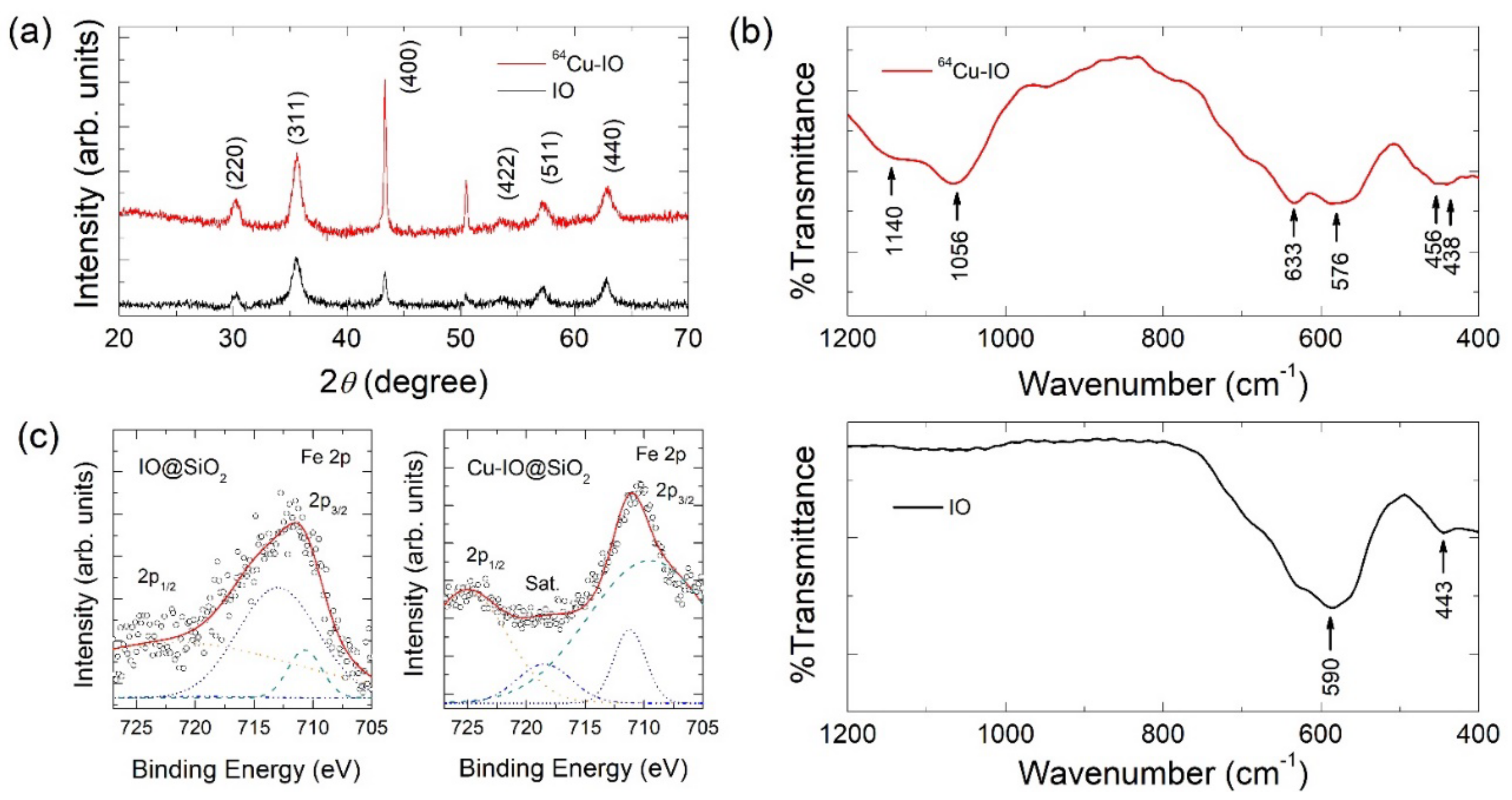
3.1. Synthesis and Characterization of 64Cu-IO@SiO2 NPs

Figure 1a displays a schematic cartoon that presents the structure of the NPs and the process of injecting them into a BALB/c mouse. Figure 1b shows TEM and SEM images of the as-synthesized IO and 64Cu-IO NPs. The mean sizes of the particles were to be 4.17 ± 0.81 nm and 4.54 ± 2.02 nm for IO and 64Cu-IO NPs, respectively, as observed by TEM images, where they dispersed well without any aggregation [9,10,11]. In Figure 1c, the lattice constant was estimated to be 0.243 nm and 0.285 nm for the IO and 64Cu-IO NPs, respectively, due to a slightly larger radius of Cu compared to Fe, implying that Cu was well incorporated into IO. EDS mapping analysis confirmed that Fe and Cu presented well in the individual 64Cu-IO NPs (Figure 1d). The Cu concentration of the 64Cu-IO NPs was assessed with an ICP-MS, confirming that the ratio of Fe to Cu is about 7 to 1. We note that, in our 64Cu-IO NPs, the radioisotope 64Cu and the stable natCu were co-incorporated into the IO NPs. Figure 1e exhibits the IO and 64Cu-IO NPs surrounded by the shell consisting of TEOS. As shown in Figure 1e, the core NPs are well surrounded by a water-soluble shell structure of SiO2, supporting our NPs to become a proper contrast agent for the living cells [2,12,13,14]. To avoid metallic toxicity for the mouse models, the shell structure has been developed to be adsorbed on the surface of the core NPs, giving us IO@SiO2 and 64Cu-IO@SiO2 NPs [15,16].
Figure 2a shows the XRD patterns of IO and 64Cu-IO NPs, confirming that the observed diffraction peaks correspond to the cubic spinel structure (ICSD card no. 98-015-8743) [2,15]. No obvious changes in the diffraction peaks were found upon the doping of both 64Cu and natCu into the IO NPs. In Figure 2b, FTIR spectra for the IO and 64Cu-IO are displayed. From a peak at 443 cm−1, we found that a small amount of Fe2O3 together with Fe3O4 was synthesized. The introduced Cu ions in 64Cu-IO NPs may give rise to a small shift of the peak from 443 cm−1 to 456 cm−1 [17]. The Cu–O vibrations were also shown from the peaks of 633 cm−1, 1056 cm−1, and 1140 cm−1 for the 64Cu-IO sample [18]. In XRD, however, the peaks of Fe2O3 and Fe3O4 would be superposed, because they have the same crystal structure. Thus, we could not find the introduced Fe2O3 phase in XRD data [7,19].
In Figure 2c, we display core-level XPS spectra of Fe 2p for IO@SiO2 and Cu-IO@SiO2 NPs, ranging from 727 eV to 705 eV. The spectra were well fitted with four Gaussian lines after the Shirley background was subtracted. For both samples, two main peaks corresponding to Fe 2p1/2 and Fe 2p3/2 appeared at ~725 eV and ~711 eV, respectively. In comparison of these two spectra, however, the data of Cu-IO@SiO2 NPs exhibit more narrowed linewidth than that of IO@SiO2 NPs, also giving us a greater satellite peak of Fe 2p3/2 located at 718.5 eV, indicative of the presence of Fe3+. Two separated peaks at 709.5 eV and 711.2 eV in Fe 2p3/2 may be attributed to Fe3+ from Fe3O4 phase and Fe2O3 phase [20,21] as in FTIR spectra. Cu 2p, O 1s, and survey spectra can be found in Figure S1 in the Supplementary Materials.

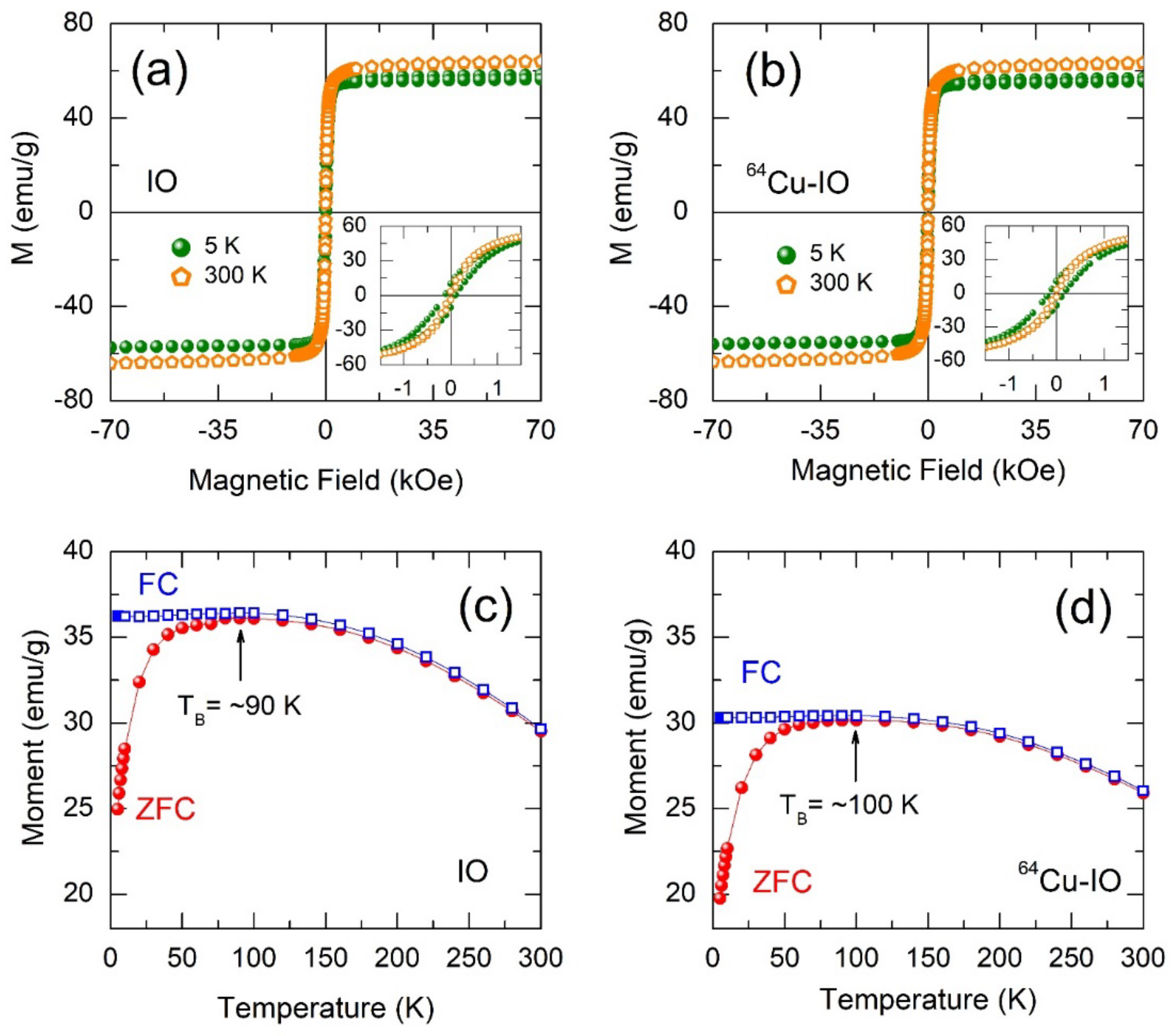
3.2. Magnetic Properties of NPs

In Figure 3a,b, the superparamagnetic behavior is observed for the IO and 64Cu-IO NPs at 300 K, whereas ferromagnetic behavior is obvious at 5 K for both samples with a remanent magnetization and a coercive field (insets of Figure 3a,b). It should be noted that, in both samples, the magnetization at 300 K is greater than that at 5 K, which may be due to the spins from the canted anti-ferromagnet (or weak ferromagnet) at 5 K resulting in spin-glass-like behavior [22]. To confirm the behavior in these samples, we performed the zero field-cooled (ZFC) and field-cooled (FC) measurements. Figure 3c,d exhibit M-T curves at ZFC and FC conditions of IO and 64Cu-IO NPs. A similar behavior between ZFC and FC magnetization was observed for both samples, in which the blocking temperature (TB) in the ZFC curve was comparably observed at ~90 K and ~100 K for IO and 64Cu-IO NPs, respectively. The TB can be defined as the temperature above which thermal energy overcomes anisotropy energy, thereby permitting the particles to rotate to their thermal equilibrium direction [23,24]. The splitting between the ZFC and FC curves denotes a typical behavior of the superparamagnetic system.
A slight increase in TB of 64Cu-IO may be due to slightly larger sizes of NPs, since TB is proportional to the particle volume V according to the relation, [24] TB = KV/ln(τM/τ0)kB, where K is the magnetic anisotropy constant, τM is the measuring time window of the experimental method, and τ0 is the attempt time. In the FC curves for both samples, very weak variation below TB indicates the presence of spin-glass-like nature due to dipole-dipole interactions between the NPs [23,24].

3.3. MR Relaxivities of IO and 64Cu-IO NPs at 4.7 T and 9.4 T

The r2 (=1/T2) and the r1 (=1/T1) relaxivities of IO and 64Cu-IO NPs were investigated at 4.7 T and 9.4 T, as shown in Figure 4. In Figure 4a–d, all the r2 and the r1 relaxivities were well fitted to a linear function with good reproducibility. In lower and higher fields, both r2 and r1 relaxivities of 64Cu-IO were greater than those of IO. Comparing the r2/r1 ratios between IO and 64Cu-IO in Figure 4f, the latter can thus be a better T2 dominant CA. We now may consider how r2 and r1 relaxivities of 64Cu-IO are greater than those of IO. In principle, r2 relaxivity may depend on the two correlation times: the diffusion correlation time, τD = rc2/D, and the static correlation time, 1/∆ω, where rc is the radius of the NPs, D is the diffusion coefficient of water, and ∆ω is the difference in Larmor frequency of the particle and that at infinity [25,26]. Considering our smaller particle size of ~4 nm, r2 relaxivity may be in a regime where motional narrowing is satisfied, which is proportional to τD. In this regime (τD ≪ 1/∆ω), diffusion is faster than a spatial variation of local field inhomogeneity induced by a single particle. For r1 relaxivity, the relaxation enhancement of 64Cu-IO compared to IO may be due to a higher diffusion of water protons along with the larger superparamagnetic particles. A greater r2 value in 64Cu-IO compared with IO may thus be associated with a slightly larger mean size of the particles (see Figure 1a). Consequently, Cu ions introduced in IO NPs may lead to slightly better performance as a T2 CA.

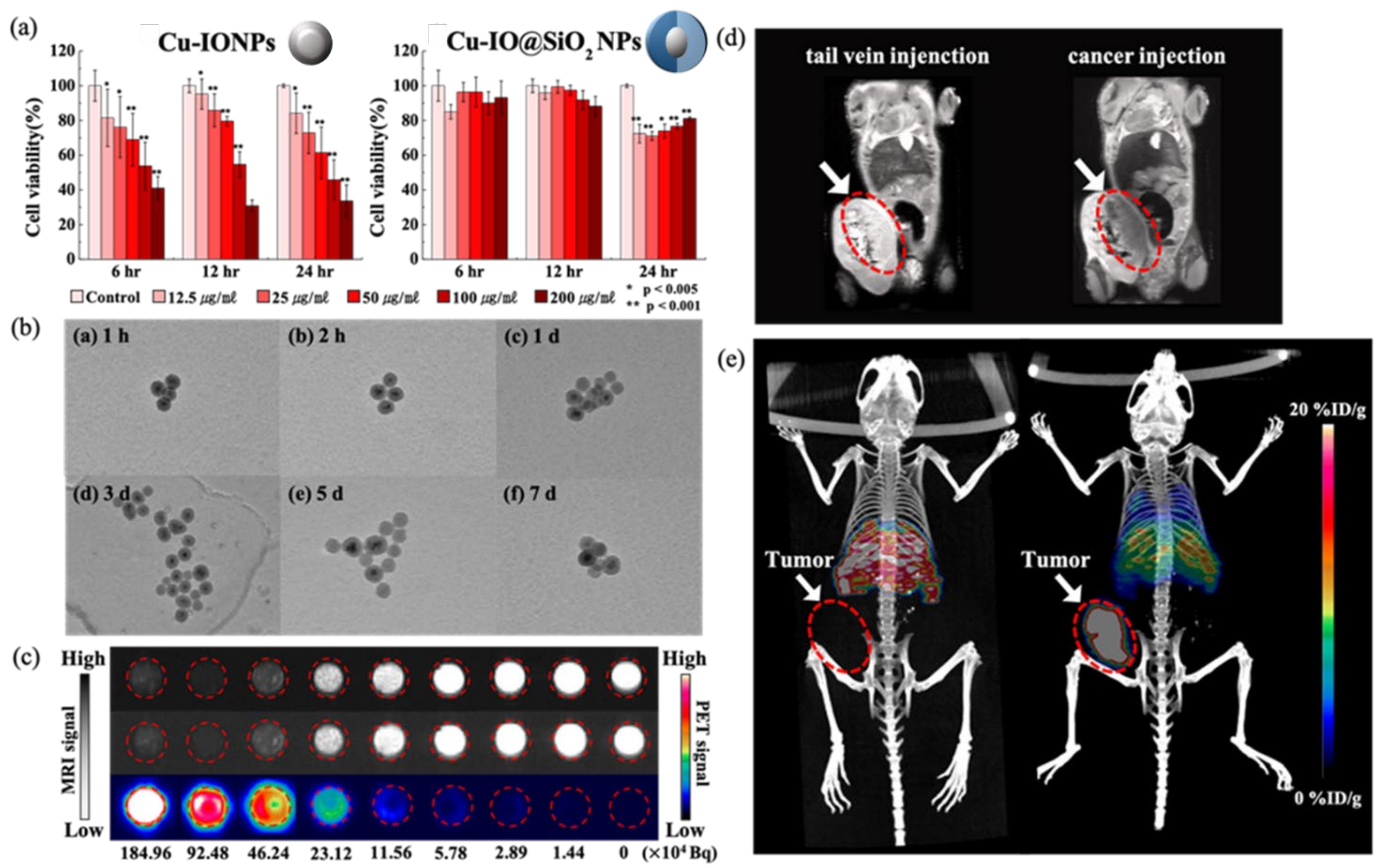
3.4. Biocompatibility of Cu-IO@SiO2 In Vitro

An essential precondition for biomedical applications is to obtain NPs with a hydrophilic surface property. We examined the cytotoxicity of the HUVECs on IO and Cu-IO NPs by CCK-8 assay with various concentrations at 6 h, 12 h, and 24 h post-injection. In Figure 5a, we display the cell viabilities of the Cu-IO NPs with and without the shell structure of SiO2. In the case of NPs without the hydrophilic shell structure, as expected, endothelial cell viabilities gradually decrease with the increasing concentration of the NPs. The asterisks indicated a significant difference relative to the control (*, p < 0.005; **, p < 0.001) based on the Student’s t-test. Error bars represent standard deviation (n = 3). The cell viabilities exhibit a similar trend depending on the delaying time, but they exhibit the lowest viability at 24 h post-injection. On the other hand, the NPs with the shell structure, Cu-IO@SiO2, exhibit no appreciable change in the cell viabilities with varying concentrations of the NPs and the delaying time. Figure 5b shows representative TEM images for Cu-IO@SiO2 NPs which are incubated in SBF at 37 °C for 1 h, 2 h, 1 d, 3 d, 5 d, and 7 d. Regardless of the incubating time, we cannot find any destruction in the shape of the surface and any aggregation for the NPs. Based on these two distinct experiments, we may ensure that our Cu-IO@SiO2 NPs have high biocompatibility and long-term stability, making them suitable for biomedical applications.

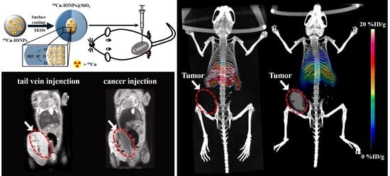
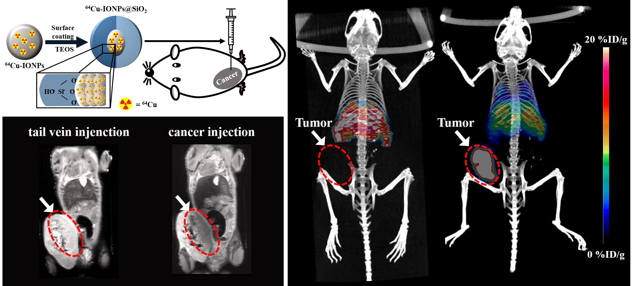
3.5. PET/MR Images of 64Cu-IO@SiO2 NPs

PET and MRI data of 64Cu-incorporated IO@SiO2 NPs are displayed in Figure 5c. Obviously, the PET signal increases with increasing 64Cu ions up to 50 μCi/mL, together with increasing concentration of 64Cu-IO@SiO2 NPs. As shown in Figure 5c, 64Cu-IO@SiO2 NPs excellently work for the MRI and PET images without any possible interference when combined into a single nanoparticle [27,28,29]. In Figure 5d, we display MR images of BALB/c mice when NPs were intravenously injected at the tail and directly injected into the cancer of the mice. When NPs were intravenously injected at the tail, the image is bright around the tumor as denoted by a red circle due to the absence of the NPs (left image). By direct injection, in contrast, the image around the tumor becomes dark when the NPs are around it, as shown in the right image of Figure 5d. However, we may not discriminate differences between these two images except for the tumor, thus not ensuring where other NPs are. Here, we predict that PET images over MR images may further provide more clear anatomical information, when using a dual-modality probe. To ensure whether PET provides clear anatomical information, we also performed the PET measurement. For the distribution of 64Cu-IO@SiO2 NPs into a body, we injected the NPs into the tail of healthy BALB/c mice living 11 weeks after they were born (left image in Figure 5e).
The NPs go towards the heart and then circulate to the whole body of the mouse. Within 3 h, we performed the PET imaging, and then successfully confirmed that the NPs are in the liver and the lung after they circulate in the body of a mouse (left image). Note that in this work we do not allow the ability of surface functionalization with specific targeting of biomarkers for tumors. In addition, we examined the PET image by injecting the NPs directly into the tumor in a mouse. As shown in the right image of Figure 5e, we successfully observed that the NPs spread around the liver and the lung after the circulation for 3 h, demonstrating that they become a good CA for PET (see Figure S2 in the Supplementary Materials). We note that a key factor in the assessment for pharmacokinetics is fast circulation within 3 h after injection [30,31,32]. The CAs staying mainly at the tumor despite their circulation in the body may be due to an effect of enhanced permeability retention [33,34]. Upon an increasing monitoring time, the radioactivity may go towards the liver and the spleen, and then would be cleared into the urine via the kidney [35,36]. We thus clearly observed a discernible image upon a distinct injection into the mice, obtaining obvious anatomical information.

4. Conclusions

In summary, we have developed chelator-free copper-64-incorporated iron oxide NPs with the core-shell structure for PET-MR imaging. We successfully synthesized 64Cu-IO NPs surrounded by the shell consisting of TEOS, exhibiting good biocompatibility and stability, as proved by in vitro SBF test and PET imaging. In both IO and 64Cu-IO NPs, superparamagnetic behaviors were equivalently observed, giving comparable blocking temperatures at ~90 K and ~100 K for the IO and 64Cu-IO NPs, respectively. In MR relaxivity, it is revealed that Cu ions introduced in IO NPS may lead to slightly better performance as a T2 CA, due solely to their slightly larger mean size of the particles. In MR imaging, we compared the images of BALB/c mice between intravenous injection at the tail and direct injection into cancer, in which intravenous injection may further need PET images to acquire clear anatomical information. From PET images, we complementarily observed that our NPs spared around the liver and the lung by intravenous injection, demonstrating that they become a good dual-modality probe for hybrid PET-MRI systems.

Supplementary Materials

The following supporting information can be downloaded at: https://www.mdpi.com/article/10.3390/nano12162791/s1, Figure S1: Survey data of (a) IO@SiO2 and (b) Cu-IO@SiO2. Fe 2p spectra of (c) IO@SiO2 and (d) Cu-IO@SiO2. Cu 2p spectra of (e) IO@SiO2 and (f) Cu-IO@SiO2. O 1s spectra of (g) IO@SiO2 and (h) Cu-IO@SiO2.; Figure S2: PET images measured immediately and in 3 h after intratumoral injection (direct injection into cancer) of the NPs for the BALB/c mice.

Author Contributions

Conceptualization, W.-J.C. and H.M.J.; methodology, H.M.J., M.H.J., J.S.L. (Jae Sang Lee) and S.-A.K.; formal analysis, H.M.J., J.S.L. (Jun Sig Lee) and J.K.P.; investigation, I.-C.L., H.I. and S.W.K.; writing—original draft preparation, H.M.J., M.H.J. and J.K.P.; writing—review and editing, H.M.J., M.H.J., W.-J.C. and J.K.P.; supervision, W.-J.C. and J.K.P.; funding acquisition, W.-J.C. All authors have read and agreed to the published version of the manuscript.

Funding

This work supported by the National Research Foundation of Korea (NRF) grant (No. NRF-2018M2A2B3A01071930) and the KOMAC (Korea Atomic Energy Research Institute) funded by the Korea government-MSIT (Ministry of Science and ICT).

Institutional Review Board Statement

The study was conducted according to the guidelines of the Declaration of Helsinki and approved by the Research Ethics Committee of the Faculty of Advanced Bio Convergence, Pohang Technopark (protocol no. ABCC2020007, 20 October 2020).

Informed Consent Statement

Not applicable.

Data Availability Statement

All data presented in this study are available upon request from the corresponding author.

Acknowledgments

The measurements of SQUID and MRI at the Korean Basic Science Institute (KBSI) are acknowledged.

Conflicts of Interest

The authors declare no conflict of interest.

References

  1. Park, J.C.; Yu, M.K.; An, G.I.; Park, S.I.; Oh, J.; Kim, H.J.; Kim, J.H.; Wang, E.K.; Hong, I.H.; Ha, Y.S.; et al. Facile Preparation of a Hybrid Nanoprobe for Triple-Modality Optical/PET/MR Imaging. Small 2010, 6, 2863–2868. [Google Scholar] [CrossRef] [PubMed]
  2. Ding, H.L.; Zhang, Y.; Wang, S.; Xu, J.M.; Xu, S.C.; Li, G.H. Fe3O4@SiO2 Core/Shell Nanoparticles: The Silica Coating Regulations with a Single Core for Different Core Sizes and Shell Thicknesses. Chem. Mater. 2012, 24, 4572–4580. [Google Scholar] [CrossRef]
  3. Chen, F.; Ellison, P.A.; Lewis, C.M.; Hong, H.; Zhang, Y.; Shi, S.; Hernandez, R.; Meyerand, M.E.; Barnhart, T.E.; Cai, W. Chelator-free synthesis of a dual-modality PET/MRI agent. Angew. Chem. Int. Ed. 2013, 52, 13319–13323. [Google Scholar] [CrossRef] [PubMed]
  4. Chakravarty, R.; Valdovinos, H.F.; Chen, F.; Lewis, C.M.; Ellison, P.A.; Luo, H.; Meyerand, M.E.; Nickles, R.J.; Cai, W. Intrinsically Germanium-69-labeled iron oxide nanoparticles: Synthesis and in-vivo dual-modality PET/MR Imaging. Adv. Mater. 2014, 26, 5119–5123. [Google Scholar] [CrossRef]
  5. Zhao, Y.; Sultan, D.; Detering, L.; Cho, S.; Sun, G.; Pierce, R.; Wooley, K.L.; Liu, Y. Copper-64-alloyed gold nanoparticles for cancer imaging: Improved radiolabel stability and diagnostic accuracy. Angew. Chem. Int. Ed. 2014, 53, 156–159. [Google Scholar] [CrossRef]
  6. Forte, E.; Fiorenza, D.; Torino, E.; di Polidoro, A.C.; Cavaliere, C.; Netti, P.A. Marco Salvatore and Marco Aiello, Radiolabeled PET/MRI Nanoparticles for Tumor Imaging. J. Clin. Med. 2019, 9, 89. [Google Scholar] [CrossRef]
  7. Wong, R.M.; Gilbert, D.; Liu, K.; Louie, A.Y. Rapid Size-Controlled Synthesis of Dextran-Coated, 64Cu-Doped Iron Oxide Nanoparticles. ACS Nano 2012, 6, 3461–3467. [Google Scholar] [CrossRef] [PubMed]
  8. Pellico, J.; Ruiz-Cabello, J.; Saiz-Alia, M.; del Rosario, G.; Caja, S.; Montoya, M.; de Manuel, L.F.; Morales, M.P.; Gurierrez, L.; Galiana, B.; et al. Fast synthesis and bioconjugation of 68Ga core-doped extremely small iron oxide nanoparticles for PET/MR imaging. Contrast Media Mol. Imaging 2015, 11, 203–210. [Google Scholar] [CrossRef]
  9. Chen, F.; Goel, S.; Valdovinos, H.F.; Luo, H.; Hernandez, R.; Barnhart, T.E.; Cai, W. In vivo integrity and biological fate of chelator-free zirconium-89-labeled mesoporous silica nanoparticles. ACS Nano 2015, 9, 7950–7959. [Google Scholar] [CrossRef] [PubMed]
  10. Tabrizian, P.; Ma, W.; Bakr, A.; Rahaman, M.S. pH-sensitive and mangetically separable Fe/Cu bimetallic nanoparticles supported by graphene oxide (GO) for high- efficiency removal of tetracyclines. J. Colloid Interface Sci. 2019, 534, 549–562. [Google Scholar] [CrossRef] [PubMed]
  11. Glaus, C.; Rossin, R.; Welch, M.J.; Bao, G. In Vivo Evaluation of 64Cu-Labled Magnetic Nanoparticles as a Dual-Modality PET/MR Imaging Agent. Bioconj. Chem. 2010, 21, 715–722. [Google Scholar] [CrossRef]
  12. Thomas, G.; Boudon, J.; Maurizi, L.; Moreau, M.; Walker, P.; Severin, I.; Oudot, A.; Goze, C.; Poty, S.; Vrigneaud, J.-M.; et al. Innovative Magnetic Nanoparticles for PET/MRI Bimodal Imaging. ACS Omega 2019, 4, 2637–2648. [Google Scholar] [CrossRef]
  13. Sodipo, B.K.; Aziz, A.A. Recent advances in synthesis and surface modification of superparamagnetic iron oxide nanoparticles with silica. J. Magn. Magn. Mater. 2016, 416, 275–291. [Google Scholar] [CrossRef]
  14. Rossi, L.M.; Costa, N.J.S.; Silva, R.P.; Wojcieszak, R. Magnetic nanomaterials in catalysis: Advanced catalysts for magnetic separation and beyond. Green Chem. 2014, 16, 2906–2933. [Google Scholar] [CrossRef]
  15. Kilic, G.; Costa, C.; Fernández-Bertólez, N.; Pásaro, E.; ao Paulo Teixeira, J.; Laf-fon, B.; Valdiglesias, V. In vitro toxicity evaluation of silica-coated iron oxide nanoparticles in human SHSY5Y neuronal cells. Toxicol. Res. 2016, 5, 235–247. [Google Scholar] [CrossRef]
  16. Malvindi, M.A.; Matteis, V.D.; Galeone, A.; Brunetti, V.; Anyfantis, G.C.; Athanas-siou, A.; Cingolani, R.; Pompa, P.P. Toxicity assessment of silica coated iron oxide nanoparticles and biocompatibility improvement by surface engineering. PLoS ONE 2014, 9, e85835. [Google Scholar] [CrossRef]
  17. Wang, Y.; Muramatsu, A.; Sugimoto, T. FTIR analysis of well-defined α-Fe203 particles. Colloid Sand Surf. A Physicochem. Eng. Asp. 1998, 134, 281–297. [Google Scholar] [CrossRef]
  18. Renuga, D.; Jeyasundari, J.; Athithan, A.S.S.; Jacob, Y.B.A. Synthesis and characterization of copper oxide nanoparticles using Brassica oleracea var. italic extract for its antifungal application. Mater. Res. Express 2020, 7, 45007. [Google Scholar] [CrossRef]
  19. Zhang, X.; Niu, Y.; Meng, X.; Li, Y.; Zhao, J. Structural evolution and characteristics of the phase transformations between α- Fe2O3, Fe3O4 and γ-Fe2O3 nanoparticles under reducing and oxidizing atmospheres. CrystEngComm 2013, 15, 8166–8172. [Google Scholar] [CrossRef]
  20. Park, J.K.; Jang, H.M.; Cho, W.-J.; Kim, C.; Suk, J.; Kim, D.S.; Lee, J.S. Enhanced anomalous magnetization in carbonyl iron by Ni+ ion beam irradiation. Sci. Rep. 2021, 11, 20118. [Google Scholar] [CrossRef]
  21. Li, Z.; Chen, H.; Liu, W. Photocatalytic activity of ZnO/CuO/ZnFe2O4 nanocomposite as a photofenton-like catalyst. Catalysts 2018, 8, 557. [Google Scholar] [CrossRef]
  22. Singh, P.; Shukla, M.; Upadhyay, C. Signatures of consolidated superparamagnetic and spin-glass behavior in magnetitie-silver core-shell nanoparticles. Nanoscale 2018, 10, 22583–22592. [Google Scholar] [CrossRef] [PubMed]
  23. Alzoubi, G.M.; Alsmadi, A.M.; Alna’washi, G.A.; Salameh, B.; Shatnawi, M.; Al-nemrat, S.; Albiss, B.A.; Bsoul, I. Coexistence of superparamagnetism and spin-glass like behavior in zinc-substituted cobalt ferrite nanoparticles. Appl. Phys. A 2020, 126, 512. [Google Scholar] [CrossRef]
  24. Greculeasa, S.G.; Palade, P.; Schinteie, G.; Leca, A.; Dumitrache, F.; Lungu, I.; Pro-dan, G.; Kuncser, A.; Kuncser, V. Tuning structural and magnetic properties of Fe oxide nanoparticles by specific hydrogenation treatments. Sci. Rep. 2020, 10, 17174. [Google Scholar] [CrossRef]
  25. Norek, M.; Kampert, E.; Zeitler, U.; Peters, J.A. Tuning of the Size of Dy2O3 Nanoparticles for Optimal Performance as an MRI Contrast Agent. J. Am. Chem. Soc. 2008, 130, 5335–5340. [Google Scholar] [CrossRef]
  26. Norek, M.; Peters, J.A. MRI contrast agents based on dysprosium or holmium. Prog. Nucl. Magn. Reson. Spectrosc. 2011, 59, 64–82. [Google Scholar] [CrossRef]
  27. Choi, J.; Park, J.C.; Nah, H.; Woo, S.; Oh, J.; Kim, K.M.; Cheon, G.J.; Chang, Y.; Yoo, J.; Cheon, J. A Hybrid Nanoparticle Probe for Dual−Modality Positron Emission Tomography and Magnetic Resonance Imaging. Angew. Chem. Int. Ed. 2008, 33, 6259–6262. [Google Scholar] [CrossRef]
  28. Madru, R.; Budassi, M.; Benveniste, H.; Lee, H.; Smith, S.D.; Schlyer, D.J.; Vaska, P.; Knutsson, L.; Strand, S.-E. Simultaneous preclinical positron emission tomography-magnetic resonance imaging study of lymphatic drainage of chelator-free 64Cu-labeled nanoparticles. Cancer Biother. Radiopharm. 2018, 33, 213–220. [Google Scholar] [CrossRef]
  29. Zhou, M.; Zhang, R.; Huang, M.; Lu, W.; Song, S.; Melancon, M.P.; Tian, M.; Liang, D.; Li, C. A chelator-free multifunctional [64Cu]CuS nanoparticles platform for simultaneous micro-PET/CT imaging and photothermal ablation therpy. J. Am. Chem. Soc. 2010, 132, 15351–15358. [Google Scholar] [CrossRef]
  30. De Kruijff, R.M.; Raavé, R.; Kip, A.; Molkenboer-Kuenen, J.; Roobol, S.J.; Essers, J.; Heskamp, S.; Denkova, A.G. Elucidating the influence of tumor presence on the poly-mersome circulation time in mice. Pharmaceutics 2019, 11, 241. [Google Scholar] [CrossRef]
  31. Duan, J.; Yu, Y.; Li, Y.; Li, Y.; Zhou, X.; Huang, P.; Sun, Z. Toxic effect of silica nanoparticles on endothelial cells through DNA damage response via Chk1-dependent G2/M checkpoint. PLoS ONE 2013, 8, e62087. [Google Scholar] [CrossRef] [PubMed]
  32. Almeida, J.P.M.; Chen, A.L.; Foster, A.; Drezek, R. In vivo biodistribution of nanoparticles. Nanomedicine 2011, 6, 815–835. [Google Scholar] [CrossRef] [PubMed]
  33. Chen, L.; Zang, F.; Wu, H.; Li, J.; Xie, J.; Ma, M.; Gu, N.; Zhang, Y. Using PEGylated magnetic nanoparticles to describe EPR effect in tumor for predicting therapeutic efficacy of micelle drugs. Nanoscale 2018, 10, 1788–1797. [Google Scholar] [CrossRef]
  34. Wang, L.; Huang, J.; Chen, H.; Wu, H.; Xu, Y.; Li, Y.; Yi, H.; Wang, Y.A.; Yang, L.; Mao, H. Exerting enhanced permeability and retention effect driven delivery by ultrafine iron oxide nanoparticles with T1–T2 switchable magnetic resonance imaging contrast. ACS Nano 2017, 11, 4582–4592. [Google Scholar] [CrossRef]
  35. Geppert, M.; Himly, M. Iron oxide nanoparticles in bioimaging-an immune perspective. Front. Immunol. 2021, 12, 688927. [Google Scholar] [CrossRef]
  36. Holder, A.L.; Vejerano, E.P.; Zhou, X.; Marr, L.C. Nanomaterial disposal by incineration. Environ. Sci. Processes Impacts 2013, 15, 1652. [Google Scholar] [CrossRef] [PubMed]
Figure 1. (a) Schematic cartoon for the experimental process for synthesizing the NPs, coating with TEOS, injecting the NPs into BALB/c mice, and PET and MRI imaging. (b) TEM images of IO and 64Cu-IO NPs. The inset displays the size distribution acquired by measuring at least 200 NPs from the corresponding TEM image of each sample. (c) The lattice constants of IO and 64Cu-IO NPs obtained from TEM images for both samples. (d) SEM images of IO NPs (upper panel) and TEM images of chelator-free 64Cu-IO NPs (lower panel) with elemental mapping analysis confirming the presence of Fe and Cu ions. (e) TEM images for core-shell-structured IO (left) and 64Cu-IO NPs (right).
Nanomaterials 12 02791 g001
Figure 2. (a) XRD patterns and (b) FTIR spectra for chelator-free 64Cu-IO (upper panel) and IO NPs (lower panel). (c) XPS spectra of IO@SiO2 (left) and Cu-IO@SiO2 (right) for Fe 2p.
Nanomaterials 12 02791 g002
Figure 3. Mass magnetization of the (a) IO and (b) chelator-free 64Cu-IO NPs obtained using a SQUID magnetometer. The insets of (a,b) show the magnified curves exhibiting a perfect superparamagnetic behavior at 300 K for both samples. The remanent magnetization and coercive field at 5 K were 10.3 emu/g and 100 Oe for (c) IO, and 10.8 emu/g and 200 Oe for (d) chelator-free 64Cu-IO, respectively. Zero-field-cooled (ZFC) and field-cooled (FC) temperature-dependent magnetization curves (M-T) for IO and chelator-free 64Cu-IO NPs under an applied magnetic field of 500 Oe. TB represents the blocking temperature.
Nanomaterials 12 02791 g003
Figure 4. Transverse (r2) relaxivity of IO and chelator-free 64Cu-IO NPs at (a) 4.7 T and (b) 9.4 T. Longitudinal (r1) relaxivity of IO and 64Cu-IO NPs at (c) 4.7 T and (d) 9.4 T. (e) The r1 and r2 relaxivities at 4.7 T and 9.4 T for IO and 64Cu-IO. (f) The r2/r1 ratios for IO and 64Cu-IO are compared at 4.7 T and 9.4 T.
Nanomaterials 12 02791 g004
Figure 5. (a) Cell viability results examined by the CCK-8 using HUVECs as a function of time with varying concentrations to assess the cytotoxicity. (b) Representative TEM images for Cu–IO@SiO2 NPs incubated in SBF at 37 °C for (b-a) 1 h, (b-b) 2 h, (b-c) 1 d, (b-d) 3 d, (b-e) 5 d, and (b-f) 7 d. SBF results confirm the stability of core-shell structures when the NPs are injected into the body. (c) MRI and PET images were measured with varying concentrations of NPs which were diluted half consecutively starting from 184.96 × 10⁴ Bq down to 1.44 × 10⁴ Bq to examine the contrast performance. (d) MR images and (e) PET images were measured in 3 h after injecting the NPs into the BALB/c mice. Intravenous injection at the tail (left of (d,e)) and direct injection into cancer (intratumoral) (right of (d,e)) of each mouse model. Tumor: white arrows; tumor size and position: red dotted ovals.
Nanomaterials 12 02791 g005
Publisher’s Note: MDPI stays neutral with regard to jurisdictional claims in published maps and institutional affiliations.

