Article

Comparative Whole-Genome Analysis of Russian Foodborne Multidrug-Resistant Salmonella Infantis Isolates

Central Research Institute of Epidemiology, Novogireevskaya str., 3a, 111123 Moscow, Russia
*
Author to whom correspondence should be addressed.
Microorganisms 2022, 10(1), 89; https://doi.org/10.3390/microorganisms10010089
Submission received: 30 November 2021 / Revised: 27 December 2021 / Accepted: 30 December 2021 / Published: 31 December 2021

Abstract

:
Non-typhoidal Salmonella infections remain a significant public health problem worldwide. In this study, we present the first detailed genomic analysis report based on short-read (Illumina) whole-genome sequencing (WGS) of 45 multidrug-resistant (MDR) Salmonella enterica subsp. enterica serotype Infantis isolates from poultry and meat product samples obtained in Russia during 2018–2020, and long-read (MinION) WGS of five more representative isolates. We sought to determine whether foodborne S. Infantis have acquired new characteristics, traits, and dynamics in MDR growth in recent years. All sequenced isolates belonged to the sequence type ST32 and more than the half of isolates was characterized by six similar antimicrobial susceptibility profiles, most of which corresponded well with the antimicrobial resistance determinants to aminoglycosides, sulphonamides, tetracycline, and chloramphenicol revealed in silico. Some of the isolates were characterized by the presence of several types of plasmids simultaneously. Plasmid typing using WGS revealed Col440I, ColpVC, ColRNAI, IncFIB, IncFII, IncX1, IncHI2, IncHI2A, and IncN replicons. The identified virulence genes for 45 whole genomes of S. Infantis were similar and included 129 genes encoding structural components of the cell, factors responsible for successful invasion of the host, and secreted products. These data will be a valuable contribution to further comparative genomics of S. Infantis circulating in Russia, as well as to epidemiological surveillance of foodborne Salmonella isolates and investigations of Salmonella outbreaks.

1. Introduction

Salmonella enterica subsp. enterica is a Gram-negative bacterium that can be divided into two groups–typhoidal Salmonella, which are adapted for humans and do not occur in other animals, and non-typhoidal Salmonella (NTS), which usually cause foodborne gastroenteritis and can infect various animals. The severity of the disease depends on the host, the Salmonella serotype, and delivery method [1,2]. More than 2500 serotypes of NTS have been identified, however, only a limited number of them are responsible for the majority of salmonellosis cases reported [3,4,5].
The pathogenesis of these organisms depends on multiple virulence and antimicrobial resistance (AMR) genes [6], mostly associated with Salmonella pathogenicity islands (SPIs), prophages, and plasmids. Some virulence factors seem to be conserved among Salmonella, while others may be serotype-specific [7]. Most cases of Salmonella-caused diseases are mild, but sometimes they can be life threatening and, therefore, require appropriate antibiotic therapy. Antimicrobial resistance is a public health problem worldwide, and Salmonella is among the microorganisms with a number of resistant strains found in the food chain. Ciprofloxacin is a common first-line antimicrobial drug for the treatment of salmonellosis, but since fluoroquinolones are not used to treat children, beta-lactams (ampicillin or third-generation cephalosporins) are equally important [8]. MDR S. enterica serotypes are associated with higher morbidity outcomes compared to drug-susceptible strains [9].
Despite the fact that over 2500 S. enterica serotypes have been documented, most investigations were focused on Salmonella enterica subsp. enterica serotypes Typhimurium and Enteritidis. However, S. Infantis has emerged as the fourth most common serotype causing human salmonellosis in Europe [10]. According to the Russian Reference Center for Salmonellosis Monitoring, in recent years S. Infantis was one of the three predominant serotypes in Russia, along with S. Enteritidis and S. Typhimurium. Most often S. Infantis was revealed in food (28.4%) and in environmental samples (12%), while the frequency of isolation from human samples was much lower (1.5%) [11]. Since 2017, S. Infantis became the most common food origin serotype in Russia [12]. The growth of S. Infantis incidence has been registered in the period of 2003–2009 and still continues. [13,14]. S. Infantis seems to be particularly important in broiler, while the prevalence in laying hens and its products is much lower. The increase in S. Infantis carriage reported in broiler was observed in some EU/EEA countries, Israel, Turkey [15,16,17], and poultry and its products have been identified as one of the most important sources of human infection with S. Infantis [18]. On the other hand, analyzed S. Infantis samples from Turkey distributed between broilers and laying hens significantly unevenly—69% and 3% correspondingly [19]. Moreover, a similar picture we observed in Japanese research, where almost 100% of broiler isolates studied belonged to S. Infantis, and laying hens isolates did not contain them [20]. Increasing incidence and dissemination of different MDR S. Infantis lead to the problems in the public health system due to the treatment issues [21]. Therefore, there is a need to monitor the growth and occurrence of S. Infantis characteristics and the ways of its spreading in order to understand its newly acquired properties and provide better treatment options.
For public health surveillance systems, susceptibility data alone may not be sufficient in some circumstances, such as in assessment of resistant foodborne pathogens [22] and the risks associated with the use of agricultural antibiotics [23]. In risk analysis, it is often necessary to carry out additional genetic testing to compare alleles and genotypes of the isolates from different environments [24]. With the advent of whole-genome sequencing (WGS) technology, it is now possible to identify and evaluate the entire bacterial DNA sequence in just a few days, making it the best surveillance tool [25,26].
The WGS has been widely used in the field of molecular biology. However, the gaps between the fragments obtained by genome assembly from short reads can lead to the incompleteness of genome structure, which is critical for determining plasmid sequences and the location of particular genes [27]. Long-read nanopore sequencing technology can facilitate the completion of the bacterial genome assembly which either does not have sufficient sequencing depth in some of the repetitive regions or has areas with zero coverage when sequenced using short-read sequencing technology [28]. However, nanopore sequencing technology exhibits higher reading error frequencies and may produce systematic errors, and thus a combination of both technologies resulting in hybrid short and long-read assembly was used to obtain the accurate genomes of representative isolates in the current study.
The aim of this work is to present a comprehensive analysis of 45 MDR foodborne S. Infantis isolates from different regions of Russia based on WGS data to determine their characteristics and traits. In this paper, we provide data for S. Infantis isolates selected from 19 different geographical regions and belonging to one sequence type (ST) using short-read (Illumina) and long-read (MinION) WGS. The majority of the isolates (n = 26) were obtained from broiler and ready-to-cook meat products (n = 8). All these isolates were multidrug-resistant (MDR) according to the commonly used definition [29]. Plasmid structures, as well as antibiotic resistance, virulence genes and CRISPR elements’ annotation, are provided. We believe that these data will greatly facilitate the investigations of the distribution and the mechanisms of antimicrobial resistance acquisition for this pathogen.

2. Materials and Methods

2.1. Sample Isolation, Identification and Antibiotic Susceptibility Testing

Isolates from poultry product samples (n = 35) and meat product samples (n = 10) were obtained in the Russian Federation during a period of 2018-2020. Sample collection was carried out as a part of regular monitoring activities by 19 Russian Federal Centers of Hygiene and Epidemiology (Table 1). The highest number of the isolates were obtained in Krasnoyarsk (n = 6), Moscow and Moscow region (n = 6), Rostov (n = 5), and Perm (n = 4). The majority of the isolates (57.8%, n = 26) were obtained from chicken, 17.8% (n = 8) from ready-to-cook meat products, 15.6 % (n = 7) pertained to ready-to-cook chicken, while meat and turkey consisted of two samples each.
A total of 45 Salmonella isolates were identified down to a species level by time-of-flight mass spectrometry (MALDI-TOF MS) using the VITEK MS system (bioMerieux, Marcy-l’Étoile, France). The susceptibility of the samples included in this study was determined by the boundary concentration method on the same analyzer. Confirmation of the serotype Infantis was carried out manually using PETAL® diagnostic Salmonella O-typing serum (Epidbiomed, Saint Petersburg, Russia). Antibiotic susceptibility testing (AST) of S. Infantis isolates was performed on the following antimicrobial panel: ampicillin (AMP), ampicillin/sulbactam (SAM), cefepime (FEP), ceftazidime (CZD/CAZ), cefuroxime axetil (CXM), ceftriaxone (CRO), cefoperazone/sulbactam (CFP), imipenem (IPM/M), ertapenem (ETP), ciprofloxacin (CIP), amikacin (AMK/AN/AKN), gentamicin (GMN/GEN), tobramycin (TOB), chloramphenicol (CHL), nitrofurantoin (NIT), trimethoprim/sulfamethoxazole (SXT), tetracycline (TET). The assessment of antimicrobial compounds included in this study was conducted according to the European Committee on Antimicrobial Susceptibility Testing interpretive standards (EUCAST). The results were interpreted according to EUCAST clinical breakpoints v.10.0 (http://www.eucast.org, accessed on 7 October 2021).

2.2. DNA Isolation and Whole Genome Sequencing

Genomic DNA was isolated with Ribo-prep Kit (Amplisens, Moscow, Russia) according to the manufacturer’s instructions. The quantity was evaluated by fluorimetry with Qubit 4.0 (Invitrogen, Waltham, MA, USA). DNA was used for paired-end library preparation with Nextera DNA Flex Library Prep Kit (Illumina, San Diego, CA, USA) according to the manufacturer’s recommendations. Short-read WGS was performed on the HiSeq 1500 platform (Illumina). Quality and quantity of libraries were confirmed by capillary gel electrophoresis (Agilent Bioanalyzer 2100, Agilent, Santa Clara, CA, USA) using a Qubit 4.0 fluorimeter (company, city, state abbreviation if USA, country).
Additionally, based on the initial analyses of short-read sequences, 5 more representative isolates were selected for long-read WGS for better characterization of plasmid structures. Libraries for long-read sequencing were prepared using the 1D Genomic DNA sequencing kit SQK-LSK108 (Oxford Nanopore, Oxford, UK). Size selection and clean-up steps were performed using AMPure XP (Beckman Coulter, Brea, CA, USA). Quantification of libraries was made using Qubit 4.0 (Thermo Fisher Scientific, Waltham, MA, USA). Sequencing of long reads was performed on MinION (Oxford Nanopore Technologies, Oxford, UK) R9 SpotON flow cell. Base calling of the raw MinION data was made using Guppy basecalling software version 4.4.1 (Oxford Nanopore Technologies) with default parameters, and demultiplexing was performed using Guppy barcoding software version 4.4.1 (Oxford Nanopore Technologies).

2.3. Genome Assembly and Sequence Analysis

Short-read genome assemblies were obtained using SPAdes version 3.12 [27] with default parameters. Hybrid short- and long-read assemblies were performed using Unicycler version 0.4.9 [28].
Assembly quality estimation, organism checking and initial annotation were performed using the custom pipeline described earlier [29]. We determined the antibiotic resistance genes in silico, performed isolate typing using multilocus sequence typing (MLST) scheme and revealed the presence of CRISPR/Cas systems and CRISPR arrays. The presence of virulence factors was also studied using the VFDB database (http://www.mgc.ac.cn/VFs/, accessed on 20 October 2021). Salmonella MLST website (https://enterobase.warwick.ac.uk/species/index/senterica, accessed on 20 October 2021) was used for sequence type detection. Salmonella enterica MLST schemes include the fragments of the following seven loci: aroC, dnaN, hemD, hisD, purE, sucA, and thrA [30]. We used the Resfinder 4.0 database with default parameters for antimicrobial gene identification [31]. Plasmid sequences were revealed and typed using PlasmidFinder with default parameters [32]. Plasmid images were built with SnapGene Viewer 6.0.0. CRISPRCasFinder with default parameters [31] was used to identify the presence of CRISPR/Cas systems and spacers in the genomes analyzed.
The assembled genome sequences for all isolates were uploaded to the NCBI Genbank under the project number PRJNA780779Salmonella.
To build the phylogenetic tree representing the relations between the isolates based on core genome sequence, we used roary version 3.13.0 [32] and RAxML version 8.2.11 [33] software with default parameters (model used: -m GTRGAMMA -p 13434345).
cgMLST analysis was performed using MentaList version 0.2.4 [34], and the minimum spanning tree was built using PHYLOViz online [33]. cgMLST scheme including 3002 targets was used (https://www.cgmlst.org/ncs/schema/schema/4792159/, accessed on 20 October 2021) [34].

2.4. CRISPR Typing and Analysis

CRISPRCasFinder [35] was used to identify the presence of CRISPR/Cas systems and spacers in the genomes studied.
Salmonella spacers were identified and downloaded from CRISPRFinder web tool [36] (http://crispr.i2bc.paris-saclay.fr/Server/, accessed on 20 October 2021), then FASTA sequences of the spacers were uploaded to Web BLAST® blastn suite (https://blast.ncbi.nlm.nih.gov, accessed on 18 September 2021) and analyzed using default parameters of MegaBLAST algorithm.
Venn diagram was constructed using a free web service (https://bioinformatics.psb.ugent.be/webtools/Venn/, accessed on 20 October 2021).

3. Results

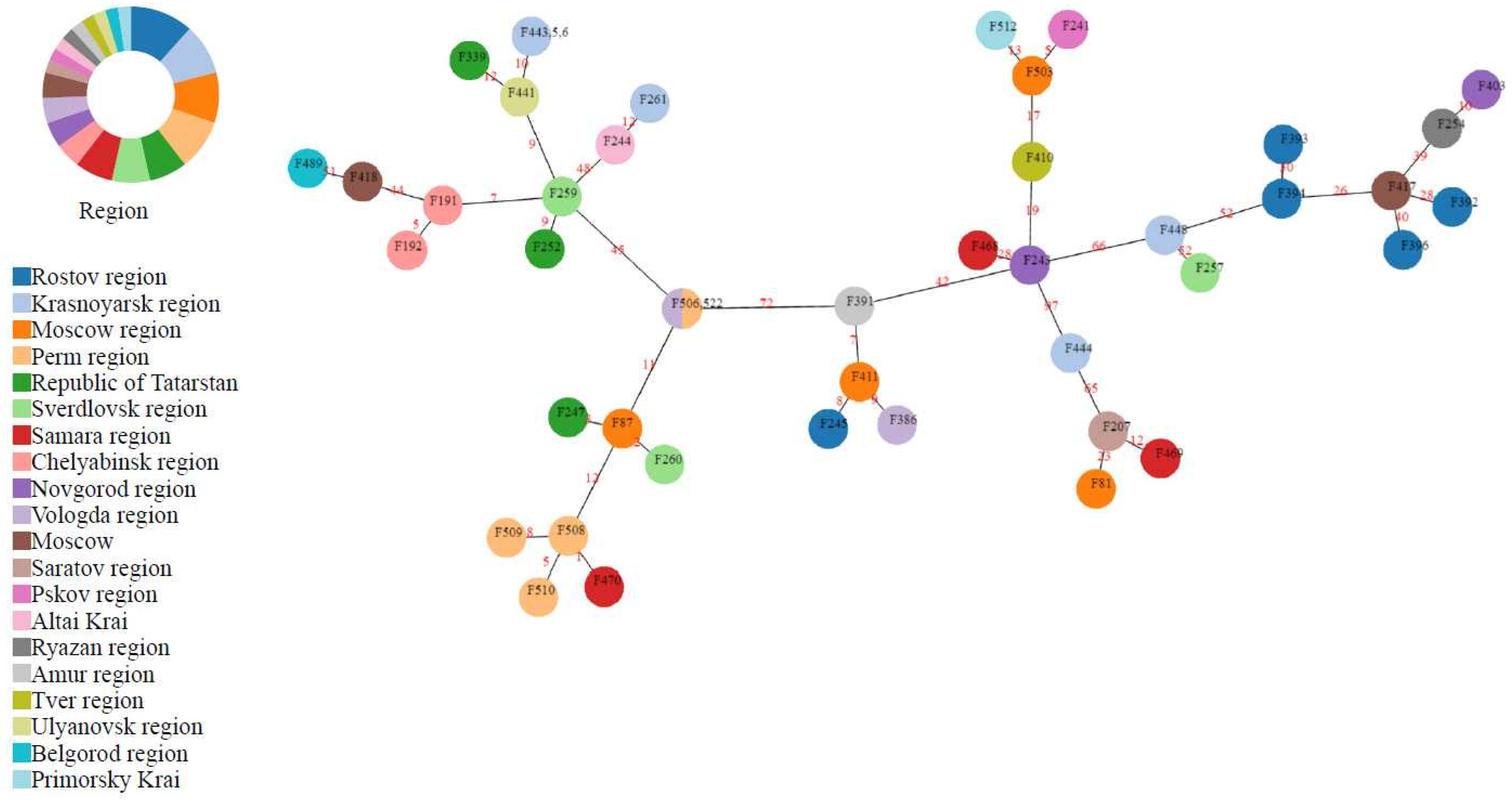
3.1. Isolation and Typing

The sources of foodborne S. Infantis isolates are described in Table 1. The majority of the isolates were obtained from Krasnoyarsk, Rostov, Moscow, and Perm Hygiene and Epidemiology Centers of Federal service on customers’ rights protection and human well-being surveillance (Rospotrebnadzor). Based on the MLST analysis, all isolates belong to the sequence type ST32.
In order to obtain additional information regarding bacteria similarity, a phylogenetic tree was built for all 45 isolates. The tree is presented below in Figure 1.
According to the phylogenetic analysis, high similarity groups of the same sequence type (ST32) were revealed regardless of the different regions. For a deeper phylogenetic relationship analysis between the 45 S. Infantis isolates, a minimum spanning tree based on core genome MLST (cgMLST) was constructed (Figure 2). The clustering seems to be unrelated to the year of isolation, source, or geographic location. However, the resistance profile appears to have some influence. For instance, Crie-F87, Crie-F247, and Crie-F260 clustered together possessed the same antimicrobial susceptibility despite the fact that they were obtained from different regions. A similar picture was observed for the isolates Crie-F244 and Crie-F261; and, in addition, for Crie-F441, Crie-F443, Crie-F445, Crie-F446, and Crie-F339.

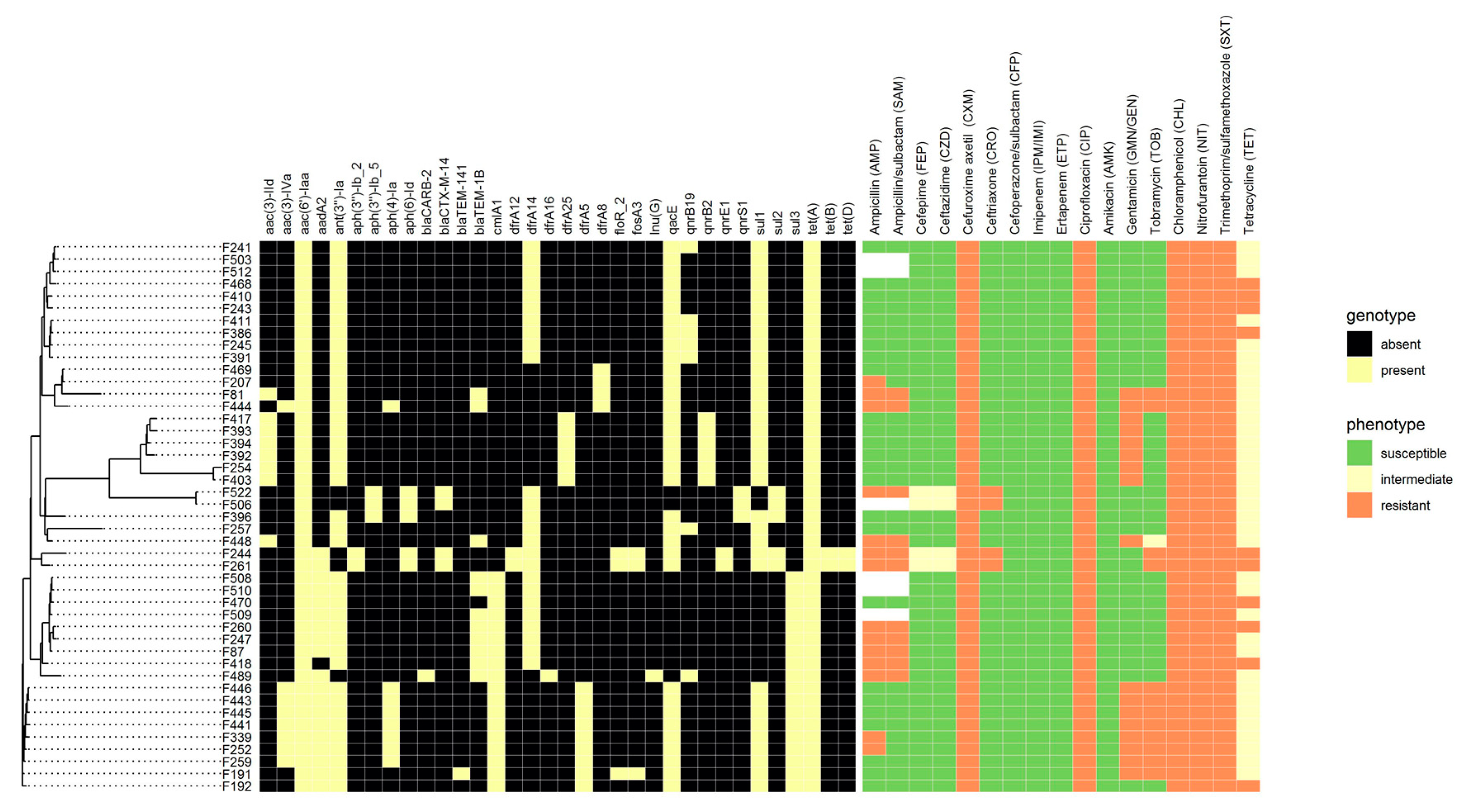
3.2. Antibiotic Resistance Determination
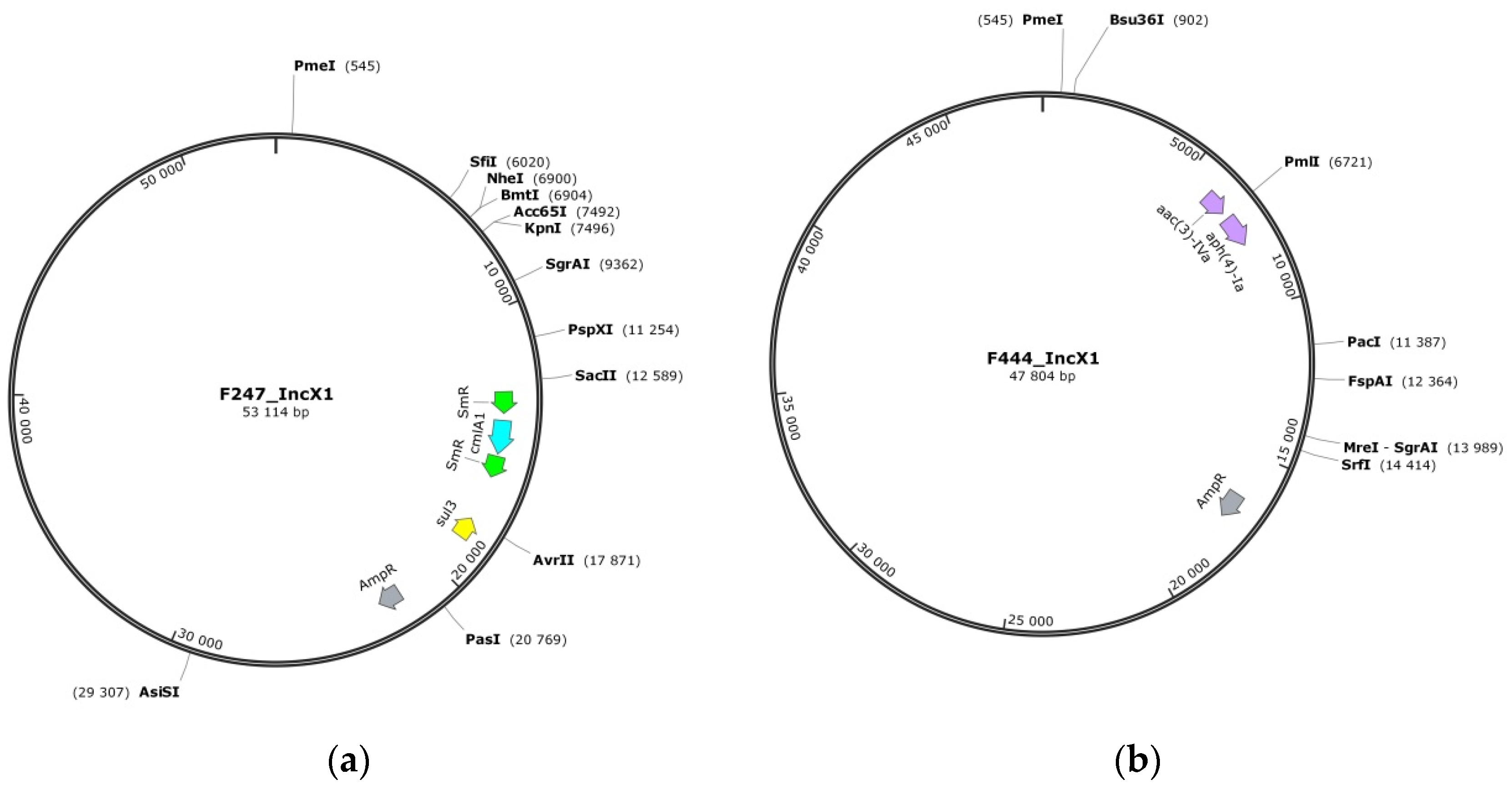
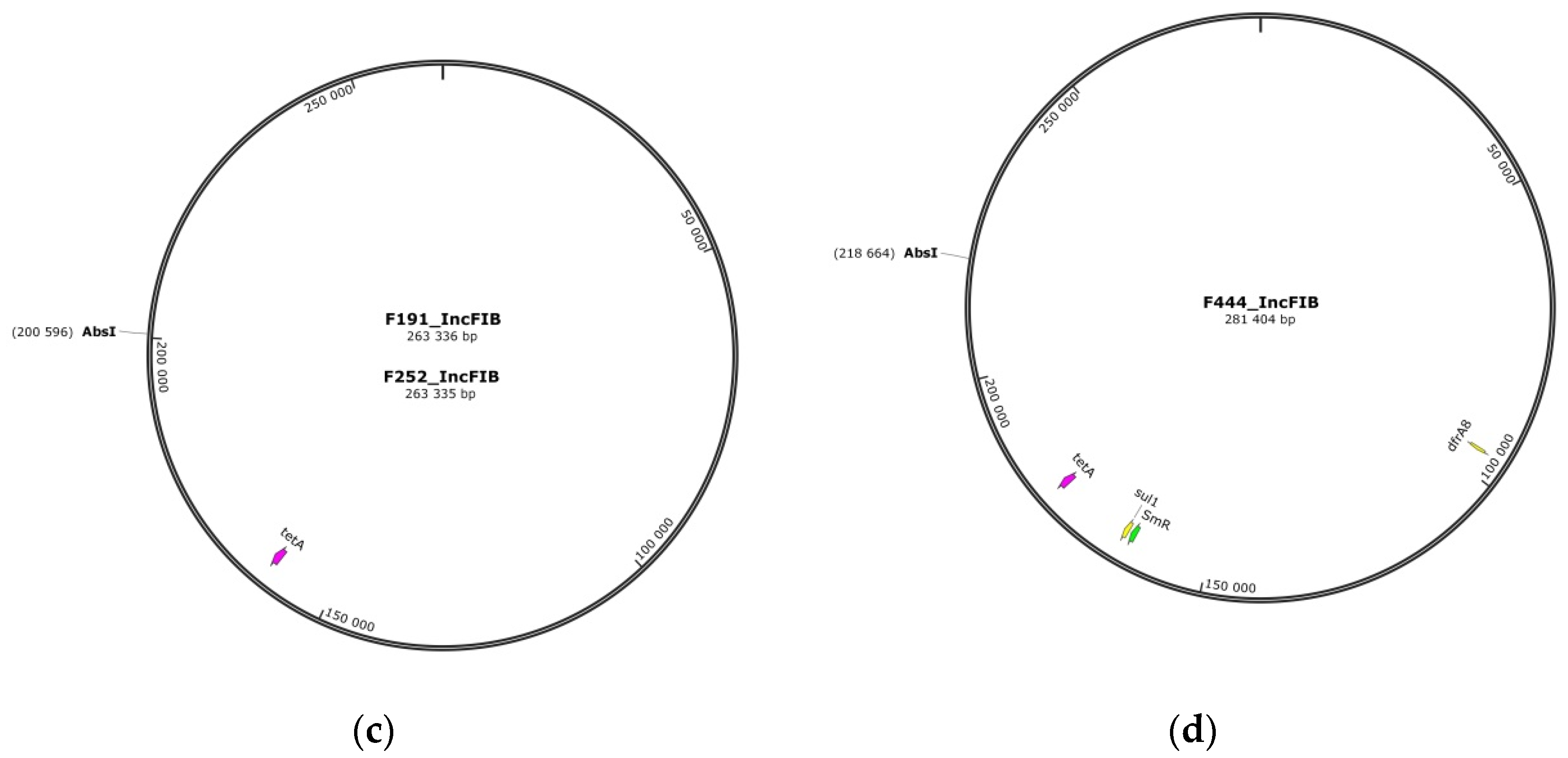
WGS technology currently allows detecting the presence of genes, but cannot reveal their expression or activation state, thus, a comprehensive comparison of empirical and theoretical susceptibility determination is required. A total of 45 S. Infantis isolates were tested for antibiotic susceptibility/resistance both by empirical (boundary concentration) method and bioinformatic analysis (searching for known acquired resistance genes in genomic sequences). A comprehensive comparison of empirical and theoretical susceptibility determination is presented in Figure 1.
All 45 isolates were resistant to six antimicrobial drugs out of 17 tested. The boundary concentration method identified resistance to CXM, CIP, CHL, NIT, SXT, and TET, which belong to different classes of antibiotics. Two isolates (Crie-F244 and Crie-F261) were resistant to 12 antimicrobial drugs, one (Crie-F522) to 11 antibiotics, and three isolates (Crie-F81, Crie-F444, Crie-F448) to 10 antibiotics from the panel used.
Moreover, with a help of WGS (Figure 1) we revealed that the main spectrum of antimicrobial resistance determinants included genes of aminoglycoside-modifying enzymes aac(6′)-Iaa and ant(3″)-Ia (45 and 40 of the isolates, respectively), as well as genes involved in tetracycline (tetABD, n = 45), and sulfonamide (sul123, dfrA14, n = 45) resistance that corresponds well to AST. However, the presence of AMR genes to chloramphenicol, quinolones, and β-lactams did not correspond well with the phenotypic profiles. For example, cmlA1 gene had 18 isolates and floR had three samples, while the susceptibility test indicated that all isolates studied possessed a resistance to CHL (Figure 2). Moreover, all isolates observed had resistance to CIP and NIT, although AMR genes for fluoroquinolones (qnrB19 and qnrB2) were revealed only in 13 isolates, and 5 isolates included quinolone AMR genes (qnrE1 and qnrS1) (Figure 2). The most common mechanism for resistance to quinolones in Salmonella is the acquisition of point mutations in chromosomal AMR gene gyrA [37]. As we did not check isolates for point mutations and intrinsic chromosomal AMR genes, we can suggest that the resistance was associated with substitutions at the Ser-83 position to Tyr, Phe, or Ala, or/and Asp-87 substitutions to Asn, Gly, or Tyr [37]. Therefore the molecular mechanism of quinolone resistance will possibly be clarified in further reports by point mutation analysis.
Interestingly, the susceptibility test identified resistance to the second generation cephalosporins (CXM) throughout the isolates studied, and just four isolates (Crie-F244, Crie-F261, Crie-F506, and Crie-F522) possessed a resistance to the third (CZD and CRO) and fourth (FEP) generation cephalosporins. At the same time, according to the AMR profile only these four isolates contain blaCTX-M-14 (Figure 2). Thus, we confirm that blaCTX-M-14 infuse resistance to 3rd and 4th generation cephalosporins in S. Infantis as has been previously described [14].

3.3. Plasmids

Since plasmids play a key role in the acquisition of antimicrobial resistance, it is important to identify plasmid sequences in the WGS data, as this information will greatly facilitate the study of resistance mechanisms. The results of in silico search for the plasmids in all genomic sequences of the isolates are presented in Table 2 below. The most common plasmid replicon groups in our collection included a range of plasmids previously associated with multidrug-resistant foodborne bacteria IncFIB (n  =  42) and widely distributed IncX1 (n = 13) previously detected in Salmonella, Klebsiella, Shigella, and E. coli species [35,36]. Some of the isolates were characterized by the presence of several plasmid types simultaneously (Crie-F386, Crie-F503, and Crie-F510). IncX1 plasmids containing a group of qnr genes (resistance to quinolones) are considered widespread [35,36].
Five isolates, Crie-F191, Crie-F247, Crie-F252, Crie-F410, and Crie-F444, had different MDR phenotypes and were resistant to eight, eight, nine, six and ten antimicrobial compounds, respectively (Figure 1). These MDR isolates were subjected to long-read WGS using the Oxford Nanopore device.
In-depth genomic analysis by hybrid assembly confirmed that five MDR isolates carried pESI-like megaplasmids with resistance and virulence gene patterns previously detected in S. Infantis from Russia. According to the WGS analysis, all samples except Crie-F191 included the IncFIB replicon, while three isolates had additional IncX1 in another plasmid. Crie-F191 had an IncFII replicon, however, hybrid assembly included the same megaplasmid as in Crie-F252 with IncFIB (Table 2, Table S1). Their size ranged from 285 kb to approximately 317 kb (Table S1). The samples Crie-F247 and Crie-F444 contained an additional plasmid of approximately 39 kb length previously found in Escherichia coli according to BLAST analysis of ‘nt’ database.
Plasmids reconstructed in silico by the hybrid assembly are presented below in Figure 3 using SnapGene tool. Visual modeling allowed us to reveal significant differences in IncX1 replicons from our samples (Figure 3a,b), while IncFIB appeared to be more similar between the isolates. For instance, plasmids of Crie-F191 and Crie-F252 contain single resistance gene tetA and have almost the same size (Figure 3c). On the other hand, IncFIB replicon of Crie-F247 has one additional dfrA14 resistance gene to SXT, and, at the same time, IncFIB replicon of Crie-F444 has also gained resistance gene to SXT (Figure S1), but in this case the gene is sul1 and it is located in different position. Moreover, Crie-F444 contains AMR gene to streptomycin (Figure 3d). It seems that IncFIB initially contained tetA genes and others could be transferred from other plasmids or microorganisms.
The pESI-like plasmids were very similar across the isolates studied: IncFIB carried virulence genes, while IncX1 carried beta-lactamase and some other resistance genes, with one to eight resistance genes in each isolate (Figure 3). All five of these isolates included a tet(A) gene, while Crie-F247, and Crie-F444 additionally had ant(3″)-Ia and blaTEM-1B. In total, we observed 12 antimicrobial resistance genes associated with pESI-like plasmids (aadA2, blaTEM-1B, dfrA14, dfrA8, sul1, sul3, tetA, cmlA1, ant(3″)-Ia, qacE, aac(3)-IVa, aph(4)-Ia).
The megaplasmids analyzed carried specific virulence gene pattern ESIv (Emergent S. Infantis virulence) [18] which consists of the genes encoding for the virulent yersiniabactin operon (fyuA, IRP1, irp2, ybtAEPQSTUX) and the genes associated with the fimbrial protein synthesis (faeCDEFHI).

3.4. Virulence Genes

The pathogenic potential of bacteria is determined by multiple factors which affect the complex interactions of Salmonella with the host [3]. These genes could be located on plasmids or within the chromosome in a form of one or several virulence genes, or large cassettes composed of a series of genes and operons (SPIs) [38]. Salmonella has five major known SPIs involved in different pathways from cell invasion and survival to the systemic phase of infection and enteropathogenesis [14,39,40].
The set of virulence determinants in the isolates studied was identical and quite extensive (up to 129). According to Figure S2, genes of fimbrial and non-fimbrial adherence gene, type III secretion system (SPI-1 and SPI-2) involved in host invasion and intracellular bacteria replication, respectively, SPI-3 required for intracellular survival or for virulence, and a gene providing resistance to antimicrobial peptides were identified. Moreover, SPI-5 which encoded effectors that were induced by distinct regulatory pathways (sopB and pipB) were observed. All 129 found virulence genes were present in all ST32 isolates, except for one gene in a sample Crie-F244 (sipD), which occurs in SPI-1 type III secretion system. The main virulence factors observed are given in Figure S2. A list of all 129 virulence determinants is provided in Table S2.

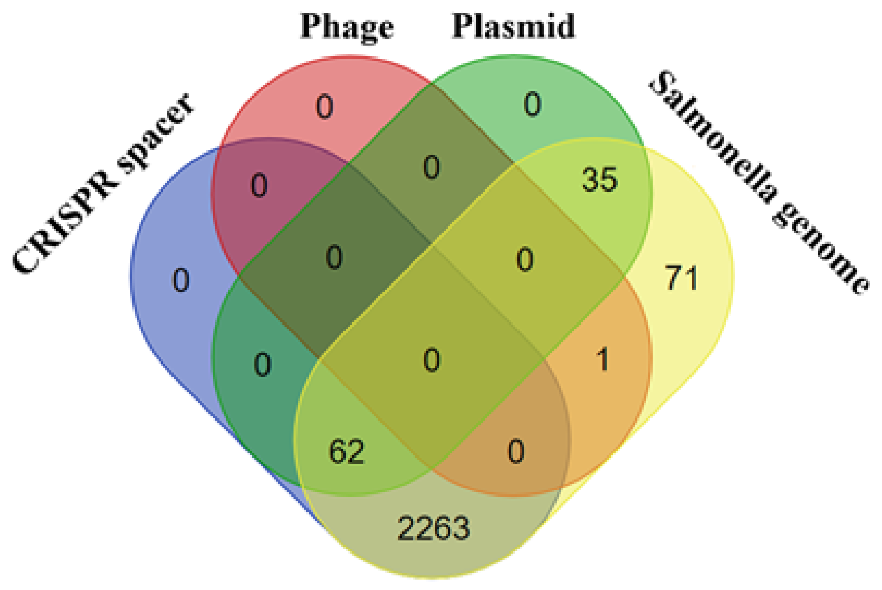
3.5. CRISPR/Cas Systems

CRISPR/Cas system might provide effective information useful for the isolate typing since Salmonella spp. serotypes carry two CRISPR loci and their motifs differ in length [41,42]. Therefore, CRISPR arrays were analyzed. All isolates carried CRISPR loci with encoded Cas proteins, which are an important component for the functioning of the putative CRISPR/Cas system, and such systems belonged to Type IE. Furthermore, all ST32 isolates included in this study contained 2, 3, or 4 CRISPRs motifs. They were formed of 29 bp, 39 bp, and 38 bp long repeats except for Crie-F417, Crie-F392 that had 23 bp length repeats, and Crie-F261 with a 51 bp repeat. We assumed that bioinformatic assay was responsible for the inconsistency in the detection of repeat length. Analysis of CRISPR arrays in S. Infantis isolates studied revealed 2432 spacers. Among them, 2424 were repeating (with 117 unique among them) and only eight spacers were unique. Unique spacers were found in CRISPR arrays of Crie-F207 (five spacers out of eight), Crie-F261 (one out of eight), Crie-F417 (one out of eight), and Crie-F468 (one out of eight) isolates (Table S3).
The vast majority (2325 of 2432) of spacers were identified by BLAST (using the megablast algorithm) as Salmonella CRISPR spacers (e.g., ‘Salmonella enterica subsp. enterica serotype Infantis strain 03-2387 CRISPR1 repeat region’). Only 0.04% of spacers (one spacer out of 2432) were identified by BLAST (using the megablast algorithm) as phage sequences (e.g., ‘Enterobacteria phage mEpX2, complete genome’), 4% of spacers—as plasmid sequences (e.g., ‘Escherichia coli isolate MSB1_4I-sc-2280412 genome assembly, plasmid: 11′) and none of the spacers—as pathogenicity island encoding sequences (Figure 4, Table S3). Interestingly, two repetitive spacers (one from Crie-F191; Crie-F192; Crie-F252; Crie-F339; Crie-F445; Crie-F446, others—from Crie-F259 and Crie-F443 CRISPR array)—were not identified by BLAST using the megablast algorithm. Using the blastn algorithm both spacers were denoted to Salmonella enterica (Table S3).
Notably, CRISPR arrays located on the one side of cas cassette from 18 out of 45 isolates were identical. On the other side of cas cassette CRISPR arrays (positions 1-152 bp) of these isolates formed two separate groups of arrays. Similarly, CRISPR arrays located on one side of cas cassette from 26 Salmonella isolates were identical and also formed two separate groups of arrays on the other side of cas cassette. One part of CRISPR arrays from Crie-F468 and Crie-F469 shared homology with the second group of Salmonella isolates. The other one formed a separate group of CRISPR arrays with partial identity.
The CRISPR/Cas loci of the 45 isolates were predicted. Interestingly, in all isolates except one putative cassettes had the same composition with some discrepancies in the combination of the cas cassette position. The loci from all of the samples except Crie-F243 were identical and consisted of eight genes encoding Cas1 endonuclease, Cas2 endoribonuclease, Cas3 helicase, and Cas5, Cas6, Cas7, Cse1, and Cse2 proteins (Table S4). At the same time, Crie-F243 did not have Cse2, and it seems that this deletion has led to cas cassette shifting (Table S4). Moreover, all systems could be grouped into two clusters: the first had a sequence of CRISPR-associated (cas) genes cas2, cas1, cas6, cas5, cas7, cse2, cse1, cas3, while the second was cas3, cse1, cse2, cas7, cas5, cas6, cas1, cas2 (Table S4). On the other hand, there is no evidence that the position of cas genes is of some functional significance.

4. Discussion

The S. enterica serotype Infantis is widespread not only in poultry, especially broilers, but also in pigs and cattle [43]. However, specifically, poultry and its products have been identified as one of the most important sources of salmonellosis infection in humans [44]. In the last few years, antimicrobial resistance has emerged in S. Infantis from various food sources and humans worldwide [44,45]. Thus, the increased dissemination of various MDR S. Infantis clones has led to the spreading of organisms in the food chain and through poultry to people in such countries as Turkey [21], Switzerland [9], Germany [18], and Russia [14]. For this reason, we aimed to find out whether the S. Infantis isolates of food origin collected in 2018−2020 have acquired new properties which resulted in changes of the pathogen characteristics and to reveal possible dynamics in the MDR development over the past few years.
In the current report, we analyzed 45 MDR S. enterica isolates obtained from various food products in 19 different geographical regions of Russia during the period of 2018–2020 within the framework of food poisoning monitoring by the regional centers of Hygiene and Epidemiology. All isolates studied belonged to ST32 and Infantis serotype, which was the most prevalent in Europe, the Middle East, and Japan [9,18,46]. Moreover, susceptibility testing showed that all of them were resistant to six antimicrobial drugs: CXM, CIP, CHL, NIT, SXT, and TET. Ciprofloxacin and β-lactams are common first-line antibiotics used for treating salmonellosis [8]. As described in the Hindermann et al., resistance to ciprofloxacin was verified in 4.6% of the isolates from Switzerland [9], while almost all isolates studied in Germany were resistant to CIP and TET [18]. Thus, we can suggest that there could be a problem with CIP therapy in Russia since our collection of ST32 isolates obtained in 2018–2020 possessed a resistance to this antibiotic according to the susceptibility testing. Moreover, the majority of the isolates from Switzerland showed a combined resistance to SXT and TET, and some samples were resistant to ampicillin [9], which was the same for our samples.
The major AMR determinants detected by WGS in the isolates studied included the genes of aminoglycoside-modifying enzymes, and genes responsible for resistance to tetracycline, sulphonamide, and chloramphenicol. Most of these genes were described in previous studies [7,18,22,47]. For example, the chromosomally encoded gene aac(6′)-Iaa was detected among all the S. Infantis samples of food origin obtained in different countries [7,18]. Various studies have shown that phenotypic resistance to tetracycline was strongly correlated with the presence of known resistance determinants predicted by the WGS with the most frequent being the tetracycline efflux transporter encoded by tet(A) [22,47], while sulfonamides resistance was predominantly encoded by sul1 and sul2 [47], which conforms well with our observations. For instance, all foodborne S. Infantis isolates from Turkey possessed tetA and sul1 genes in combination and carried aac(6′)-Iaa and ant(3″)-Ia genes [21] encoding aminoglycoside modifying enzymes, just as our samples described above. However, most of the isolates were sensitive to aminoglycoside tested drugs (gentamicin, tobramycin) in the current study. At the same time, all our isolates included AMR gene pattern specific for the MDR strains revealed in Germany in 2010, which consisted of ant(3″)-Ia, tet(A), and sul1 [18].
The current study also showed that the antimicrobial resistance phenotypes and genotypes correlated well in general, however, some discrepancies were found. This can be explained by the fact that the presence of a resistance gene in a genome cannot be considered as single evidence for its expression and activity in the given isolate, which is determined by a complex of factors [48]. For example, discrepancies were observed in low presence of β-lactam (bla family) and quinolone (qnr) genes, while the susceptibility test showed widespread resistance to some antibiotics of these classes. Interestingly, all isolates studied possessed CXM resistance and were quite susceptible to third and fourth-generation cephalosporins (Figure 1). It can be explained that third and fourth-generation cephalosporins have specific modifications to penetrate through the outer membrane of Gram-negative bacteria. However, we found β-lactamase AMR genes that could made S. Infantis resistant to CRO, CZD, and FEP. Surprisingly, according to the data from the USA, dfrA12 and dfrA14 genes were unique to the human isolates of Salmonella [22], while our foodborne samples carried both these resistance genes, and thus we may suggest that they could be transferred from humans to food products via manual handling. However, the data from Turkey showed similarity to Russian genotypes of S. Infantis isolates in a sense that more than half of Turkish isolates had dfrA14 [21]. Moreover, WGS results revealed that three out of 45 S. Infantis isolates harbored the floR gene, for which disagreement of phenotypic resistance to chloramphenicol was observed, while foodborne samples from Turkey showed resistance to chloramphenicol/florfenicol without the presence of the floR gene [21]. In our case, phenotypic resistance of some isolates could be explained by cmlA1 occurrence. However, the possible resistance mechanism requires a more detailed study. AMR phenotypes and genotypes information is important for epidemiological studies and accumulation of general data on resistance genes circulating in the bacterial phavingopulation.
Short-read sequencing indicated the presence of IncFIB replicon, which belongs to the family of IncF, in 93.3% S. Infantis isolates. IncF plasmids are widely distributed in the Enterobacteriaceae family and carry virulence factors, AMR, cytotoxins, and adhesion factors [49], which were confirmed by the presence of the main traits of resistance and virulence found in the German isolates of S. Infantis which were positive for the IncFIB replicon [18]. Moreover, for example, IncF replicon families have been associated with the sudden worldwide emergence of resistance to quinolones and aminoglycosides due to the presence of aac(6′)-Ib-cr, which was absent in our AMR profile, and qnr genes [36,49]. Resistance to aminoglycosides in our isolates was possibly encoded by the aac(6′)-Iaa gene revealed in all of them, which differs from the resistance genes usually present on the replicons of IncF plasmids [49]. Thus we can assume that the aac(6′)-Iaa gene can be located on the chromosome. As previously described, it was supposed that ampicillin resistance can be carried not only by plasmids belonging to the incompatibility group I1 (IncI1) [50,51], but also by other plasmid families, such as the small plasmids Col and IncX possessing blaTEM resistance genes [52,53]. The genes ant(3″)-Ia, sul1, sul3, tetA were found by long-read sequencing in pESI-like plasmids. Interestingly, sul genes and ant(3″)-Ia presented in our study were located in different incompatibility replicons, while replicon typing identified the IncF incompatibility group that carried sul genes (sul1, sul2, and sul3) in E. coli isolates of food origin from China [54]. At the same time, we suggest that ant(3″)-Ia could be presented in both megaplasmids and small plasmids as described by Kürekci et al. [21]. Moreover, the IncFIB replicons contain tet(A), sul1 and dfr A14 genes which represent widespread elements of S. Infantis [14,21].
Whereas the majority of genes for full virulence are found in the chromosome as SPIs [38], we analyzed 129 virulence genes harbored by all S. Infantis isolates of our sample collection (Table S2). All 45 samples showed the same virulence profile regardless of the year, geographical region or source of isolation (Figure S2), except for one gene in a sample Crie-F244 (sipD). However, this is most likely a sequencing inaccuracy, since Crie-F261 has the same profile of the AMR genes and plasmids, and included the whole spectrum of virulence genes presented. The fimbrial adherence gene operons bcf, fim, inv, csg, the type III secretion system 1 (T3SS-1) genes: sipABC, sopABE2 and spaOPQRS, the type III secretion system 2 (T3SS-2) genes: spiC, sifA, sscAB, sse, and ssa operons were similar to the most common strains in Tunisia [7]. Type III secretion systems (SPI-1 and SPI-2) were identified among the set of virulence determinants in the isolates studied. SPIs located on plasmids or in chromosome consist of multiple genes for full virulence and might have been acquired by horizontal transfer from phage or plasmids of other organisms [38,55]. Additionally, they are highly conserved between the different Salmonella serotypes [38]. A single SPI can turn a normally non-hazardous microorganism into a pathogen, but the acquisition of SPI cannot ensure such a transformation [56].
In addition, we detected the S. Infantis plasmid-encoded fimbria lpf operon and genes encoding for the virulent yersiniabactin operon (fyuA, irp1, irp2, ybt), which were associated with the specific gene pattern ESIv (Emergent S. Infantis virulence) [18,57]. As previously reported [14], similar resistance and virulence gene patterns have been determined by the presence of a pESI-like plasmid. Thus, we decided to perform genomic detection and further characterization of pESI-like plasmids among our most representative isolates. ESIv of S. Infantis isolates studied consists of the genes encoding for the virulent yersiniabactin operon (fyuA, irp1, irp2, ybtAEPQSTUX) and genes associated with the fimbrial protein synthesis (faeCDEFHI). S. Infantis from Turkey displayed the presence of fimbrial operons such as Agf/Csg, fim, and lpf, and many of them were located on pESI-like plasmid, as well as YbtTU and Irp1 regulatory and transport operons of Yersinia pestis [21]. Interestingly, the yersiniabactin biosynthetic protein-coding cassette from Klebsiella pneumoniae was located in the chromosome, as mentioned earlier [58].
Notably, all S. Infantis isolates studied carried CRISPR/Cas systems belonging to Type IE. In our study, the spacers targeting S. enterica genomes were also analyzed. According to CRISPRminer Self-targeting (http://www.microbiome-bigdata.com/CRISPRminer/index.php/Home/Index/selfTarget accessed on 13 May 2021) 870 of 22110 self-targeting spacers belong to S. enterica. Among the 71 spacers which were targeting S. enterica genomes (Table S3) one spacer was unique and 70 were repeating (with five unique ones among repeating spacers). On the one hand, it is known that CRISPR/Cas systems can acquire self-targeting spacers from the host chromosome which results in autoimmunity and cell death [59], but, on the other hand, such spacers are suggested to be involved in mRNA degradation that allows evading immune detection [60]. CRSIPR/Cas systems possessing self-targeting spacers may require tight regulation to properly balance the danger of autoimmunity with the risk of phage infection, and thus deserve further investigation [60]. Taking into account all of the above, we can assume that CRISPR sequences are serotype-specific and, thus CRISPR/Cas system might provide the information useful for effective typing [41,42,61].

5. Conclusions

In the current report we presented a comprehensive analysis of 45 MDR foodborne S. Infantis isolates from different regions of Russia based on WGS data including molecular typing, antibiotic resistance profiling, virulence factors, and plasmid descriptions. Notably, Russia is not a member of the PulseNet International (PulseNet Europe) surveillance system, that probably may be one of the reasons of an insufficiency in genetic diversity data of foodborne Salmonella serotypes [62]. Moreover, the few studies carried out in Russia are mainly limited to the observation of strains isolated from salmonellosis outbreaks [62]. In addition, we found only one confirmation of WGS analysis for foodborne Salmonella [14]. Thus, to the best of our knowledge, our WGS-based study is the most extensive in terms of the number of the isolates in Russia in general, and of a single serotype in particular. Additionally, the long-read sequencing was performed for the first time for such isolates, which allowed getting some insights into the mechanisms of antibiotic resistance transmission.
We believe that the data obtained will greatly facilitate further epidemiological surveillance of S. Infantis as an important foodborne pathogen in terms of genome dynamics and monitoring the newly acquired resistance and virulence characteristics.

Supplementary Materials

The following are available online at www.mdpi.com/article/10.3390/microorganisms10010089/s1, Figure S1: Plasmid replicon IncFIB (270 kb) harbored by Crie-F247 S. Infantis isolate determined by hybrid assembly, Figure S2: Virulence genes presence for the isolates studied revealed by bioinformatics methods. The number of representative genes was selected specific to S. enterica, Table S1: in silico megaplasmid data provided by hybrid assembly, Table S2: List of 129 virulence genes observed by VFDB database. Table S3: Spacers of S. Infantis isolates studied found using blastn algorithm, Table S4: Data providing by CRISPRCasFinder. Group of similar genes is shown by one color.

Author Contributions

All authors contributed to this study. A.C. performed susceptibility testing; Y.M. and S.S. performed the sequencing part; M.T., A.T. and K.K. performed CRISPR/Cas analysis; A.S. analyzed the data; A.E. and Y.M. wrote the manuscript; I.M. and V.A. supervised the project and obtained funding. All authors have read and agreed to the current version of the manuscript.

Funding

This research was funded by a grant from the Ministry of Science and Higher Education of the Russian Federation (agreement No. 075-15-2019-1666).

Institutional Review Board Statement

Not applicable.

Informed Consent Statement

Not applicable.

Data Availability Statement

The assembled genome sequences for all isolates were uploaded to the NCBI Genbank, the project number PRJNA780779, under the following accession numbers: JAJMUC000000000 (Crie-F81), JAJJZC000000000 (Crie-F87), JAJMUB000000000 (Crie-F191), JAJJZB000000000 (Crie-F192), JAJJZA000000000 (Crie-F207), JAJMUA0000 F2400000 (Crie-F244), JAJMTX000000000 (Crie-F245), JAJMTW000000000 (Crie-F247), JAJMTV000000000 (Crie-F252), JAJMTU000000000 (Crie-F254), JAJMTT000000000 (J250000A F257) Crie-F260), JAJMTQ000000000 (Crie-F261), JAJMTP000000000 (Crie-F339), JAJMTO000000000 (Crie-F386), JAJMTN000000000 (Crie-F391), JAJJYZ000000000 (JrieMJTM000000000 (CrieAJTM000000) -F394), JAJMTL000000000 (Crie-F396), JAJMTK000000000 (Crie-F403), JAJMTJ000000000 (Crie-F410), JAJMTI000000000 (Crie-F411), JAJMTH000000000 (Crie-FM4000000) F441), JAJMTF000000000 (Crie-F443), JAJMTE000000000 (Crie-F444), JAJMTD000000000 (Crie-F445), JAJMTC000000000 (Crie-F446), JAJJYW000000000 (Crie-F448000000) JAJ (Crie-F468), JAJMTA000000000 (Crie-F469), JAJMSZ000000000 (Crie-F470), JAJMSY000000000 (Crie-F489), JAJJYV000000000 (Crie-F503), JAJMSX000000000 (Crie-F503), JAJMSX000000000 (Crie-C000000000), JAMSV000000 Crie-F509), JAJMSU000000000 (Crie-F510), JAJJYU000000000 (Crie-F512), JAJJYT000000000 (Crie-F522) Salmonella.

Acknowledgments

The authors wish to thank K. Kuleshov for fruitful discussions.

Conflicts of Interest

The authors declare no conflict of interest.

References

  1. Coburn, B.; Grassl, G.A.; Finlay, B.B. Salmonella, the host and disease: A brief review. Immunol. Cell Biol. 2007, 85, 112–118. [Google Scholar] [CrossRef]
  2. Santos, R.L.; Tsolis, R.M.; Bäumler, A.J.; Adams, L.G. Pathogenesis of Salmonella-induced enteritis. Braz. J. Med. Biol. Res. 2003, 36, 3–12. [Google Scholar] [CrossRef] [Green Version]
  3. Pfeifer, C.G.; Marcus, S.L.; Steele-Mortimer, O.; Knodler, L.A.; Finlay, B.B. Salmonella Typhimurium virulence genes are induced upon bacterial invasion into phagocytic and nonphagocytic cells. Infect. Immun. 1999, 67, 5690–5698. [Google Scholar] [CrossRef] [PubMed] [Green Version]
  4. Langridge, G.C.; Fookesa, M.; Connora, T.R.; Feltwella, T.; Feaseya, N.; Parsonsb, B.N.; Seth-Smitha, H.M.B.; Barquista, L.; Stedmana, A.; Humphreyd, T.; et al. Patterns of genome evolution that have accompanied host adaptation in Salmonella. Proc. Natl. Acad. Sci. USA 2015, 112, 863–868. [Google Scholar] [CrossRef] [PubMed] [Green Version]
  5. Mead, P.S.; Slutsker, L.; Dietz, V.; McCaig, L.F.; Bresee, J.S.; Shapiro, C.; Griffin, P.M.; Tauxe, R.V. Food-related illness and death in the United States. Emerg. Infect. Dis. 1999, 5, 607–625. [Google Scholar] [CrossRef] [PubMed]
  6. Ngoi, S.T.; Ju Teh, C.S.; Chai, L.C.; Thong, K.L. Overview of Molecular Typing Tools for the Characterization of Salmonella enterica in Malaysia. Biomed. Environ. Sci. 2015, 28, 751–764. [Google Scholar] [CrossRef] [PubMed]
  7. Ben Hassena, A.; Haendiges, J.; Zormati, S.; Guermazi, S.; Gdoura, R.; Gonzalez-Escalona, N.; Siala, M. Virulence and resistance genes profiles and clonal relationships of non-typhoidal food-borne Salmonella strains isolated in Tunisia by whole genome sequencing. Int. J. Food Microbiol. 2021, 337, 108941. [Google Scholar] [CrossRef] [PubMed]
  8. Medalla, F.; Gu, W.; Mahon, B.E.; Judd, M.; Folster, J.; Griffin, P.M.; Hoekstra, R.M. Estimated incidence of antimicrobial drug-resistant nontyphoidal Salmonella infections, United States, 2004–2012. Emerg. Infect. Dis. 2017, 23, 29–37. [Google Scholar] [CrossRef] [Green Version]
  9. Hindermann, D.; Gopinath, G.; Chase, H.; Negrete, F.; Althaus, D.; Zurfluh, K.; Tall, B.D.; Stephan, R.; Nüesch-Inderbinen, M. Salmonella enterica serovar Infantis from food and human infections, Switzerland, 2010-2015: Poultry-related multidrug resistant clones and an emerging ESBL producing clonal lineage. Front. Microbiol. 2017, 8, 1322. [Google Scholar] [CrossRef] [Green Version]
  10. ECDC Annual Epidemiological Report 2016 [2014 data]. EFSA J. 2016, 2016–2017. Available online: https://www.ecdc.europa.eu/en/publications-data/malaria-annual-epidemiological-report-2016 (accessed on 29 November 2021).
  11. Rozhnova, S.S.; Kuleshov, K.V.; Pavlova, A.S.; Guseva, A.N.; Kozhakhmetova, T.A.; Akulova, N.K.; Podkolzin, A.T. Heterogeneity of Salmonella isolates obtained from various sources in Russia 2010–2019. Epidemiol. Infect. Dis. 2020, 25, 26–34. [Google Scholar] [CrossRef]
  12. Federal Service for Consumer Protection and Human Welfare Federal Report “On the State of the Sanitary-Epidemiological Well-Being of the Population in the Russian Federation in 2018.” 2019, 254. Available online: https://www.rospotrebnadzor.ru/documents/details.php?ELEMENT_ID=12053 (accessed on 4 June 2019).
  13. Rozhnova, S.H.; Akulova, N.K.; Khristukhina, O.A. Prospects for the organization of an expanded surveillance system for salmonellosis in Russia. Epidemiol. Vaccine 2015, 14, 28–34. [Google Scholar] [CrossRef] [Green Version]
  14. Bogomazova, A.N.; Gordeeva, V.D.; Krylova, E.V.; Soltynskaya, I.V.; Davydova, E.E.; Ivanova, O.E.; Komarov, A.A. Mega-plasmid found worldwide confers multiple antimicrobial resistance in Salmonella Infantis of broiler origin in Russia. Int. J. Food Microbiol. 2020, 319, 108497. [Google Scholar] [CrossRef]
  15. Acar, S.; Bulut, E.; Durul, B.; Uner, I.; Kur, M.; Avsaroglu, M.D.; Kirmaci, H.A.; Tel, Y.O.; Zeyrek, F.Y.; Soyer, Y. Phenotyping and genetic characterization of Salmonella enterica isolates from Turkey revealing arise of different features specific to geography. Int. J. Food Microbiol. 2017, 241, 98–107. [Google Scholar] [CrossRef]
  16. Aviv, G.; Tsyba, K.; Steck, N.; Salmon-Divon, M.; Cornelius, A.; Rahav, G.; Grassl, G.A.; Gal-Mor, O. A unique megaplasmid contributes to stress tolerance and pathogenicity of an emergent Salmonella enterica serovar Infantis strain. Environ. Microbiol. 2014, 16, 977–994. [Google Scholar] [CrossRef] [PubMed]
  17. The European Union Summary Report on Antimicrobial Resistance in zoonotic and indicator bacteria from humans, animals and food in 2017/2018. EFSA J. 2020, 18. Available online: https://efsa.onlinelibrary.wiley.com/doi/full/10.2903/j.efsa.2020.6007 (accessed on 29 November 2021).
  18. García-Soto, S.; Abdel-Glil, M.Y.; Tomaso, H.; Linde, J.; Methner, U. Emergence of Multidrug-Resistant Salmonella enterica Subspecies enterica Serovar Infantis of Multilocus Sequence Type 2283 in German Broiler Farms. Front. Microbiol. 2020, 11, 1741. [Google Scholar] [CrossRef]
  19. Karacan Sever, N.; Akan, M. Molecular analysis of virulence genes of Salmonella Infantis isolated from chickens and turkeys. Microb. Pathog. 2019, 126, 199–204. [Google Scholar] [CrossRef]
  20. Kudaka, J.; Itokazu, K.; Taira, K.; Iwai, A.; Kondo, M.; Susa, T.; Iwanaga, M. Characterization of Salmonella Isolated in Okinawa, Japan. Jpn. J. Infect. Dis 2006, 59, 15–19. [Google Scholar]
  21. Kürekci, C.; Sahin, S.; Iwan, E.; Kwit, R.; Bomba, A.; Wasyl, D. Whole-genome sequence analysis of Salmonella Infantis isolated from raw chicken meat samples and insights into pESI-like megaplasmid. Int. J. Food Microbiol. 2021, 337, 108956. [Google Scholar] [CrossRef]
  22. McDermott, P.F.; Tyson, G.H.; Kabera, C.; Chen, Y.; Li, C.; Folster, J.P.; Ayers, S.L.; Lam, C.; Tate, H.P.; Zhao, S. Whole-genome sequencing for detecting antimicrobial resistance in nontyphoidal Salmonella. Antimicrob. Agents Chemother. 2016, 60, 5515–5520. [Google Scholar] [CrossRef] [Green Version]
  23. Gilbert, J.M.; White, D.G.; McDermott, P.F. The US National Antimicrobial Resistance Monitoring System. Future Microbiol. 2007, 2, 493–500. [Google Scholar] [CrossRef] [PubMed]
  24. Foley, S.L.; White, D.G.; McDermott, P.F.; Walker, R.D.; Rhodes, B.; Fedorka-Cray, P.J.; Simjee, S.; Zhao, S. Comparison of subtyping methods for differentiating Salmonella enterica serovar Typhimurium isolates obtained from food animal sources. J. Clin. Microbiol. 2006, 44, 3569–3577. [Google Scholar] [CrossRef] [PubMed] [Green Version]
  25. Punina, N.V.; Makridakis, N.M.; Remnev, M.A.; Topunov, A.F. Whole-genome sequencing targets drug-resistant bacterial infections. Hum. Genom. 2015, 9, 1–20. [Google Scholar] [CrossRef] [PubMed]
  26. Egorova, S.; Kuleshov, K.; Kaftyreva, L. Modern Salmonella subtyping methods in outbreak investigations. Immunopathol. Allergol. Infectology 2019, 36–42. [Google Scholar] [CrossRef]
  27. Ashton, P.M.; Nair, S.; Dallman, T.; Rubino, S.; Rabsch, W.; Mwaigwisya, S.; Wain, J.; O’Grady, J. MinION nanopore sequencing identifies the position and structure of a bacterial antibiotic resistance island. Nat. Biotechnol. 2015, 33, 296–302. [Google Scholar] [CrossRef] [Green Version]
  28. Madoui, M.A.; Engelen, S.; Cruaud, C.; Belser, C.; Bertrand, L.; Alberti, A.; Lemainque, A.; Wincker, P.; Aury, J.M. Genome assembly using Nanopore-guided long and error-free DNA reads. BMC Genom. 2015, 16, 1–11. [Google Scholar] [CrossRef] [Green Version]
  29. Parry, C.M.; Threlfall, E.J. Antimicrobial resistance in typhoidal and nontyphoidal Salmonellae. Curr. Opin. Infect. Dis. 2008, 21, 531–538. [Google Scholar] [CrossRef]
  30. Achtman, M.; Wain, J.; Weill, F.X.; Nair, S.; Zhou, Z.; Sangal, V.; Krauland, M.G.; Hale, J.L.; Harbottle, H.; Uesbeck, A.; et al. Multilocus Sequence Typing as a Replacement for Serotyping in Salmonella enterica. PLOS Pathog. 2012, 8, e1002776. [Google Scholar] [CrossRef] [Green Version]
  31. Bortolaia, V.; Kaas, R.S.; Ruppe, E.; Roberts, M.C.; Schwarz, S.; Cattoir, V.; Philippon, A.; Allesoe, R.L.; Rebelo, A.R.; Florensa, A.F.; et al. ResFinder 4.0 for predictions of phenotypes from genotypes. J. Antimicrob. Chemother. 2020, 75, 3491–3500. [Google Scholar] [CrossRef] [PubMed]
  32. Carattoli, A.; Zankari, E.; Garciá-Fernández, A.; Larsen, M.V.; Lund, O.; Villa, L.; Aarestrup, F.M.; Hasman, H. In silico detection and typing of plasmids using PlasmidFinder and plasmid multilocus sequence typing. Antimicrob. Agents Chemother. 2014, 58, 3895–3903. [Google Scholar] [CrossRef] [Green Version]
  33. Nascimento, M.; Sousa, A.; Ramirez, M.; Francisco, A.P.; Carriço, J.A.; Vaz, C. PHYLOViZ 2.0: Providing scalable data integration and visualization for multiple phylogenetic inference methods. Bioinformatics 2017, 33, 128–129. [Google Scholar] [CrossRef]
  34. Alikhan, N.F.; Zhou, Z.; Sergeant, M.J.; Achtman, M. A genomic overview of the population structure of Salmonella. PLOS Genet. 2018, 14, e1007261. [Google Scholar] [CrossRef] [Green Version]
  35. Dolejska, M.; Villa, L.; Minoia, M.; Guardabassi, L.; Carattoli, A. Complete sequences of IncHI1 plasmids carrying blaCTX-M-1 and qnrS1 in equine Escherichia coli provide new insights into plasmid evolution. J. Antimicrob. Chemother. 2014, 69, 2388–2393. [Google Scholar] [CrossRef] [Green Version]
  36. Dobiasova, H.; Dolejska, M. Prevalence and diversity of IncX plasmids carrying fluoroquinolone and β-lactam resistance genes in Escherichia coli originating from diverse sources and geographical areas. J. Antimicrob. Chemother. 2016, 71, 2118–2124. [Google Scholar] [CrossRef] [Green Version]
  37. Dimitrov, T.; Dashti, A.A.; Albaksami, O.; Udo, E.E.; Jadaon, M.M.; Albert, M.J. Ciprofloxacin-resistant Salmonella enterica serovar Typhi from Kuwait with novel mutations in gyrA and parC genes. J. Clin. Microbiol. 2009, 47, 208–211. [Google Scholar] [CrossRef] [PubMed] [Green Version]
  38. Marcus, S.L.; Brumell, J.H.; Pfeifer, C.G.; Finlay, B.B. Salmonella pathogenicity islands: Big virulence in small packages. Microbes Infect. 2000, 2, 145–156. [Google Scholar] [CrossRef]
  39. Riquelme, S.; Varas, M.; Valenzuela, C.; Velozo, P.; Chahin, N.; Aguilera, P.; Sabag, A.; Labra, B.; Álvarez, S.A.; Chávez, F.P.; et al. Relevant genes linked to virulence are required for Salmonella Typhimurium to survive intracellularly in the social amoeba Dictyostelium discoideum. Front. Microbiol. 2016, 7, 1305. [Google Scholar] [CrossRef] [PubMed]
  40. Kombade, S.; Kaur, N. Pathogenicity Island in Salmonella. In Salmonella-a Challenge From Farm to Fork; IntechOpen: London, UK, 2021. [Google Scholar] [CrossRef]
  41. Bugarel, M.; den Bakker, H.; Grout, J.; Vignaud, M.L.; Loneragan, G.H.; Fach, P.; Brisabois, A. CRISPR-based assay for the molecular identification of highly prevalent Salmonella serotypes. Food Microbiol. 2018, 71, 8–16. [Google Scholar] [CrossRef] [PubMed]
  42. Bratčikov, M.; Mauricas, M. The use of high-resolution melting analysis for Salmonella spp. CRISPR sequence genotyping. Acta Med. Litu. 2009, 16, 98–102. [Google Scholar] [CrossRef]
  43. Lindqvist, N.; Pelkonen, S. Genetic surveillance of endemic bovine Salmonella Infantis infection. Acta Vet. Scand. 2007, 49, 1–9. [Google Scholar] [CrossRef] [Green Version]
  44. European Food Safety Authority; European Centre for Disease Prevention and Control. The European Union One Health 2020 Zoonoses Report. EFSA J. 2021, 19, e06971. [Google Scholar] [CrossRef]
  45. Nógrády, N.; Király, M.; Davies, R.; Nagy, B. Multidrug resistant clones of Salmonella Infantis of broiler origin in Europe. Int. J. Food Microbiol. 2012, 157, 108–112. [Google Scholar] [CrossRef]
  46. Sakano, C.; Kuroda, M.; Sekizuka, T.; Ishioka, T.; Morita, Y.; Ryo, A.; Tsukagoshi, H.; Kawai, Y.; Inoue, N.; Takada, H.; et al. Genetic analysis of non-hydrogen sulfide-producing Salmonella enterica serovar Typhimurium and S. enterica Serovar Infantis isolates in Japan. J. Clin. Microbiol. 2013, 51, 328–330. [Google Scholar] [CrossRef] [PubMed] [Green Version]
  47. Chen, Z.; Kuang, D.; Xu, X.; González-Escalona, N.; Erickson, D.L.; Brown, E.; Meng, J. Genomic analyses of multidrug-resistant Salmonella Indiana, Typhimurium, and Enteritidis isolates using MinION and MiSeq sequencing technologies. PLoS ONE 2020, 15, e0235641. [Google Scholar] [CrossRef]
  48. Shelenkov, A.; Mikhaylova, Y.; Yanushevich, Y.; Samoilov, A.; Petrova, L.; Fomina, V.; Gusarov, V.; Zamyatin, M.; Shagin, D.; Akimkin, V. Molecular typing, characterization of antimicrobial resistance, virulence profiling and analysis of whole-genome sequence of clinical Klebsiella pneumoniae isolates. Antibiotics 2020, 9, 261. [Google Scholar] [CrossRef] [PubMed]
  49. Villa, L.; García-Fernández, A.; Fortini, D.; Carattoli, A. Replicon sequence typing of IncF plasmids carrying virulence and resistance determinants. J. Antimicrob. Chemother. 2010, 65, 2518–2529. [Google Scholar] [CrossRef] [Green Version]
  50. Carattoli, A. Resistance plasmid families in Enterobacteriaceae. Antimicrob. Agents Chemother. 2009, 53, 2227–2238. [Google Scholar] [CrossRef] [PubMed] [Green Version]
  51. García-Fernández, A.; Chiaretto, G.; Bertini, A.; Villa, L.; Fortini, D.; Ricci, A.; Carattoli, A. Multilocus sequence typing of IncI1 plasmids carrying extended-spectrum β-lactamases in Escherichia coli and Salmonella of human and animal origin. J. Antimicrob. Chemother. 2008, 61, 1229–1233. [Google Scholar] [CrossRef] [PubMed] [Green Version]
  52. Johnson, T.J.; Bielak, E.M.; Fortini, D.; Hansen, L.H.; Hasman, H.; Debroy, C.; Nolan, L.K.; Carattoli, A. Expansion of the IncX plasmid family for improved identification and typing of novel plasmids in drug-resistant Enterobacteriaceae. Plasmid 2012, 68, 43–50. [Google Scholar] [CrossRef]
  53. Ahmed, D.; Nahid, M.A.; Sami, A.B.; Halim, F.; Akter, N.; Sadique, T.; Rana, M.S.; Elahi, M.S.B.; Rahman, M.M. Bacterial etiology of bloodstream infections and antimicrobial resistance in Dhaka, Bangladesh, 2005–2014. Antimicrob. Resist. Infect. Control 2017, 6, 2. [Google Scholar] [CrossRef] [Green Version]
  54. Jiang, H.; Cheng, H.; Liang, Y.; Yu, S.; Yu, T.; Fang, J.; Zhu, C. Diverse Mobile Genetic Elements and Conjugal Transferability of Sulfonamide Resistance Genes (sul1, sul2, and sul3) in Escherichia coli Isolates From Penaeus vannamei and Pork From Large Markets in Zhejiang, China. Front. Microbiol. 2019, 10, 1787. [Google Scholar] [CrossRef] [Green Version]
  55. Conner, C.P.; Heithoff, D.M.; Julio, S.M.; Sinsheimer, R.L.; Mahan, M.J. Differential patterns of acquired virulence genes distinguish Salmonella strains. Proc. Natl. Acad. Sci. USA 1998, 95, 4641. [Google Scholar] [CrossRef] [Green Version]
  56. Groisman, E.A.; Ochman, H. Pathogenicity islands: Bacterial evolution in quantum leaps. Cell 1996, 87, 791–794. [Google Scholar] [CrossRef] [Green Version]
  57. Aviv, G.; Elpers, L.; Mikhlin, S.; Cohen, H.; Vitman Zilber, S.; Grassl, G.A.; Rahav, G.; Hensel, M.; Gal-Mor, O. The plasmid-encoded Ipf and Klf fimbriae display different expression and varying roles in the virulence of Salmonella enterica serovar Infantis in mouse vs. avian hosts. PLOS Pathog. 2017, 13, e1006559. [Google Scholar] [CrossRef] [PubMed] [Green Version]
  58. Shaidullina, E.; Shelenkov, A.; Yanushevich, Y.; Mikhaylova, Y.; Shagin, D.; Alexandrova, I.; Ershova, O.; Akimkin, V.; Kozlov, R.; Edelstein, M. Antimicrobial resistance and genomic characterization of OXA-48-and CTX-M-15-co-producing hypervirulent Klebsiella pneumoniae st23 recovered from nosocomial outbreak. Antibiotics 2020, 9, 862. [Google Scholar] [CrossRef] [PubMed]
  59. McGinn, J.; Marraffini, L.A. Molecular mechanisms of CRISPR–Cas spacer acquisition. Nat. Rev. Microbiol. 2019, 17, 7–12. [Google Scholar] [CrossRef] [PubMed]
  60. Wimmer, F.; Beisel, C.L. CRISPR-Cas Systems and the Paradox of Self-Targeting Spacers. Front. Microbiol. 2020, 10, 3078. [Google Scholar] [CrossRef] [PubMed] [Green Version]
  61. Yin, Z.; Liu, J.; Du, B.; Ruan, H.-H.; Huo, Y.-X.; Du, Y.; Qiao, J. Whole-Genome-Based Survey for Polyphyletic Serovars of Salmonella enterica subsp. Enterica Provides New Insights into Public Health Surveillance. Int. J. Mol. Sci. 2020, 21, 5226. [Google Scholar] [CrossRef]
  62. Rakov, A.V.; Kuznetsova, N.A.; Yakovlev, A.A. Genetic diversity of Salmonella enterica subsp. enterica serovar Enteritidis in the Siberia and Far East of Russia based on plasmid profiles. AIMS Microbiol. 2020, 6, 106–120. [Google Scholar] [CrossRef]
Figure 1. Phenotypic and genotypic antibiotic resistance profiles of the S. Infantis isolates studied. The tree shown on the left was built using ad hoc core genomes calculated by roary with the total number of core genes being 4371. White color means missing results for particular antibiotics.
Microorganisms 10 00089 g001
Figure 2. Minimum spanning tree (MST) based on cgMLST profiles for ST32 S. Infantis isolates. The number of different alleles between the profiles is shown on the branches connecting the respective isolates. The node CrieF443 had a completely identical profile to Crie-F445 and Crie-F446, thus they are combined in one node, as well as Crie-F522, whose profile was the same as for Crie-F506. Nodes are colored by the region of isolate collection.
Microorganisms 10 00089 g002
Figure 3. Plasmid replicons IncX1 and IncFIB determined by hybrid assembly. (a) Small IncX1plasmid (53 kb) harbored by Crie-F247 S. Infantis isolate. (b) IncX1plasmid (47 kb) located in Crie-F444 S. Infantis isolate. (c) IncFIB plasmid (263 kb) harbored by Crie-F191 and Crie-F252. (d) IncFIB plasmid (281 kb) from Crie-F444. Resistance to doxycycline and tetracycline is highlighted with pink color, resistance to SXT—with yellow, resistance to streptomycin—with green, chloramphenicol AMR genes are shown in blue, ampicillin resistance—in gray, and AMR to aminoglycosides is highlighted with purple. The plasmids were visualized via SnapGene, annotation was performed manually.
Microorganisms 10 00089 g003aMicroorganisms 10 00089 g003b
Figure 4. Homology of CRISPR spacers for the Salmonella isolates studied.
Microorganisms 10 00089 g004
Table 1. The metadata for the foodborne S. Infantis isolates studied.
IsolateYear CollectedRegionSource
Crie-F812018Moscow regionChicken
Crie-F872018Moscow regionChicken
Crie-F1912019Chelyabinsk regionReady-to-cook chicken
Crie-F1922019Chelyabinsk regionChicken
Crie-F2072019Saratov regionTurkey
Crie-F2412019Pskov regionReady-to-cook chicken cutlets
Crie-F2432019Novgorod regionMeat
Crie-F2442019Altai KraiChicken
Crie-F2452019Rostov regionReady-to-cook meat
Crie-F2472019Republic of TatarstanReady-to-cook meat
Crie-F2522019Republic of TatarstanChicken
Crie-F2542019Ryazan regionReady-to-cook chicken cutlets
Crie-F2572019Sverdlovsk regionReady-to-cook meat cutlets
Crie-F2592019Sverdlovsk regionChicken
Crie-F2602019Sverdlovsk regionReady-to-cook meat
Crie-F2612019Krasnoyarsk regionReady-to-cook meat cutlets
Crie-F3392019Republic of TatarstanChicken
Crie-F3862019Vologda regionReady-to-cook meat cutlets
Crie-F3912019Amur regionReady-to-cook chicken cutlets
Crie-F3922019Rostov regionChicken
Crie-F3932019Rostov regionChicken
Crie-F3942019Rostov regionReady-to-cook chicken cutlets
Crie-F3962019Rostov regionChicken
Crie-F4032019Novgorod regionChicken
Crie-F4102019Tver regionReady-to-cook chicken cutlets
Crie-F4112019Moscow regionChicken
Crie-F4172019MoscowChicken
Crie-F4182019MoscowChicken
Crie-F4412020Ulyanovsk regionChicken
Crie-F4432020Krasnoyarsk regionChicken
Crie-F4442020Krasnoyarsk regionChicken
Crie-F4452020Krasnoyarsk regionChicken
Crie-F4462020Krasnoyarsk regionChicken
Crie-F4482020Krasnoyarsk regionChicken
Crie-F4682020Samara regionTurkey
Crie-F4692020Samara regionPork
Crie-F4702020Samara regionReady-to-cook meat
Crie-F4892020Belgorod regionChicken
Crie-F5032020Moscow regionReady-to-cook beef cutlets
Crie-F5062020Perm regionReady-to-cook chicken cutlets
Crie-F5082020Perm regionChicken
Crie-F5092020Perm regionChicken
Crie-F5102020Perm regionChicken
Crie-F5122020Primorsky KraiChicken
Crie-F5222020Vologda regionChicken
Table 2. Plasmid replicons found in the isolates.
IsolatePlasmid Replicon
ColIncFIncXOther
Crie-F81-IncFIBIncX1-
Crie-F87-IncFIBIncX1-
Crie-F191-IncFII--
Crie-F192----
Crie-F207Col440I---
Crie-F241-IncFIB--
Crie-F243-IncFIB--
Crie-F244-IncFIB-IncHI2, IncHI2A, IncN
Crie-F245-IncFIB--
Crie-F247-IncFIBIncX1-
Crie-F252-IncFIB--
Crie-F254-IncFIB--
Crie-F257-IncFIB--
Crie-F259-IncFIB--
Crie-F260-IncFIBIncX1-
Crie-F261-IncFIB-IncHI2A, IncHI2, IncN
Crie-F339-IncFIB--
Crie-F386ColpVCIncFIB--
Crie-F391-IncFIB--
Crie-F392-IncFIB--
Crie-F393-IncFIB--
Crie-F394-IncFIB--
Crie-F396-IncFIB--
Crie-F403-IncFIB--
Crie-F410-IncFIBIncX1-
Crie-F411-IncFIB--
Crie-F417-IncFIB--
Crie-F418-IncFIBIncX1-
Crie-F441-IncFIB--
Crie-F443-IncFIB--
Crie-F444-IncFIBIncX1-
Crie-F445-IncFIB--
Crie-F446-IncFIB--
Crie-F448-IncFIBIncX1-
Crie-F468-IncFIBIncX4-
Crie-F469-IncFIB--
Crie-F470-IncFIBIncX1-
Crie-F489-IncFIBIncX1-
Crie-F503ColRNAI, ColpVCIncFIB--
Crie-F506-IncFIB--
Crie-F508-IncFIBIncX1-
Crie-F509-IncFIBIncX1-
Crie-F510Col(pHAD28), ColRNAIIncFIBIncX1-
Crie-F512-IncFIB--
Crie-F522-IncFIB--
Publisher’s Note: MDPI stays neutral with regard to jurisdictional claims in published maps and institutional affiliations.

