Article

Pathogenic APC Variants in Latvian Familial Adenomatous Polyposis Patients

1
Institute of Oncology, Riga Stradiņš University, LV-1007 Riga, Latvia
2
Department of Surgery, Pauls Stradiņš Clinical University Hospital, LV-1002 Riga, Latvia
3
Department of Biology and Microbiology, Riga Stradiņš University, LV-1007 Riga, Latvia
4
Department of Surgery, Riga Stradiņš University, LV-1007, Riga, Latvia
*
Author to whom correspondence should be addressed.
Medicina 2019, 55(10), 612; https://doi.org/10.3390/medicina55100612
Submission received: 27 June 2019 / Revised: 16 August 2019 / Accepted: 18 September 2019 / Published: 20 September 2019

Abstract

:
Background and objectives: Familial adenomatous polyposis is one of the APC-associated polyposis conditions described as genetically predetermined colorectal polyposis syndrome with a variety of symptoms. The purpose of this study was to determine sequence variants of the APC gene in patients with familial adenomatous polyposis (FAP) phenotype and positive or negative family history. Materials and Methods: Eight families with defined criteria of adenomatous polyposis underwent molecular genetic testing. Coding regions and flanking intron regions of the APC gene were analyzed by Sanger sequencing. Results: Eight allelic variants of the APC gene coding sequence were detected. All allelic variants of the APC gene were predicted to be pathogenic based on criteria according to the “Joint Consensus Recommendation of the American College of Medical Genetics and Genomics and the Association for Molecular Pathology” (2015), four of them c.1586_1587insAT, c.2336delT, c.3066_3067insGA, and c.4303_4304insC, were considered novel. Conclusions: The timely molecular genetic analysis of APC germline variants and standardized interpretation of the pathogenicity of novel allelic variants has a high impact on choice for treatment, cancer prevention, and family genetic counseling.

1. Introduction

APC-associated polyposis conditions include three main clinical phenotypes of polyposis: familial adenomatous polyposis (FAP), attenuated FAP (AFAP), and gastric adenocarcinoma and proximal polyposis of the stomach (GAPPS) [1]. The Online Mendelian Inheritance in Man (OMIM) database in phenotype familial adenomatous polyposis (FAP1, # 175100) also includes Gardner syndrome (GS) and brain tumor-polyposis syndrome 2 (BTPS2) [2]. All these clinical phenotypes are caused by heterozygous pathogenic variants of the APC gene (APC regulator of Wnt signaling pathway), encoding a multifunctional gatekeeper tumor suppressor protein with ubiquitous expression in brain, colon and 23 other tissues [1,3,4].
Familial adenomatous polyposis (FAP) is a genetically predetermined colorectal polyposis syndrome with a variety of symptoms. Approximately 1% of colorectal cancer (CRC) cases are due to FAP [5]. The reported incidence of FAP among CRC patients in Latvia is 0.08% [6]. The main findings in FAP patients are hundreds to thousands of polyps and adenomas in the colorectum as well as extra-colonic findings, such as desmoid tumors, osteomas, epidermoid cysts, etc. The development of adenomas can be seen in late childhood and early adolescence. Most FAP patients (almost 100%, if left untreated) develop colorectal cancer at a mean age of 40 to 50 years. In the case of AFAP, the development of polyps starts later (between the age of 40 and 70), and fewer polyps are present (around 30). The development of CRC in AFAP patients is estimated to be 70% by age 80 [3,5].
FAP is caused by germline variants of the APC gene in about 80% to 95% of patients. In this case, the phenotype is considered to be associated with APC-associated polyposis conditions. The inheritance of FAP is autosomal dominant. Approximately 20% to 25% of allelic variants of the APC gene arise de novo; the rest are inherited [1]. Various online databases and scientific articles widely describe allelic variants in the gene. Based on information from the Leiden Open Variation Database (LOVD) database, 1692 unique allelic variants of the APC gene are included in the database (information retrieved on 16/06/2019) [7]. The rest of the FAP phenotype is caused by biallelic variants in the MUTYH gene. The clinical phenotype of MUTYH-associated polyposis (MAP) is similar to AFAP. MAP has an autosomal recessive type of inheritance [8]. In this article, only FAP cases with allelic variants of the APC gene will be considered. This article concerns only APC germline variants associated with mainly classical FAP phenotype.
APC is a tumor-suppressor gene with a central role in the Wnt signaling pathway. The gene is located on chromosome 5 locus q21-q22 and consists of 18 exons (LRG_130t1; NM_000038.4). The gene has three transcripts and encodes for a 2843 amino acid long polypeptide [4,5,9]. The APC protein (UniProtKB-P25054) acts as an antagonist in the Wnt signaling pathway and is also involved in processes of cell migration and adhesion, activation of transcription, and apoptosis [4,10]. Around 95% of germline variants of the APC gene are nonsense or frameshift variants leading to a truncated protein. Most of the germline variants occurring in the 5’ coding half of the gene cause loss of the β-catenin level regulating domain, axin binding domain as well as C-terminus microtubule and EB1 binding domains [11].
The main goal of this study was to summarize current findings of the APC germline variants in Latvian FAP patients, report novel variants, and evaluate their pathogenicity.

2. Materials and Methods

2.1. Patients

Patients corresponding to defined criteria of adenomatous polyposis were sent to Riga Stradins University Institute of Oncology for molecular genetics analysis of the APC gene. Eight unrelated patients were involved from Pauls Stradins Clinical University Hospital, Liepaja Regional Hospital, and University Children’s Hospital. After the FAP diagnosis was confirmed, the family genetic consultation and cascade testing were performed. None of the patients’ family members had previously been screened for APC mutations. The genetic study was performed with the approval of the Central Committee of Medical Ethics (decision No1/26-05-17 of 26 January 2017) and written informed consent was obtained from all involved patients.

2.2. Methods

DNA was extracted from whole blood using the QIAgen FlexiGene DNA Kit (Qiagen, Hilden, Germany). All DNA samples were subjected to the Sanger sequencing of coding regions and flanking intron regions of the APC gene as described earlier [12] and according to the manufacturer’s protocol (Life Technologies, Carlsbad, CA, USA). Allelic variants were confirmed by sequencing both DNA strands on an independent PCR product. Reactions were analyzed using the Applied Biosystems genetic analyzer ABI3130 (Life Technologies, Carlsbad, CA, USA). Large deletions or duplications of the APC gene were not analyzed. For the annotation of APC variants the following databases were used: INSIGHT-group database (http://www.insight-group.org/mutations/), National Center for Biotechnology Information (NCBI) SNP database, LOVD database, The Universal Mutation Database (UMD-APC), NCBI ClinVar database, and in-silico prediction tools MutationTaster and Mutalyzer [7,13,14,15,16]. Interpretation of the germline variants was made according to the “Joint Consensus Recommendation of the American College of Medical Genetics and Genomics and the Association for Molecular Pathology” (2015) [17].

3. Results

Genetic analysis of the APC gene was requested for eight patients. All eight had positive clinical findings confirming polyposis, and all but one patient had a positive family history of colorectal polyposis or colorectal cancer, according to diagnostic criteria for FAP. The patient family histories of cancer are summarized in Figure 1. Five had carpeted polyposis throughout the colon and rectum, and one had diffuse polyposis of the rectum, stomach, and duodenum. Six out of eight patients had developed colorectal cancer, two of them diagnosed only during total proctocolectomy, with no previous suspicion of this diagnosis, and two patients were without a clinical cancer diagnosis. Two of the patients underwent total proctocolectomy, one patient had left-side colon resection, and one patient has not had colorectal surgery. No information about surgical management was available for three patients. The age range at diagnosis was from 30 to 55 years, with the average age of patient age being 38.88 years (± 8.84). Based on the number of polyps and the age of onset, two of the patients (J751 and TA698) corresponded to the AFAP phenotype. Despite phenotype similarities with AFAP, patient J751 developed an early manifestation of CRC.
DNA sequencing revealed APC germline variants in all eight patients. The identified germline variants and clinical data of patients are summarized in Table 1. All allelic variants were classified as pathogenic, as results in a truncated protein. One of the variants is nonsense and all other frameshifts, leading to shortening of the polypeptide by half or more amino acids. The location of variants according to the exons and codons of the APC gene is depicted in Figure 2.
Allelic variant c.1433T>G is described in the ClinVar database as likely pathogenic, but no further information about the clinical background of the patient is available either in databases or the literature. Germline allelic variant c.3942delG is not found in databases but has been described previously in the Latvian population [6]. There is no information available about the known link between families harboring the same allelic variant. Variant c.4826delC is mentioned in the UMD-APC database. Variant c.4393_4394delAG is listed in the NCBI dbSNP with the database number rs387906234. The other four APC variants are novel, to the best of our knowledge, previously undescribed. The pathogenicity of novel germline variants was evaluated based on standard recommendations for interpretation of variants observed in patients with suspected inherited disorders or conditions for a clinical diagnostic purpose [17]. The combined criteria for supporting pathogenicity are summarized in Table 2. Genotype–phenotype evaluation showed the consistency of clinical phenotype with the position of allelic variant described earlier, for seven out of eight patients. One phenotype showed overlapping of FAP and AFAP (J751).
Family cascade testing was done in five families. Genetic analysis of APC variants was done in children under 18, in two of the families. In one family, with allelic variant c.3066insGA, one of the children was negative for allelic variant in APC, while the other was positive and colonoscopy was positive for polyposis in the rectum. In a family with allelic variant c.3942delG, one child out of three was negative for APC variant, but two were positive. The eldest daughter at age 18 had polyposis of the rectum. For the youngest daughter, no clinical investigations were done. All other patients from families available for family cascade genetic testing were adults, but no clinical investigation results are available after the results of genetic testing.

4. Discussion

The reported incidence of genetically confirmed FAP among CRC patients in Latvia is far lower than that reported worldwide. Most of our reported cases were clinically recognized and requested for molecular genetic testing by surgeons. Only one case was requested by a clinical geneticist. This situation is not unusual and is limited by the age of onset and specific phenotype.
The spectrum of the APC gene germline variants in European descent populations is reported extensively [21,22,23,24]. The reported mutational hotspots of the APC gene are located in the 5′ part at codons 1309 and 1061, accounting for approximately 17% and 11% of all described germline APC variants, respectively. Due to the accumulation of variants from codon 1250 to 1464, this region is termed the mutation cluster region (MCR) [9]. In our study, we did not find any of the two most common germline variants; however, our study group was rather small. Only three out of eight APC variants reported by our group are in the MCR.
The population-specific variants are known to be reported from the population with Ashkenazi Jewish ancestry, with two missense variants, which do not lead to polyposis but may develop CRC with a lifetime risk of around 20% [9]. All revealed APC variants detected in our study were unique for unrelated patients; however, the size of the study group should be taken into account here. Acquired results demonstrate high heterogeneity of APC variants in Latvian FAP patients.
Interpretation of the germline variants was made according to the «Joint Consensus Recommendation of the American College of Medical Genetics and Genomics and the Association for Molecular Pathology» (2015) [17]. This is the most appropriate standardized up-to-date approach for interpretation of sequence variants for clinical use. No functional studies were performed within the scope of this evaluation of reported novel sequence variants, but all available molecular biology data and clinical data were taken into account.
The genotype impact on phenotype is studied by many research groups, leading to the correlation of mutated codons with a clinical phenotype of APC-associated polyposis conditions. The detected genotype–phenotype correlation plays an important role in clinical decisions for patient treatment strategy. The clinical phenotype of our patients was consistent with the genotype–phenotype correlation described earlier (Figure 2).

5. Conclusions

The study of clinically selected patients revealed eight pathogenic variants of the APC gene, four of them—c.1586_1587insAT, c.2336delT, c.3066_3067insGA and c.4303_4304insC, are considered novel. The timely molecular genetic analysis of germline variants and standardized interpretation of the pathogenicity of novel APC variants has a high impact on choice for treatment, cancer prevention, and family genetic counseling.

Author Contributions

Formal analysis and writing—original draft preparation, Z.D.; molecular genetics investigation, D.B. and E.B.-S.; clinical investigation, V.B., Z.K., L.K.-S., A.G., and J.G.; writing—review and editing, E.M.

Funding

This research was funded by the state research program “Biomedicine for Public Health (BIOMEDICINE)” project 5 “Personalized cancer diagnostics and treatment effectiveness evaluation”.

Conflicts of Interest

The authors declare no conflict of interest.

References

  1. Jasperson, K.W.; Patel, S.G.; Ahnen, D.J. APC-Associated Polyposis Conditions; University of Washington: Seattle, WA, USA, 1993. [Google Scholar]
  2. McKusick-Nathans Institute of Genetic Medicine. Online Mendelian Inheritance in Man, OMIM® n.d. Available online: https://omim.org/ (accessed on 16 June 2019).
  3. Papp, J.; Kovacs, M.E.; Matrai, Z.; Orosz, E.; Kásler, M.; Børresen-Dale, A.L.; Olah, E. Contribution of APC and MUTYH mutations to familial adenomatous polyposis susceptibility in Hungary. Fam. Cancer 2016, 15, 85–97. [Google Scholar] [CrossRef] [PubMed]
  4. Bethesda (MD). Gene. Natl Libr Med (US), Natl Cent Biotechnol Information; 2004. Available online: https://www.ncbi.nlm.nih.gov/gene/ (accessed on 16 June 2019).
  5. Talseth-Palmer, B.A. The genetic basis of colonic adenomatous polyposis syndromes. Hered. Cancer Clin. Pract. 2017, 15, 5. [Google Scholar] [CrossRef] [PubMed]
  6. Borosenko, V.; Irmejs, A.; Melbărde-Gorkusa, I.; Gardovskis, A.; Pavărs, M.; Vanags, A.; Trofimovics, G.; Miklasevics, E.; Gardovskis, J. Initial results of colorectal polyposis research in Latvia. Anticancer Res. 2009, 29, 711–715. [Google Scholar] [PubMed]
  7. Fokkema, I.F.A.C.; Taschner, P.E.M.; Schaafsma, G.C.P.; Celli, J.; Laros, J.F.J.; den Dunnen, J.T. LOVD v.2.0: The next generation in gene variant databases. Hum. Mutat. 2011, 32, 557–563. [Google Scholar] [CrossRef] [PubMed]
  8. Araujo, L.F.; Molfetta, G.A.; Vincenzi, O.C.; Huber, J.; Teixeira, L.A.; Ferraz, V.E.; Silva, W.A. Molecular basis of familial adenomatous polyposis in the southeast of Brazil: Identification of six novel mutations. Int. J. Biol. Mark. 2019, 34, 80–89. [Google Scholar] [CrossRef] [PubMed]
  9. Leoz, M.L.; Carballal, S.; Moreira, L.; Ocaña, T.; Balaguer, F. The genetic basis of familial adenomatous polyposis and its implications for clinical practice and risk management. Appl. Clin. Genet. 2015, 8, 95–107. [Google Scholar] [CrossRef] [PubMed]
  10. UniProt Consortium. UniProt: The universal protein knowledgebase. Nucleic Acids Res. 2017, 45, D158–D169. [Google Scholar] [CrossRef] [PubMed]
  11. Heinen, C.D. Genotype to phenotype: Analyzing the effects of inherited mutations in colorectal cancer families. Mutat. Res. Fundam. Mol. Mech. Mutagen. 2010, 693, 32–45. [Google Scholar] [CrossRef] [PubMed] [Green Version]
  12. Miyoshi, Y.; Ando, H.; Nagase, H.; Nishisho, I.; Horii, A.; Miki, Y.; Mori, T.; Utsunomiya, J.; Baba, S.; Petersen, G. Germ-line mutations of the APC gene in 53 familial adenomatous polyposis patients. Proc. Natl. Acad. Sci. USA 1992, 89, 4452–4456. [Google Scholar] [CrossRef] [PubMed]
  13. Sherry, S.T.; Ward, M.H.; Kholodov, M.; Baker, J.; Phan, L.; Smigielski, E.M.; Sirotkin, K. dbSNP: The NCBI database of genetic variation. Nucleic Acids Res. 2001, 29, 308–311. [Google Scholar] [CrossRef] [PubMed]
  14. Landrum, M.J.; Lee, J.M.; Benson, M.; Brown, G.R.; Chao, C.; Chitipiralla, S.; Gu, B.; Hart, J.; Hoffman, D.; Jang, W.; et al. ClinVar: Improving access to variant interpretations and supporting evidence. Nucleic Acids Res. 2018, 46, D1062–D1067. [Google Scholar] [CrossRef] [PubMed]
  15. Schwarz, J.M.; Cooper, D.N.; Schuelke, M.; Seelow, D. MutationTaster2: Mutation prediction for the deep-sequencing age. Nat. Methods 2014, 11, 361–362. [Google Scholar] [CrossRef] [PubMed]
  16. Wildeman, M.; van Ophuizen, E.; den Dunnen, J.T.; Taschner, P.E.M. Improving sequence variant descriptions in mutation databases and literature using the Mutalyzer sequence variation nomenclature checker. Hum. Mutat. 2008, 29, 6–13. [Google Scholar] [CrossRef] [PubMed]
  17. Richards, S.; Aziz, N.; Bale, S.; Bick, D.; Das, S.; Gastier-Foster, J.; Grody, W.W.; Hegde, M.; Lyon, E.; Spector, E.; et al. Standards and guidelines for the interpretation of sequence variants: A joint consensus recommendation of the American College of Medical Genetics and Genomics and the Association for Molecular Pathology. Genet. Med. 2015, 17, 405–424. [Google Scholar] [CrossRef] [PubMed]
  18. Unicancer Cancer Genetic Group. The UMD-APC Mutations Database n.d. Available online: http://www.umd.be/APC/ (accessed on 17 June 2019).
  19. Chung, D.C.; Mino, M.; Shannon, K.M. Case 34-2003. N. Engl. J. Med. 2003, 349, 1750–1760. [Google Scholar] [CrossRef] [PubMed]
  20. De Queiroz Rossanese, L.B.; De Lima Marson, F.A.; Ribeiro, J.D.; Coy, C.S.R.; Bertuzzo, C.S. APC germline mutations in families with familial adenomatous polyposis. Oncol. Rep. 2013, 30, 2081–2088. [Google Scholar] [CrossRef] [PubMed] [Green Version]
  21. Friedl, W.; Aretz, S. Familial Adenomatous Polyposis: Experience from a Study of 1164 Unrelated German Polyposis Patients. Hered. Cancer Clin. Pract. 2005, 3, 95. [Google Scholar] [CrossRef] [PubMed]
  22. Plawski, A.; Slomski, R. APC gene mutations causing familial adenomatous polyposis in Polish patients. J. Appl. Genet. 2008, 49, 407–414. [Google Scholar] [CrossRef] [PubMed]
  23. Torrezan, G.T.; Da Silva, F.C.C.; Santos, É.M.M.; Krepischi, A.C.V.; Achatz, M.I.W.; Junior, S.A.; Rossi, B.M.; Carraro, D.M. Mutational spectrum of the APC and MUTYH genes and genotype-phenotype correlations in Brazilian FAP, AFAP, and MAP patients. Orphanet J. Rare Dis. 2013, 8, 54. [Google Scholar] [CrossRef] [PubMed]
  24. Vandrovcová, J.; Štekrová, J.; Kebrdlová, V.; Kohoutová, M. Molecular analysis of the APC and MYH genes in Czech families affected by FAP or multiple adenomas: 13 novel mutations. Hum. Mutat. 2004, 23, 397. [Google Scholar] [CrossRef] [PubMed]
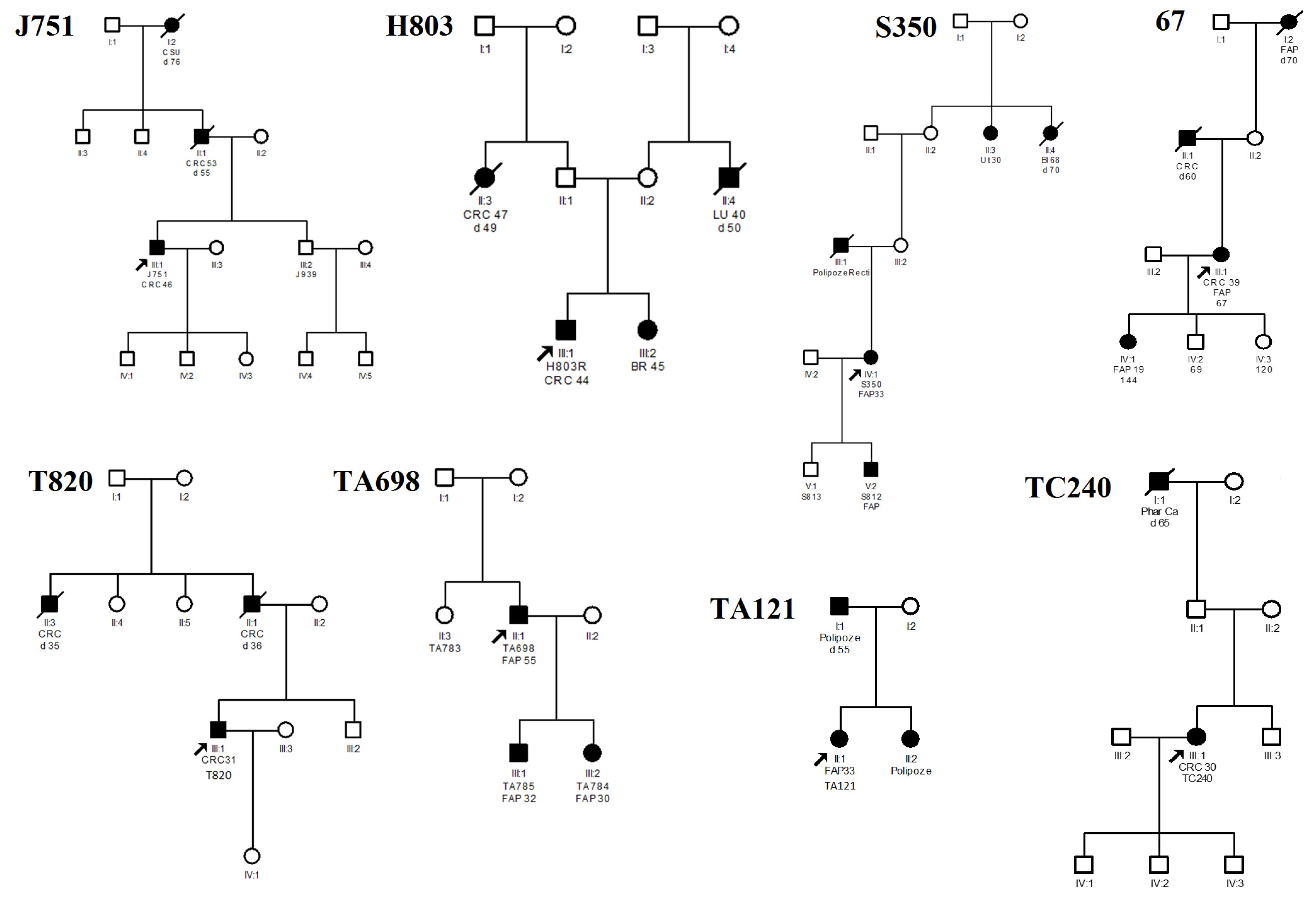
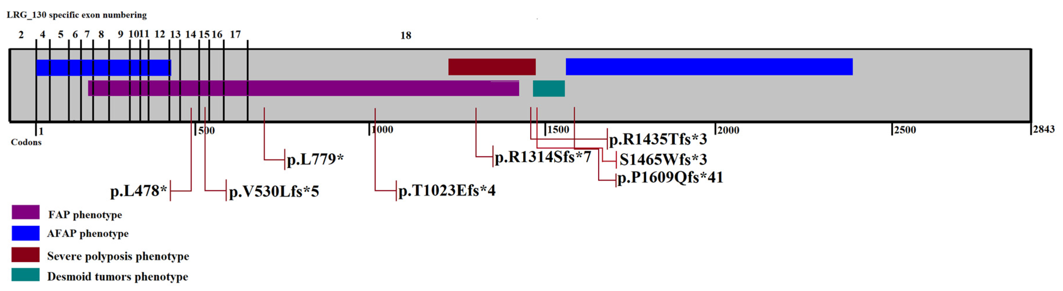
Figure 1. Medical history of cancers in the patients’ families. Abbreviations: CRC—colorectal cancer; Ut—cancer; C Su—cancer site unknown; Li – liver cancer; BR – breast cancer; Phar – pharyngeal cancer; Bl – bladder cancer; d – died.
Medicina 55 00612 g001
Figure 2. APC germline variants identified by exon and codon location. The color bars represent genotype–phenotype correlation described earlier [19,20].
Medicina 55 00612 g002
Table 1. Identified germline variants and clinical data of probands.
Family (Proband)Nucleotide Change 1AA ChangeType of VariantAge of DiagnosisPolyposisCRCRef.
J751c.1433T>Gp.L478*Nonsense46<100+[14]
H803c.1586_1587insATp.V530Lfs*5Frameshift44Carpeted polyposis+novel
TA121c.2336delTp.L779*Frameshift33Carpeted polyposis-novel
S350c.3066_3067insGAp.T1023Efs*4Frameshift33Carpeted polyposis+novel
67c.3942delGp.R1314Sfs*7Frameshift39Carpeted polyposis+[6]
T820c.4303_4304insCp.R1435Tfs*3Frameshift31Carpeted polyposis+novel
TC240c.4393_4394delAGS1465Wfs*3Frameshift30Diffuse polyposis+[13]
TA698c.4826delCp.P1609Qfs*41Frameshift55<100-[18]
1 LRG_130t1.
Table 2. Combined criteria for the classification of novel APC variants based on the «Joint Consensus Recommendation of the American College of Medical Genetics and Genomics and the Association for Molecular Pathology» (2015) [17].
Nucleotide Change 1Type of VariantCriteriaPathogenicity
c.1586_1587insATFrameshiftPVS1, PM1, PP1, PP3, PP4Pathogenic
c.2336delTFrameshiftPVS1, PM1, PP1, PP3, PP4Pathogenic
c.3066_3067insGAFrameshiftPVS1, PM1, PP1, PP3, PP4Pathogenic
c.4303_4304insCFrameshiftPVS, PM1, PP1, PP3, PP4Pathogenic
1LRG_130t1. PVS1 – frameshift variant, PM1 – located in the critical and well-established functional domain, PP1 – co-segregation with the disease in multiple affected family members in a gene definitively known to cause the disease, PP3 – computational evidence support a deleterious effect on the gene product, PP4 – patient’s phenotype is highly specific for a disease.

