Article

Novel Antioxidant Peptides Identified from Arthrospira platensis Hydrolysates Prepared by a Marine Bacterium Pseudoalteromonas sp. JS4-1 Extracellular Protease

1
State Key Laboratory of Medical Genetics, School of Life Sciences, Central South University, Changsha 410013, China
2
Department of Biochemistry, Changzhi Medical College, Changzhi 046000, China
3
Institute of Crop Germplasm Resources, Shandong Academy of Agricultural Sciences, Jinan 250100, China
*
Authors to whom correspondence should be addressed.
These authors contributed equally to this work.
Mar. Drugs 2023, 21(2), 133; https://doi.org/10.3390/md21020133
Submission received: 30 December 2022 / Revised: 16 February 2023 / Accepted: 16 February 2023 / Published: 20 February 2023
(This article belongs to the Special Issue Marine Bioactive Compounds against Oxidative Stress and Inflammation)

Abstract

:
Crude enzymes produced by a marine bacterium Pseudoalteromonas sp. JS4-1 were used to hydrolyze phycobiliprotein. Enzymatic productions showed good performance on DPPH radical and hydroxyl radical scavenging activities (45.14 ± 0.43% and 65.11 ± 2.64%, respectively), especially small peptides with MWCO <3 kDa. Small peptides were fractioned to four fractions using size-exclusion chromatography and the second fraction (F2) had the highest activity in hydroxyl radical scavenging ability (62.61 ± 5.80%). The fraction F1 and F2 both exhibited good antioxidant activities in oxidative stress models in HUVECs and HaCaT cells. Among them, F2 could upregulate the activities of SOD and GSH-Px and reduce the lipid peroxidation degree to scavenge the ROS to protect Caenorhabditis elegans under adversity. Then, 25 peptides total were identified from F2 by LC-MS/MS, and the peptide with the new sequence of INSSDVQGKY as the most significant component was synthetized and the ORAC assay and cellular ROS scavenging assay both illustrated its excellent antioxidant property.

1. Introduction

The microalgae Arthrospira belongs to the cyanobacteria division, the cyanophycean class, and the Oscillatoriaceae family. Arthrospira sp. is a photosynthetic cyanobacterium that has recently grown in value as a nutritional supplement and food additive due to its high protein content of around 60–70% in their biomass and various valuable natural compounds, such as pigments, β-carotenes, polysaccharides and peptides. In addition, Arthrospira sp. has a wide range of uses in the pharmaceutical, cosmetic, and nutritional industries. Because of its high protein content and nutritive value of amino acid composition, Arthrospira sp. was evaluated as a potential meat substitute and has received increased attention [1]. Phycobiliprotein is a class of proteins with antioxidant, antitumor and anti-inflammatory properties, but the heat sensitivity and allergenicity limit its wider applications. Phycobiliprotein is highly abundant in Arthrospira, accounting for about 20% of its dry weight [2]. Using proteases to hydrolyze the phycobiliprotein to produce antioxidant peptides is an efficient way to reach higher bioactive potential and promote high value utilization of marine proteins while avoiding its natural limitations. Moreover, the proteins of microalgae can be converted by enzymatic hydrolysis into value-added products with better functional properties such as antioxidant peptides (APs), anti-hypertension peptides, and antibacterial peptides, etc. Therefore, more and more researchers are interested in obtaining effective bioactive peptides (BPs) from Arthrospira sp. hydrolysates.
Enzymatic hydrolysis of the dietary proteins generates BPs, which are short chains of 2–15 amino acids residues, can enhance nutritional value, safety, bioactive function and reduce the allergenicity [3,4]. Numerous studies have demonstrated that BPs with antioxidant activity produced by proteolysis can achieve antioxidation by scavenging free radicals, chelating transition metals, and enhancing the activity of endogenous antioxidant enzymes such as catalase (CAT), superoxide dismutase (SOD), and glutathione peroxidase (GSH-Px). Redox homeostasis (balance) is an important cellular process that plays a vital role in maintaining the normal physiological steady state of the human body. A disturbance of balance between oxidants and antioxidants results in oxidative stress. Reactive oxygen species (ROS) are molecules derived from aerobic cellular metabolism; in the oxidative stress condition excessive production of ROS can disrupt the intracellular redox balance and cause numerous diseases such as cancer, diabetes, atherosclerosis, cardiovascular and neurodegenerative [4]. A growing number of studies have demonstrated that APs from protease hydrolysates can effectively scavenge free radicals or inhibit the production of ROS. Meanwhile, food-derived APs also performed nontoxicity, are low cost, absorb efficiency advantages, and were considered as potential substitutes for commercial synthetic antioxidants [5,6].
Since different proteases have specific hydrolyzed performance on protein substrate and can produce diverse BPs with different length, sequence, and composition, these hydrolyzes may exhibit various hydrophobicity and functionalities [7,8]. Therefore, it is necessary to find a new enzymes resource to hydrolyze Arthrospira platensis (A. platensis) and produce novel APs with high antioxidant activity and bioactive properties. Our previous study suggested using extracellular proteases secreted by a marine bacterium Pseudoalteromonas sp. JS4-1 to hydrolyze chlorella can obtain hydrolysates with high antioxidant activity [9]. In this study, proteases from Pseudoalteromonas sp. JS4-1 were used to hydrolyze A. platensis proteins and produced diverse Aps; a series of biochemical assays, cell models, and animal models were applied for testing the bioactivity of the APs.

2. Results

2.1. Screening of Proteases for Enzymatic Hydrolysis

Pseudoalteromonas is a strictly marine genus and able to produce extracellular enzymes, which have good advantages in degrading natural marine proteins [10]. In order to evaluate which enzyme is more suitable for hydrolyzing Arthrospira sp. proteins, the extracellular enzymes from eight strains named Pseudoalteromonas sp. B27-3, Pseudoalteromonas sp. B47-6, Pseudoalteromonas sp. B62-3, Pseudoalteromonas sp. WH05-1, Pseudoalteromonas sp. WH06-2, Pseudoalteromonas sp. WH16-2, Pseudoalteromonas sp. JS4-1 and Pseudoalteromonas sp. ZB23-2 were extracted and screened by zymography. The enzymatic hydrolysates were identified by SDS-PAGE and their DPPH RSA and OH RSA activities were also detected. The zymography showed with the exception of Pseudoalteromonas sp. WH06-2 and Pseudoalteromonas sp. WH16-2, all the other strains can secrete a variety of proteases, indicating that most marine bacteria could produce extracellular proteases with a variety of cleavage sites, and suitable for enzymatic hydrolysis (Figure 1A). Proteases from Pseudoalteromonas sp. B27-3 and Pseudoalteromonas sp. JS4-1 have stronger hydrolysis activity than other bacterial proteases toward Arthrospira sp. proteins. SDS-PAGE showed that the hydrolysates existed in smaller fragments compared with the control protein, and almost no protein band left on the gel, while the hydrolysates produced by other bacterial proteases still left visible bands on the gel (Figure 1B). The hydrolysates of Pseudoalteromonas sp. JS4-1 proteases have the highest DPPH RSA (45.14 ± 0.43%) and OH RSA (65.11 ± 2.64%) than other productions (Figure 1C,D). Therefore, proteases from Pseudoalteromonas sp. JS4-1 were selected as a candidate for Arthrospira sp. proteins hydrolyzing.

2.2. Optimization of Enzymatic Hydrolyzing Parameters

Enzymatic hydrolyzation was optimized using the one-variable-at-a-time (OVAT) approach by changing one parameter while keeping other parameters constant. First, the hydrolyzing was performed at 50 °C from 1 h to 7 h with 1 h at intervals, at a E: S ratio of 1:10, to estimate the optimal hydrolyzing time. Data showed that the hydrolysis degree increased along with the hydrolyzing process, especially in the initial three hours. After five hours hydrolyzation, the hydrolysis degree was not improved significantly. DPPH RSA as an evaluation index suggested that when the E/S ratio ranged from 1:6 to 1:10, the DPPH RSA of the hydrolytes exhibited no significant difference. While the E/S ratio adjusted to 1:12 and 1:14, the DPPH RSA of the hydrolytes dropped 6.72% and 15.95%, respectively, indicating that the substrate exceeded the catalytic capacity of the enzymes. When the hydrolyzing temperature increased, the DPPH RSA improved and reached the maximal activity at 50 °C, but when the temperature exceeded >50 °C the DPPH RSA decreased, probably due to the enzymes being inactivated at higher temperatures. After OVAT optimization, when E:S ratio was 1:10 and enzymatic hydrolyzing performed at 50 °C for 4 h, the DPPH RSA and the OH RSA of the hydrolysates achieved the maximal level of 48.51 ± 2.42% and 67.16 ± 3.21%, respectively. The ninhydrin colorimetry was used to determine the hydrolysis degree, and data showed that about 0.67 ± 0.02 mM of •NH2 was released after hydrolyzing reaction.
In order to evaluate the free RSA of small peptides, the hydrolysates were separated into 2 parts by ultrafiltration. The DPPH RSA of < 3 kDa and > 3 kDa parts were 48.26 ± 1.34% and 42.60 ± 0.25%, respectively. The OH RSA of < 3 kDa and > 3 kDa parts were 63.30 ± 1.11% and 44.30 ± 0.68%, respectively (Figure 1E). It has been proven that the small peptides (<3 kDa) contained more antioxidant components and were more easy to exert biological effects [11]. The bioactive peptides with small molecular weight have broad application potential in food, medicine, cosmetics and other industries due to their good absorbability and fewer side effects besides their higher bioactivity [12].

2.3. Antioxidant Activities of Size-Exclusion Liquid Chromatography Fractions of Phycobiliprotein Hydrolysis Products

In order to investigate the antioxidant components of the small peptides, the peptides with MWCO <3 kDa were further separated using size-exclusion liquid chromatography, and in total four fractions defined as F1 to F4 were obtained (Figure 1F). By measuring the DPPH RSA (F1 to F4 were 52.06 ± 2.13%, 52.56 ± 1.38%, 24.43 ± 0.85% and 27.58 ± 2.24%, respectively; Figure 1G) and the OH RSA (F1 to F4 were 40.75 ± 14.35%, 62.61 ± 5.80%, 30.86 ± 10.90%, and 10.21 ± 7.91%, respectively; Figure 1H), F1 and F2 showed higher antioxidant activity than F3 and F4. We could obtain about 40 mg of F1 and 40 mg of F2 lyophilized powder after the size-exclusion liquid chromatography from 1 g of A. Platensis powder.
Then, the ORAC assay was performed to compare the antioxidant activity of F1 and F2. At peptide concentration of 0.015 mg/mL, both F1 and F2 could significantly slow down the decrease rate of the fluorescence decay compared with PBS. The Trolox equivalent antioxidant capacity of F1 and F2 were 0.27 ± 0.02 mmol TE/g and 0.41 ± 0.04 mmol TE/g, respectively (Figure 1I). F2 exhibited higher antioxidant activity than F1.
The hydroxyl radicals could open the circular of supercoiled DNA (SC DNA) and make it become partly opened circular DNA (OC DNA), which can slow down the DNA mobility on agarose gel. Comparing with the no damaged plasmid DNA pET-22b (Figure 1J, lane 2), when the plasmid DNA has no protection, it can be completely degraded by hydroxyl radicals into small fragments and no band observed on the gel (Figure 1J, lane 3). An amount of 0.2 mg/mL of Vitamin C can protect the plasmid DNA from oxidative damage, but the protection is limited and the OC DNA is the major form retained on the gel (Figure 1J, lane 10). F1 and F2 concentrations range from 0.2 mg/mL to 0.4 mg/mL; all can significantly protect plasmid DNA from oxidative damage and these protections were far better than Vitamin C, which make the SC DNA and OC DNA co-existing on the gel (Figure 1J, lane 4, 5, 7, 8). So, this study proved that both F1 and F2 have strong antioxidant activity to protect DNA from oxidative stress.

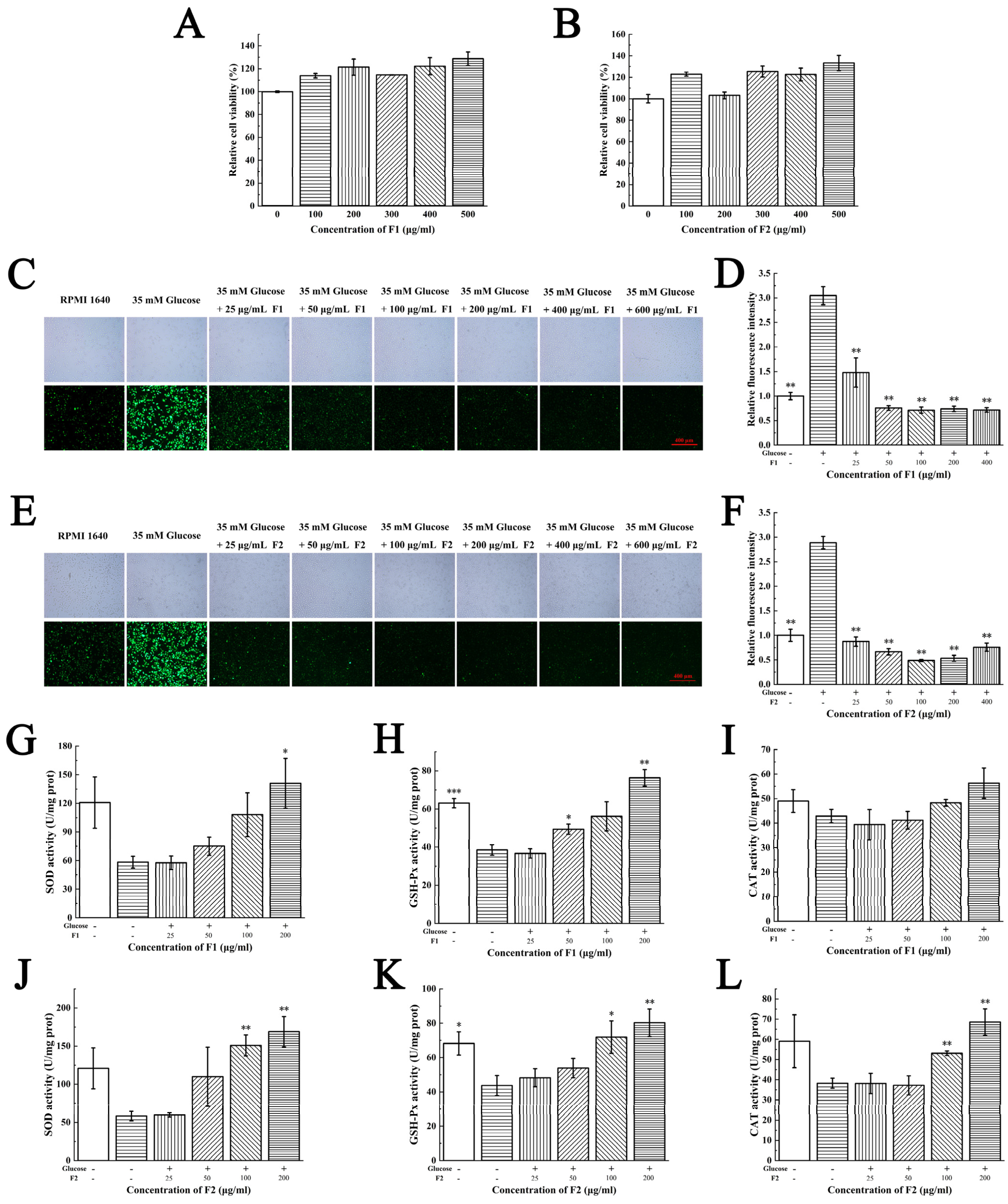
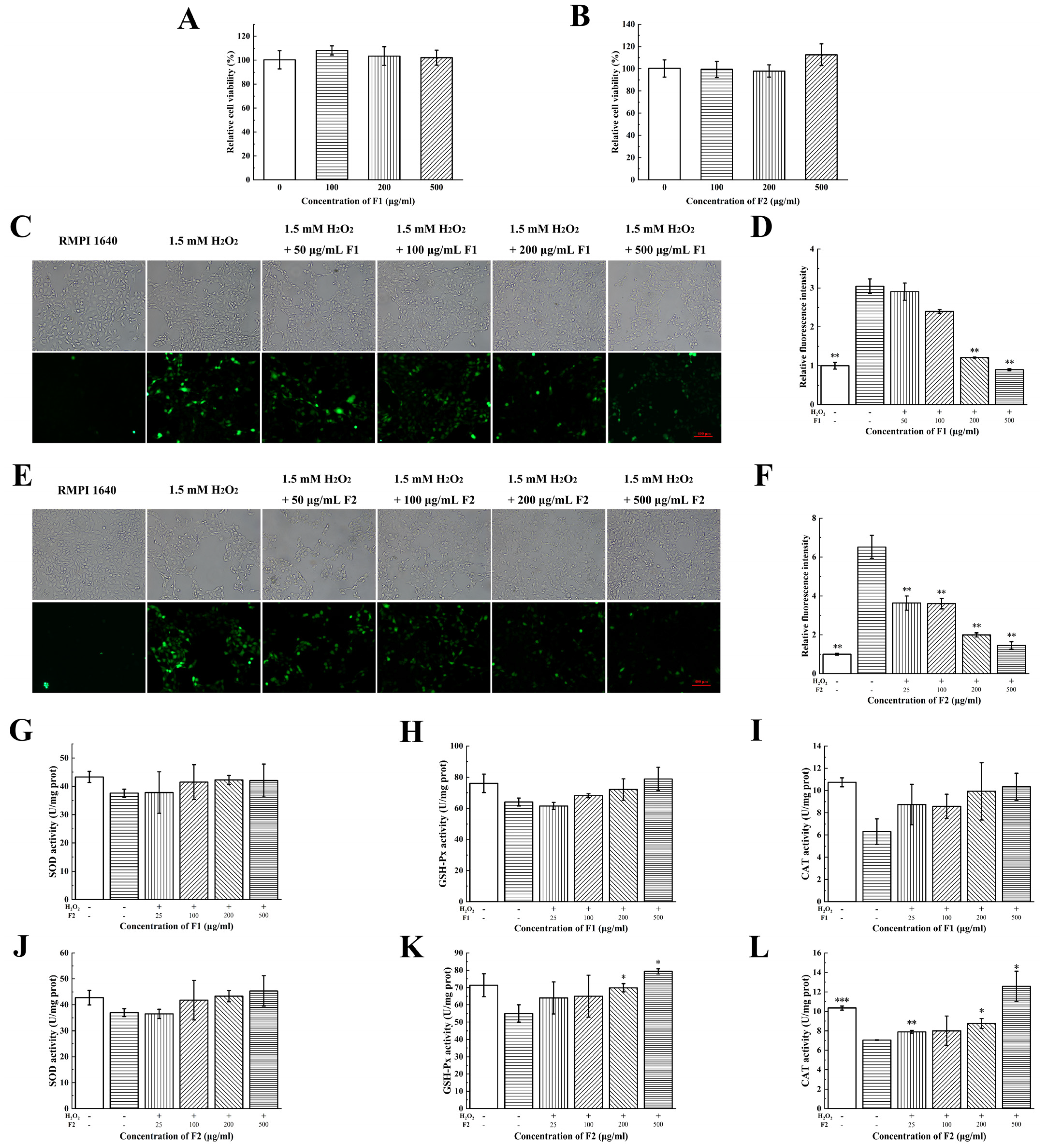
2.4. Antioxidant Activities of F1 and F2 at Cellular Levels

The intracellular oxidized cell model is a common method that is used to assess the antioxidative capacity of compounds. Vascular endothelial cell injury is associated with several factors, among them oxidative stress is an important cause of cardiovascular disease. APs are able to protect vascular endothelial cell function which is a key point in the prevention and treatment for cardiovascular diseases. The skin is the first immune defense of the body and susceptible to a variety of physical and chemical stimuli. Protecting skin cells could be an effective strategy to prevent and treat dermatosis. Therefore, we chose HUVECs and HaCaT cells as cellular models to investigate the intracellular antioxidant effects of F1 and F2 [13,14,15,16].
MTT assay indicated that both F1 and F2 had no significant cytotoxicity on HUVECs and HaCaT cells; moreover, they can obviously increase the cell viability at a concentration range of 100–500 μg/mL (Figure 2A,B and Figure 3A,B).
An amount of 35 mM of glucose was added to the HUVECs and 1.5 mM H2O2 was used in the HaCaT cells. Cells treated with glucose and H2O2 exhibited higher fluorescence intensity than control groups, indicating the oxidative stress models have been successfully established (Figure 2C,E and Figure 3C,E). The intracellular ROS were labelled by DCFH-DA probe. Cells treated with low concentration of F1 and F2 (25 μg/mL) displayed less fluorescence intensity than the glucose group in HUVECs, suggesting the fractions were able to scavenge intracellular ROS to protect HUVECs from hyperglycemia-induced oxidative stress (Figure 2D,F). Cells treated with a high concentration of F1 (100 μg/mL) and a low concentration of F2 (50 μg/mL) displayed less fluorescence intensity than the H2O2 group in HaCaT cells, suggesting that the fractions were able to scavenge intracellular ROS to protect HaCaT cells from H2O2 induced oxidative stress (Figure 3D,F). The antioxidant activities of peptides on a cellular level in this study were close to those reported extracts of plants [17,18,19], which suggested the peptides in our study may also have great potential applications.
To further investigate the underlying mechanisms of fractions scavenging intracellular ROS, we detected the activities of antioxidant enzymes such as SOD, GSH-Px and CAT after being treated with fractions due to their role in scavenging ROS and suppressing oxidative stress [20,21]. The glucose and H2O2 reduced the activities of SOD, GSH-Px and CAT in HUVECs and HaCaT cells. The administration of F1 and F2 dose-dependently increased the activities of these enzymes at a concentration range of 25–200 μg/mL (Figure 2G–L and Figure 3G–L).

2.5. Effects of F2 on Resistance to Oxidative Stress in C. elegans

Caenorhabditis elegans (C. elegans) have many highly conserved genes and signaling pathways involved in the regulation of aging and oxidative stress [22,23]. The genes and pathways in aging, oxidative stress and inflammation between C. elegans and human are highly homologous [24]. C. elegans as an animal model is widely used for anti-aging and anti-oxidative studies.
In this study, 0.5 mg/mL of F1 and F2 had no toxicity and no significant life-extending effect on N2 (Figure 4A). The in vivo ROS assay showed that 0.5 mg/mL of F1 had no significant ROS scavenging effects, while F2 could scavenge ROS at low concentration (0.2 mg/mL, Figure 4E,F). The fluorescence accumulation assay of 80 nematodes showed similar results (Figure 4G).
Then, we speculate whether the APs can enhance the stress resistance of N2. Oxidizing agent H2O2 (10 mM), pesticide paraquat (10 mM) and heat (35 °C) were chosen as stress conditions. Considering F2 has higher anti-oxidative activity than F1, in this study N2 were treated with F2 and then exposed to stress conditions. Data showed that the F2 can extend the stressed N2 survival time in all stress conditions, and the survival rate was positively correlated with the addition of F2. An amount of 0.2 mg/mL of F2 increased 35% survival rate after 7 h of H2O2 stress, 50% after 120 h of paraquat stress and 24% under heat stress (Figure 4B–D). Although F2 did not help the C. elegans live longer, it really improved the resistant ability of C. elegans obviously to the oxidation, pesticide and heat stress.
We then investigated the MDA levels and activities of antioxidant enzymes in nematodes. The MDA level is an important biomarker of lipid peroxidation [25]. F2 could significantly reduce the MDA levels in nematodes (Figure 4H). As shown in Figure 4I–K, F2 could have dose-dependently increased the activities of SOD and GSH-Px, while it had no effects on CAT. These results suggested that F2 could upregulate the activities of SOD and GSH-Px and reduce the lipid peroxidation degree to scavenge the ROS in the organisms. Activating these antioxidant enzymes contributes to maintaining the redox balance, delaying aging process and prolonging lifetime [26]. Antioxidant enzymes with high activity are able to reduce ROS levels, maintain normal physiological steady state and prevent numerous diseases such as cancer, diabetes, neurodegenerative diseases and so on [4].

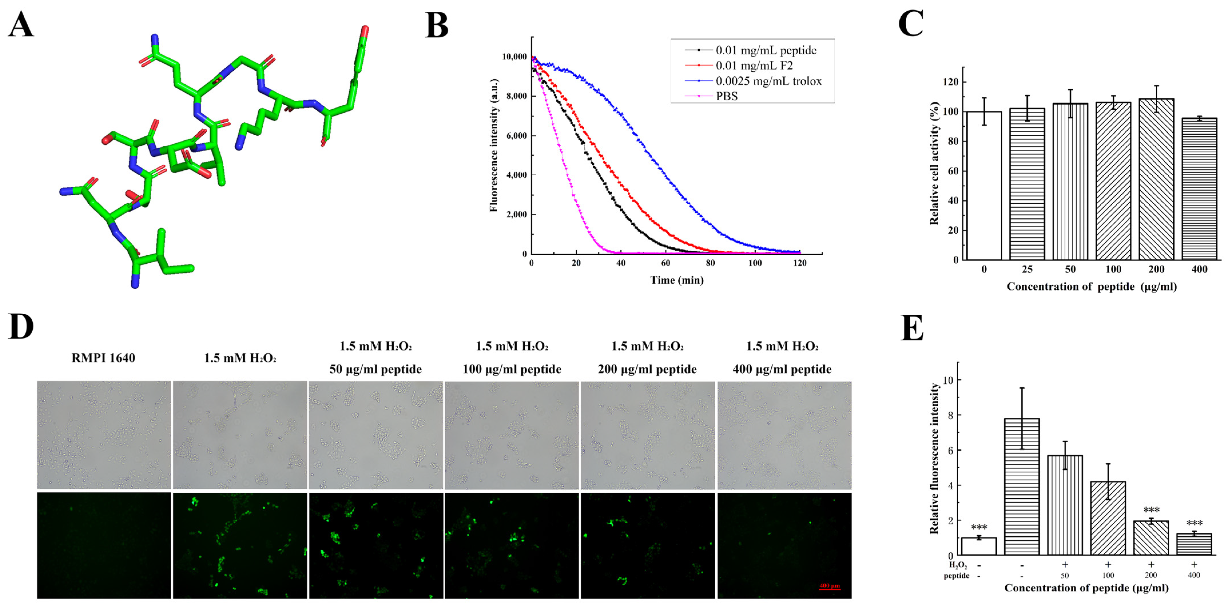
2.6. Antioxidant Activities of Synthesized Peptide INSSDVQGKY

Since F2 has the highest antioxidant activity in all fractions, its components were investigated using LC-MS/MS. The total ion chromatography of F2 was shown in Figure S1. A total of 25 peptides with relative abundance >1% were identified, and their sequence and abundance were listed in Table 1. Among them, one peptide with the sequence of INSSDVQGKY accounting for 17.83% was considered as the major component of F2. The MS/MS of INSSDVQGKY was shown in Figure S2.
Sequence alignment showed that the peptide INSSDVQGKY was a part of a beta-subunit of allophycocyanin. This was a novel AP first be sequenced and proved with antioxidant activity. The structure of INSSDVQGKY was predicted and it was a linear peptide with a little helix (Figure 5A). In order to validate the antioxidant function of this peptide, it was synthetized. ORAC assay of the synthetized peptide was 0.32 ± 0.03 mmol TE/g and slightly lower than F2 (0.41 ± 0.04 mmol TE/g) (Figure 5B). The peptide had no significant cytotoxicity on HaCaT cells within 400 μg/mL (Figure 5C). The peptide dose-dependently reduced the intracellular ROS levels at a concentration range from 100 μg/mL to 400 μg/mL (Figure 5D,E). This peptide is therefore hypothesized to be the major antioxidant component of F2. The antioxidant activity of INSSDVQGKY is close to peptide PNN which was obtained from hydrolysis of phycobiliprotein by protease K [27] and may have potential applications in further study.

3. Discussion

Phycobiliprotein is an ideal marine protein resource with antioxidant properties widely spread in Rhodophyta and Cyanophyta, which could be used in bioactive peptides preparation [28]. This study aimed to apply the bacterial extracellular protease in preparing antioxidant peptides from phycobiliprotein. To utilize phycobiliprotein more comprehensively and avoid its natural limitations, we reported bacterial extracellular protease of Pseudoalteromonas sp. JS4–1 hydrolyzed phycobiliprotein to obtain antioxidant peptides in a single process. The bioactive fractions exhibited antioxidant activities both in vitro and in vivo. The biochemical mechanisms of the antioxidant activities of the fractions at cellular and C. elegans levels may be through activating the activities of antioxidant enzymes and scavenging the oxidative stress in the organisms. The molecular mechanism underlying this biological progress still need to be further studied in the future.
In this study, extracellular proteases from marine bacteria were used to hydrolyze phycobiliprotein. Marine proteases can degrade organics in the ocean naturally; thus, they have extraordinary advantages in digesting marine-sourced proteins such as fish and plants compared with land proteases [9,29,30]. Compared with commercial proteases, marine proteases have higher catalyzing efficiency and shorter hydrolyzing time. The unique cleavage sites of marine proteases also produced peptides with different amino acid sequences which is helpful to select more bioactive peptides and develop various applications of phycobiliprotein [31]. The identified peptide INSSDVQGKY exhibited antioxidant activities at a concentration of 100–200 μg/mL which was close to some newly found antioxidant peptides derived from other resources [32,33]. The cellular ROS scavenging assay is strong proof of the outstanding antioxidant activity of the peptide during biological process.
ROS are important mediators of biological process. The increased formation of ROS could cause oxidative stress which is associated with oxidative damage to biomolecules [34]. Oxidative stress happens in various pathophysiological processes and is an important pathophysiological mechanism underlying many diseases. Thus, decreasing the ROS levels to inhibit the oxidative stress has become an efficient and promising way in medicine [35]. Activating endogenous antioxidant enzymes such as SOD, CAT and GSH-Px by small molecules is an effective and protective approach in diseases [36]. The enzymatic hydrolysates and newly identified peptide in this study were able to activate the antioxidant enzymes to scavenge ROS under exogenous stress such as high glucose, and H2O2. Li G et al. identified a novel peptide EDEQKFWGK with DPPH RSA 26.76% and the OH RSA 32.19% from porcine plasma hydrolysate, and this peptide could increase SOD, CAT, and GSH-Px activities in HepG2 cells [37]. Park YR et al. identified a novel peptide NCWPFQGVPLGFQAPP with DPPH RSA 50% from clam worms and exhibited good antioxidant and anti-inflammation effects in RAW264 7 cells [38]. Compared with antioxidant peptides from other resources, phycobiliprotein hydrolysates also showed good antioxidant activities (DPPH RSA: 45.14 ± 0.43% and OH RSA: 65.11 ± 2.64%). Further, phycobiliprotein can be easily obtained and produced massively in the industry. So, exploring bioactive peptides from phycobiliprotein is of great economic value.
Conclusively, the enzymatic hydrolysis of phycobiliprotein is a promising and efficient method to produce antioxidant peptides. The peptides hydrolyzed from phycobiliprotein by marine proteases exhibited antioxidant activities both at cellular levels and in C. elegans.

4. Materials and Methods

4.1. Strains and Reagents

The A. platensis powder was purchased from a local supplier in Shanghai. Strains used in this study were isolated from the sediment of the South China Sea and stored in the lab: Pseudoalteromonas sp. JS4-1 (GenBank accession number: MT116988), Pseudoalteromonas sp. B27-3, Pseudoalteromonas sp. B47-6, Pseudoalteromonas sp. B62-3, Pseudoalteromonas sp. WH05-1, Pseudoalteromonas sp. WH06-2, and Pseudoalteromonas sp. ZB23-2. Immortal human umbilical vein endothelial cell line (HUVECs) and human immortal keratinocyte line (HaCaT) were obtained from the National Collection of Authenticated Cell Cultures (Shanghai, China). Wild type C. elegans (N2) were obtained from Caenorhabditis Genetics Center (CGC).
Ninhydrin, penicillin-streptomycin solution (cell culture grade), trypsin-EDTA solution, dimethyl sulfoxide (DMSO), thiazolyl blue tetrazolium bromide (MTT), and phosphate buffered saline (PBS) were purchased from Sangon Biotech (Shanghai, China). Trisodium citrate dihydrate, SnCl2, n-propanol, FeSO4, 30% H2O2, cholesterol and glucose were purchased from Sinopharm Chemical Reagent Co., Ltd. (Shanghai, China). In addition, 1,1-diphenyl-2-picrylhydrazyl (DPPH), 1,10-phenanthroline monohydrate (OP), fluorescein sodium salt, 2,20-azobis (2-amidinopropane) dihydrochloride (AAPH), 6-hydroxy-2,5,7,8-tetramethylchroman-2-carboxylic acid (Trolox), L-leucine, antifade mounting medium and NaN3 were purchased from Sigma-Aldrich, Ltd. (St. Louis, MO, USA). AmiconTM Ultra-15 centrifugal filter units were purchased from Millipore® (Billerica, MA, USA). Sephadex LH-20 was purchased from GE Healthcare Life Sciences (Uppsala, Sweden). Fetal bovine serum (FBS) was purchased from PAN-BiotechGibco company (Aidenbach, Germany). RPMI-1640 medium was purchased from Gibco® ThermoFisher Scientific Company (Waltham, MA, USA). Methyl viologen dichloride was purchased from Aladdin® (Shanghai, China). BCA assay kit, cell lysis buffer, ROS assay kit, catalase (CAT) assay kit, total superoxide dismutase (SOD) assay kit with NBT, cellular glutathione peroxidase (GSH-Px) assay kit with NADPH and lipid peroxidation MDA assay kit were purchased from Beyotime Biotechnology (Shanghai, China).

4.2. Extraction of Phycobiliprotein and Screening of Proteases

4.2.1. Extraction of Phycobiliprotein from A. platensis

One gram of A. platensis powder was stirred in 100 mL of sterilized ultrapure water, then frozen in −20 °C and thawed in 37 °C-water bath repeatedly 3–4 times to help proteins released. The protein content was measured by BCA method [39]. The soluble phycobiliprotein (crude extract) was obtained by centrifugation at 12,000× g for 20 min, and stored at −20 °C for future use. We could obtain 100 mL of 3.75 mg/mL protein solution from 1 g of A. platensis powder and the yield rate was 37.5%.

4.2.2. Preparation of Crude Bacterial Extracellular Protease

The activated seed inoculum was prepared by adding bacterial strains preserved in glycerin into 5 mL 2216E liquid medium (tryptone 5 g/L, yeast extract 1 g/L, Fe2(SO4)3 0.01 g/L, dissolved in artificial sea water, pH 7.8), and incubating at 16 °C for 16–24 h with 180 rpm shaking. Then, 2% of seed inoculum was transformed to fresh fermentation medium (bean powder 20 g/L, corn powder 20 g/L, wheat bran 10 g/L, Na2HPO4·12H2O 1 g/L, KH2PO4 0.3 g/L, CaCl2 1 g/L, Na2CO3 1 g/L, dissolved in artificial sea water, pH 7.8) and continuously culturing at the same condition. After 5 days cultivation, the crude enzyme was obtained by collecting the supernatant of the fermentation broth at 12,000× g centrifugation for 10 min at 4 °C, and stored at −20 °C prior to use.

4.2.3. Substrate Immersing Zymography

Substrate immersing zymography was conducted to determine the enzymatic activities against casein according to a previous study, with minor modifications [40]. The SDS-PAGE gel was immersed in the pre-warmed 0.5% (w/v) casein solution and incubated at 37 °C for 1 h with 75 rpm shaking. The gel was then stained with 0.1% of Coomassie Brilliant Blue dye solution for 4 h, and then decolored with 30% of ethanol and 70% of acetic acid (v/v) until the bands were clearly visible.

4.3. Antioxidant Activities of Hydrolysates

4.3.1. Optimization of Hydrolysis Conditions

First, the enzyme/substrate (E: S) ratio (v/w, mL/mg) was designed as 1:6, 1:8, 1:10, 1:12 and 1:14 with 1 mg/mL enzyme at 50 °C, to optimize the E/S ratio during the hydrolyzing. Then, the hydrolysis reaction was carried out at 35 to 60 °C, with 5 °C at intervals, to estimate the optimal temperature for hydrolyzing. Finally, hydrolyzing was taken from 1 h to 7 h at hourly intervals at 50 °C to optimize the reaction time. The reaction was terminated by heating at 95 °C for 10 min. The reaction buffer was ultrapure water. The hydrolysate was centrifuged at 12,000× g and 4 °C for 20 min and the hydrolysis degree was measured by ninhydrin coloration method [41]. The •NH2 in hydrolysate could react with ninhydrin and have absorption peak at 570 nm. The standard curve was determined using L-leucine at a concentration of 0, 1, 1.5, 2, 2.5 and 3 mM.

4.3.2. Purification of Hydrolysates

After digestion, the hydrolysates were added into the upper casing of the AmiconTM Ultra-15 centrifugal filter units and centrifuged at 5000× g for 30 min to separate peptides with MWCO (molecular weight cut-off) > 3 kDa (in the upper casing) and MWCO < 3 kDa (in the lower casing).
The peptides with MWCO < 3 kDa was further fractionated by NGC Scout Plus fast protein liquid chromatography (FPLC) system (Bio-Rad, USA) and a XK16/70 column (1.6 × 60 cm) equipped with Sephadex LH-20 gel. One mL of sample was injected into the loading loop and the column was continuously eluted with filtered ultrapure water at a flow rate of 0.75 mL/min. The protein absorbance was monitored at 220 nm with a UV detection all the time. Fractions were lyophilized and redissolved in ultrapure water at the same concentration for further assay.

4.3.3. Determination of Antioxidant Activities

To evaluate antioxidant activities of hydrolytes in vitro, DPPH free radical scavenging activity (DPPH RSA) assay, hydroxyl free radical (OH) scavenging activity (OH RSA) assay and oxygen radical absorbance capacity (ORAC) assay were conducted according to previous studies, with minor modifications [42,43,44]. The DPPH and OH radical scavenging rate were calculated.
The ORAC was defined as Trolox equivalents (mmol TE/g sample or mmol TE/mmol sample) according to the area under the curve (AUC) and calculated using the following formula:
ORAC = (AUCsample − AUCcontrol)/(AUCTrolox − AUCcontrol) × (MTrolox/Msample)
where AUCsample, AUCcontrol and AUCTrolox were the integral areas under the fluorescence decay curve of the sample, PBS and Trolox, and MTolox and Msample were the concentrations of the Trolox and sample, respectively.

4.3.4. Protective Effects on Oxidative Damage of Plasmid DNA

To evaluate the protective effects of peptides on the oxidative damage of biological macromolecules (proteins and DNA), the protection effect assay was conducted according to a previous method, with minor modifications [15]. An amount of 8 μL of pET-22b DNA, 2 μL of FeSO4 (2 mM), 8 μL of sample and 2 μL of 0.1% H2O2 were mixed in a sterilized tube and incubated at 37 °C for 10 min. For the blank, 12 μL of sterilized water replaced other reagents incubating with the DNA. For the negative control, sample was replaced with sterilized water. For the positive control, the sample was replaced with Vitamin C (200 μg/mL). The reaction solution was then analyzed by 1% agarose gel electrophoresis under 120 V for 30 min.

4.3.5. Determination of Intracellular ROS Levels and the Activities of Intracellular Oxidative Enzymes

HUVECs and HaCaT cells were cultured in RPMI-1640 medium with 10% FBS, 100 U/mL penicillin and 100 μg/mL streptomycin (complete medium) at 37 °C in a humidified atmosphere of 5% CO2. Cell viability was measured by MTT assay.
The 2′,7′-dichloro-fluorescence diacetate (DCFH-DA) probe can be oxidized by ROS to produce green fluorescent materials which can be observed under fluorescence microscope. An amount of 1 × 10⁵ of cells were plated in a 24-well plate and incubated overnight. For the HUVECs cells, the medium was replaced with RPMI-1640 medium (without FBS instead, added 35 mM of glucose) for 24 h. For the HaCaT cells, the medium was replaced with RPMI-1640 medium (without FBS, but added 1.5 mM of H2O2) for 4 h. For the blank, cells were treated with RPMI-1640 medium (without FBS). An amount of 10 μM DCFH-DA probe was added to each well and incubated for 1 h. Cells were washed with PBS for 3 times and re-immersed in 500 μL of PBS. Images were taken using an inverted fluorescence microscope (Axio vert. A1, Carl Zeiss, Oberkochen, Germany).
Next, 2 × 10⁶ of cells were plated in a 6-well plate and treated with the same condition. After incubating for 24 h, cells were washed with PBS for 3 times, then incubating with 150 μL of cell lysis buffer for 30 min on ice. The suspension was centrifuged at 12,000× g for 10 min at 4 °C. The protein concentration was measured by BCA method. The CAT, SOD and GSH-Px activities were determined according to the manufacturer’s instructions.

4.3.6. Antioxidant Activities Evaluation Using C. elegans Model

Maintenance of C. elegans and Lifespan Assay

N2 was cultured on nematode growth medium (NGM) plates supplied with E. coli OP50 as food at 20 °C. The age-synchronized C. elegans were prepared using the alkaline hypochlorite method according to Phaniendra et al. [45].
Fresh NGM plates were coated with E. coli OP50 containing a different concentration of APs (0.2 and 0.5 mg/mL) and incubated at 37 °C for 48 h to prepare the sample plates. Age-synchronized nematodes (L4 stage) were plated in blank plates and sample plates (80 worms on each plate), as Day 0 for lifespan assay. Nematodes on each plate were transferred to a new plate to avoid new-born worms and counted every day until all nematodes were dead. The survival rate was calculated.

Determination of ROS Levels and Activities of Antioxidant Enzymes in C. elegans

After 3 days of treatment on the sample plates, nematodes were collected into a M9 buffer (added 100 μM DCFH-DA probe) and incubated at 20 °C for 30 min without light. Nematodes were washed with M9 buffer 3 times. For inverted fluorescence microscope observation, nematodes were anesthetized with 1 μL of NaN3 (25 mM) and 20 μL of suspension was dropped onto the center of the slide, and 5 μL of antifade mounting medium was added for observation.
The ROS accumulation was conducted according to previous studies, with minor modifications [46]. The fluorescence intensity was measured at 15 min intervals for 6 h with excitation 485 nm and emission 530 nm at 25 °C.
For intracellular proteins extraction, nematodes were ultrasonicated on ice and the suspension was centrifuged at 12,000× g for 10 min at 4 °C. The protein concentration was measured by BCA method. The CAT, SOD and GSH-Px activities and MDA levels in the supernatant were determined according to the manufacturer’s instructions.

Oxidative Stress Experiment

After 3 days of treatment, nematodes were transferred onto NGM plates supplied with 10 mM H2O2 or 10 mM paraquat, and each plate with 80 worms. The number of survival nematodes was counted per 30 min intervals (the H2O2 plates) and per 12 h intervals (the paraquat plates) until all the nematodes were dead [47]. The survival rate was calculated.
After 3 days of treatment, nematodes were transferred onto the fresh NGM plates (80 worms on each plate) and incubated at 35 °C for 10 h [48]. Numbers of survival nematodes were counted after the heat shock and the survival rate was calculated.

4.3.7. Identification and Solid Phase Synthesis of APs

The sequence and composition of peptides in the active fractions were analyzed by liquid chromatography-mass spectrometry (LC-MS/MS) in Sangon Biotech Co., Ltd. (Shanghai, China). The LC-MS/MS was developed on an UltiMate 3000 RSLCnano system (Thermo, Waltham, MA, USA) coupled with a Q Exactive Plus HPLC-Mass Spectrometer (Thermal, Riverside County, CA, USA). The chromatographic separation was achieved using a C18 column (3 μm, 120 Å, 100 μm × 20 mm). The analysis was performed on a C18 analytical column (2 μm, 120 Å, 750 μm × 150 mm). Mobile phase A was 3% DMSO, 0.1% formic acid, and 97% H2O and mobile phase B was 3% DMSO, 0.1% formic acid, and 97% ACN. The flow rate was set as 300 nL/min. The MS data was processed with MaxQuant (V1.6.2.10) to identify detected peptides in Arthrospira platensis proteomics database. Peptide with maximal abundance was synthesized by solid phase synthesis in ChinaPeptides Co., Ltd. (Shanghai, China) according to the sequence information given by LC-MS/MS.
The structure of peptide was predicted by Phyre2 (http://www.sbg.bio.ic.ac.uk/phyre2 (accessed on 20 December 2022)).

4.4. Statistical Analysis

All the experiments and analysis were carried out in triplicate. The Normality of experimental data was analyzed by Shapiro–Wilk test. Normal experimental data were analyzed with by Student t-test. Non-parametric data were analyzed by Mann–Whitney test. Data were presented as the mean ± standard deviation (SD) and P < 0.05 was considered as a statistically significant difference.

5. Conclusions

Algal protein resources are considered to be a huge treasury of bioactive peptides. More and more researchers are attempting to prepare novel bioactive peptides from algal protein through protease hydrolysis. In addition, novel proteases from marine bacteria also possess great potential in antioxidant peptide preparation, due to their high efficiency and low cost. In this study, phycobiliprotein was hydrolyzed by extracellular proteases from Pseudoalteromonas sp. JS4-1 to produce antioxidant peptides. A novel antioxidant peptide INSSDVQGKY was separated and identified from the hydrolysates. The hydrolysates and peptide INSSDVQGKY exhibited excellent antioxidant activities both at cellular levels and in C. elegans.

Supplementary Materials

The following supporting information can be downloaded at: https://www.mdpi.com/article/10.3390/md21020133/s1, Figure S1: Total ion chromatography of F2; Figure S2: MS/MS of INSSDVQGKY.

Author Contributions

C.L.: conceptualization, methodology and validation. G.C.: writing—original draft and visualization. H.R., X.X., Y.C., C.W.: investigation. F.B.: writing—review and editing. H.H.: supervision and funding acquisition. All authors have read and agreed to the published version of the manuscript.

Funding

This work was supported by the National Natural Science Foundation of China (31900038 and 31500048), the Natural Science Foundation of Hunan Province, China (2021JJ30029), the Research Fund of The State Key Laboratory of Coal Resources and Safe Mining (SKLCRSM22KF020), The Independent Exploration and Innovation project for postgraduates of Hunan Province (CX20220357, CX20220316), the Independent Exploration and Innovation project for postgraduates of Central South University (2022ZZTS0996), and the Open Sharing Fund for the Large-scale Instruments and Equipments of Central South University (CSUZC202244).

Institutional Review Board Statement

Not applicable.

Informed Consent Statement

Not applicable.

Data Availability Statement

The data presented in this study are available on request from the corresponding author. The data are not publicly available due to privacy.

Conflicts of Interest

The authors declare no conflict of interest.

References

  1. Lupatini, A.L.; Colla, L.M.; Canan, C.; Colla, E. Potential application of microalga Spirulina platensis as a protein source. J. Sci. Food Agric. 2017, 97, 724–732. [Google Scholar] [CrossRef] [PubMed]
  2. Romay, C.; González, R.; Ledón, N.; Remirez, D.; Rimbau, V. C-phycocyanin: A biliprotein with antioxidant, anti-inflammatory and neuroprotective effects. Curr. Protein Pept. Sci. 2003, 4, 207–216. [Google Scholar] [CrossRef] [PubMed]
  3. Marthandam Asokan, S.; Wang, T.; Su, W.T.; Lin, W.T. Antidiabetic Effects of a Short Peptide of Potato Protein Hydrolysate in STZ-Induced Diabetic Mice. Nutrients 2019, 11, 779. [Google Scholar] [CrossRef] [PubMed] [Green Version]
  4. Qiao, Q.; Chen, L.; Li, X.; Lu, X.; Xu, Q. Roles of Dietary Bioactive Peptides in Redox Balance and Metabolic Disorders. Oxid. Med. Cell Longev. 2021, 2021, 5582245. [Google Scholar] [CrossRef] [PubMed]
  5. Amigo, L.; Hernández-Ledesma, B. Current Evidence on the Bioavailability of Food Bioactive Peptides. Molecules 2020, 25, 4479. [Google Scholar] [CrossRef]
  6. Wang, K.; Han, L.; Hong, H.; Pan, J.; Liu, H.; Luo, Y. Purification and identification of novel antioxidant peptides from silver carp muscle hydrolysate after simulated gastrointestinal digestion and transepithelial transport. Food Chem. 2021, 342, 128275. [Google Scholar] [CrossRef]
  7. Chalamaiah, M.; Dinesh Kumar, B.; Hemalatha, R.; Jyothirmayi, T. Fish protein hydrolysates: Proximate composition, amino acid composition, antioxidant activities and applications: A review. Food Chem. 2012, 135, 3020–3038. [Google Scholar] [CrossRef]
  8. Lu, X.; Zhang, L.; Sun, Q.; Song, G.; Huang, J. Extraction, identification and structure-activity relationship of antioxidant peptides from sesame (Sesamum indicum L.) protein hydrolysate. Food Res. Int. 2019, 116, 707–716. [Google Scholar] [CrossRef]
  9. Olena, Z.; Yang, Y.; TingTing, Y.; XiaoTao, Y.; HaiLian, R.; Xun, X.; Dong, X.; CuiLing, W.; HaiLun, H. Simultaneous preparation of antioxidant peptides and lipids from microalgae by pretreatment with bacterial proteases. Bioresour. Technol. 2022, 348, 126759. [Google Scholar] [CrossRef]
  10. Xu, F.; Cha, Q.Q.; Zhang, Y.Z.; Chen, X.L. Degradation and Utilization of Alginate by Marine Pseudoalteromonas: A Review. Appl. Environ. Microbiol. 2021, 87, e0036821. [Google Scholar] [CrossRef]
  11. Kou, X.; Gao, J.; Xue, Z.; Zhang, Z.; Wang, H.; Wang, X. Purification and identification of antioxidant peptides from chickpea (Cicer arietinum L.) albumin hydrolysates. LWT—Food Sci. Technol. 2013, 50, 591–598. [Google Scholar] [CrossRef]
  12. Hou, H.; Wang, J.; Wang, J.; Tang, W.; Shaikh, A.S.; Li, Y.; Fu, J.; Lu, L.; Wang, F.; Sun, F.; et al. A Review of Bioactive Peptides: Chemical Modification, Structural Characterization and Therapeutic Applications. J. Biomed. Nanotechnol. 2020, 16, 1687–1718. [Google Scholar] [CrossRef] [PubMed]
  13. Cai, S.; Lu, C.; Liu, Z.; Wang, W.; Lu, S.; Sun, Z.; Wang, G. Derivatives of gecko cathelicidin-related antioxidant peptide facilitate skin wound healing. Eur. J. Pharmacol. 2021, 890, 173649. [Google Scholar] [CrossRef] [PubMed]
  14. Hseu, Y.C.; Vudhya Gowrisankar, Y.; Wang, L.W.; Zhang, Y.Z.; Chen, X.Z.; Huang, P.J.; Yen, H.R.; Yang, H.L. The in vitro and in vivo depigmenting activity of pterostilbene through induction of autophagy in melanocytes and inhibition of UVA-irradiated α-MSH in keratinocytes via Nrf2-mediated antioxidant pathways. Redox Biol. 2021, 44, 102007. [Google Scholar] [CrossRef]
  15. Wu, R.; Wu, C.; Liu, D.; Yang, X.; Huang, J.; Zhang, J.; Liao, B.; He, H. Antioxidant and anti-freezing peptides from salmon collagen hydrolysate prepared by bacterial extracellular protease. Food Chem. 2018, 248, 346–352. [Google Scholar] [CrossRef]
  16. Zou, J.; Fei, Q.; Xiao, H.; Wang, H.; Liu, K.; Liu, M.; Zhang, H.; Xiao, X.; Wang, K.; Wang, N. VEGF-A promotes angiogenesis after acute myocardial infarction through increasing ROS production and enhancing ER stress-mediated autophagy. J. Cell Physiol. 2019, 234, 17690–17703. [Google Scholar] [CrossRef]
  17. Nensat, C.; Songjang, W.; Tohtong, R.; Suthiphongchai, T.; Phimsen, S.; Rattanasinganchan, P.; Metheenukul, P.; Kumphune, S.; Jiraviriyakul, A. Porcine placenta extract improves high-glucose-induced angiogenesis impairment. BMC Complement. Med. Ther. 2021, 21, 66. [Google Scholar] [CrossRef]
  18. Rezabakhsh, A.; Rahbarghazi, R.; Malekinejad, H.; Fathi, F.; Montaseri, A.; Garjani, A. Quercetin alleviates high glucose-induced damage on human umbilical vein endothelial cells by promoting autophagy. Phytomedicine 2019, 56, 183–193. [Google Scholar] [CrossRef]
  19. Son, D.H.; Yang, D.J.; Sun, J.S.; Kim, S.K.; Kang, N.; Kang, J.Y.; Choi, Y.H.; Lee, J.H.; Moh, S.H.; Shin, D.M.; et al. A Novel Peptide, Nicotinyl-Isoleucine-Valine-Histidine (NA-IVH), Promotes Antioxidant Gene Expression and Wound Healing in HaCaT Cells. Mar. Drugs 2018, 16, 262. [Google Scholar] [CrossRef] [Green Version]
  20. Dong, H.; Liu, M.; Wang, L.; Liu, Y.; Lu, X.; Stagos, D.; Lin, X.; Liu, M. Bromophenol Bis (2,3,6-Tribromo-4,5-dihydroxybenzyl) Ether Protects HaCaT Skin Cells from Oxidative Damage via Nrf2-Mediated Pathways. Antioxidants 2021, 10, 1436. [Google Scholar] [CrossRef]
  21. Wang, G.; Hao, M.; Liu, Q.; Jiang, Y.; Huang, H.; Yang, G.; Wang, C. Protective effect of recombinant Lactobacillus plantarum against H(2)O(2)-induced oxidative stress in HUVEC cells. J. Zhejiang Univ. Sci. B 2021, 22, 348–365. [Google Scholar] [CrossRef] [PubMed]
  22. Lin, C.; Xiao, J.; Xi, Y.; Zhang, X.; Zhong, Q.; Zheng, H.; Cao, Y.; Chen, Y. Rosmarinic acid improved antioxidant properties and healthspan via the IIS and MAPK pathways in Caenorhabditis elegans. Biofactors 2019, 45, 774–787. [Google Scholar] [CrossRef] [PubMed]
  23. Nakagawa, H.; Shiozaki, T.; Kobatake, E.; Hosoya, T.; Moriya, T.; Sakai, F.; Taru, H.; Miyazaki, T. Effects and mechanisms of prolongevity induced by Lactobacillus gasseri SBT2055 in Caenorhabditis elegans. Aging Cell 2016, 15, 227–236. [Google Scholar] [CrossRef] [Green Version]
  24. Ayuda-Durán, B.; González-Manzano, S.; González-Paramás, A.M.; Santos-Buelga, C. Caernohabditis elegans as a Model Organism to Evaluate the Antioxidant Effects of Phytochemicals. Molecules 2020, 25, 3194. [Google Scholar] [CrossRef] [PubMed]
  25. Tsikas, D. Assessment of lipid peroxidation by measuring malondialdehyde (MDA) and relatives in biological samples: Analytical and biological challenges. Anal. Biochem. 2017, 524, 13–30. [Google Scholar] [CrossRef] [PubMed]
  26. Shi, Y.; Meng, X.; Zhang, J. Multi- and trans-generational effects of N-butylpyridium chloride on reproduction, lifespan, and pro/antioxidant status in Caenorhabditis elegans. Sci. Total Environ. 2021, 778, 146371. [Google Scholar] [CrossRef] [PubMed]
  27. Yu, J.; Hu, Y.; Xue, M.; Dun, Y.; Li, S.; Peng, N.; Liang, Y.; Zhao, S. Purification and Identification of Antioxidant Peptides from Enzymatic Hydrolysate of Spirulina platensis. J. Microbiol. Biotechnol. 2016, 26, 1216–1223. [Google Scholar] [CrossRef] [Green Version]
  28. Pagels, F.; Guedes, A.C.; Amaro, H.M.; Kijjoa, A.; Vasconcelos, V. Phycobiliproteins from cyanobacteria: Chemistry and biotechnological applications. Biotechnol. Adv. 2019, 37, 422–443. [Google Scholar] [CrossRef]
  29. Gómez-Estaca, J.; Bravo, L.; Gómez-Guillén, M.C.; Alemán, A.; Montero, P. Antioxidant properties of tuna-skin and bovine-hide gelatin films induced by the addition of oregano and rosemary extracts. Food Chem. 2009, 112, 18–25. [Google Scholar] [CrossRef] [Green Version]
  30. Kim, S.Y.; Je, J.Y.; Kim, S.K. Purification and characterization of antioxidant peptide from hoki (Johnius belengerii) frame protein by gastrointestinal digestion. J. Nutr. Biochem. 2007, 18, 31–38. [Google Scholar] [CrossRef]
  31. Wu, R.; Chen, L.; Liu, D.; Huang, J.; Zhang, J.; Xiao, X.; Lei, M.; Chen, Y.; He, H. Preparation of Antioxidant Peptides from Salmon Byproducts with Bacterial Extracellular Proteases. Mar. Drugs 2017, 15, 4. [Google Scholar] [CrossRef] [PubMed] [Green Version]
  32. Fontoura, R.; Daroit, D.J.; Corrêa, A.P.F.; Moresco, K.S.; Santi, L.; Beys-da-Silva, W.O.; Yates, J.R., 3rd; Moreira, J.C.F.; Brandelli, A. Characterization of a novel antioxidant peptide from feather keratin hydrolysates. New Biotechnol. 2019, 49, 71–76. [Google Scholar] [CrossRef] [PubMed]
  33. Ma, Y.; Huang, K.; Wu, Y. In Vivo/In Vitro Properties of Novel Antioxidant Peptide from Pinctada fucata. J. Microbiol. Biotechnol. 2021, 31, 33–42. [Google Scholar] [CrossRef] [PubMed]
  34. Li, R.; Jia, Z.; Trush, M.A. Defining ROS in Biology and Medicine. React. Oxyg Species 2016, 1, 9–21. [Google Scholar] [CrossRef] [PubMed] [Green Version]
  35. He, L.; He, T.; Farrar, S.; Ji, L.; Liu, T.; Ma, X. Antioxidants Maintain Cellular Redox Homeostasis by Elimination of Reactive Oxygen Species. Cell Physiol. Biochem. 2017, 44, 532–553. [Google Scholar] [CrossRef] [PubMed]
  36. Christofidou-Solomidou, M.; Muzykantov, V.R. Antioxidant strategies in respiratory medicine. Treat Respir. Med. 2006, 5, 47–78. [Google Scholar] [CrossRef] [PubMed]
  37. Li, G.; Zhan, J.; Hu, L.; Yuan, C.; Ying, X.; Hu, Y. Identification of novel antioxidant peptide from porcine plasma hydrolysate and its effect in in vitro digestion/HepG2 cells model. J. Food Biochem. 2022, 46, e13853. [Google Scholar] [CrossRef]
  38. Park, Y.R.; Park, C.I.; Soh, Y. Antioxidant and Anti-Inflammatory Effects of NCW Peptide from Clam Worm (Marphysa sanguinea). J. Microbiol. Biotechnol. 2020, 30, 1387–1394. [Google Scholar] [CrossRef]
  39. Walker, J.M. The bicinchoninic acid (BCA) assay for protein quantitation. Methods Mol. Biol. 1994, 32, 5–8. [Google Scholar]
  40. Liu, D.; Yang, X.; Huang, J.; Wu, R.; Wu, C.; He, H.; Li, H. In situ demonstration and characteristic analysis of the protease components from marine bacteria using substrate immersing zymography. Appl. Biochem. Biotechnol. 2015, 175, 489–501. [Google Scholar] [CrossRef]
  41. Leach, A.A. The determination of the substitution achieved at the alpha-amino, epsilon-amino and imidazole groups of proteins with special reference to derivatives of gelatin. Biochem. J. 1966, 98, 506–512. [Google Scholar] [CrossRef] [PubMed] [Green Version]
  42. Dávalos, A.; Gómez-Cordovés, C.; Bartolomé, B. Extending applicability of the oxygen radical absorbance capacity (ORAC-fluorescein) assay. J. Agric. Food Chem. 2004, 52, 48–54. [Google Scholar] [CrossRef] [PubMed]
  43. Shimada, K.; Fujikawa, K.; Yahara, K.; Nakamura, T. Antioxidative properties of xanthan on the autoxidation of soybean oil in cyclodextrin emulsion. J. Agric. Food Chem. 1992, 40, 945–948. [Google Scholar] [CrossRef]
  44. Wang, B.; Li, Z.R.; Chi, C.F.; Zhang, Q.H.; Luo, H.Y. Preparation and evaluation of antioxidant peptides from ethanol-soluble proteins hydrolysate of Sphyrna lewini muscle. Peptides 2012, 36, 240–250. [Google Scholar] [CrossRef]
  45. Alugoju, P.; Janardhanshetty, S.S.; Subaramanian, S.; Periyasamy, L.; Dyavaiah, M. Quercetin Protects Yeast Saccharomyces cerevisiae pep4 Mutant from Oxidative and Apoptotic Stress and Extends Chronological Lifespan. Curr. Microbiol. 2018, 75, 519–530. [Google Scholar] [CrossRef]
  46. Lin, C.; Su, Z.; Luo, J.; Jiang, L.; Shen, S.; Zheng, W.; Gu, W.; Cao, Y.; Chen, Y. Polysaccharide extracted from the leaves of Cyclocarya paliurus (Batal.) Iljinskaja enhanced stress resistance in Caenorhabditis elegans via skn-1 and hsf-1. Int. J. Biol. Macromol. 2020, 143, 243–254. [Google Scholar] [CrossRef]
  47. Sakamoto, T.; Imai, H. Hydrogen peroxide produced by superoxide dismutase SOD-2 activates sperm in Caenorhabditis elegans. J. Biol. Chem. 2017, 292, 14804–14813. [Google Scholar] [CrossRef] [Green Version]
  48. Yasuda, K.; Kubo, Y.; Murata, H.; Sakamoto, K. Cortisol promotes stress tolerance via DAF-16 in Caenorhabditis elegans. Biochem. Biophys. Rep. 2021, 26, 100961. [Google Scholar] [CrossRef]
Figure 1. Antioxidant activities of phycobiliprotein hydrolysis products with JS4-1. (A) Comparison of catalytic ability on casein of proteases from Pseudoalteromonas sp. B27-3, Pseudoalteromonas sp. B47-6, Pseudoalteromonas sp. B62-3, Pseudoalteromonas sp. WH05-1, Pseudoalteromonas sp. WH06-2, Pseudoalteromonas sp. WH16-2, Pseudoalteromonas sp. JS4-1 and Pseudoalteromonas sp. ZB23-2. (B) SDS-PAGE electrophoresis of phycobiliprotein and hydrolysates of different proteases (Line M: protein marker, Line PC: phycobiliprotein). (C) DPPH radical and (D) hydroxyl radical scavenging activity of phycobiliprotein hydrolysates of different proteases. (E) Antioxidant activities of the ultrafiltration fraction of phycobiliprotein hydrolysis products treated with optimized hydrolysis condition. Data were expressed as mean ± SD (n = 3). (F) Size-exclusion liquid chromatography of the <3 kDa ultrafiltration fractions on a Sephadex LH-20 column. The F1–F4 represent purified fractions. (G) DPPH radical scavenging activity of F1-F4 compared with Vitamin C. (H) Hydroxyl radical scavenging activity of F1–F4. (I) Peroxyl radical scavenging activity (ORAC) of F1 and F2 compared with PBS and Trolox. (J) Protective effects of F1 and F2 on hydroxyl radical-induced oxidation of plasmid DNA compared with Vitamin C. Data were expressed as mean ± SD (n = 3).
Marinedrugs 21 00133 g001
Figure 2. Antioxidant activities of F1 and F2 on HUVEC cells. The effects of different concentration of (A) F1 and (B) F2 on cell viability were measured by MTT assay. ROS scavenging capacities of (C) F1 and (E) F2 on HUVEC cells indicated as a green DCFH-DA fluorescence. Images were taken using fluorescence microscope (magnification, 10×). (D,F) Statistical analysis was performed to quantify relative fluorescence density accordingly (Student t-test). (G,J) Superoxide dismutase (SOD), (H,K) glutathione peroxidase (GSH-Px) and (I,L) catalase (CAT) activities in HUVEC cells were detected after treatment with F1 and F2, respectively. The Normality of data was analyzed by Shapiro–Wilk test. Data were expressed as mean ± SD (n = 3). * represents p < 0.05, ** represents p < 0.01 and *** represents p < 0.001, compared with glucose group (Student t-test).
Marinedrugs 21 00133 g002
Figure 3. Antioxidant activities of F1 and F2 on HaCaT cells. The effects of different concentration of (A) F1 and (B) F2 on cell viability were measured by MTT assay. ROS scavenging capacities of (C) F1 and (E) F2 on HaCaT cells indicated as green DCFH-DA fluorescence. Images were taken using fluorescence microscope (magnification, 10×). (D,F) Statistical analysis was performed to quantify relative fluorescence density accordingly (Student t-test). (G,J) Superoxide dismutase (SOD), (H,K) glutathione peroxidase (GSH-Px) and (I,L) catalase (CAT) activities in HaCaT cells were detected after treatment with F1 and F2, respectively. The Normality of data was analyzed by Shapiro–Wilk test. Data were expressed as mean ± SD (n = 3). * represents p < 0.05, ** represents p < 0.01 and *** represents p < 0.001, compared with H2O2 group (G,J Mann–Whitney test; H,I,K,L Student t-test).
Marinedrugs 21 00133 g003
Figure 4. Effects of F2 on resistance to oxidative stress in Caenorhabditis elegans. (A) Survival curves of C. elegans treated with F1 and F2. Survival curves of C. elegans pre-treated with F2 at given concentrations for 3 days and then exposed to (B) 10 mM H2O2 and (C) 10 mM paraquat. * represents p < 0.05, ** represents p < 0.01 and *** represents p < 0.001, compared with H2O2 and paraquat group (Student t-test). (D) Survival rate of C. elegans pre-treated with F2 at given concentrations for 3 days and then cultured at 35 °C for 10 h. (E) Antioxidant activities of F2 on C. elegans were detected by DCFH-DA probe. Images were taken using fluorescence microscope (magnification, 5×). (F) Statistical analysis was performed to quantify relative fluorescence density (Student t-test). (G) The ROS content in C. elegans was determined based on the average integrated area of the fluorescene intensity of probes during 6 h intervals. (H) MDA levels, (I) superoxide dismutase (SOD), (J) glutathione peroxidase (GSH-Px) and (K) catalase (CAT) activities in C. elegans were detected after treated with F2 for 3 days. The Normality of data was analyzed by Shapiro–Wilk test. Data were expressed as mean ± SD (n = 3). * represents p < 0.05, ** represents p < 0.01 and *** represents p < 0.001, compared with blank control group (Student t-test).
Marinedrugs 21 00133 g004
Figure 5. Antioxidant activities of synthesized peptide INSSDVQGKY. (A) Predicted structure of peptide INSSDVQGKY. (B) ORAC assay of synthesized peptide INSSDVQGKY compared with Trolox and PBS. (C) The effects of different concentration of synthesized peptide INSSDVQGKY on HaCaT cell viability were measured by MTT assay. (D) Antioxidant activities of synthesized peptide INSSDVQGKY on HaCaT cells were detected by DCFH-DA probe. Images were taken using fluorescence microscope (magnification, 10×). (E) Statistical analysis was performed to quantify relative fluorescence density. The Normality of data was analyzed by Shapiro–Wilk test. Data were expressed as mean ± SD (n = 3). *** represents p < 0.001, compared with H2O2 group (Student t-test).
Marinedrugs 21 00133 g005
Table 1. Peptides identified through liquid chromatography mass spectrum (> 1%).
Amino Acid SequenceMass (Da)Source (Position)Counts (%)
INSSDVQGKY1109.535Allophycocyanin beta-subunit [Arthrospira platensis qy3] (9–18)17.83
VGGSVPREY962.482Elongation factor G [Arthrospira platensis] (531–539)9.12
SSYPNRPVP1015.509Photosystem II lipoprotein Psb27 [Arthrospira platensis C1] (102–110)8.81
GIFDTMNH933.401ATP-dependent Clp protease proteolytic subunit [Arthrospira platensis C1] (74–81)6.91
YTPDYTPK983.460Ribulose bisphosphate carboxylase large chain [Arthrospira platensis C1] (26–33)3.34
IDPSHGTGF929.424Phospho-2-dehydro-3-deoxyheptonate aldolase [Arthrospira platensis] (277–285)3.33
TPEPKPEPKPEPKPEP1795.936CAAD domain-containing protein [Arthrospira platensis C1] (52–67)3.25
YEQMPEPKY1183.522NAD(P)H-quinone oxidoreductase subunit K [Arthrospira platensis C1] (105–113)3.14
DVDWSDYQKQ1282.547Ferredoxin-NADP reductase [Arthrospira platensis] (380–389)2.78
VGGSVPKEY934.476Elongation factor G [Arthrospira platensis C1] (496–504)2.76
TDVPANHPY1012.461S-layer domain protein [Arthrospira platensis C1] (283–291)2.42
QGTLEKYI950.507Putative adenylate cyclase [Arthrospira platensis C1] (375–382)2.40
AGIDEINRT987.499Phycocyanin alpha-subunit [Arthrospira platensis qy3] (113–121)2.32
SESPNLILMDIQMP1586.768Multi-sensor hybrid histidine kinase [Arthrospira platensis C1] (1700–1713)2.32
APYDESEVVFH1291.572Rod linker polypeptide CpcI [Arthrospira platensis] (107–117)2.20
IDTIPTGGK900.49250S ribosomal protein L13 [Arthrospira platensis C1] (144–152)1.96
LIAGGTGPM815.421Phycocyanin alpha-subunit [Arthrospira platensis qy3] (99–107)1.92
DVDWSDYQK1154.488Ferredoxin-NADP reductase [Arthrospira platensis] (380–388)1.90
LPEEPMTGK1000.490Transcriptional regulator AbrB family [Arthrospira platensis C1] (9–17)1.80
VINSSDVQGKY1208.604Allophycocyanin beta-subunit [Arthrospira platensis qy3] (8–18)1.68
LVTQQPLGGKAQ1238.698DNA-directed RNA polymerase subunit beta [Arthrospira platensis] (992–1003)1.52
KELTKKSPNSP1227.682NYN domain-containing protein [Arthrospira platensis] (398–408)1.37
IETKEIPVPT1125.628Assimilatory sulfite reductase (ferredoxin) [Arthrospira platensis C1] (592–601)1.34
VDYQEQPREY1325.589Major membrane protein I [Arthrospira platensis C1] (81–90)1.30
GANYEDEWK1110.462Transketolase [Arthrospira platensis] (308–316)1.30
Disclaimer/Publisher’s Note: The statements, opinions and data contained in all publications are solely those of the individual author(s) and contributor(s) and not of MDPI and/or the editor(s). MDPI and/or the editor(s) disclaim responsibility for any injury to people or property resulting from any ideas, methods, instructions or products referred to in the content.

