
Presence of Lactic Acid Bacteria in the Intestinal Tract of the Mediterranean Trout (Salmo macrostigma) in Its Natural Environment

 ,
,  , , , , and
Abstract
:1. Introduction
2. Materials and Methods
2.1. Lactic Acid Bacteria (LAB) Isolation
2.2. Phenotypic and Biochemical Characterization
2.3. Genotypic Identification
3. Results
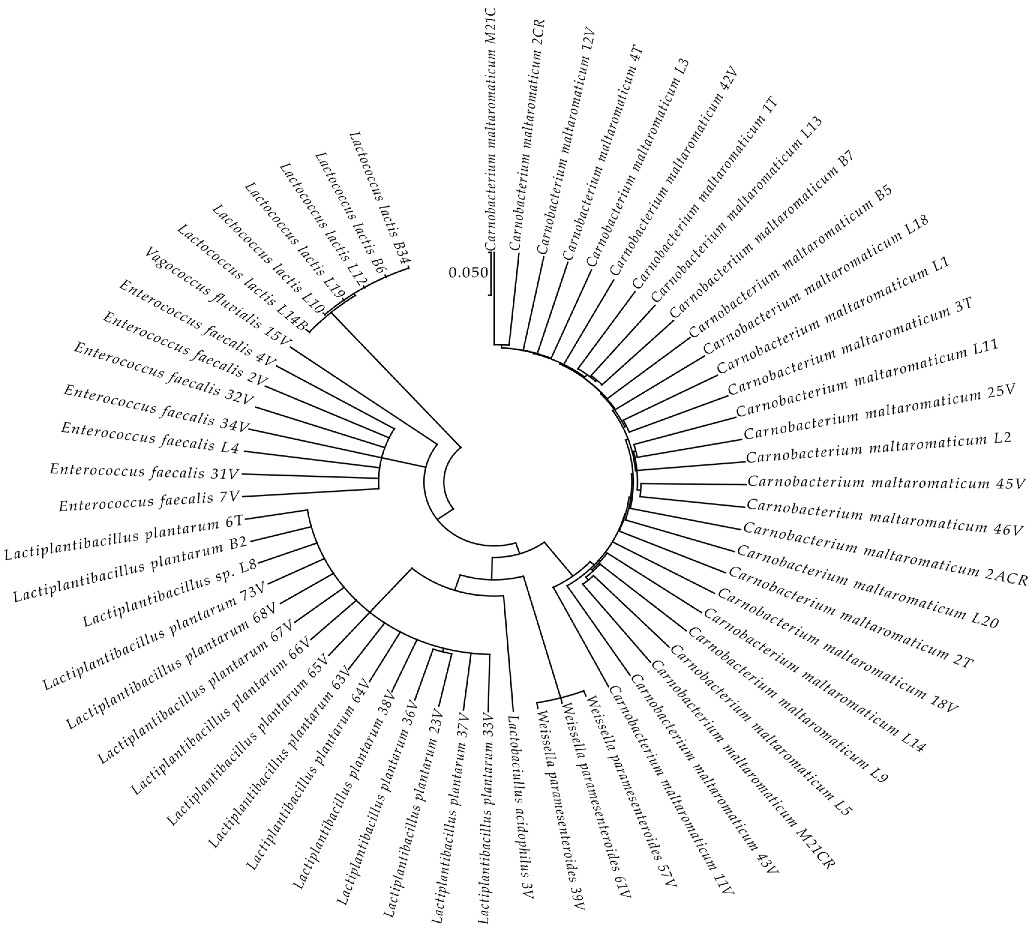
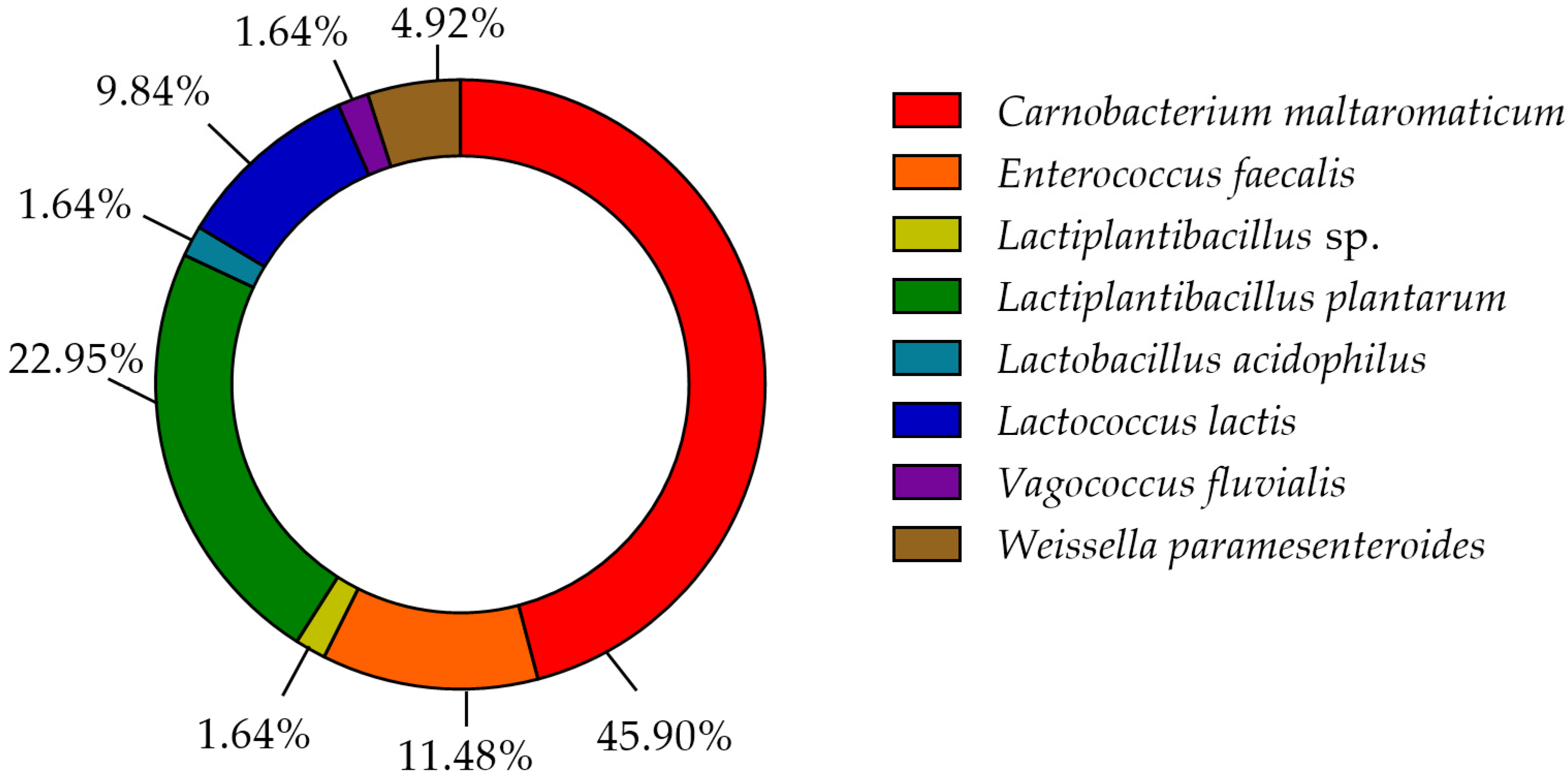
3.1. LAB Species Diversity
3.2. Biochemical Characterization
4. Discussion
Supplementary Materials
Author Contributions
Funding
Acknowledgments
Conflicts of Interest
References
- Tarnecki, A.; Burgos, F.; Ray, C.; Arias, C. Fish intestinal microbiome: Diversity and symbiosis unravelled by metagenomics. J. Appl. Microbiol. 2017, 123, 2–17. [Google Scholar] [CrossRef] [PubMed] [Green Version]
- Wang, A.R.; Ran, C.; Ringø, E.; Zhou, Z.G. Progress in fish gastrointestinal microbiota research. Rev. Aquac. 2018, 10, 626–640. [Google Scholar] [CrossRef] [Green Version]
- Ringø, E.; Hoseinifar, S.H.; Ghosh, K.; Van Doan, H.; Beck, B.R.; Song, S.K. Lactic Acid Bacteria in Finfish—An Update. Front. Microbiol. 2018, 9, 1818. [Google Scholar] [CrossRef]
- Butt, R.L.; Volkoff, H. Gut Microbiota and Energy Homeostasis in Fish. Front. Endocrinol. 2019, 10, 9. [Google Scholar] [CrossRef] [PubMed] [Green Version]
- Ringø, E.; Van Doan, H.; Lee, S.H.; Soltani, M.; Hoseinifar, S.H.; Harikrishnan, R.; Song, S.K. Probiotics, lactic acid bacteria and bacilli: Interesting supplementation for aquaculture. J. Appl. Microbiol. 2020, 129, 116–136. [Google Scholar] [CrossRef] [PubMed] [Green Version]
- Pessione, E. Lactic acid bacteria contribution to gut microbiota complexity: Lights and shadows. Front. Cell. Infect. Microbiol. 2012, 2, 86. [Google Scholar] [CrossRef] [PubMed] [Green Version]
- Merrifield, D.L.; Balcázar, J.L.; Daniels, C.; Zhou, Z.; Carnevali, O.; Sun, Y.-Z.; Hoseinifar, S.H.; Ringø, E. Indigenous Lactic Acid Bacteria in Fish and Crustaceans. Aquac. Nutr. Gut Health Probiotics Prebiotics 2014, 128–168. [Google Scholar] [CrossRef]
- Sullam, K.E.; Essinger, S.D.; Lozupone, C.A.; O’Connor, M.P.; Rosen, G.L.; Knight, R.; Kilham, S.S.; Russell, J.A. Environmental and ecological factors that shape the gut bacterial communities of fish: A meta-analysis. Mol. Ecol. 2012, 21, 3363–3378. [Google Scholar] [CrossRef] [Green Version]
- Ringo, E.; Zhou, Z.; Vecino, J.L.G.; Wadsworth, S.; Romero, J.P.; Krogdahl, A.; Olsen, R.; Dimitroglou, A.; Foey, A.; Davies, S.G.; et al. Effect of dietary components on the gut microbiota of aquatic animals. A never-ending story? Aquac. Nutr. 2015, 22, 219–282. [Google Scholar] [CrossRef] [Green Version]
- Pedrotti, F.S.; Davies, S.; Merrifield, D.; Marques, M.R.F.; Fraga, A.P.M.; Mouriño, J.L.P.; Fracalossi, D.M. The autochthonous microbiota of the freshwater omnivores jundiá (Rhamdia quelen) and tilapia (Oreochromis niloticus) and the effect of dietary carbohydrates. Aquac. Res. 2015, 46, 472–481. [Google Scholar] [CrossRef]
- González, C.-J.; López-Díaz, T.-M.; García-López, M.-L.; Prieto, M.; Otero, A. Bacterial Microflora of Wild Brown Trout (Salmo trutta), Wild Pike (Esox lucius), and Aquacultured Rainbow Trout (Oncorhynchus mykiss). J. Food Prot. 1999, 62, 1270–1277. [Google Scholar] [CrossRef]
- González, C.; Encinas, J.; García-López, M.; Otero, A. Characterization and identification of lactic acid bacteria from freshwater fishes. Food Microbiol. 2000, 17, 383–391. [Google Scholar] [CrossRef]
- Bucio, A.; Hartemink, R.; Schrama, J.W.; Verreth, J.; Rombouts, F.M. Presence of lactobacilli in the intestinal content of freshwater fish from a river and from a farm with a recirculation system. Food Microbiol. 2006, 23, 476–482. [Google Scholar] [CrossRef] [PubMed]
- Skrodenyte-Arbaciauskiene, V.; Sruoga, A.; Butkauskas, D. Assessment of microbial diversity in the river trout Salmo trutta fario L. intestinal tract identified by partial 16S rRNA gene sequence analysis. Fish. Sci. 2006, 72, 597–602. [Google Scholar] [CrossRef]
- Amato, K. Co-evolution in context: The importance of studying gut microbiomes in wild animals. Microbiome Sci. Med. 2013, 1, 1. [Google Scholar] [CrossRef]
- Directive, H. Council Directive 92/43/EEC of 21 May 1992 on the Conservation of Natural Habitats and of Wild Fauna and Flora. Off. J. Eur. Union 1992, 206, 7–50. [Google Scholar]
- Réalis-Doyelle, E.; Pasquet, A.; De Charleroy, D.; Fontaine, P.; Teletchea, F. Strong Effects of Temperature on the Early Life Stages of a Cold Stenothermal Fish Species, Brown Trout (Salmo trutta L.). PLoS ONE 2016, 11, e0155487. [Google Scholar] [CrossRef] [PubMed] [Green Version]
- Ojanguren, A.; Braña, F. Thermal dependence of embryonic growth and development in brown trout. J. Fish Biol. 2003, 62, 580–590. [Google Scholar] [CrossRef]
- Réalis-Doyelle, E.; Gisbert, E.; Alcaraz, C.; Teletchea, F.; Pasquet, A. Temperature affects growth allometry and development patterns in brown trout (Salmo trutta) fry: A multitrait approach. Can. J. Fish. Aquat. Sci. 2018, 75, 714–722. [Google Scholar] [CrossRef]
- Almodóvar, A.; Nicola, G.G.; Ayllón, D.; Elvira, B. Global warming threatens the persistence of Mediterranean brown trout. Glob. Chang. Biol. 2012, 18, 1549–1560. [Google Scholar] [CrossRef] [Green Version]
- Powers, E.M. Efficacy of the Ryu nonstaining KOH technique for rapidly determining gram reactions of food-borne and waterborne bacteria and yeasts. Appl. Environ. Microbiol. 1995, 61, 3756–3758. [Google Scholar] [CrossRef] [Green Version]
- Muyzer, G.; de Waal, E.C.; Uitterlinden, A.G. Profiling of complex microbial populations by denaturing gradient gel electrophoresis analysis of polymerase chain reaction-amplified genes coding for 16S rRNA. Appl. Environ. Microbiol. 1993, 59, 695–700. [Google Scholar] [CrossRef] [PubMed] [Green Version]
- Hou, Q.; Bai, X.; Li, W.; Gao, X.; Zhang, F.; Sun, Z.; Zhang, H. Design of Primers for Evaluation of Lactic Acid Bacteria Populations in Complex Biological Samples. Front. Microbiol. 2018, 9, 2045. [Google Scholar] [CrossRef] [Green Version]
- Zhang, Z.; Schwartz, S.; Wagner, L.; Miller, W. A Greedy Algorithm for Aligning DNA Sequences. J. Comput. Biol. 2000, 7, 203–214. [Google Scholar] [CrossRef]
- NCBI Resource Coordinators. Database resources of the National Center for Biotechnology Information. Nucleic Acids Res. 2018, 46, D8–D13. [Google Scholar] [CrossRef] [PubMed] [Green Version]
- Johnson, J.S.; Spakowicz, D.J.; Hong, B.-Y.; Petersen, L.M.; Demkowicz, P.; Chen, L.; Leopold, S.R.; Hanson, B.M.; Agresta, H.O.; Gerstein, M.; et al. Evaluation of 16S rRNA gene sequencing for species and strain-level microbiome analysis. Nat. Commun. 2019, 10, 5029. [Google Scholar] [CrossRef] [Green Version]
- Kumar, S.; Stecher, G.; Li, M.; Knyaz, C.; Tamura, K. MEGA X: Molecular evolutionary genetics analysis across computing platforms. Mol. Biol. Evol. 2018, 35, 1547–1549. [Google Scholar] [CrossRef]
- Hasegawa, M.; Kishino, H.; Yano, T.-A. Dating of the human-ape splitting by a molecular clock of mitochondrial DNA. J. Mol. Evol. 1985, 22, 160–174. [Google Scholar] [CrossRef]
- Fei, Y.-T.; Liu, D.-M.; Luo, T.-H.; Chen, G.; Wu, H.; Li, L.; Yu, Y.-G. Molecular Characterization of Lactobacillus plantarum DMDL 9010, a Strain with Efficient Nitrite Degradation Capacity. PLoS ONE 2014, 9, e113792. [Google Scholar] [CrossRef]
- Torriani, S.; Felis, G.E.; Dellaglio, F. Differentiation of Lactobacillus plantarum, L. pentosus, and L. paraplantarum by recA Gene Sequence Analysis and Multiplex PCR Assay with recA Gene-Derived Primers. Appl. Environ. Microbiol. 2001, 67, 3450–3454. [Google Scholar] [CrossRef] [PubMed] [Green Version]
- Wong, S.; Rawls, J.F. Intestinal microbiota composition in fishes is influenced by host ecology and environment. Mol. Ecol. 2012, 21, 3100–3102. [Google Scholar] [CrossRef] [PubMed] [Green Version]
- Kashinskaya, E.; Simonov, E.; Kabilov, M.; Izvekova, G.; Andree, K.; Solovyev, M. Diet and other environmental factors shape the bacterial communities of fish gut in an eutrophic lake. J. Appl. Microbiol. 2018, 125, 1626–1641. [Google Scholar] [CrossRef] [PubMed]
- Ingerslev, H.-C.; von Gersdorff Jørgensen, L.; Strube, M.L.; Larsen, N.; Dalsgaard, I.; Boye, M.; Madsen, L. The development of the gut microbiota in rainbow trout (Oncorhynchus mykiss) is affected by first feeding and diet type. Aquaculture 2014, 424–425, 24–34. [Google Scholar] [CrossRef] [Green Version]
- Huyben, D.; Sun, L.; Moccia, R.; Kiessling, A.; Dicksved, J.; Lundh, T. Dietary live yeast and increased water temperature influence the gut microbiota of rainbow trout. J. Appl. Microbiol. 2018, 124, 1377–1392. [Google Scholar] [CrossRef] [PubMed]
- Araújo, C.; Muñoz-Atienza, E.; Nahuelquin, Y.; Poeta, P.; Igrejas, G.; Hernández, P.E.; Herranz, C.; Cintas, L.M. Inhibition of fish pathogens by the microbiota from rainbow trout (Oncorhynchus mykiss, Walbaum) and rearing environment. Anaerobe 2015, 32, 7–14. [Google Scholar] [CrossRef]
- Balcázar, J.L.; de Blas, I.; Ruiz-Zarzuela, I.; Vendrell, D.; Gironés, O.; Muzquiz, J.L. Sequencing of variable regions of the 16S rRNA gene for identification of lactic acid bacteria isolated from the intestinal microbiota of healthy salmonids. Comp. Immunol. Microbiol. Infect. Dis. 2007, 30, 111–118. [Google Scholar] [CrossRef]
- Ringo, E.; Bendiksen, H.; Wesmajervi, M.; Olsen, R.; Jansen, P.; Mikkelsen, H. Lactic acid bacteria associated with the digestive tract of Atlantic salmon (Salmo salar L.). J. Appl. Microbiol. 2000, 89, 317–322. [Google Scholar] [CrossRef] [Green Version]
- Ringø, E.; Wesmajervi, M.S.; Bendiksen, H.R.; Berg, A.; Olsen, R.E.; Johnsen, T.; Mikkelsen, H.; Seppola, M.; Strøm, E.; Holzapfel, W. Identification and Characterization of Carnobacteria Isolated from Fish Intestine. Syst. Appl. Microbiol. 2001, 24, 183–191. [Google Scholar] [CrossRef]
- Nicholson, W.L.; Krivushin, K.; Gilichinsky, D.; Schuerger, A.C. Growth of Carnobacterium spp. from permafrost under low pressure, temperature, and anoxic atmosphere has implications for Earth microbes on Mars. Proc. Natl. Acad. Sci. USA 2013, 110, 666–671. [Google Scholar] [CrossRef] [Green Version]
- Kim, D.-H.; Austin, B. Characterization of probiotic carnobacteria isolated from rainbow trout (Oncorhynchus mykiss) intestine. Lett. Appl. Microbiol. 2008, 47, 141–147. [Google Scholar] [CrossRef]
- Leisner, J.J.; Laursen, B.G.; Prévost, H.; Drider, D.; Dalgaard, P. Carnobacterium:positive and negative effects in the environment and in foods. FEMS Microbiol. Rev. 2007, 31, 592–613. [Google Scholar] [CrossRef] [Green Version]
- Schaffer, P.A.; Lifland, B.; Van Sommeran, S.; Casper, D.R.; Davis, C.R. Meningoencephalitis Associated with Carnobacterium maltaromaticum–Like Bacteria in Stranded Juvenile Salmon Sharks (Lamna ditropis). Vet. Pathol. 2013, 50, 412–417. [Google Scholar] [CrossRef]
- Loch, T.P.; Xu, W.; Fitzgerald, S.M.; Faisal, M. Isolation of a Carnobacterium maltaromaticum-like bacterium from systemically infected lake whitefish (Coregonus clupeaformis). FEMS Microbiol. Lett. 2008, 288, 76–84. [Google Scholar] [CrossRef] [Green Version]
- Pastorino, P.; Colussi, S.; Pizzul, E.; Varello, K.; Menconi, V.; Mugetti, D.; Tomasoni, M.; Esposito, G.; Bertoli, M.; Bozzetta, E.; et al. The unusual isolation of carnobacteria in eyes of healthy salmonids in high-mountain lakes. Sci. Rep. 2021, 11, 1–10. [Google Scholar] [CrossRef] [PubMed]
- Roh, H.; Kim, B.S.; Lee, M.K.; Park, C.; Kim, D. Genome-wide comparison of Carnobacterium maltaromaticum derived from diseased fish harbouring important virulence-related genes. J. Fish Dis. 2020, 43, 1029–1037. [Google Scholar] [CrossRef] [PubMed]
- Siezen, R.J.; Tzeneva, V.A.; Castioni, A.; Wels, M.; Phan, H.T.K.; Rademaker, J.L.; Starrenburg, M.J.C.; Kleerebezem, M.; Molenaar, D.; Vlieg, J.E.T.V.H. Phenotypic and genomic diversity ofLactobacillus plantarumstrains isolated from various environmental niches. Environ. Microbiol. 2010, 12, 758–773. [Google Scholar] [CrossRef]
- Iorizzo, M.; Pannella, G.; Lombardi, S.J.; Ganassi, S.; Testa, B.; Succi, M.; Sorrentino, E.; Petrarca, S.; De Cristofaro, A.; Coppola, R.; et al. Inter- and Intra-Species Diversity of Lactic Acid Bacteria in Apis mellifera ligustica Colonies. Microorganisms 2020, 8, 1578. [Google Scholar] [CrossRef]
- Maji, U.J.; Mohanty, S. Genotypic characterization of Lactic acid bacteria in gut microbiome of freshwater fish. Microbiology 2017, 86, 276–285. [Google Scholar] [CrossRef]
- Mohamad, N.; Manan, H.; Sallehhuddin, M.; Musa, N.; Ikhwanuddin, M. Screening of Lactic Acid Bacteria isolated from giant freshwater prawn (Macrobrachium rosenbergii) as potential probiotics. Aquac. Rep. 2020, 18, 100523. [Google Scholar] [CrossRef]
- Foysal, J.; Fotedar, R.; Siddik, M.A.B.; Tay, A. Lactobacillus acidophilus and L. plantarum improve health status, modulate gut microbiota and innate immune response of marron (Cherax cainii). Sci. Rep. 2020, 10, 1–13. [Google Scholar] [CrossRef] [PubMed] [Green Version]
- Michel, C.; Pelletier, C.; Boussaha, M.; Douet, D.-G.; Lautraite, A.; Tailliez, P. Diversity of Lactic Acid Bacteria Associated with Fish and the Fish Farm Environment, Established by Amplified rRNA Gene Restriction Analysis. Appl. Environ. Microbiol. 2007, 73, 2947–2955. [Google Scholar] [CrossRef] [PubMed] [Green Version]
- Didinen, B.; Onuk, E.; Metin, S.; Cayli, O. Identification and characterization of lactic acid bacteria isolated from rainbow trout (Oncorhynchus mykiss, Walbaum 1792), with inhibitory activity against Vagococcus salmoninarum and Lactococcus garvieae. Aquac. Nutr. 2018, 24, 400–407. [Google Scholar] [CrossRef]
- Kelly, W.; Ward, L. Genotypic vs. phenotypic biodiversity in Lactococcus lactis. Microbiology 2002, 148, 3332–3333. [Google Scholar] [CrossRef] [PubMed]
- Ghanbari, M.; Rezaei, M.; Jami, M.; Nazari, R. Isolation and Characterization of Lactobacillus Species from Intestinal Contents of Beluga (Huso Huso) and Persian Sturgeon (Acipenser Persicus). Iran. J. Vet. Res. 2009, 10, 152–157. [Google Scholar] [CrossRef]
- Hagi, T.; Tanaka, D.; Iwamura, Y.; Hoshino, T. Diversity and seasonal changes in lactic acid bacteria in the intestinal tract of cultured freshwater fish. Aquaculture 2004, 234, 335–346. [Google Scholar] [CrossRef]
- Ghiasi, F. Predominant lactic acid bacteria isolated from the intestines of silver carp in low water temperature. Afr. J. Biotechnol. 2011, 10, 12717–12721. [Google Scholar] [CrossRef] [Green Version]
- Itoi, S.; Abe, T.; Washio, S.; Ikuno, E.; Kanomata, Y.; Sugita, H. Isolation of halotolerant Lactococcus lactis subsp. lactis from intestinal tract of coastal fish. Int. J. Food Microbiol. 2008, 121, 116–121. [Google Scholar] [CrossRef]
- Itoi, S.; Yuasa, K.; Washio, S.; Abe, T.; Ikuno, E.; Sugita, H. Phenotypic variation inLactococcus lactissubsp.lactisisolates derived from intestinal tracts of marine and freshwater fish. J. Appl. Microbiol. 2009, 107, 867–874. [Google Scholar] [CrossRef]
- Rademaker, J.L.W.; Herbet, H.; Starrenburg, M.J.C.; Naser, S.M.; Gevers, D.; Kelly, W.J.; Hugenholtz, J.; Swings, J.; Van Hylckama Vlieg, J.E.T. Diversity Analysis of Dairy and Nondairy Lactococcus lactis Isolates, Using a Novel Multilocus Sequence Analysis Scheme and (GTG) 5 -PCR Fingerprinting. Appl. Environ. Microbiol. 2007, 73, 7128–7137. [Google Scholar] [CrossRef] [Green Version]
- Mortezaei, F.; Royan, M.; Noveirian, H.A.; Babakhani, A.; KordGhashlaghi, H.A.; Balcázar, J. In vitro assessment of potential probiotic characteristics of indigenous Lactococcus lactis and Weissella oryzae isolates from rainbow trout (Oncorhynchus mykiss Walbaum). J. Appl. Microbiol. 2020, 129, 1004–1019. [Google Scholar] [CrossRef] [PubMed]
- Devriese, L.A.; Van De Kerckhove, A.; Kilpper-Balz, R.; Schleifer, K.H. Characterization and Identification of Enterococcus Species Isolated from the Intestines of Animals. Int. J. Syst. Evol. Microbiol. 1987, 37, 257–259. [Google Scholar] [CrossRef] [Green Version]
- Dubin, K.; Pamer, E.G. Enterococci and Their Interactions with the Intestinal Microbiome. Microbiol. Spectr. 2017, 5. [Google Scholar] [CrossRef] [PubMed] [Green Version]
- Cai, Y.; Suyanandana, P.; Saman, P.; Benno, Y. Classification and characterization of lactic acid bacteria isolated from the intestines of common carp and freshwater prawns. J. Gen. Appl. Microbiol. 1999, 45, 177–184. [Google Scholar] [CrossRef] [PubMed] [Green Version]
- García-Solache, M.; Rice, L.B. The Enterococcus: A Model of Adaptability to Its Environment. Clin. Microbiol. Rev. 2019, 32, e00058-18. [Google Scholar] [CrossRef] [Green Version]
- Sica, M.G.; Brugnoni, L.I.; Marucci, P.L.; Cubitto, M.A. Characterization of probiotic properties of lactic acid bacteria isolated from an estuarine environment for application in rainbow trout (Oncorhynchus mykiss, Walbaum) farming. Antonie van Leeuwenhoek 2012, 101, 869–879. [Google Scholar] [CrossRef]
- Román, L.; Real, F.; Sorroza, L.; Padilla, D.; Acosta, B.; Grasso, V.; Bravo, J.; Acosta, F. The in vitro effect of probiotic Vagococcus fluvialis on the innate immune parameters of Sparus aurata and Dicentrarchus labrax. Fish Shellfish Immunol. 2012, 33, 1071–1075. [Google Scholar] [CrossRef] [PubMed]
- Lyons, P.P.; Turnbull, J.; Dawson, K.A.; Crumlish, M. Exploring the microbial diversity of the distal intestinal lumen and mucosa of farmed rainbow troutOncorhynchus mykiss(Walbaum) using next generation sequencing (NGS). Aquac. Res. 2017, 48, 77–91. [Google Scholar] [CrossRef]
- Zeng, A.; Tan, K.; Gong, P.; Lei, P.; Guo, Z.; Wang, S.; Gao, S.; Zhou, Y.; Shu, Y.; Zhou, X.; et al. Correlation of microbiota in the gut of fish species and water. 3 Biotech 2020, 73, 7128–7137. [Google Scholar] [CrossRef]
- Lauzon, H.L.; Ringø, E. Prevalence and Application of Lactic Acid Bacteria in Aquatic Environments. In Lactic Acid Bacteria, 4th ed.; CRC Press: Boca Raton, FL, USA, 2012; pp. 593–631. [Google Scholar]
- George, F.; Daniel, C.; Thomas, M.; Singer, E.; Guilbaud, A.; Tessier, F.J.; Revol-Junelles, A.-M.; Borges, F.; Foligné, B. Occurrence and Dynamism of Lactic Acid Bacteria in Distinct Ecological Niches: A Multifaceted Functional Health Perspective. Front. Microbiol. 2018, 9, 2899. [Google Scholar] [CrossRef] [Green Version]
- Shannon, A.; Attwood, G.; Hopcroft, D.; Christeller, J. Characterization of lactic acid bacteria in the larval midgut of the keratinophagous lepidopteran, Hofmannophila pseudospretella. Lett. Appl. Microbiol. 2008, 32, 36–41. [Google Scholar] [CrossRef]
- Liu, H.; Guo, X.; Gooneratne, S.R.; Lai, R.; Zeng, C.; Zhan, F.; Wang, W. The gut microbiome and degradation enzyme activity of wild freshwater fishes influenced by their trophic levels. Sci. Rep. 2016, 6, 24340. [Google Scholar] [CrossRef] [Green Version]
- Sepulveda, J.; Moeller, A.H. The Effects of Temperature on Animal Gut Microbiomes. Front. Microbiol. 2020, 11, 384. [Google Scholar] [CrossRef]
- Zarkasi, K.Z.; Taylor, R.; Abell, G.C.J.; Tamplin, M.L.; Glencross, B.D.; Bowman, J. Atlantic Salmon (Salmo salar L.) Gastrointestinal Microbial Community Dynamics in Relation to Digesta Properties and Diet. Microb. Ecol. 2016, 71, 589–603. [Google Scholar] [CrossRef]
- Banerjee, G.; Ray, A.K. Bacterial symbiosis in the fish gut and its role in health and metabolism. Symbiosis 2017, 72, 1–11. [Google Scholar] [CrossRef]
- Tremaroli, V.; Bäckhed, F. Functional interactions between the gut microbiota and host metabolism. Nature 2012, 489, 242–249. [Google Scholar] [CrossRef] [PubMed]
- Ray, A.; Ghosh, K.; Ringø, E. Enzyme-producing bacteria isolated from fish gut: A review. Aquac. Nutr. 2012, 18, 465–492. [Google Scholar] [CrossRef]
- Bairagi, A.; Ghosh, K.S.; Sen, S.K.; Ray, A.K. Enzyme producing bacterial flora isolated from fish digestive tracts. Aquac. Int. 2002, 10, 109–121. [Google Scholar] [CrossRef]
- Mohammadian, T.; Alishahi, M.; Tabande, M.R.; Ali, Z.D.; Nejad, A.J. Effect of Different Levels of Lactobacillus Casei on Growth Performance and Digestive Enzymes Activity of Shirbot (Barbus Gryprus). J. Vet. Res. 2017, 72, 43–52. [Google Scholar]
- Raftari, M.; Ghafourian, S.; Bakar, F.A. Metabolic engineering ofLactococcus lactisinfluence of the overproduction of lipase enzyme. J. Dairy Res. 2013, 80, 490–495. [Google Scholar] [CrossRef]
- Hernández-Mosqueira, C.; Velez-Delvalle, C.; Kuri-Harcuch, W. Tissue alkaline phosphatase is involved in lipid metabolism and gene expression and secretion of adipokines in adipocytes. Biochim. Biophys. Acta 2015, 1850, 2485–2496. [Google Scholar] [CrossRef] [PubMed]
- Mir, I.H.; Channa, A. Histochemical Distribution of Lipase and Acid Phosphatase in the Intestinal Tract of the Snow Trout, Schizothorax curvifrons Heckel. J. Biol. Sci. 2010, 10, 643–647. [Google Scholar] [CrossRef] [Green Version]
- Leisner, J.J.; Vogensen, F.K.; Kollmann, J.; Aideh, B.; Vandamme, P.; Vancanneyt, M.; Ingmer, H. α-Chitinase activity among lactic acid bacteria. Syst. Appl. Microbiol. 2008, 31, 151–156. [Google Scholar] [CrossRef] [PubMed]
- Moussian, B. Chitin: Structure, Chemistry and Biology. In Targeting Chitin-Containing Organisms; Yang, Q., Fukamizo, T., Eds.; Springer: Singapore, 2019; pp. 5–18. ISBN 9789811373183. [Google Scholar]
- García-Cano, I.; Rocha-Mendoza, D.; Ortega-Anaya, J.; Wang, K.; Kosmerl, E.; Jiménez-Flores, R. Lactic acid bacteria isolated from dairy products as potential producers of lipolytic, proteolytic and antibacterial proteins. Appl. Microbiol. Biotechnol. 2019, 103, 5243–5257. [Google Scholar] [CrossRef] [Green Version]
- Nayak, S.K. Role of gastrointestinal microbiota in fish. Aquac. Res. 2010, 41, 1553–1573. [Google Scholar] [CrossRef]
- Ganguly, S.; Paul, I.; Mukhopadhayay, S. Immunostimulant, Probiotic and Prebiotic–Their Applications and Effectiveness in Aquaculture: A Review. Isr. J. Aquacult. Bamid. 2010, 62, 130–138. [Google Scholar]
- Das, P.; Mandal, S.; Khan, A.; Manna, S.K.; Ghosh, K. Distribution of extracellular enzyme-producing bacteria in the digestive tracts of 4 brackish water fish species. Turk. J. Zool. 2014, 38, 79–88. [Google Scholar] [CrossRef]
- Michl, S.C.; Beyer, M.; Ratten, J.-M.; Hasler, M.; Laroche, J.; Schulz, C. A diet-change modulates the previously established bacterial gut community in juvenile brown trout (Salmo trutta). Sci. Rep. 2019, 9, 2339. [Google Scholar] [CrossRef] [Green Version]
- Stephens, W.Z.; Burns, A.R.; Stagaman, K.; Wong, S.; Rawls, J.; Guillemin, K.; Bohannan, B.J.M. The composition of the zebrafish intestinal microbial community varies across development. ISME J. 2016, 10, 644–654. [Google Scholar] [CrossRef] [PubMed] [Green Version]
- Yan, Q.; Li, J.; Yu, Y.; Wang, J.; He, Z.; Van Nostrand, J.D.; Kempher, M.L.; Wu, L.; Wang, Y.; Liao, L.; et al. Environmental filtering decreases with fish development for the assembly of gut microbiota. Environ. Microbiol. 2016, 18, 4739–4754. [Google Scholar] [CrossRef]
- Wang, W.; Wu, S.; Zheng, Y.; Cheng, Y.; Li, W.; Zou, H.; Wang, G. Characterization of the bacterial community associated with early-developmental stages of grass carp (Ctenopharyngodon idella). Aquac. Res. 2015, 46, 2728–2735. [Google Scholar] [CrossRef]
- Gildberg, A.; Mikkelsen, H.; Sandaker, E.; Ringø, E. Probiotic effect of lactic acid bacteria in the feed on growth and survival of fry of Atlantic cod (Gadus morhua). Hydrobiologia 1997, 352, 279–285. [Google Scholar] [CrossRef]
- Alonso-Fernández, S.; Castro, M.C.; Berdasco, M.; De La Banda, I.G.; Moreno-Ventas, X.; De Rojas, A.H. Isolation and Partial Characterization of Lactic Acid Bacteria from the Gut Microbiota of Marine Fishes for Potential Application as Probiotics in Aquaculture. Probiotics Antimicrob. Proteins 2018, 11, 569–579. [Google Scholar] [CrossRef]
- Ringø, E.; Zhou, Z.; Olsen, R.; Song, S. Use of chitin and krill in aquaculture—The effect on gut microbiota and the immune system: A review. Aquac. Nutr. 2012, 18, 117–131. [Google Scholar] [CrossRef]
- Dehler, C.E.; Secombes, C.J.; Martin, S.A. Environmental and physiological factors shape the gut microbiota of Atlantic salmon parr (Salmo salar L.). Aquaculture 2017, 467, 149–157. [Google Scholar] [CrossRef] [Green Version]
- Boutin, S.; Bernatchez, L.; Audet, C.; Derôme, N. Network Analysis Highlights Complex Interactions between Pathogen, Host and Commensal Microbiota. PLoS ONE 2013, 8, e84772. [Google Scholar] [CrossRef]
- Tan, C.K.; Natrah, I.; Suyub, I.B.; Edward, M.J.; Kaman, N.; Samsudin, A.A. Comparative study of gut microbiota in wild and captive Malaysian Mahseer (Tor tambroides). MicrobiologyOpen 2019, 8, e00734. [Google Scholar] [CrossRef] [PubMed] [Green Version]
- Lavoie, C.; Courcelle, M.; Redivo, B.; Derome, N. Structural and compositional mismatch between captive and wild Atlantic salmon (Salmo salar) parrs’ gut microbiota highlights the relevance of integrating molecular ecology for management and conservation methods. Evol. Appl. 2018, 11, 1671–1685. [Google Scholar] [CrossRef]


| LAB Species | Number of Strain Isolates | Host Trout Specimens | Total Trout Specimens | Frequency (%) |
|---|---|---|---|---|
| Carnobacterium maltaromaticum | 28 | 12 | 18 | 66.7 |
| Lactiplantibacillus plantarum | 14 | 8 | 18 | 44.4 |
| Lactococcus lactis | 6 | 5 | 18 | 27.8 |
| Enterococcus faecalis | 7 | 4 | 18 | 27.8 |
| Lactobacillus acidophilus | 1 | 1 | 18 | 5.5 |
| Lactiplantibacillus sp. | 1 | 1 | 18 | 5.5 |
| Vagococcus fluvialis | 1 | 1 | 18 | 5.5 |
| Weisella paramesenteroides | 3 | 1 | 18 | 5.5 |
| Enzyme Assayed | C. maltoaromaticum | E. faecalis | Lactiplantibacillus sp. | Lp. plantarum | L. acidophilus | Lc. lactis | V. fluvialis | W. paramesenteroides | Sparkline Chart (%) |
|---|---|---|---|---|---|---|---|---|---|
| (28) | (7) | (1) | (14) | (1) | (6) | (1) | (3) | ||
| Alkaline phosphatase | ● | ● | ● | ● | ● | ● | ● | ● |  |
| Esterase (C4) | ● | ● | ● | ◐ | ● | ● | ● | ● |  |
| Esterase lipase (C8) | ● | ● | ● | ◐ | ● | ● | ● | ● |  |
| Lipase (C14) | ● | ● | ● | ● | ● | ◐ | ● | ● |  |
| Leucine arylamidase | ● | ● | ● | ◐ | ● | ● | ● | ● |  |
| Valine arylamidase | ● | ● | ● | ◐ | ● | ● | ● | ● |  |
| Cystine arylamidase | ● | ● | ● | ● | ● | ● | ● | ● |  |
| Trypsin | ● | ● | ● | ● | ● | ● | ● | ● |  |
| α-chymotrypsin | ● | ● | ● | ● | ● | ◐ | ● | ● |  |
| Acid phosphatase | ● | ● | ● | ● | ● | ● | ● | ● |  |
| Naphthol-AS-BI-phosphohydrolase | ● | ● | ● | ◐ | ● | ● | ● | ● |  |
| α-galactosidase | ◐ | ● | ● | ◐ | ● | ◐ | ● | ● |  |
| β-galactosidase | ◐ | ● | ● | ● | ● | ◐ | ● | ● |  |
| β-glucuronidase | ● | ● | ● | ● | ● | ● | ● | ● |  |
| α-glucosidase | ◐ | ● | ● | ● | ● | ◐ | ● | ● |  |
| β-glucosidase | ◐ | ● | ● | ● | ● | ◐ | ● | ● |  |
| N-acetil-β-glucosaminidase | ● | ● | ● | ● | ● | ◐ | ● | ● |  |
| α-mannosidase | ● | ● | ● | ● | ● | ● | ● | ● |  |
| α-fucosidase | ◐ | ● | ● | ◐ | ● | ● | ● | ● |  |
Publisher’s Note: MDPI stays neutral with regard to jurisdictional claims in published maps and institutional affiliations. |
© 2021 by the authors. Licensee MDPI, Basel, Switzerland. This article is an open access article distributed under the terms and conditions of the Creative Commons Attribution (CC BY) license (https://creativecommons.org/licenses/by/4.0/).
Share and Cite
Iorizzo, M.; Albanese, G.; Testa, B.; Ianiro, M.; Letizia, F.; Succi, M.; Tremonte, P.; D’Andrea, M.; Iaffaldano, N.; Coppola, R. Presence of Lactic Acid Bacteria in the Intestinal Tract of the Mediterranean Trout (Salmo macrostigma) in Its Natural Environment. Life 2021, 11, 667. https://doi.org/10.3390/life11070667
Iorizzo M, Albanese G, Testa B, Ianiro M, Letizia F, Succi M, Tremonte P, D’Andrea M, Iaffaldano N, Coppola R. Presence of Lactic Acid Bacteria in the Intestinal Tract of the Mediterranean Trout (Salmo macrostigma) in Its Natural Environment. Life. 2021; 11(7):667. https://doi.org/10.3390/life11070667
Chicago/Turabian StyleIorizzo, Massimo, Gianluca Albanese, Bruno Testa, Mario Ianiro, Francesco Letizia, Mariantonietta Succi, Patrizio Tremonte, Mariasilvia D’Andrea, Nicolaia Iaffaldano, and Raffaele Coppola. 2021. "Presence of Lactic Acid Bacteria in the Intestinal Tract of the Mediterranean Trout (Salmo macrostigma) in Its Natural Environment" Life 11, no. 7: 667. https://doi.org/10.3390/life11070667