Article

Primary Composition and Pigments of 11 Red Seaweed Species from the Center of Portugal

1
MARE/ARNET, ESTM, Polytechnic of Leiria, Edifício Cetemares, Av. Porto de Pesca, 2520-641 Peniche, Portugal
2
MARE—Marine and Environmental Sciences Centre, Department of Life Sciences, University of Coimbra, Calçada Martim de Freitas, 3000-456 Coimbra, Portugal
3
ESTM—School of Tourism and Maritime Technology, Polytechnic of Leiria, Rua do Conhecimento 4, 2520-614 Peniche, Portugal
4
Department of Biology, University of Minho, Braga, Portugal, Campus de Gualtar, 4710-057 Braga, Portugal
*
Author to whom correspondence should be addressed.
J. Mar. Sci. Eng. 2022, 10(9), 1168; https://doi.org/10.3390/jmse10091168
Submission received: 1 July 2022 / Revised: 17 August 2022 / Accepted: 20 August 2022 / Published: 23 August 2022
(This article belongs to the Section Marine Biology)

Abstract

:
Macroalgae have been regarded as a natural food source since ancient times, their nutritional value being not only proven by recent studies, but also triggering further in-depth research efforts on the matter. The present study aims to provide an insight into the nutritional potential of selected red seaweed species collected in central Portugal by specifically comparing the moist yield and ash content, crude protein, total lipids, carbohydrates and pigment content between species and, ultimately, finding out if there are differences between taxa. The results obtained highlighted the most nutritionally appealing species, namely, Plocamium cartilagineum with respect to protein content (23.18% dw) and Sphaerocococcus coronopifolius with respect to carbohydrate content (40.23% dw), while none of the species studied showed a lipid content higher than 1.80% dw. Regarding pigment content, the highest concentrations of phycoerythrin, carotenoid and chlorophyll a were obtained, respectively, from P. cartilagineum (0.09 mg.mL−1), Porphyra umbilicalis (1.88 µg.g−1 fw) and Jania rubens (38.41 µg.mL−1). We concluded that there are significant differences between the species studied regarding their nutritional profile, with a marked difference between Corallinales and all other species not belonging to this order; regarding pigment content, this variation between orders was not observed. Nevertheless, all the studied species may act as promising complements in a human healthy diet.

1. Introduction

Algae, like all photosynthetic organisms, are able to convert solar energy through photosynthesis into chemical energy, which is then stored as chemical compounds [1]. These compounds have been the target of recent research endeavors and are known to provide health benefits. The abundance and diversity of the compounds within a seaweed follow geographical distribution [2,3,4], seasonal patterns [2,5,6,7], taxonomic position [8,9,10] and processing methodologies [11]. Among algae, red seaweeds (Rhodophyta) are highly valued as a natural food and feed source, and the compounds extracted from them are reportedly notorious for their biotechnological significance [12,13,14]. The high content of proteins, vitamins, carotenoids (β-carotene, lutein and zeaxanthin), phycobilin pigments (especially phycoerythrin), polysaccharides and dietary fibers, coupled with a low calorie content (yet rich in omega 3 and omega 6 fatty acids) [15], renders Rhodophyta particularly appealing from a nutritional perspective. Furthermore, there has been an increasing effort to find novel, biologically active compounds from red seaweeds with cost-efficient and economically viable nutraceutical, cosmeceutical and pharmaceutical applicability [13].
Rhodophyta species have been acknowledged and used for ages, especially species such as Porphyra sp., Chondrus crispus, Palmaria palmata and the agarophytes Gracilaria sp. and Gelidium sp. Porphyra sp. have held a key position in the history and tradition of several Asian countries, in a medicinal and culinary context, since ancient times; the market for nori, the pressed sheet that is prepared from dried Porphyra/Pyropia biomass, is widespread worldwide [16]. C. crispus, commonly known as the Irish moss, is mainly supplied from Ireland and provides the phycocolloid carragenaan, which is widely sought after as an ingredient in the manufacture of numerous culinary ingredients and meals [17]. P. palmata, commonly known as dulse, and also known for its pleasant edibility, is one of the few seaweeds with an old and well-documented use in human consumption in Europe, especially in Irish and British traditional cuisine [18]. Both Gracilaria sp. and Gelidium sp. are famed worldwide for their high-quality agar yields, with Gracilaria being cultivated around the globe mainly to supply the agar market; although Gelidium provides agar of superior quality, it presents cultivation challenges [19]. The literature provides plenty of studies targeting and considering the potential health benefits of either Porphyra sp. [10,20,21,22,23,24,25], C. crispus (e.g., [26,27,28,29,30,31]), P. palmata (e.g., [24,32,33,34,35,36]), Gracilaria sp. (e.g., [37,38,39,40,41,42]) or Gelidium sp. [6,10,43,44,45] as well.
Also fairly well known, or, at least, acknowledged and targeted from this very same perspective, are species such as Sphaerococcus coronopifolius (e.g., [10,46,47]), Plocamium cartilagineum (e.g., [3,12,48,49]), Osmundea pinnatifida (e.g., [10,50]) and Jania rubens [51,52,53]; all these species are considered edible as well in certain cultures around the world [15]. Coralline seaweeds (Corallinales, Rhodophyta) have been studied mainly as a potential calcium dietary supplement to promote bone health and structure (e.g., [54,55,56]). On the other hand, there are a number of species that have not yet been studied to this extent and, therefore, are yet to be acknowledged for their potential value as a nutritional agent.
Speaking regionally, although Portugal′s continental shores are extremely rich in seaweed abundance and diversity, society in general no longer uses seaweeds traditionally in cuisine, has yet to recognize their potential as a healthy food source and still looks at the idea of seaweed edibility with a fair degree of suspicion. This is most curious as, factually, Portugal has exploited seaweeds during very specific events over the course of a long and historical timeline for various purposes, including as fertilizers, feed, food (during periods of famine) and for agar extraction [57], reaching second place in world agar production in 1971 [58]. Seaweed harvesting is an ancient profession, being noteworthy enough to earn legal documentation; specialized tools for the trade were invented, and harvesting activities were strictly regulated [57]. Nowadays, those traditional activities and knowledge are lost for the most part; seaweed fertilizers were replaced by chemical alternatives, and agar extraction activity abruptly declined [57]. Even in coastal settlements, such as fishing towns and city ports, which are known for their historical and close relationship with the sea and maritime activities, there is no record of seaweed being used as food, with the exception of on the Azores islands [15]. Therefore, to date, on a European scale, and unlike other coastal countries such as France (the first European country to regulate the use of seaweed for human consumption [59]), Portugal has not yet played a significant role in the ever-increasing global seaweed market, which reached USD 6.73 billion in 2021 [60].
Therefore, in the current work, we aim to provide the basic nutritional profile, as well as the pigment content, namely, phycobiliproteins (phycoerythrin and phycocyanin), total carotenoid content and chlorophyll a content, for eleven species of red seaweeds commonly found on the central coast of Portugal. Specifically, we chose Porphyra umbilicalis, Ceramium ciliatum, Osmundea pinnatifida, Chondrus crispus, Sphaerococcus coronopifolius, Plocamium cartilagineum, Ellisolandia elongata, Amphiroa rigida, Jania rubens, Mesophyllum lichenoides and Liagora viscida as our study targets. The results found may shed light on the nutritional and biotechnological value that these species are likely to offer and, ultimately, draw interest on a regional and worldwide scale and drive people to consider them further not only from an industrial and commercial perspective but also in their daily diet routine. The protocol for the cultivation of species not yet farmable is under development to allow for the commercial exploitation of the most interesting species if it proves worthwhile.

2. Materials and Methods

2.1. Biomass Harvesting and Processing

Healthy fronds of eleven red seaweed species abundant in central Portugal were harvested from several beaches around the region of Peniche (São Marcos: 39°19′10″ N, 9°21′24″ W; Quebrado: 39°22′3″ N, 9°22′26″ W; Consolação: 39°19′27″ N, 9°21′39″ W; Portinho da Areia Norte: 39°22′07″ N, 9°22′41″ W) and Buarcos (Buarcos: 40°09′57″ N, 8°53′05″ W) during low tide and transported to the laboratory inside cooled boxes in the dark. The red seaweed species selected were Porphyra umbilicalis, Ceramium ciliatum, Osmundea pinnatifida, Chondrus crispus, Sphaerococcus coronopifolius, Plocamium cartilagineum, Ellisolandia elongata, Amphiroa rigida, Jania rubens, Mesophyllum lichenoides and Liagora viscida. All species belong to the class Florideophyceae, except P. umbilicalis, which belongs to class Bangiophyceae. The season and coordinates for each species were collected and are shown in Table 1.
In the laboratory, filtered seawater was used to wash the collected biomass, which was subsequently meticulously cleaned by removing debris, epiphytes, other adherent organisms and unhealthy tissue. The healthy, clean biomass was then stored at −20 °C to be used in all subsequent analyses described below that required either frozen or dried (25 °C, 48 h) (Binder, FD115) seaweed biomass.

2.2. Yield of Moisture Content and Ash Quantification

The determination of the moisture yield and ash content in seaweed biomass was performed according to AOAC [61]. Briefly, the yield of the moisture content was determined by oven-drying (Binder, FD115) a portion of fresh biomass (105 °C, 48 h), which was then left to cool until a constant weight was achieved. The ash content was assessed by heating the dried biomass in a muffle furnace (525 °C, 5 h) (Nabertherm, B170) and allowing it to cool to a constant weight. The moisture yield and ash content were expressed as percentages of fresh weight (fw) and dry weight (dw), respectively.

2.3. Crude Protein Determination

The crude protein content in the seaweed biomass was estimated by assessing its nitrogen content according to the Kjeldahl method [62]. An amount of dried and ground biomass (0.5 g) was processed in digestor (Foss, Digestor2006, Hillerød, Germany) and distilling (Foss, Kjeltec2100, Hillerød, Germany) units before its titration to determine the ammonia (and thus, nitrogen) present in the sample. The protein content was then estimated by multiplying this nitrogen value by the conversion factor of 5 [63] and was expressed as percentage of dry weight (% dw).

2.4. Lipid Content Quantification

The lipid content in the seaweed biomass was determined according to Folch [64]. An amount of previously dried and ground biomass (1 g) was rehydrated in 0.8 mL of ultrapure water and homogenized in a solution of chloroform and methanol (2:1 v/v) for 6 min. Afterwards, the mixture was cleaned with 0.8% NaCl, and the lipid content was extracted twice through centrifugation (4025× g, 10 min, 4 °C) (Eppendorf, 5810R Horsholm, Denmark), followed by the separation of the chloroform phase through a funnel with sodium sulphate. The chloroform fraction, containing the lipids, was removed on a rotary evaporator (Heidolph, Laborota 4000, Burladingen, Germany), and the remaining lipid content was then weighed and calculated. Lipid content was expressed as percentage of dry weight (% dw).

2.5. Carbohydrate Content Quantification

The carbohydrate content in seaweed biomass was determined according to Dubois’ method [65]. The extraction was performed by adding an amount of previously dried and ground biomass (5 mg) to a solution of sulphur acid (H2SO4, 1 M), followed by incubation (90 °C, 60 min) (Selecta, Precisterm 6000387, Barcelona, Spain) and centrifugation (1005× g, 2 min) (Eppendorf, 5804 Horsholm, Denmark). Afterwards, 0.5 mL phenol (5%) and 3 mL H2SO4 (96%) were added to the sample; the sample was left to cool at room temperature before 6 mL of ultrapure water was added. The absorbance was then read at 485 nm by an UV–visible spectrophotometer (Evolution201, Waltham, MA, USA). A galactose solution was used as standard to calculate the carbohydrate content, which was expressed as percentage of dry weight (% dw).

2.6. Pigment Content Determination

2.6.1. Phycobiliproteins

Phycobiliprotein (PBP) extraction was adapted from the methods of Dumay et al. [66] and Beattie et al. [67]. Briefly, an amount of frozen algal sample was mixed with sodium phosphate buffer following a biomass-to-volume ratio of 1:20. The mixture was ground with a food processor for 1–2 min and manually ground with mortar and pestle for at least 10 min or until completely macerated. Finally, the mixture was homogenized under constant stirring for 30 min, having been enveloped in ice to keep the temperature as low as possible. The samples were then centrifuged (12.500× g, 20 min, 4 °C) (Eppendorf, 5810R). The supernatant was collected, filtered whenever necessary (1.2 µm) to remove lingering debris and scanned between 300–800 nm with a UV–visible spectrophotometer (Thermo Scientific, Evolution 201) to screen the absorption curve and to obtain the necessary absorbance values to calculate the PBP concentrations. These PBP parameters were calculated according to the methods of Beer and Eshel [68], Román et al. [69] and Beattie et al. [67], and the PBPs were phycoerythrin from red seaweeds (except Bangiales) (R-PE), phycoerythrin from Bangiales (B-PE) and phycocyanin (R-PC).

2.6.2. Carotenoid and Chlorophyll a Content Determination

The extraction of chlorophyll a and carotenoids was adapted from the methods of Dumay et al. [66] and Beattie et al. [67], with acetone 90% as the solvent extractor, suitable for extracting both carotenoids and chlorophyll a from seaweeds according to other authors [25,53]. Briefly, an amount of frozen algal sample was mixed with acetone (90%) following a biomass-to-volume ratio of 1:20. The mixture was ground with a food processor for 1–2 min and manually ground with mortar and pestle for at least 10 min or until completely macerated. Finally, the mixture was homogenized under constant stirring for 30 min. The samples were then centrifuged (8000× g, 20 min, RT) (Eppendorf, 5810R). The supernatant was collected, filtered whenever necessary (1.2 µm) to remove lingering debris and scanned between 300–800 nm with a UV–visible spectrophotometer (Thermo Scientific, Evolution 201) to screen the absorption curve and to obtain the necessary absorbance values to calculate the total carotenoid concentration, as described by Kirk and Allen [70], and the chlorophyll a concentration, as described by Ritchie et al. [71].

2.7. Statistical Analysis

All assays and analyses were performed at least in triplicate (n = 3). One-way analysis of variance (ANOVA) was executed upon all treatments following validation of normality and homogeneity of variances. Whenever this validation was not achieved, the non-parametric Kruskal–Wallis test was executed. All differences were considered significant at p-value < 0.05. Data were expressed as mean ± standard deviation. All statistical assessments were performed in SPSS Statistics 28 (IBM Corporation, New York, USA).

3. Results

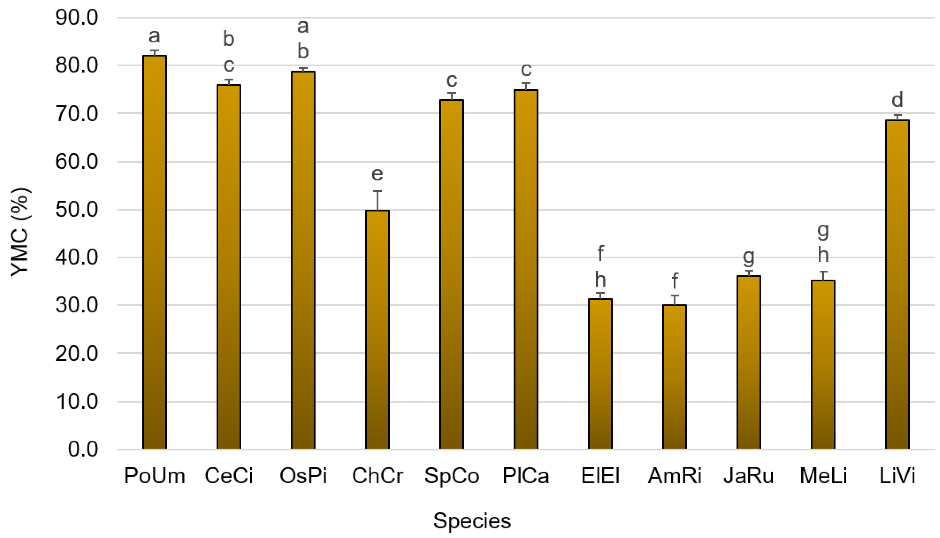
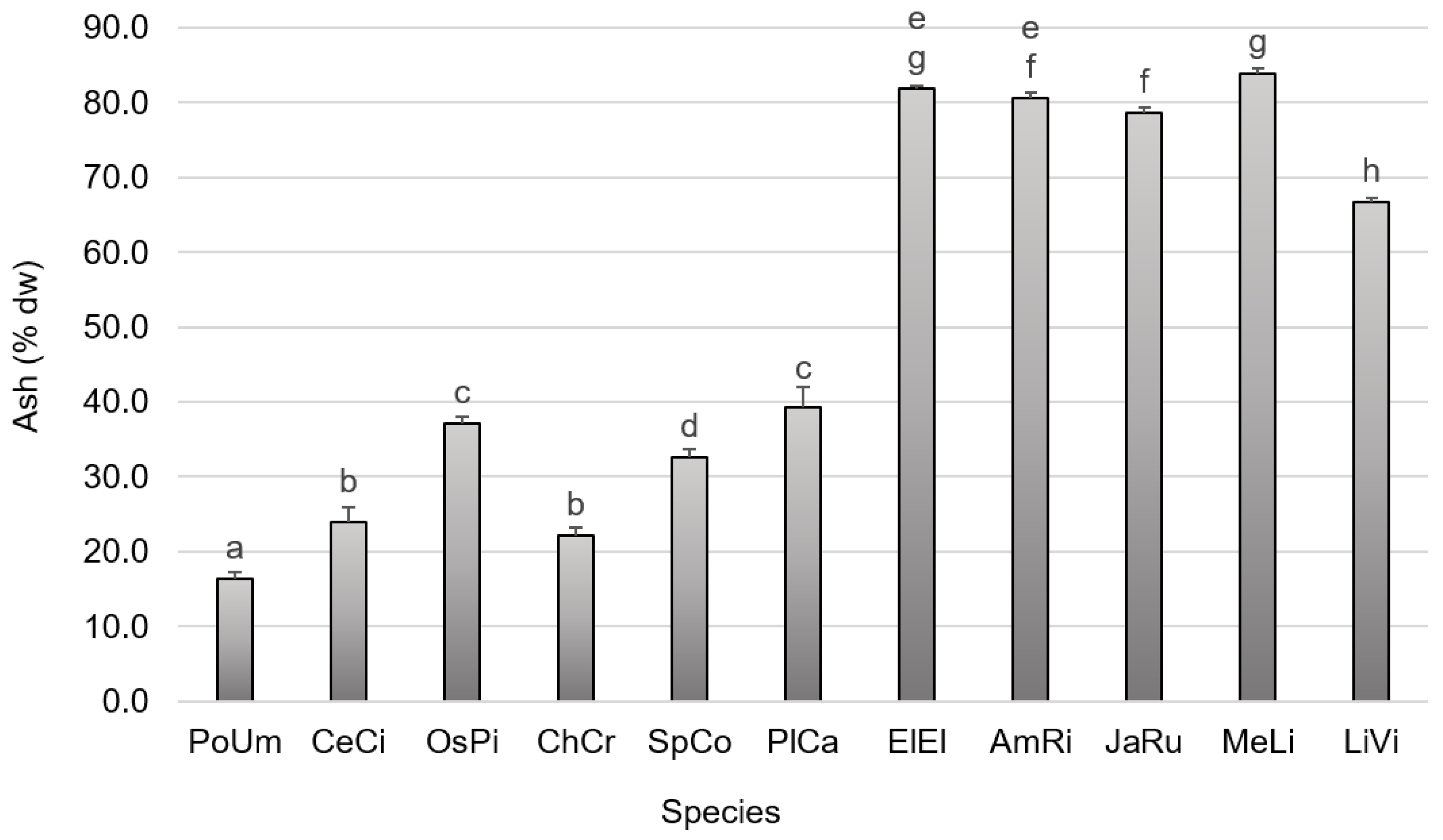
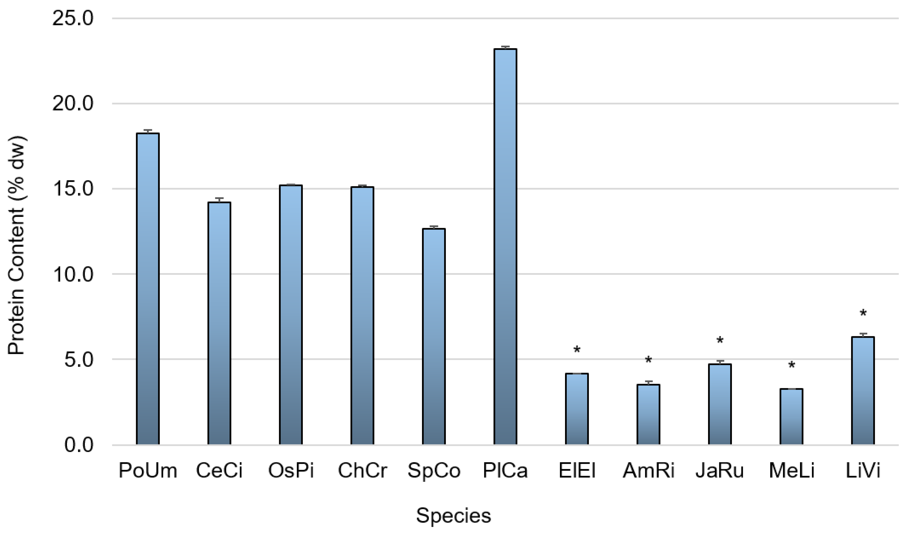
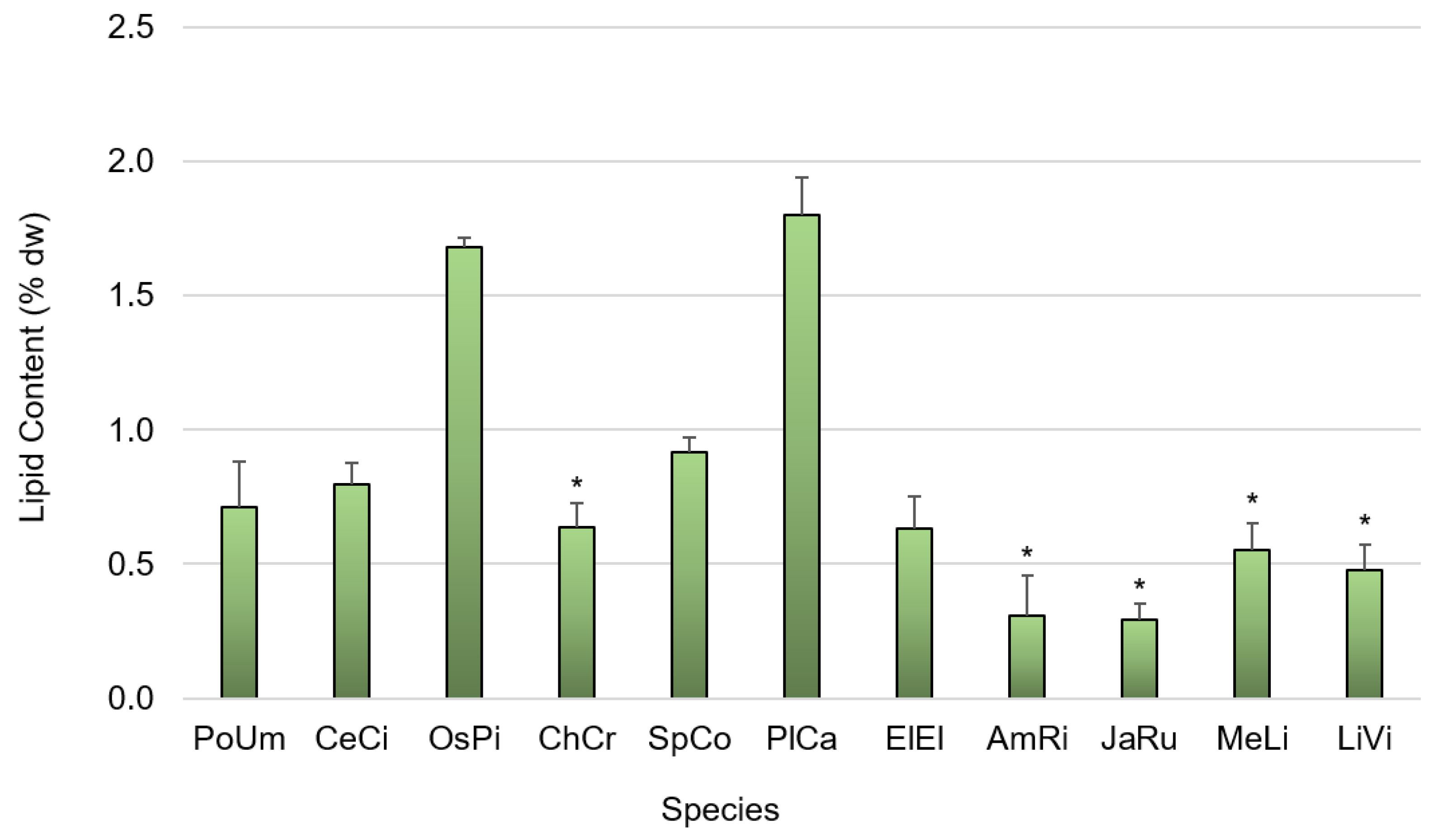
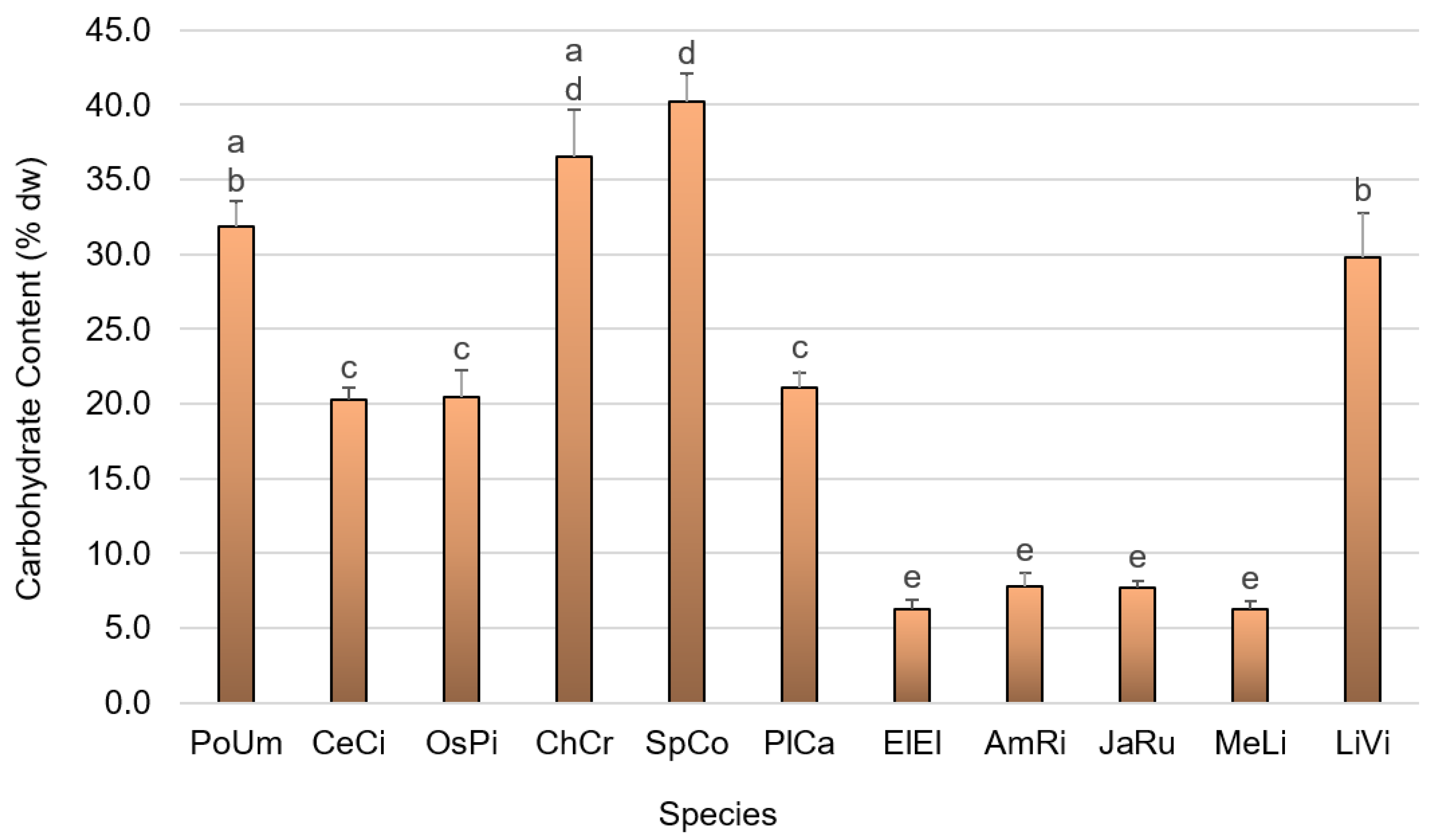
3.1. Primary Composition

The analysis of the primary composition of the red seaweeds considered in the present study revealed distinct results according to species. Regarding the variation of the yield of moisture content (YMC) according to species, as shown in Figure 1, the values obtained ranged from 30.05 ± 1.94% (A. rigida) to 82.00 ± 1.11% (P. umbilicalis). Different letters above the bars indicate statistically different results (Tukey HSD test (F(10,43) = 580.877; p = 0.00)), and the difference is evident between the results of all coralline species (E. elongata, A. rigida, J. rubens and M. lichenoides) and all other seaweed species considered, with the former group showing significantly lower values.
The results regarding the variation of ash content according to species, as shown in Figure 2, showed that the values obtained ranged from 16.38 ± 0.93% dw (P. umbilicalis) to 81.93 ± 0.34% dw (E. elongata), with significant differences found between species (Tukey HSD test (F(10,23) = 1791.090; p = 0.00)). Similar to the results found for YMC, it is evident that there was a significant difference M. between the ash content of coralline species (E. elongata, A. rigida, J. rubens and Mesophyllum lichenoides) and all other seaweed species considered, with the former group presenting radically higher values. By observing both Figure 1 and Figure 2, we can see that, generally, species with high YMC have low ash content and vice versa. The exception lies with Chondrus crispus and Liagora viscida, species whose aforementioned values do not follow this pattern; C. crispus had both a YMC and ash content lower than 50%, while, in L. viscida, both values were higher than 60%.
As for the protein content found in the species analyzed, Figure 3 shows that the values obtained ranged from 3.27 ± 0.01% dw (M. lichenoides) to 23.18 ± 0.17% dw (Plocamium cartilagineum), with significant differences between all species pinpointed by the Kruskal–Wallis test (χ2(10) = 31.670; p = 0.000). P. umbilicalis also presented a noteworthy protein content (18.27 ± 0.19% dw), and, again, the coralline species all showed very low values (<5% dw) when compared to the majority of the remaining species, with the exception of Liagora viscida.
The values obtained for the lipid content in the species analyzed (Figure 4) were generally quite low for all species, ranging from 0.29 ± 0.06% dw (J. rubens) to 1.80 ± 0.14% dw (P. cartilagineum), with significant differences between all species pinpointed by the Kruskal–Wallis test (χ2(10) = 28.848; p = 0.001). Plocamium cartilagineum and O. pinnatifida had the highest lipid content, whereas coralline algae showed remarkably low lipid values overall.
The results showed that the species analyzed had variable carbohydrate levels (Figure 5), ranging from 6.24 ± 0.56% dw (M. lichenoides) to 40.23 ± 1.87% dw (S. coronopifolius), with significant differences among all species pinpointed by the Tukey HSD test (F(10,32) = 170.637; p = 0.00). Following S. coronopifolius, C. crispus and P. umbilicalis also showed appreciable carbohydrate content (36.58 ± 3.06% and 31.89 ± 1.65% dw, respectively), while all coralline algae showed values no higher than 7.78 ± 0.90% dw. Ceramium ciliatum, O. pinnatifida and P. cartilagineum had similar carbohydrate content (~20% dw).

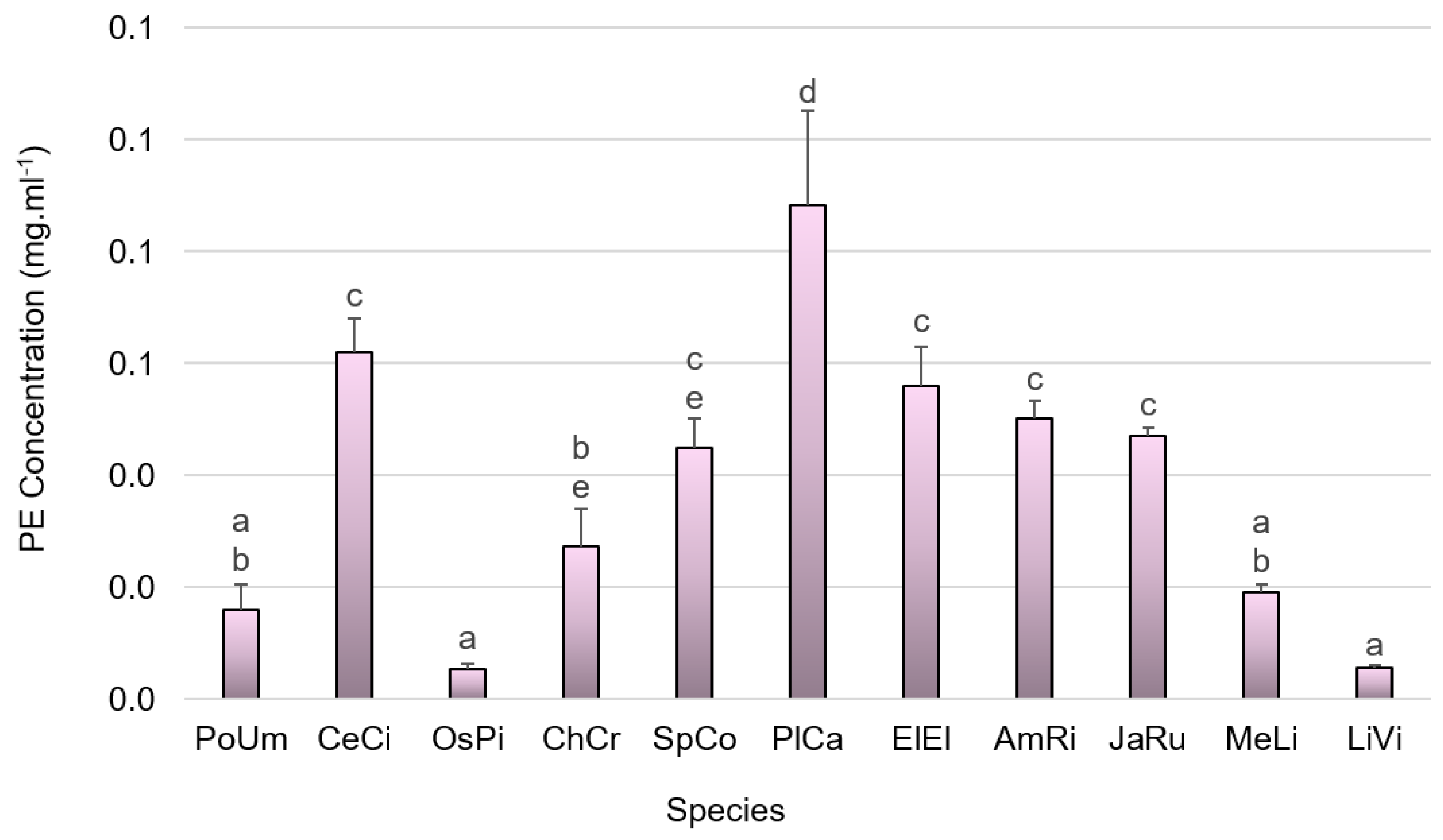
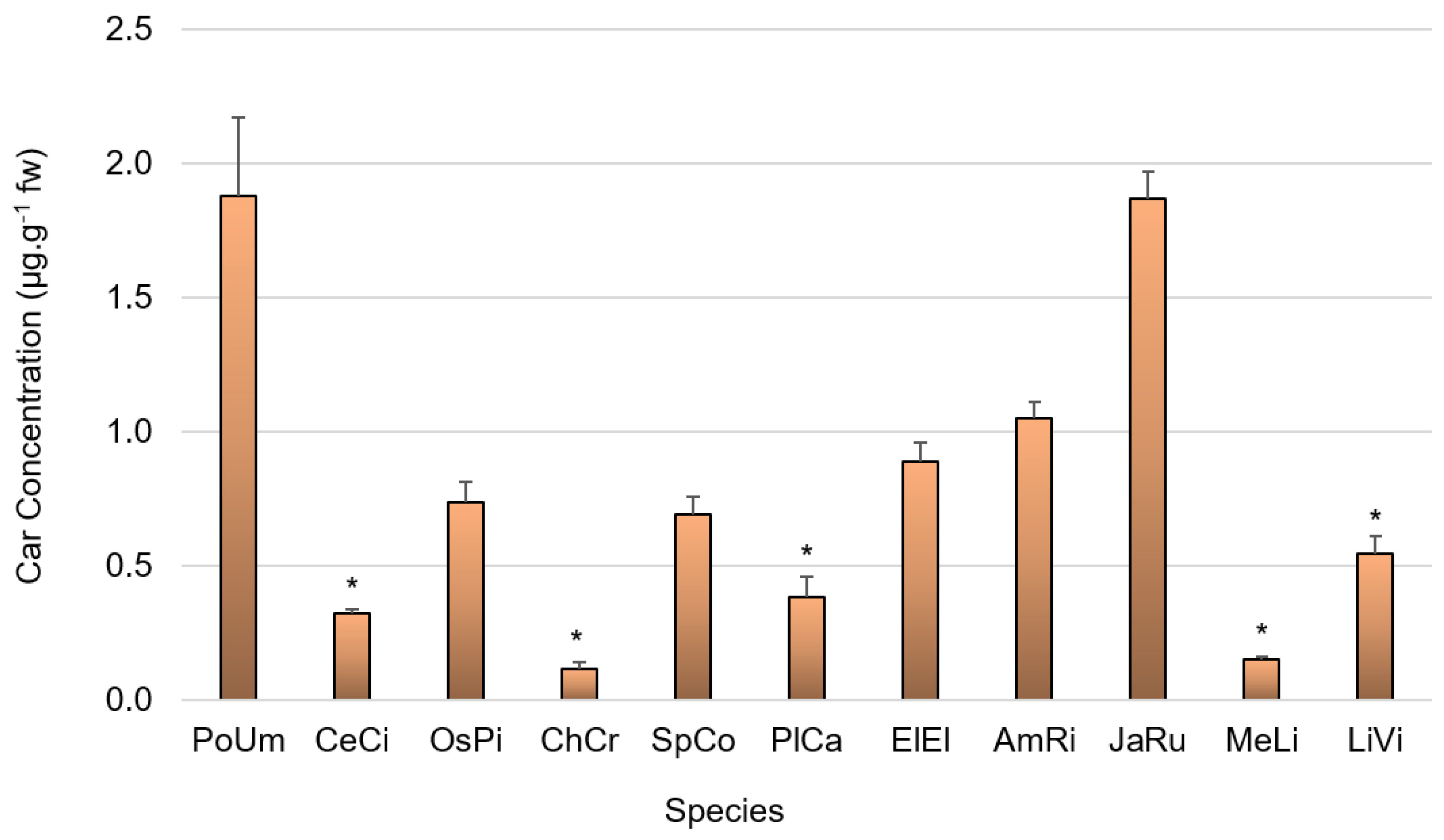
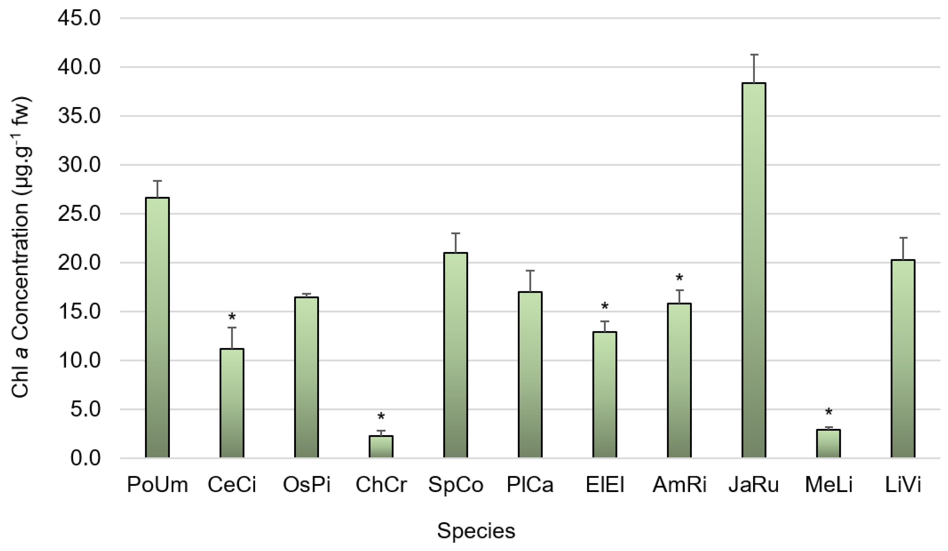
3.2. Pigment Composition

The analysis of the pigment composition, specifically the PBP phycoerythrin and phycocyanin, carotenoids and chlorophyll a, for all algae studied, showed marked differences between species. Specifically, phycoerythrin concentrations, shown in Figure 6, ranged from 0.01 mg.mL−1 (both in O. pinnatifida and L. viscida) to 0.09 ± 0.02 mg.mL−1 in Plocamium cartilagineum, with significant differences found between species (Tukey HSD test (F(10,32) = 46.407; p = 0.00)). Most of the coralline algae studied, specifically E. elongata, A. rigida and J. rubens, showed values around 0.05–0.1 mg.mL−1, which were comparable to C. ciliatum and S. coronopifolius.
Phycocyanin concentrations and yields were found to be quite low for all species studied (Table 2), not exceeding 0.01 mg.mL−1 for P. umbilicalis, C. ciliatum, O. pinnatifida and P. cartilagineum, while all the remaining studied species did not show readable phycocyanin content; significant differences were shown by the Kruskal–Wallis test (χ2(10) = 29.271; p = 0.001).
Regarding carotenoid content, shown in Figure 7, differences were found among all species studied (Kruskal–Wallis test (χ2(10) = 31.451; p = 0.000). The values ranged from 1.88 ± 0.29 µg.g−1 for P. umbilicalis to 0.12 ± 0.02 µg.g−1 for C. crispus. Interestingly, Jania rubens presented a carotenoid content of 1.87 ± 0.10 µg.g−1, a value reached by no other coralline algae, which showed values ranging from 0.15 ± 0.01 µg.g−1 (M. lichenoides) to 1.05 ± 0.06 µg.g−1 (A. rigida).
As for chlorophyll a, differences in this pigment concentration (Figure 8) were also found between all species studied (Kruskal–Wallis test (χ2(10) = 30.930; p = 0.001). Values ranged from 2.27 ± 0.50 µg.g−1 for C. crispus to 38.41 ± 2.84 µg.g−1 for J. rubens. Again, J. rubens stood out, with a value of 1.87 ± 0.10 µg.g−1, a value reached by no other coralline algae; others presented values ranging from 2.89 ± 0.25 µg.g−1 (M. lichenoides) to 15.86 ± 1.29 µg.g−1 (A. rigida). Generally, the pattern obtained was similar to that found for carotenoid concentrations, with the species showing a higher carotenoid content also presenting a higher chlorophyll a content.

4. Discussion

In the present work, we provided the basic nutritional profile and pigment content determination for eleven seaweed species that commonly occur across the central coast of Portugal. Our target species, Porphyra umbilicalis, Ceramium ciliatum, Osmundea pinnatifida, Chondrus crispus, Sphaerococcus coronopifolius, Plocamium cartilagineum, Ellisolandia elongata, Amphiroa rigida, Jania rubens, Mesophyllum lichenoides and Liagora viscida, were widely available for collection, although they seemingly had a specific season when they are most easily found. Specifically, all species were easily found during winter (and colder waters), with the exception of C. crispus, which was collected during spring, and C. ciliatum, S. coronopifolius and L. viscida, which were collected during summer.
It is known that the biochemical profile of a seaweed is shaped not only according to taxonomic classification [8,9,10] but also according to seasonal patterns and environmental gradients [2,5,6,7], as previously stated. In this sense, although our working hypothesis in the present study revolved around differences in the basic nutritional profile and pigment composition according to species, we are aware that, since these species were collected during specific seasons, we should take this into account as well, not only when discussing our findings, but also when comparing them with other studies. Furthermore, the collection of each species from only one locality also limited our understanding of the natural variability that these species can show.

4.1. Primary Composition

Regarding the primary composition of the eleven seaweeds studied, we found that there were differences between species for all the analyses performed, backed up by statistical tests, all stated previously. This is not surprising, as several authors that endeavored to study the nutritional profile and/or pigment composition of a range of species in a single study also discovered differences according to species and pinpointed the most promising ones.

4.1.1. Yield of Moisture Content and Ash Content

We observed that higher YMC corresponded, generally, to a lower ash content and vice versa. The ash content measures the total mineral content in a biological sample, and, in seaweeds, this fraction usually comprises sodium, potassium, iron, zinc and magnesium, all of which are essential in human nutrition and not as easily found in edible land plants [31,72].
Regarding ash content, it is noteworthy to point out the clear distinction between coralline algae and all other species studied, with the exception of L. viscida, which also had values similar to corallines. Factually, coralline algae have highly mineralized fronds and are a species otherwise known to be poor in other elements that commonly occur in other Rhodophyta families. These species obtain and accumulate carbonate salts from seawater [54], and we assume that the resulting calcium content might have contributed to the overall ash content observed. Although not a coralline algae, L. viscida is a species that has also a fair amount of calcium carbonate in its structure, and, therefore, it was not surprising to find it grouped with other coralline with respect to ash content. Among the other remaining algae studied, P. cartilagineum and O. pinnatifida presented the highest amount of ash, respectively, 39.23% dw and 37.06% dw, which shows their value in providing essential minerals other than calcium.
On the other hand, we found P. umbilicalis had 16.38% ash content; previous studies reported variable amounts of ash content for P. umbilicalis, namely 12% dw [73] and 28.16% dw (for Porphyra sp. [74]). C. ciliatum showed an ash content of 23.97% dw, while other authors found lower values for Ceramium spp., namely, 11.35% dw for Ceramium diaphanum [75] and 27.1% dw for Ceramium sp. [21]. O. pinnatifida’s ash content of 37.06% dw was similar to the previously reported values, namely, 30.62% dw [8], 32.3% dw [21] and 38.55% dw [74]. C. crispus had an ash content of 22.19% dw, in range of that reported previously (21.44% dw [76] and 29.46% dw [26]). Jania rubens presented an ash content of 78.60% dw, higher than that found in another study (up to 48.82% dw) [52]. Values regarding the other studied species were not found in literature for comparison purposes.

4.1.2. Protein Content

The protein found in seaweeds is an excellent source of essential amino acids, which represent almost half of the total amino acids they naturally own. Therefore, seaweeds are particularly interesting from a nutritional standpoint and can potentially minimize the issue of protein malnutrition in the human diet [72]. Protein values can reportedly vary according to taxa, although red seaweeds are known to have the highest protein content, which can reach up to 47% dw, of all phyla [77]; in turn, the protein content can be heavily shaped by geographic location, season, environment and even methodologies [10].
In our study, the highest value obtained was 23.18% dw for P. cartilagineum but, as stated, several factors play into calculating the metabolite content and diversity even within species, namely, seasonal conditions and the methodologies adopted. On the other hand, the lowest values were found for all the coralline species considered.
P. umbilicalis presented 18.27% dw of protein content, only surpassed by Plocamium cartilagineum, but this species regularly showed higher protein content throughout the literature. For example, P. umbilicalis protein extracts with 22.6% yield were reported by Harrysson et al. [22], while dry biomass had highly variable protein values among the literature, with authors reporting, for example, 24.11% dw [78], 24.82% dw [74], 25.80% dw [10], 28.29% dw [76], 31.4% dw (all for Porphyra sp.) [79], 40% dw (for P. umbilicalis) [73] and 44% dw (for Porphyra sp.) [21]. In this sense, values found for the P. umbilicalis presently studied were indeed low in comparison, but again, this can be explained not only according to the geographic variation and season at the time of harvest but also by the different extraction methodologies adopted by each author. C. ciliatum showed a protein content of 14.21% dw, which is in range of that found by other authors for Ceramium spp., namely, 14% dw for C. diaphanum [75], although higher protein concentrations were also found (31.2% dw for Ceramium sp. [21]). Osmundea pinnatifida yielded 15% dw of protein, a value lower than that previously reported by other authors, who found a protein content of 20.64% dw [10], 20.79% dw [74], 23.8% [8] and 27.3% dw [21]. Chondrus crispus presented a protein content of 15.10% dw, in range of that found by previous studies (17% dw [26] and 20.10% dw [76]). Sphaerococcus coronopifolius presented a protein content of 12.65% dw in our study, lower than that obtained in previous works, namely, 19.56% dw [10]. Jania rubens presented a low protein content (4.70% dw) similar to that found by another study (up to 3.41% dw) [52]. For the remaining species analyzed, we did not find studies that allowed comparisons with our data.

4.1.3. Lipid Content

In red seaweeds, lipid content is known to be low, with values that are generally nutritionally adequate for a healthy diet [5], although they poorly contribute as energy providers [80].
Similar to what was observed for protein content, all corallines showed lower values than any other species, whereas P. cartilagineum and O. pinnatifida stood out with, respectively, 1.80% dw and 1.68% dw of fat content. Nevertheless, all values found were below 1.80% dw, thus being within range of what is usually reported for red seaweeds [5,8,26,38,79,81] and seaweeds in general [21,52,79,82].

4.1.4. Carbohydrate Content

In seaweeds, the abundance and composition of carbohydrates varies across species; within Rhodophyta, we typically find floridean starch, cellulose, xylan and mannan [72]. The soluble fiber fraction is, in turn, rich in sulphur-containing galactans such as agar and carrageenan, both earning a worldwide standing in global food industries [72,83]. Specifically, in the order Bangiales genus Porphyra/Pyropia, we found the sulphated polysaccharide porphyrin, for which there has been well-documented research on its bioactivity potential (such as anti-tumoral and anti-viral activity) and its possibilities as a promoter of human health [72].
In the present study, S. coronopifolius, C. crispus, P. umbilicalis and L. viscida presented remarkably high carbohydrate values (40.23%, 36.58%, 31.89% and 29.80% dw) when compared to the other remaining species, thus, being good candidates for future research regarding the diversity and composition of the carbohydrates presently measured and their suitability for human nutrition. On the other hand, all coralline algae showed very low values when compared to the other seaweeds studied.
Each species analyzed presented distinct values when compared to their counterparts reported throughout the literature. Specifically, P. umbilicalis presented a carbohydrate value of 31.89% dw, as stated, while lower values were found in previous studies; a value of 25.37% dw [74] was reported, for example. Ceramium ciliatum presented a carbohydrate content of 20.31% dw, a value that stands between values obtained by other authors (18.70% dw for C. diaphanum [75] and 32.33% dw for Ceramium virgatum). Osmundea pinnatifida presented a carbohydrate content of 20.44% dw, which stands between those previously reported by other authors, namely, 17.61% dw [74] and 32.4% for O. pinnatifida [8]. Chondrus crispus, in the present study, presented one of the highest carbohydrate contents among all the algae analyzed (36.58% dw), but the literature reported even higher values (53.43% dw [26]). For the other species analyzed, we did not find values in literature that allowed comparisons across studies.

4.2. Pigment Composition

Red seaweeds are rich in phycoerythrin and carotenoids and also have a small degree of phycocyanin and chlorophyll a. Altogether, these pigmented metabolites provide these algae their unique red colors, hues and variations and, hence, their own place within the phylum Rhodophyta. These natural pigments, regardless of their source (as none is exclusively found in red macroalgae), have been widely studied recently, having a range of well-documented applications in biomedical research, therapeutics, clinical diagnosis and cosmeceutical/pharmaceutical industries, as well as application in food as both a coloring agent [84,85,86,87,88,89,90,91] and nutritional booster [92,93], and effectiveness as an antioxidant agent [94,95,96,97,98], among other bioactivities [34,95,99,100,101].

4.2.1. Phycobiliprotein Content

Phycobiliproteins (PBP) are pinkish-red pigments known for their noteworthy spectroscopic properties, exhibiting high excitation/emission spectra, a high absorption coefficient, high quenching stability and water solubility [102,103]. These qualities render PBP suitable for applications in flow cytometry, immunofluorescence microscopy [104] and cancer therapy [105]. As such, these natural pigments have proven their usefulness in not only the cosmeceutical and pharmaceutic industries, as mentioned earlier (where they shine as bioactive agents), but also in biomedical research, clinical diagnostics and therapeutics [84,85,86,106].
Regarding phycoerythrin (PE) and phycocyanin (PC) content, P. cartilagineum provided 0.09 mg.mL−1 of PE, the highest value obtained for all the species considered, while both O. pinnatifida and L. viscida presented the lowest values (0.01 mg.mL−1). In fact, the difference between P. cartilagineum and all other species considered is noteworthy and provides clues about the potential of this species in providing PE; although their results were not close to P. cartilagineum, the species C. ciliatum, S. coronopifolius, E. elongata, A. rigida and J. rubens are worth due consideration as well. In fact, of all the coralline studied, E. elongata had the highest PE content, and it is a known as a potential candidate for PE extraction and applications [72,107]. These results may, therefore, pave the way for future studies regarding the biotechnological potential of the PE extracted from these species, namely, in terms of bioactivities.
Considering comparisons with the results obtained in the literature, in our study, P. umbilicalis presented one of the lowest PE values (0.21 mg.mL−1) when compared to other algae, while previous studies reported distinct values for P. umbilicalis concentrations. To mention a few examples, previous studies reported that Porphyra spp. yielded variable PE and PC concentrations, namely, PE: 0.35 mg.mL−1 dw for Porphyra sp. [108], PE: 8319 µg.g−1 and PC: 5305 µg.g−1 for Porphyra spp. [25]. Kim et al. [109] reported that lower temperatures enhance PE contents in P. umbilicalis, having achieved values of 26 mg.g−1 fw. However, it must be noted that several authors possibly considered, calculated and discussed Porphyra PE as R-PE and not the B-PE that Bangiophyceae possess. These pigments are fundamentally different in their structure and spectral data, namely, absorption peaks (λ max-B-PE: 546, 565 nm; R-PE: 496, 546, 565) and extinction coefficients (B-PE: 2.410.000 cm−1 M−1; R-PE: 1.960.000 cm−1 M−1) [110].
For the other species, such as C. ciliatum, we obtained a PE content of 1.23 mg.mL−1, the second highest PE content obtained for all the species considered in the current study, while previous studies reported a variable PE content for Ceramium spp., namely, 0.383% dw for Ceramium isogonium [111] and 2.13% dw for Ceramium tenuicorne [112]. E. elongata and J. rubens yielded values of 1.11 mg.mL−1 and 0.94 mg.mL−1, the former achieving the highest value obtained among the coralline presently considered; both values were consistent with those reported by Ismail and Osman [113], who obtained >1 mg.g−1 fw for E. elongata and 0.91 mg.mL−1 for J. rubens. To date, we have not found published records for the remaining seaweed species considered.

4.2.2. Carotenoid Content

Regarding carotenoid content, P. umbilicalis and J. rubens provided the highest values among all species considered, namely, 1.88 µg.g−1 fw and 1.88 µg.g−1 fw, while C. crispus showed the lowest value (0.15 µg.g−1 fw). Both P. umbilicalis and J. rubens are species worth considering in future studies regarding carotenoid extraction and evaluation of its potential in biotechnological applications. Ranking next, we found that the coralline algae A. rigida and E. elongata also presented appreciable carotenoid values (1.05 µg.g−1 fw and 0.89 µg.g−1 fw, respectively).
Concerning the differences across studies, for P. umbilicalis, we reported a total carotenoid content of 1.88 µg.g−1 fw, as stated, while other authors reported values up to 74.5 µg.g−1 dw. For Ceramium sp., we obtained a carotenoid content of 0.323 µg.g−1 fw, while previous authors reported a total carotenoid content of 8.77 µg.g−1 dw [114] and 23 mg.g−1 dw (for Ceramium rubrum) [115]. To date, we have found no published studies for the remaining seaweed species considered in the present study.
Although we only analyzed the total carotenoid content, Takaichi et al. [116] stated that carotenoid composition is related to Rhodophyta phylogeny, as Bangiophyceae (such as P. umbilicalis) contain α-carotene, lutein-type and zeaxanthin-type carotenoids, with lutein constituting more than 50% of the total carotenoid composition. Within Florideophyceae, Takaichi et al. [116] found that carotenoid profiles differ between subclasses. Specifically, Nemaliophycidae (such as L. viscida) reportedly contain lutein-type carotenoids similarly to Bangiophyceae. Most orders within Rhodymeniophycidae (such as C. ciliatum, O. pinnatifida, C. crispus, S. coronopifolius and P. cartilagineum) contain lutein-type carotenoids; an exception is Gracilariales, which we did not include in the present study. Among Corallinophycidae, more specifically, those in the order Corallinales, Lithophylloideae (such as A. rigida) contain lutein-type carotenoids, and Corallinoideae (such as E. elongata and J. rubens) contain anteraxanthin-type carotenoids, while the order Hapalidiales (such as M. lichenoides) contains lutein-type carotenoids [116,117]. The variation between the carotenoid profiles according to taxonomic position alone may explain the differences found between some of the species considered, such as the difference between the carotenoid content in P. umbilicalis and that of all other species. Yet, if this is the case, we would also have found a close relation between J. rubens and E. elongata, but both had highly discrepant carotenoid values. Another example that stands out is C. crispus, which presented low carotenoid values when compared to the other species within Rhodymeniophycidae, although they reportedly share the same carotenoid profile. However, it is a Gigartinales, highly rich in sulphated galactan carrageenan, which is a phycocolloid that may somehow prevent efficient extraction of any other compound.
In this sense, other hypotheses come into play when discussing carotenoid variation between species, specifically, geographic distribution, season pattern and extraction methodologies, which have not yet been adapted on a species-specific basis. In fact, even the same species of algae can show distinct carotenoid levels and composition shaped by the environmental conditions maintained during their growth [114] and even shaped by the extraction and analytical methods employed to study these metabolites.

4.2.3. Chlorophyll a Content

The study of chlorophylls from seaweeds is highly important, given that these metabolites possess a range of biological activities, such as anti-cancer activity [118], and might potentially serve as a magnesium source since this compound is highly bioavailable from chlorophylls [119].
For chlorophyll a, in our study, P. umbilicalis presented 26.68 µg.g−1, which was lower than in previous studies, which reported a Chl a content of up to 542.2 µg.g−1 [25]. Similarly, C. ciliatum yielded a Chl a content of 11.15 µg.g−1, whereas previous studies reported a content of 90 µg.g−1 (for Ceramium rubrum). On the other hand, A. rigida showed a Chl a content of 15.855 µg.g−1, a higher value than that obtained by previous studies, which reported values of 13.65 mg.g−1 for A. rigida [120] and 0.4 to 1.6 mg.g−1 for Amphiroa fragilissima [121]. We did not find published registers for the remaining seaweed species considered.

4.2.4. Additional Considerations

Although the differences in pigment content between our species and those studied by other authors can be explained by geographical and seasonal differences, several other factors certainly played a role as well. The molecular structure of the biological sample must be taken into account. Ana-Marija et al. [100] mentioned that red seaweeds have a distinct chemical and cellular structure when compared to other organisms, such as other macroalgae and microalgae taxa, but we hypothesize that these discrepancies can also be found within Rhodophyta according to taxonomic classification and divergence in time. Because a simpler chemical and cellular structure facilitates solvent penetration [100], this might explain the different concentrations obtained between species for all the pigments considered.
On the other hand, differences in algal processing and in extracting the solvent itself (regarding type and/or concentration) can also produce quite distinct results. For example, although we chose to use fresh algae for all pigment measurements, as advised by Beattie et al. [67], many authors presented their results in units of dry weight, which renders comparisons across studies not entirely immediate or intuitive. Next, as an example of the differences driven by the use of different solvents, both acetone and methanol are popular solvents for carotenoid extraction and would certainly yield different results (e.g., [25]). Lastly, the importance of the method itself must be stressed, not only regarding the extraction but also with respect to the readings (with UV–Vis and HPLC popularly applied for this matter) and the final calculations (such as differences between the equation formulae used to calculate the concentrations of any given pigment).

4.3. Species-Specific Notes

In our study, specifically, Porphyra umbilicalis stood out in several analyses, presenting the highest YMC and the lowest ash content and one of the highest protein and carbohydrate contents when compared to all other species analyzed. Regarding pigment content, although it did not present appreciable B-phycoerythrin contents, it did stand out remarkably regarding carotenoid content and, to a lesser degree, chlorophyll a content. Taxonomically speaking, P. umbilicalis is a Bangiophyceae, while all the other species considered belong to class Florideophyceae, and both, according to Brawley et al. [122], are highly divergent; this fact alone might explain why P. umbilicalis stood out in the analysis performed. One of the traits that distinguishes this species from all the other species considered is the nature of the pigment PE, which is B-PE in Bangiophyceae, such as Porphyra spp., while Florideophyceae contain R-PE—the analysis we performed required distinct equations that specifically targeted each pigment. P. umbilicalis’ commercial importance has triggered studies on a molecular level, an example being the endeavor to sequence its entire genome by Brawley et al. [122]. With such a study, these authors sought out not only to find insights into P. umbilicalis’ appealing nutritional profile but also into its success in growing in intertidal zones, specifically mid-to-high regions which are known to be physically stressful for other algal species and organisms [122]. Porphyra is, thus, an ancient red alga with a unique biochemical profile likely shaped by millions of years of thriving in environments that suffer daily fluctuations in temperature, salinity and irradiance. P. umbilicalis is well known for its nutritional value [123], and this present study supports that fact.
Chondrus crispus is a renowned carrageenan worldwide, and the original source of this phycocolloid, and it is a species also known for its pleasant taste, exploited as a food ingredient in a number of countries [15]. In the present study, C. crispus was one of the seaweeds with high carbohydrate content, appreciable protein content and low fatty acid content, whereas, regarding pigments, it stood out as one of the species with the lowest concentrations of all pigments analyzed. As C. crispus is a carragenophyte, which is a phycocolloid with high gelling properties, we wondered whether this particular substance hindered the effectiveness of the several extracting methodologies we applied to assess the biochemical basic profile for this species; this was particularly evident when assessing pigment content, where the values we obtained were remarkably low. Therefore, we hypothesize that this particular alga may be an example of how protocols and extraction methods need to be adapted depending on the species.
Sphaerococcus coronopifolius is widely reported as a source of important metabolites with biological activity, namely antioxidant [124], antimicrobial [125,126] and anti-tumoral activities [46,49,126]. This study provides, on the other hand, insight into the nutritional profile of S. coronopifolius, which showed a carbohydrate content similar to that which we obtained for C. crispus. We consider that this species may be worth exploring further to assess its potential value as a food capable of enriching human nutritional diets.
Plocamium cartilagineum, similarly to S. coronopifolius, has been regularly studied regarding its potential as a bioactive agent. Studies have assessed its antioxidant potential [48], as well as its ability to synthetize phycobiliproteins according to light source [127]. P. cartilagineum, in the current study, ranked first in many of the analyses performed; this was a curious finding, because this species is not as extensively studied as other Rhodophyta species, such as P. umbilicalis or C. crispus, studied presently. The present study offers an eye-opening insight regarding the potential of this species from a nutraceutical perspective, given its high content in protein and phycobiliproteins, fatty acids, a fair degree of mineral content and carbohydrates and, therefore, will also earn further consideration from us.
Coralline algae (e.g., E. elongata, A. rigida, J. rubens and M. lichenoides) are particularly important from an ecological standpoint, known as ecosystem engineers, and their strong physical structure provides habitat and shelter for numerous forms of aquatic life [128,129]. As stated, coralline algae are one of the most important vegetable sources of calcium and have the ability to accumulate carbonate salts from seawater [54], and this ability, coupled with the resulting high mineral content, has been explored in a nutritional context by several authors, within the context of bone structure and function [54,130,131,132,133]. These studies ultimately promote the use of this group of seaweeds as a food supplement to enrich diets low in essential minerals. In comparison to other substances, coralline algae are generally poor in the metabolites that commonly occur in other seaweed species [54]; this was shown by the current study, where all coralline studied had a higher ash content but were substantially lower content in protein, lipids, carbohydrates and pigments when compared to the other species studied. However, it must be noted that J. rubens showed a comparatively high content in carotenoids and chlorophyll a compared to other coralline and even other species.
Liagora viscida remains, to this day, one of the most poorly studied species (from among those presently studied), although it is, reportedly, an important potential source of metabolites with antimicrobial activity against bacteria and fungi [134]. Although taxonomically distinct from coralline algae, L. viscida presented similar results to coralline algae with regard to ash content, protein and fatty acids. In fact, Liagora viscida also has the ability to accumulate calcium carbonate in its biological tissue, although to a lesser extent than a coralline alga. One therefore wonders whether its ash content corresponds to a substantial calcium content which, as stated, is essential to promoting bone health and structure. L. viscida also showed a high carbohydrate content, similar to that observed for P. umbilicalis, a fact that renders this alga worthy of consideration as a complement to healthy human diets.

5. Conclusions

The results found shed light on the nutritional and biotechnological value of Porphyra umbilicalis, Ceramium ciliatum, Osmundea pinnatifida, Chondrus crispus, Sphaerococcus coronopifolius, Plocamium cartilagineum, Ellisolandia elongata, Amphiroa rigida, Jania rubens, Mesophyllum lichenoides and Liagora viscida by providing their primary nutritional composition and pigments. Of these eleven Rhodophyta species that have been studied, five are consumed in Europe, namely, P. umbilicalis, O. pinnatifida, C. crispus, P. cartilagineum and J. rubens [135]. Of these, only Porphyra sp. and C. crispus are listed in the Novel Food Catalogue kept by the European Commission (Regulation (EU) 2017/2470), although this non-exhaustive list includes merely 22 species of seaweeds [136]. Species such as S. coronopifolius and M. lichenoides are also considered edible in certain parts of the world [137]. To date, we have not found records of the remaining species in our list of target species for human consumption.
We hope that the present study contributes to the vast knowledge already existing for species such as P. umbilicalis and C. crispus and that it has shed light on other species that are not so well recognized, such as C. ciliatum, O. pinnatifida, S. coronopifolius, P. cartilagineum, E. elongata, A. rigida, J. rubens, M. lichenoides and L. viscida. Differences were found across these species regarding their primary composition and pigment concentrations, which were likely shaped by seasonal patterns, taxonomic position, processing methodologies and data treatment. Nevertheless, the present study highlighted the potential of P. cartilagineum (regarding protein content and phycoerythrin), S. coronopifolius (regarding carbohydrate content), P. umbilicalis (regarding carotenoid content) and J. rubens (regarding chlorophyll a) as food sources capable of enriching human nutritional diets. Whether the human organism can effectively and safely incorporate these metabolites remains to be assessed by performing bioavailability and biotoxicity assays upon these species.
All these species occur in relative abundance throughout central Portugal and remain, to date, mostly unexplored from a nutritional and biotechnological perspective when, in fact, most of them may be potentially of noteworthy value in relation to these topics. Furthermore, and as stated before, the protocol for the cultivation of species not yet farmable is under development to allow the commercial exploitation of the most interesting species, if it proves worthwhile, and thus preserve natural populations.

Author Contributions

Conceptualization, M.V.F., T.M., C.A. and L.P.; methodology, M.V.F., T.M. and C.A.; validation, M.V.F., L.G.I., T.M. and C.A.; formal analysis, C.A., M.V.F., L.G.I. and M.M.; investigation, M.V.F., L.G.I. and M.M.; writing—original draft preparation, M.V.F.; writing—review and editing, M.V.F., T.M., C.A. and L.P.; supervision, T.M., C.A. and L.P. All authors have read and agreed to the published version of the manuscript.

Funding

This work was funded by national funds through FCT—Fundação para a Ciência e a Tecnologia for I.P., within the scope of the project MARE (UIDB/04292/2020 and UIDP/04292/2020), the project LA/P/0069/2020, granted to the Associate Laboratory ARNET, and the individual doctoral grant awarded to Marta V. Freitas (UI/BD/150957/2021).

Institutional Review Board Statement

Not applicable.

Informed Consent Statement

Not applicable.

Data Availability Statement

Not applicable.

Conflicts of Interest

The authors declare no conflict of interest. The funders had no role in the design of the study; in the collection, analyses or interpretation of data; in the writing of the manuscript or in the decision to publish the results.

References

  1. Ariede, M.B.; Candido, T.M.; Jacome, A.L.M.; Velasco, M.V.R.; de Carvalho, J.C.M.; Baby, A.R. Cosmetic attributes of algae—A review. Algal Res. 2017, 25, 483–487. [Google Scholar] [CrossRef]
  2. Hagen Rødde, R.S.; Vårum, K.M.; Larsen, B.A.; Myklestad, S.M. Seasonal and geographical variation in the chemical composition of the red alga Palmaria palmata (L.) Kuntze. Bot. Mar. 2004, 47, 125–133. [Google Scholar] [CrossRef]
  3. Young, R.M.; Von Salm, J.L.; Amsler, M.O.; Lopez-Bautista, J.; Amsler, C.D.; McClintock, J.B.; Baker, B.J. Site-specific variability in the chemical diversity of the Antarctic red alga Plocamium cartilagineum. Mar. Drugs 2013, 11, 2126–2139. [Google Scholar] [CrossRef] [PubMed] [Green Version]
  4. Takeshi, S.; Yumiko, Y.-S.; Joko, S. Mineral Components and Anti-oxidant Activities of Tropical Seaweeds. J. Ocean Univ. China 2005, 4, 205–208. [Google Scholar] [CrossRef]
  5. Afonso, C.; Correia, A.P.; Freitas, M.V.; Baptista, T.; Neves, M.; Mouga, T. Seasonal changes in the nutritional composition of Agarophyton vermiculophyllum (Rhodophyta, Gracilariales) from the center of Portugal. Foods 2021, 10, 1145. [Google Scholar] [CrossRef]
  6. Cavaco, M.; Duarte, A.; Freitas, M.V.; Afonso, C.; Bernardino, S.; Pereira, L.; Martins, M.; Mouga, T. Seasonal nutritional profile of Gelidium corneum (Rhodophyta, Gelidiaceae) from the center of Portugal. Foods 2021, 10, 2394. [Google Scholar] [CrossRef]
  7. Kumar, K.S.; Kumari, S.; Singh, K.; Kushwaha, P. Influence of Seasonal Variation on Chemical Composition and Nutritional Profiles of Macro- and Microalgae. In Recent Advances in Micro and Macroalgal Processing; Rajauria, G., Yuan, Y.V., Eds.; Wiley-Blackwell: Hoboken, NJ, USA, 2021; pp. 14–71. [Google Scholar]
  8. Rodrigues, D.; Freitas, A.C.; Pereira, L.; Rocha-Santos, T.A.P.; Vasconcelos, M.W.; Roriz, M.; Rodríguez-Alcalá, L.M.; Gomes, A.M.P.; Duarte, A.C. Chemical composition of red, brown and green macroalgae from Buarcos bay in Central West Coast of Portugal. Food Chem. 2015, 183, 197–207. [Google Scholar] [CrossRef]
  9. Kreissig, K.J.; Hansen, L.T.; Jensen, P.E.; Wegeberg, S.; Geertz-Hansen, O.; Sloth, J.J. Characterisation and chemometric evaluation of 17 elements in ten seaweed species from Greenland. PLoS ONE 2021, 16, e0243672. [Google Scholar] [CrossRef]
  10. Patarra, R.F.; Paiva, L.; Neto, A.I.; Lima, E.; Baptista, J. Nutritional value of selected macroalgae. J. Appl. Phycol. 2011, 23, 205–208. [Google Scholar] [CrossRef] [Green Version]
  11. Nisizawa, K.; Noda, H.; Kikuchi, R.; Watanabe, T. The main seaweed foods in Japan. Hydrobiologia 1987, 151–152, 5–29. [Google Scholar] [CrossRef]
  12. Santos, M.A.Z.; Colepicolo, P.; Pupo, D.; Fujii, M.T.; de Pereira, C.M.P.; Mesko, M.F. Antarctic red macroalgae: A source of polyunsaturated fatty acids. J. Appl. Phycol. 2017, 29, 759–767. [Google Scholar] [CrossRef]
  13. Cotas, J.; Leandro, A.; Pacheco, D.; Gonçalves, A.M.M.; Pereira, L. A Comprehensive Review of the Nutraceutical and Therapeutic Applications of Red Seaweeds (Rhodophyta). Life 2020, 10, 19. [Google Scholar] [CrossRef] [PubMed] [Green Version]
  14. Ismail, M.M.; Alotaibi, B.S.; EL-Sheekh, M.M. Therapeutic Uses of Red Macroalgae. Molecules 2020, 25, 4411. [Google Scholar] [CrossRef] [PubMed]
  15. Pereira, L. Edible Seaweeds of the World, 1st ed.; Pereira, L., Ed.; CRC Press: Boca Raton, FL, USA, 2016; ISBN 978-1-4987-3047-1. [Google Scholar]
  16. Isaka, S.; Cho, K.; Nakazono, S.; Abu, R.; Ueno, M.; Kim, D.; Oda, T. Antioxidant and anti-inflammatory activities of porphyran isolated from discolored nori (Porphyra yezoensis). Int. J. Biol. Macromol. 2015, 74, 68–75. [Google Scholar] [CrossRef]
  17. Pereira, L. A review of the nutrient composition of selected edible seaweeds. In Seaweed: Ecology, Nutrient Composition and Medicinal Uses; Pomin, V.H., Ed.; Nova Science Publishers, Inc.: Hauppauge, NY, USA, 2011; pp. 15–47. ISBN 978-1-61470-878-0. [Google Scholar]
  18. Mouritsen, O.G.; Dawczynski, C.; Duelund, L.; Jahreis, G.; Vetter, W.; Schröder, M. On the human consumption of the red seaweed dulse (Palmaria palmata (L.) Weber & Mohr). J. Appl. Phycol. 2013, 25, 1777–1791. [Google Scholar] [CrossRef]
  19. Gioele, C.; Marilena, S.; Valbona, A.; Nunziacarla, S.; Andrea, S.; Antonio, M. Gracilaria gracilis, Source of Agar: A Short Review. Curr. Org. Chem. 2017, 21, 380–386. [Google Scholar] [CrossRef]
  20. López-López, I.; Bastida, S.; Ruiz-Capillas, C.; Bravo, L.; Larrea, M.T.; Sánchez-Muniz, F.; Cofrades, S.; Jiménez-Colmenero, F. Composition and antioxidant capacity of low-salt meat emulsion model systems containing edible seaweeds. Meat Sci. 2009, 83, 492–498. [Google Scholar] [CrossRef]
  21. Marsham, S.; Scott, G.W.; Tobin, M.L. Comparison of nutritive chemistry of a range of temperate seaweeds. Food Chem. 2007, 100, 1331–1336. [Google Scholar] [CrossRef]
  22. Harrysson, H.; Hayes, M.; Eimer, F.; Carlsson, N.-G.; Toth, G.B.; Undeland, I. Production of protein extracts from Swedish red, green, and brown seaweeds, Porphyra umbilicalis Kützing, Ulva lactuca Linnaeus, and Saccharina latissima (Linnaeus) J. V. Lamouroux using three different methods. J. Appl. Phycol. 2018, 30, 3565–3580. [Google Scholar] [CrossRef] [Green Version]
  23. Cao, J.; Wang, J.; Wang, S.; Xu, X. Porphyra species: A mini-review of its pharmacological and nutritional properties. J. Med. Food 2016, 19, 111–119. [Google Scholar] [CrossRef]
  24. Yanshin, N.; Kushnareva, A.; Lemesheva, V.; Birkemeyer, C.; Tarakhovskaya, E. Chemical Composition and Potential Practical Application of 15 Red Algal Species from the White Sea Coast (the Arctic Ocean). Molecules 2021, 26, 2489. [Google Scholar] [CrossRef] [PubMed]
  25. Osório, C.; Machado, S.; Peixoto, J.; Bessada, S.; Pimentel, F.B.; Alves, R.C.; Oliveira, M.B.P.P. Pigments content (Chlorophylls, fucoxanthin and phycobiliproteins) of different commercial dried algae. Separations 2020, 7, 33. [Google Scholar] [CrossRef]
  26. Carpena, M.; Caleja, C.; Pereira, E.; Pereira, C.; Ćirić, A.; Soković, M.; Soria-Lopez, A.; Fraga-Corral, M.; Simal-Gandara, J.; Ferreira, I.C.F.R.; et al. Red Seaweeds as a Source of Nutrients and Bioactive Compounds: Optimization of the Extraction. Chemosensors 2021, 9, 132. [Google Scholar] [CrossRef]
  27. Pereira, L.; Gheda, S.F.; Ribeiro-Claro, P.J.A. Analysis by Vibrational Spectroscopy of Seaweed Polysaccharides with Potential Use in Food, Pharmaceutical, and Cosmetic Industries. Int. J. Carbohydr. Chem. 2013, 2013, 1–7. [Google Scholar] [CrossRef]
  28. Santos, S.A.O.; Vilela, C.; Freire, C.S.R.; Abreu, M.H.; Rocha, S.M.; Silvestre, A.J.D. Chlorophyta and Rhodophyta macroalgae: A source of health promoting phytochemicals. Food Chem. 2015, 15, 122–128. [Google Scholar] [CrossRef]
  29. Nunes, N.; Ferraz, S.; Valente, S.; Barreto, M.C.; Pinheiro de Carvalho, M.A.A. Biochemical composition, nutritional value, and antioxidant properties of seven seaweed species from the Madeira Archipelago. J. Appl. Phycol. 2017, 29, 2427–2437. [Google Scholar] [CrossRef]
  30. Mendes, M.; Pereira, R.; Sousa Pinto, I.; Carvalho, A.P.; Gomes, A.M. Antimicrobial activity and lipid profile of seaweed extracts from the North Portuguese Coast. Int. Food Res. J. 2013, 20, 3337–3345. [Google Scholar]
  31. Rupérez, P. Mineral content of edible marine seaweeds. Food Chem. 2002, 79, 23–26. [Google Scholar] [CrossRef]
  32. Schmid, M.; Guihéneuf, F.; Stengel, D.B. Fatty acid contents and profiles of 16 macroalgae collected from the Irish Coast at two seasons. J. Appl. Phycol. 2014, 26, 451–463. [Google Scholar] [CrossRef]
  33. Lopes, D.; Melo, T.; Meneses, J.; Abreu, M.H.; Pereira, R.; Domingues, P.; Lillebø, A.I.; Calado, R.; Rosário Domingues, M. A New Look for the Red Macroalga Palmaria palmata: A seafood with polar lipids rich in EPA and with antioxidant properties. Mar. Drugs 2019, 17, 533. [Google Scholar] [CrossRef] [Green Version]
  34. Lee, D.; Nishizawa, M.; Shimizu, Y.; Saeki, H. Anti-inflammatory effects of dulse (Palmaria palmata) resulting from the simultaneous water-extraction of phycobiliproteins and chlorophyll a. Food Res. Int. 2017, 100, 514–521. [Google Scholar] [CrossRef] [PubMed]
  35. Galland-Irmouli, A.V.; Fleurence, J.; Lamghari, R.; Luçon, M.; Rouxel, C.; Barbaroux, O.; Bronowicki, J.P.; Villaume, C.; Guéant, J.L. Nutritional value of proteins from edible seaweed Palmaria palmata (Dulse). J. Nutr. Biochem. 1999, 10, 353–359. [Google Scholar] [CrossRef]
  36. Mishra, V.K.; Temelli, F.; Ooraikul, B.; Shacklock, P.F.; Craigie, J.S. Lipids of the Red Alga, Palmaria palmata. Bot. Mar. 2009, 36, 169–174. [Google Scholar] [CrossRef]
  37. De Almeida, C.L.F.; Falcão, H.D.S.; Lima, G.R.D.M.; Montenegro, C.D.A.; Lira, N.S.; de Athayde-Filho, P.F.; Rodrigues, L.C.; de Souza, M.F.V.; Barbosa-Filho, J.M.; Batista, L.M. Bioactivities from marine algae of the genus Gracilaria. Int. J. Mol. Sci. 2011, 12, 4550–4573. [Google Scholar] [CrossRef] [PubMed]
  38. Francavilla, M.; Franchi, M.; Monteleone, M.; Caroppo, C. The red seaweed Gracilaria gracilis as a multi products source. Mar. Drugs 2013, 11, 3754–3776. [Google Scholar] [CrossRef] [PubMed] [Green Version]
  39. Afonso, C.; Correia, A.P.; Freitas, M.V.; Mouga, T.; Baptista, T. In Vitro evaluation of the antibacterial and antioxidant activities of extracts of Gracilaria gracilis with a view into its potential use as an additive in fish feed. Appl. Sci. 2021, 11, 6642. [Google Scholar] [CrossRef]
  40. Freitas, M.V.; Mouga, T.; Correia, A.P.; Afonso, C.; Baptista, T.; Patr, A.; Baptista, T. New Insights on the Sporulation, Germination, and Nutritional Profile of Gracilaria gracilis (Rhodophyta) Grown under Controlled Conditions. J. Mar. Sci. Eng. 2021, 9, 562. [Google Scholar] [CrossRef]
  41. Zepeda, E.; Freile-Pelegrín, Y.; Robledo, D. Nutraceutical assessment of Solieria filiformis and Gracilaria cornea (Rhodophyta) under light quality modulation in culture. J. Appl. Phycol. 2020, 32, 2363–2373. [Google Scholar] [CrossRef]
  42. Mantri, V.A.; Veeragurunthan, V.; Sambhwani, K.; Anisoddin Kazi, M. Concise review of industrially important red seaweed Gracilaria dura (C. Agardh) J. Agardh. J. Appl. Phycol. 2022, 34, 1825–1841. [Google Scholar] [CrossRef]
  43. Matias, M.; Pinteus, S.; Martins, A.; Silva, J.; Alves, C.; Mouga, T.; Gaspar, H.; Pedrosa, R. Gelidiales Are Not Just Agar—Revealing the Antimicrobial Potential of Gelidium corneum for Skin Disorders. Antibiotics 2022, 11, 481. [Google Scholar] [CrossRef]
  44. Mittal, R.; Raghavarao, K.S.M.S. Extraction of R-Phycoerythrin from marine macro-algae, Gelidium pusillum, employing consortia of enzymes. Algal Res. 2018, 34, 1–11. [Google Scholar] [CrossRef]
  45. Kang, M.C.; Kang, N.; Kim, S.Y.; Lima, I.S.; Ko, S.C.; Kim, Y.T.; Kim, Y.B.; Jeung, H.D.; Choi, K.S.; Jeon, Y.J. Popular edible seaweed, Gelidium amansii prevents against diet-induced obesity. Food Chem. Toxicol. 2016, 90, 181–187. [Google Scholar] [CrossRef] [PubMed]
  46. Alves, C.; Serrano, E.; Silva, J.; Rodrigues, C.; Pinteus, S.; Gaspar, H.; Botana, L.M.; Alpoim, M.C.; Pedrosa, R. Sphaerococcus coronopifolius bromoterpenes as potential cancer stem cell-targeting agents. Biomed. Pharmacother. 2020, 128, 110275. [Google Scholar] [CrossRef] [PubMed]
  47. Pinteus, S.; Alves, C.; Rodrigues, A.; Mouga, T.; Pedrosa, R. Algae from the Peniche coast (Portugal) exhibit new promising antibacterial activities against fish pathogenic bacteria. Curr. Opin. Biotechnol. 2011, 22, 33–34. [Google Scholar] [CrossRef]
  48. Valentão, P.; Trindade, P.; Gomes, D.; Guedes de Pinho, P.; Mouga, T.; Andrade, P.B. Codium tomentosum and Plocamium cartilagineum: Chemistry and antioxidant potential. Food Chem. 2010, 119, 1359–1368. [Google Scholar] [CrossRef]
  49. Alves, C.; Pinteus, S.; Rodrigues, A.; Horta, A.; Pedrosa, R. Algae from Portuguese Coast Presented High Cytotoxicity and Antiproliferative Effects on an in vitro Model of Human Colorectal Cancer. Pharmacognosy Res. 2018, 10, 24–30. [Google Scholar] [CrossRef] [PubMed]
  50. Campos, A.M.; Matos, J.; Afonso, C.; Gomes, R.; Bandarra, N.M.; Cardoso, C. Azorean macroalgae (Petalonia binghamiae, Halopteris scoparia and Osmundea pinnatifida) bioprospection: A study of fatty acid profiles and bioactivity. Int. J. Food Sci. Technol. 2019, 54, 880–890. [Google Scholar] [CrossRef]
  51. Hegazi, M.M.; Pérez-Ruzafa, A.; Almela, L.; Candela, M.E. Separation and identification of chlorophylls and carotenoids from Caulerpa prolifera, Jania rubens and Padina pavonica by reversed-phase high-performance liquid chromatography. J. Chromatogr. A 1998, 829, 153–159. [Google Scholar] [CrossRef]
  52. Polat, S.; Ozogul, Y. Seasonal proximate and fatty acid variations of some seaweeds from the northeastern Mediterranean coast. Oceanologia 2013, 55, 375–391. [Google Scholar] [CrossRef] [Green Version]
  53. Mohy El-Din, S.M.; El-Ahwany, A.M.D. Bioactivity and phytochemical constituents of marine red seaweeds (Jania rubens, Corallina mediterranea and Pterocladia capillacea). J. Taibah Univ. Sci. 2016, 10, 471–484. [Google Scholar] [CrossRef] [Green Version]
  54. Aslam, M.N.; Kreider, J.M.; Paruchuri, T.; Bhagavathula, N.; DaSilva, M.; Zernicke, R.F.; Goldstein, S.A.; Varani, J. A mineral-rich extract from the red marine algae Lithothamnion calcareum preserves bone structure and function in female mice on a western-style diet. Calcif. Tissue Int. 2010, 86, 313–324. [Google Scholar] [CrossRef] [PubMed] [Green Version]
  55. Brennan, O.; Stenson, B.; Widaa, A.; O′Gorman, D.M.; O′Brien, F.J. Incorporation of the natural marine multi-mineral dietary supplement Aquamin enhances osteogenesis and improves the mechanical properties of a collagen-based bone graft substitute. J. Mech. Behav. Biomed. Mater. 2015, 47, 114–123. [Google Scholar] [CrossRef] [PubMed]
  56. Pereira Da Silva, R.; Sayuri, G.; Kawai, D.; Dias De Andrade, F.R.; Danilo, V.; Bezzon, N.; Gomes Ferraz, H. Characterisation and Traceability of Calcium Carbonate from the Seaweed Lithothamnium calcareum. Solids 2021, 2, 192–211. [Google Scholar] [CrossRef]
  57. Cavaco, M.; Duarte, A.; Bernardino, S.; Afonso, C.; Mouga, T. Sustainable use of seaweeds from S. Martinho do Porto Portugal – past present and future perspective. In Proceedings of the International Conference on Water, Energy, Food and Sustainability; Galvão, J., Ed.; Springer: Leiria, Portugal, 2021; pp. 1–10. [Google Scholar]
  58. Santos, R.; Duarte, P. Marine plant harvest in Portugal. J. Appl. Phycol. 1991, 3, 11–18. [Google Scholar] [CrossRef]
  59. Mabeau, S.; Fleurence, J. Seaweed in Food Products: Biochemical and Nutritional Aspects. Trends Food Sci. Technol. 1993, 4, 103–107. [Google Scholar] [CrossRef]
  60. IMARC Group. Seaweed Market: Global Industry Trends, Share, Size, Growth, Opportunity and Forecast 2022–2027; IMARC: Uttar Pradesh, India, 2022. [Google Scholar]
  61. AOAC. Official Methods of Analysis of AOAC International, 20th ed.; Latimer, G.W., Jr., Ed.; Association of Official Analytical Chemists: Rockville, Maryland, 2016; ISBN 0935584870. [Google Scholar]
  62. Kjeldahl, J. Neue Methode zur Bestimmung des Stickstoffs in organischen Körpern. J. Anal. Chem. 1883, 22, 366–382. [Google Scholar] [CrossRef] [Green Version]
  63. Angell, A.R.; Mata, L.; de Nys, R.; Paul, N.A. The protein content of seaweeds: A universal nitrogen-to-protein conversion factor of five. J. Appl. Phycol. 2016, 28, 511–524. [Google Scholar] [CrossRef]
  64. Folch, J.; Lees, M.; Sloane Stanley, G.H. A simple method for the isolation and purification of total lipides from animal tissues. J. Biol. Chem. 1957, 226, 497–509. [Google Scholar] [CrossRef]
  65. Dubois, M.; Gilles, K.A.; Hamilton, J.K.; Rebers, P.A.; Smith, F. Colorimetric Method for Determination of Sugars and Related Substances. Anal. Chem. 1956, 28, 350–356. [Google Scholar] [CrossRef]
  66. Dumay, J.; Morançais, M.; Nguyen, H.P.T.; Fleurence, J. Extraction and Purification of R-phycoerythrin from Marine Red Algae. In Natural Products From Marine Algae. Methods in Molecular Biology; Stengel, D., Connan, S., Eds.; Humana Press: New York, NY, USA, 2015; Volume 1308. [Google Scholar]
  67. Beattie, S.W.; Morançais, M.; Déléris, P.; Fleurence, J.; Dumay, J. Extraction of phycocyanin and phycoerythrin pigments. In Protocols for Macroalgae Research; Charrier, B., Wichard, T., Reddy, C.R.K., Eds.; CRC Press: Boca Raton, FL, USA, 2018; pp. 249–265. [Google Scholar]
  68. Beer, S.; Eshel, A. Determining Phycoerythrin and Phycocyanin Concentrations in Aqueous Crude Extracts of Red Algae. Mar. Freshw. Res. 1985, 36, 785–792. [Google Scholar] [CrossRef]
  69. Román, R.B.; Alvárez-Pez, J.M.; Fernández, F.G.A.; Grima, E.M. Recovery of pure B-phycoerythrin from the microalga Porphyridium cruentum. J. Biotechnol. 2002, 93, 73–85. [Google Scholar] [CrossRef]
  70. Kirk, J.T.O.; Allen, R.L. Dependence of chloroplast pigment synthesis on protein synthesis: Effect of actidione. Biochem. Biophys. Res. Commun. 1965, 21, 523–530. [Google Scholar] [CrossRef]
  71. Ritchie, R.J. Universal chlorophyll equations for estimating chlorophylls a, b, c, and d and total chlorophylls in natural assemblages of photosynthetic organisms using acetone, methanol, or ethanol solvents. Photosynthetica 2008, 46, 115–126. [Google Scholar] [CrossRef]
  72. Pereira, L. Nutritional Composition of the Main Edible Algae. In Therapeutic and Nutritional Uses of Algae; Pereira, L., Ed.; CRC Press: Boca Raton, FL, 2018; pp. 65–127. [Google Scholar]
  73. Cofrades, S.; López-López, I.; Bravo, L.; Ruiz-Capillas, C.; Bastida, S.; Larrea, M.T.; Jiménez-Colmenero, F. Nutritional and Antioxidant Properties of Different Brown and Red Spanish Edible Seaweeds. Food Sci. Technol. Int. 2010, 16, 361–370. [Google Scholar] [CrossRef] [PubMed]
  74. Paiva, L.; Lima, E.; Patarra, R.F.; Neto, A.I.; Baptista, J. Edible Azorean macroalgae as source of rich nutrients with impact on human health. Food Chem. 2014, 164, 128–135. [Google Scholar] [CrossRef] [PubMed]
  75. Frikha, F.; Kammoun, M.; Hammami, N.; Mchirgui, R.; Belbahri, L.; Gargouri, Y.; Miled, N.; Ben–Rebah, F. Composición química y algunas actividades biológicas de algas marinas recolectadas en Túnez. Ciencias Mar. 2011, 37, 113–124. [Google Scholar] [CrossRef] [Green Version]
  76. Rupérez, P.; Saura-Calixto, F.; Rupérez, P.; Saura-Calixto, F. Dietary fibre and physicochemical properties of edible Spanish seaweeds. Eur. Food Res. Technol. 2001, 212, 349–354. [Google Scholar] [CrossRef]
  77. Fleurence, J. Seaweed proteins: Biochemical, nutritional aspects and potential uses. Trends Food Sci. Technol. 1999, 10, 25–28. [Google Scholar] [CrossRef]
  78. Sánchez-Machado, D.I.; López-Cervantes, J.; López-Hernández, J.; Paseiro-Losada, P. Fatty acids, total lipid, protein and ash contents of processed edible seaweeds. Food Chem. 2004, 85, 439–444. [Google Scholar] [CrossRef]
  79. Dawczynski, C.; Schubert, R.; Jahreis, G. Amino acids, fatty acids, and dietary fibre in edible seaweed products. Food Chem. 2007, 103, 891–899. [Google Scholar] [CrossRef]
  80. Lordan, S.; Ross, R.P.P.; Stanton, C. Marine bioactives as functional food ingredients: Potential to reduce the incidence of chronic diseases. Mar. Drugs 2011, 9, 1056–1100. [Google Scholar] [CrossRef] [PubMed] [Green Version]
  81. Gamero-Vega, G.; Palacios-Palacios, M.; Quitral, V. Nutritional Composition and Bioactive Compounds of Red Seaweed: A Mini-Review. J. Food Nutr. Res. 2020, 8, 431–440. [Google Scholar] [CrossRef]
  82. Bocanegra, A.; Bastida, S.; Benedi, J.; Rodenas, S.; Sanchez-Muniz, F.J. Characteristics and Nutritional and Cardiovascular-Health Properties of Seaweeds. J. Med. Food 2013, 12, 236–258. [Google Scholar] [CrossRef] [PubMed]
  83. Pereira, L. Therapeutic Uses of Phycocolloids. In Therapeutic and Nutritional Uses of Algae; Pereira, L., Ed.; CRC Press: Boca Raton, FL, USA, 2017; pp. 128–142. ISBN 9781315152844. [Google Scholar]
  84. Li, W.; Su, H.N.; Pu, Y.; Chen, J.; Liu, L.N.; Liu, Q.; Qin, S. Phycobiliproteins: Molecular structure, production, applications, and prospects. Biotechnol. Adv. 2019, 37, 340–353. [Google Scholar] [CrossRef] [PubMed]
  85. Fleurence, J. R-phycoerythrin from red macroalgae: Strategies for extraction and potential application in biotechnology. Appl. Biotechnol. Food Sci. Policy 2003, 1, 1–6. [Google Scholar]
  86. Pagels, F.; Guedes, A.C.; Amaro, H.M.; Kijjoa, A.; Vasconcelos, V. Phycobiliproteins from cyanobacteria: Chemistry and biotechnological applications. Biotechnol. Adv. 2019, 37, 422–443. [Google Scholar] [CrossRef]
  87. Dagnino-Leone, J.; Figueroa, C.P.; Castañeda, M.L.; Youlton, A.D.; Vallejos-Almirall, A.; Agurto-Muñoz, A.; Pavón Pérez, J.; Agurto-Muñoz, C. Phycobiliproteins: Structural aspects, functional characteristics, and biotechnological perspectives. Comput. Struct. Biotechnol. J. 2022, 20, 1506–1527. [Google Scholar] [CrossRef]
  88. Fernández-Rojas, B.; Hernández-Juárez, J.; Pedraza-Chaverri, J. Nutraceutical properties of Phycocyanin. J. Funct. Foods 2014, 11, 375–392. [Google Scholar] [CrossRef]
  89. Eriksen, N.T. Production of phycocyanin - A pigment with applications in biology, biotechnology, foods and medicine. Appl. Microbiol. Biotechnol. 2008, 80, 1–14. [Google Scholar] [CrossRef]
  90. Ramu Ganesan, A.; Kannan, M.; Karthick Rajan, D.; Pillay, A.; Shanmugam, M.; Sathishkumar, P.; Johansen, J.; Tiwari, B.K. Phycoerythrin: A pink pigment from red sources (Rhodophyta) for a greener biorefining approach to food applications. Crit. Rev. Food Sci. Nutr. 2022, 0, 1–19. [Google Scholar] [CrossRef]
  91. Kannaujiya, V.K.; Kumar, D.; Richa, J.P.; Sonker, A.S.; Rajneesh, V.S.; Sundaram, S.; Sinha, R.P. Recent advances in production and the biotechnological significance of phycobiliproteins. In New Approaches in Biological Research; Sinha, R.P., Ed.; Nova Science Publishers: Hauppauge, NY, USA, 2017. [Google Scholar]
  92. Rapoport, A.; Guzhova, I.; Bernetti, L.; Buzzini, P.; Kieliszek, M.; Kot, A.M. Carotenoids and some other pigments from fungi and yeasts. Metabolites 2021, 11, 92. [Google Scholar] [CrossRef] [PubMed]
  93. Meléndez-Martínez, A.J.; Mandić, A.I.; Bantis, F.; Böhm, V.; Borge, G.I.A.; Brnčić, M.; Bysted, A.; Cano, M.P.; Dias, M.G.; Elgersma, A.; et al. A comprehensive review on carotenoids in foods and feeds: Status quo, applications, patents, and research needs. Crit. Rev. Food Sci. Nutr. 2020, 5, 1–51. [Google Scholar] [CrossRef] [PubMed]
  94. Pangestuti, R.; Kim, S.K. Biological activities and health benefit effects of natural pigments derived from marine algae. J. Funct. Foods 2011, 3, 255–266. [Google Scholar] [CrossRef]
  95. Soni, B.; Kalavadia, B.; Trivedi, U.; Madamwar, D. Extraction, purification and characterization of phycoerythrin from Michrochaete and its biological activities. Biocatal. Agric. Biotechnol. 2018, 13, 84–89. [Google Scholar] [CrossRef]
  96. Yabuta, Y.; Fujimura, H.; Kwak, C.S.; Enomoto, T.; Watanabe, F. Antioxidant activity of the phycoerythrobilin compound formed from a dried Korean purple laver (Porphyra sp.) during in vitro digestion. Food Sci. Technol. Res. 2010, 16, 347–352. [Google Scholar] [CrossRef] [Green Version]
  97. Nagaraj, S.; Arulmurugan, P.; Rajaram, M.G.; Karuppasamy, K.; Jayappriyan, K.R.; Sundararaj, R.; Vijayanand, N.; Rengasamy, R. Hepatoprotective and antioxidative effects of C-phycocyanin from Arthrospira maxima SAG 25780 in CCl4-induced hepatic damage rats. Biomed. Prev. Nutr. 2012, 2, 81–85. [Google Scholar] [CrossRef]
  98. Lanfer-Marquez, U.M.; Barros, R.M.C.; Sinnecker, P. Antioxidant activity of chlorophylls and their derivatives. Food Res. Int. 2005, 38, 885–891. [Google Scholar] [CrossRef]
  99. Arun Kumar, S.; Brown, L. Seaweeds as potential therapeutic interventions for the metabolic syndrome. Rev. Endocr. Metab. Disord. 2013, 14, 299–308. [Google Scholar] [CrossRef]
  100. Cikoš, A.-M.; Šubarić, D.; Roje, M.; Babić, J.; Jerković, I.; Jokić, S. Recent advances on macroalgal pigments and their biological activities (2016–2021). Algal Res. 2022, 65, 102748. [Google Scholar] [CrossRef]
  101. Lee, H.G.; Lu, Y.A.; Je, J.G.; Jayawardena, T.U.; Kang, M.C.; Lee, S.H.; Kim, T.H.; Lee, D.S.; Lee, J.M.; Yim, M.J.; et al. Effects of Ethanol Extracts from Grateloupia elliptica, a Red Seaweed, and Its Chlorophyll Derivative on 3T3-L1 Adipocytes: Suppression of Lipid Accumulation through Downregulation of Adipogenic Protein Expression. Mar. Drugs 2021, 19, 91. [Google Scholar] [CrossRef]
  102. Kannaujiya, V.K.; Kumar, D.; Singh, V.; Sinha, R.P. Advances in Phycobiliproteins Research: Innovations and Commercialization. In Natural Bioactive Compounds: Technological Advancements; Sinha, R.P., Häder, D.-P., Eds.; Academic Press: London, UK, 2021; pp. 57–81. [Google Scholar]
  103. Niu, J.; Xu, M.; Wang, G.; Zhang, K.; Peng, G. Comprehensive extraction of agar and R-phycoerythrin from Gracilaria lemaneiformis (Bangiales, Rhodophyta). Indian J. Mar. Sci. 2013, 42, 21–28. [Google Scholar]
  104. Leney, A.C.; Tschanz, A.; Heck, A.J.R. Connecting color with assembly in the fluorescent B-phycoerythrin protein complex. FEBS J. 2018, 285, 178–187. [Google Scholar] [CrossRef] [PubMed] [Green Version]
  105. Bei, H.; Guang-Ce, W.; Chen-Kui, Z.; Zhen-Gang, L. The experimental research of R-phycoerythrin subunits on cancer treatment: A new photosensitizer in PDT. Cancer Biother. Radiopharm. 2002, 17, 35–42. [Google Scholar] [CrossRef] [PubMed]
  106. Dagnino-Leone, J.; Figueroa, M.; Uribe, E.; Hinrichs, M.V.; Ortiz-López, D.; Martínez-Oyanedel, J.; Bunster, M. Biosynthesis and characterization of a recombinant eukaryotic allophycocyanin using prokaryotic accessory enzymes. Microbiologyopen 2020, 9, e989. [Google Scholar] [CrossRef] [PubMed] [Green Version]
  107. Pereira, L. Guia ilustrado das macroalgas: Conhecer e reconhecer algumas espécies da flora portuguesa; Pereira, L., Ed.; Imprensa da Universidade de Coimbra: Coimbra, Portugal, 2009; ISBN 978-989-26-0397-1. [Google Scholar]
  108. Huang, D.; Boxin, O.U.; Prior, R.L. The Chemistry behind Antioxidant Capacity Assays. J. Agric. Food Chem. 2005, 53, 1841–1856. [Google Scholar] [CrossRef]
  109. Kim, J.K.; Kraemer, G.P.; Neefus, C.D.; Chung, I.K.; Yarish, C. Effects of temperature and ammonium on growth, pigment production and nitrogen uptake by four species of Porphyra (Bangiales, Rhodophyta) native to the New England coast. J. Appl. Phycol. 2007, 19, 431–440. [Google Scholar] [CrossRef]
  110. Spectral Data for B-PE, R-PE and APC—Table 6.2 | Thermo Fisher Scientific—PT. Available online: https://www.thermofisher.com/pt/en/home/references/molecular-probes-the-handbook/tables/spectral-data-for-b-pe-r-pe-and-apc.html (accessed on 29 June 2022).
  111. Kaixian, Q.; Franklin, M.; Borowitzka, M.A. The study for isolation and purification of R-phycoerythrin from a red alga. Appl. Biochem. Biotechnol. 1993, 43, 133–139. [Google Scholar] [CrossRef]
  112. Saluri, M.; Kaldmäe, M.; Tuvikene, R. Reliable quantification of R-phycoerythrin from red algal crude extracts. J. Appl. Phycol. 2020, 32, 1421–1428. [Google Scholar] [CrossRef]
  113. Ismail, M.M.; Osman, M.E.H. Seasonal fluctuation of photosynthetic pigments of most common red seaweeds species collected from Abu Qir, Alexandria, Egypt. Rev. Biol. Mar. Oceanogr. 2016, 51, 515–525. [Google Scholar] [CrossRef] [Green Version]
  114. Bhat, I.; Haripriya, G.; Jogi, N.; Mamatha, B.S. Carotenoid composition of locally found seaweeds of Dakshina Kannada district in India. Algal Res. 2021, 53, 102154. [Google Scholar] [CrossRef]
  115. Bjørnland, T.; Aguilar-Martinez, M. Carotenoids in red algae. Phytochemistry 1976, 15, 291–296. [Google Scholar] [CrossRef]
  116. Takaichi, S.; Yokoyama, A.; Mochimaru, M.; Uchida, H.; Murakami, A. Carotenogenesis diversification in phylogenetic lineages of Rhodophyta. J. Phycol. 2016, 52, 329–338. [Google Scholar] [CrossRef]
  117. Esteban, R.; Martínez, B.; Fernández-Marín, B.; Becerril, J.M.; García-Plazaola, J.I. Carotenoid composition in Rhodophyta: Insights into xanthophyll regulation in Corallina elongata. Eur. J. Phycol. 2009, 44, 221–230. [Google Scholar] [CrossRef]
  118. Halim, R.; Hosikian, A.; Lim, S.; Danquah, M.K. Chlorophyll Extraction from Microalgae: A Review on the Process Engineering Aspects. Int. J. Chem. Eng. 2010, 2010, 391632. [Google Scholar] [CrossRef] [Green Version]
  119. Chen, K.; Roca, M. In Vitro bioavailability of chlorophyll pigments from edible seaweeds. J. Funct. Foods 2018, 41, 25–33. [Google Scholar] [CrossRef] [Green Version]
  120. Cikoš, A.M.; Flanjak, I.; Bojanić, K.; Babić, S.; Čižmek, L.; Čož-Rakovac, R.; Jokić, S.; Jerković, I. Bioprospecting of Coralline Red Alga Amphiroa rigida J.V. Lamouroux: Volatiles, Fatty Acids and Pigments. Molecules 2021, 26, 520. [Google Scholar] [CrossRef]
  121. Yang, F.; Wei, Z.; Long, L. Transcriptomic and Physiological Responses of the Tropical Reef Calcified Macroalga Amphiroa fragilissima to Elevated Temperature. J. Phycol. 2021, 57, 1254–1265. [Google Scholar] [CrossRef]
  122. Brawley, S.H.; Blouin, N.A.; Ficko-Blean, E.; Wheeler, G.L.; Lohr, M.; Goodson, H.V.; Jenkins, J.W.; Blaby-Haas, C.E.; Helliwell, K.E.; Chan, C.X.; et al. Insights into the red algae and eukaryotic evolution from the genome of Porphyra umbilicalis (Bangiophyceae, Rhodophyta). Proc. Natl. Acad. Sci. USA 2017, 114, E6361–E6370. [Google Scholar] [CrossRef] [Green Version]
  123. Wells, M.L.; Potin, P.; Craigie, J.S.; Raven, J.A.; Merchant, S.S.; Helliwell, K.E.; Smith, A.G.; Camire, M.E.; Brawley, S.H. Algae as nutritional and functional food sources: Revisiting our understanding. J. Appl. Phycol. 2017, 29, 949–982. [Google Scholar] [CrossRef]
  124. Paiva, L.; Patarra, R.; Neto, A.; Lima, E.; Baptista, J. Antioxidant activity of macroalgae from the Azores. Arquipelago. Life Mar. Sci. 2012, 29, 1–6. [Google Scholar]
  125. Pinteus, S.; Alves, C.; Monteiro, H.; Araújo, E.; Horta, A.; Pedrosa, R. Asparagopsis armata and Sphaerococcus coronopifolius as a natural source of antimicrobial compounds. World J. Microbiol. Biotechnol. 2015, 31, 445–451. [Google Scholar] [CrossRef] [PubMed]
  126. Rodrigues, D.; Alves, C.; Horta, A.; Pinteus, S.; Silva, J.; Culioli, G.; Thomas, O.P.; Pedrosa, R. Antitumor and antimicrobial potential of bromoditerpenes isolated from the red alga, Sphaerococcus coronopifolius. Mar. Drugs 2015, 13, 713–726. [Google Scholar] [CrossRef] [PubMed] [Green Version]
  127. López-Figueroa, F.; Niell, F.X. Effects of light quality on chlorophyll and biliprotein accumulation in seaweeds. Mar. Biol. 1990, 104, 321–327. [Google Scholar] [CrossRef]
  128. Williamson, C.J.; Brodie, J.; Goss, B.; Yallop, M.; Lee, S.; Perkins, R. Corallina and Ellisolandia (Corallinales, Rhodophyta) photophysiology over daylight tidal emersion: Interactions with irradiance, temperature and carbonate chemistry. Mar. Biol. 2014, 161, 2051–2068. [Google Scholar] [CrossRef] [PubMed] [Green Version]
  129. Nelson, W.A. Calcified macroalgae critical to coastal ecosystems and vulnerable to change: A review. Mar. Freshw. Res. 2009, 60, 787–801. [Google Scholar] [CrossRef]
  130. Adluri, R.S.; Zhan, L.; Bagchi, M.; Maulik, N.; Maulik, G. Comparative effects of a novel plant-based calcium supplement with two common calcium salts on proliferation and mineralization in human osteoblast cells. Mol. Cell. Biochem. 2010, 340, 73–80. [Google Scholar] [CrossRef]
  131. Frestedt, J.L.; Kuskowski, M.A.; Zenk, J.L. A natural seaweed derived mineral supplement (Aquamin F) for knee osteoarthritis: A randomised, placebo controlled pilot study. Nutr. J. 2009, 8, 7. [Google Scholar] [CrossRef] [Green Version]
  132. Nguyen, M.H.T.; Jung, W.K.; Kim, S.K. Marine algae possess therapeutic potential for CA-mineralization via osteoblastic differentiation. Adv. Food Nutr. Res. 2011, 64, 429–441. [Google Scholar] [CrossRef]
  133. Walsh, P.J.; Buchanan, F.J.; Dring, M.; Maggs, C.; Bell, S.; Walker, G.M. Low-pressure synthesis and characterisation of hydroxyapatite derived from mineralise red algae. Chem. Eng. J. 2008, 137, 173–179. [Google Scholar] [CrossRef]
  134. Sayın, S. Antimicrobial Activities of Some Marine Macroalgae Species from Iskenderun Bay. Turkish J. Agric-Food Sci. Technol. 2021, 9, 792–796. [Google Scholar] [CrossRef]
  135. Caria Mendes, M.; Navalho, S.; Ferreira, A.; Paulino, C.; Figueiredo, D.; Silva, D.; Gao, F.; Gama, F.; Bombo, G.; Jacinto, R.; et al. Algae as Food in Europe: An Overview of Species Diversity and Their Application. Foods 2022, 11, 1871. [Google Scholar] [CrossRef] [PubMed]
  136. European Commision EU Novel Food Catalogue. Available online: https://webgate.ec.europa.eu/fip/novel_food_catalogue/ (accessed on 29 June 2022).
  137. Freitas, M.V.; Pereira, L.; Afonso, C.; Mouga, T. Sabores do Mar: Algas Edíveis do Centro de Portugal; Politécnico de Leiria: Leiria, Portugal, 2021; ISBN 9789895339006. [Google Scholar]
Figure 1. Yield of moisture content (YMC) of the eleven studied red seaweed species expressed in % of fresh weight. PoUm: Porphyra umbilicalis, CeCi: Ceramium ciliatum, OsPi: Osmundea pinnatifida, ChCr: Chondrus crispus, SpCo: Sphaerococcus coronopifolius, PlCa: Plocamium cartilagineum, ElEl: Elisolandia elongata, AmRi: Amphiroa rigida, JaRu: Jania rubens, MeLi: Mesophyllum lichenoides, LiVi: Liagora viscida. Values are presented as means ± SD (n = 4), and lower-case letters (a to h) indicate statistically significant differences in the Tukey HSD test (F(10,43) = 580.877; p = 0.00).
Jmse 10 01168 g001
Figure 2. Ash content of the eleven studied red seaweed species expressed in % of dry weight. PoUm: Porphyra umbilicalis, CeCi: Ceramium ciliatum, OsPi: Osmundea pinnatifida, ChCr: Chondrus crispus, SpCo: Sphaerococcus coronopifolius, PlCa: Plocamium cartilagineum, ElEl: Elisolandia elongata, AmRi: Amphiroa rigida, JaRu: Jania rubens, MeLi: Mesophyllum lichenoides, LiVi: Liagora viscida. Values are presented as means ± SD (n = 4), and lower-case letters (a to h) indicate statistically significant differences in the Tukey HSD test (F(10,23) = 1791.090; p = 0.00).
Jmse 10 01168 g002
Figure 3. Protein content of the eleven studied red seaweed species expressed in % of dry weight. PoUm: Porphyra umbilicalis, CeCi: Ceramium ciliatum, OsPi: Osmundea pinnatifida, ChCr: Chondrus crispus, SpCo: Sphaerococcus coronopifolius, PlCa: Plocamium cartilagineum, ElEl: Elisolandia elongata, AmRi: Amphiroa rigida, JaRu: Jania rubens, MeLi: Mesophyllum lichenoides, LiVi: Liagora viscida. Values are presented as means ± SD (n = 3), and asterisks (*) indicate statistically significant differences between the species with the highest protein content (PlCa) and all other remaining species (Kruskal–Wallis test (χ2(10) = 31.670; p = 0.000)).
Jmse 10 01168 g003
Figure 4. Lipid content of the eleven studied red seaweed species expressed in % of dry weight. PoUm: Porphyra umbilicalis, CeCi: Ceramium ciliatum, OsPi: Osmundea pinnatifida, ChCr: Chondrus crispus, SpCo: Sphaerococcus coronopifolius, PlCa: Plocamium cartilagineum, ElEl: Elisolandia elongata, AmRi: Amphiroa rigida, JaRu: Jania rubens, MeLi: Mesophyllum lichenoides, LiVi: Liagora viscida. Values are presented as means ± SD (n = 3), and asterisks (*) indicate statistically significant differences between the species with the highest lipid content (PlCa) and all other remaining species (Kruskal–Wallis test (χ2(10) = 28.848; p = 0.001)).
Jmse 10 01168 g004
Figure 5. Carbohydrate content of the eleven studied red seaweed species expressed in % of dry weight. PoUm: Porphyra umbilicalis, CeCi: Ceramium ciliatum, OsPi: Osmundea pinnatifida, ChCr: Chondrus crispus, SpCo: Sphaerococcus coronopifolius, PlCa: Plocamium cartilagineum, ElEl: Elisolandia elongata, AmRi: Amphiroa rigida, JaRu: Jania rubens, MeLi: Mesophyllum lichenoides, LiVi: Liagora viscida. Values are presented as means ± SD (n = 3), and lower-case letters (a to e) indicate statistically significant differences in the Tukey HSD test (F(10,32) = 170.637; p = 0.00).
Jmse 10 01168 g005
Figure 6. Phycoerythrin concentration of the eleven studied red seaweed species expressed in mg.mL−1 of fresh weight. PoUm: Porphyra umbilicalis, CeCi: Ceramium ciliatum, OsPi: Osmundea pinnatifida, ChCr: Chondrus crispus, SpCo: Sphaerococcus coronopifolius, PlCa: Plocamium cartilagineum, ElEl: Elisolandia elongata, AmRi: Amphiroa rigida, JaRu: Jania rubens, MeLi: Mesophyllum lichenoides, LiVi: Liagora viscida. Values are presented as means ± SD (n = 3), and lower-case letters (ae) indicate statistically significant differences in the Tukey HSD test (F(10,32) = 46.407; p = 0.00).
Jmse 10 01168 g006
Figure 7. Carotenoid concentration of the eleven studied red seaweed species expressed in µg.g−1 fresh weight. PoUm: Porphyra umbilicalis, CeCi: Ceramium ciliatum, OsPi: Osmundea pinnatifida, ChCr: Chondrus crispus, SpCo: Sphaerococcus coronopifolius, PlCa: Plocamium cartilagineum, ElEl: Elisolandia elongata, AmRi: Amphiroa rigida, JaRu: Jania rubens, MeLi: Mesophyllum lichenoides, LiVi: Liagora viscida. Values are presented as means ± SD (n = 3), and asterisks (*) indicate statistically significant differences between the species with the highest carotenoid content (PoUm) and all other remaining species (Kruskal–Wallis test (χ2(10) = 31.451; p = 0.000)).
Jmse 10 01168 g007
Figure 8. Chlorophyll a concentration of the eleven studied red seaweed species expressed in µg.mL−1 fresh weight. PoUm: Porphyra umbilicalis, CeCi: Ceramium ciliatum, OsPi: Osmundea pinnatifida, ChCr: Chondrus crispus, SpCo: Sphaerococcus coronopifolius, PlCa: Plocamium cartilagineum, ElEl: Elisolandia elongata, AmRi: Amphiroa rigida, JaRu: Jania rubens, MeLi: Mesophyllum lichenoides, LiVi: Liagora viscida. Values are presented as means ± SD (n = 3), and asterisks (*) indicate statistically significant differences between the species with the highest chlorophyll a content (JaRu) and all other remaining species (Kruskal–Wallis test (χ2(10) = 30.930; p = 0.001)).
Jmse 10 01168 g008
Table 1. List of the species presently studied, corresponding code names and harvesting season.
SpeciesOrderCodeHarvest SeasonCoordinates
Porphyra umbilicalis (Linnaeus) J.AgadhBangialesPoUmWinter39°19′10″ N, 9°21′24″ W
Ceramium ciliatum (J.Ellis) Ducluzeau CeramialesCeCiSummer39°19′27″ N, 9°21′39″ W
Osmundea pinnatifida (Hudson) StackhouseCeramialesOsPiWinter39°22′07″ N, 9°22′41″ W
Chondrus crispus StackhouseGigartinalesChCrSpring40°09′57″ N, 8°53′05″ W
Sphaerococcus coronopifolius StackhouseGigartinalesSpCoSummer39°22′3″ N, 9°22′26″ W
Plocamium cartilagineum (Linnaeus) P.S.DixonPlocamialesPlCaWinter39°19′10″ N, 9°21′24″ W
Ellisolandia elongata (J.Ellis & Solander) K.R.Hind & G.W.SaundersCorallinalesElElWinter39°19′10″ N, 9°21′24″ W
Amphiroa rigida J.V.LamourouxCorallinalesAmRiWinter39°19′10″ N, 9°21′24″ W
Jania rubens (Linnaeus) J.V.LamourouxCorallinalesJaRuWinter39°19′10″ N, 9°21′24″ W
Mesophyllum lichenoides (J.Ellis) Me.LemoineHapalidialesMeLiWinter39°19′10″ N, 9°21′24″ W
Liagora viscida (Forsskål) C.AgardhNemalialesLiViSummer39°22′3″ N, 9°22′26″ W
Table 2. Pigment concentration (mg.mL−1) of the eleven species presently studied expressed in mean ± standard deviation (n = 3).
SpeciesPC (mg.mL−1)
Porphyra umbilicalis0.009 ± 0.003
Ceramium ciliatum0.006 ± 0.001
Osmundea pinnatifida0.008 ± 0.001
Chondrus crispus0.004 ± 0.001
Sphaerococcus coronopifolius0.001 ± 0.000
Plocamium cartilagineum0.006 ± 0.001
Ellisolandia elongata0
Amphiroa rigida0
Jania rubens0
Mesophyllum lichenoides0
Liagora viscida0
Publisher’s Note: MDPI stays neutral with regard to jurisdictional claims in published maps and institutional affiliations.

