Article

Genome-Wide Pleiotropy Study Identifies Association of PDGFB with Age-Related Macular Degeneration and COVID-19 Infection Outcomes

1
Department of Medicine (Biomedical Genetics), Boston University Chobanian & Avedisian School of Medicine, Boston, MA 02118, USA
2
Department of Ophthalmology, Boston University Chobanian & Avedisian School of Medicine, Boston, MA 02118, USA
3
Department of Medicine (Pulmonary & Critical Care), Boston University Chobanian & Avedisian School of Medicine, Boston, MA 02118, USA
4
Department of Population Health Sciences and Department of Ophthalmology and Visual Sciences, University of Utah School of Medicine, Salt Lake City, UT 84112, USA
5
Department of Ophthalmology, Jacobs School of Medicine and Biomedical Sciences, State University of New York at Buffalo and VA Research Service, Veterans Affairs Western New York Healthcare System, Buffalo, NY 14203, USA
6
Department of Neurology, Boston University Chobanian & Avedisian School of Medicine, Boston, MA 02118, USA
7
Departments of Epidemiology and Biostatistics, Boston University School of Public Health, Boston, MA 02118, USA
*
Authors to whom correspondence should be addressed.
J. Clin. Med. 2023, 12(1), 109; https://doi.org/10.3390/jcm12010109
Submission received: 15 November 2022 / Accepted: 16 December 2022 / Published: 23 December 2022
(This article belongs to the Special Issue State-of-the-Art Research on Vitreoretinal Disorders)

Abstract

:
Age-related macular degeneration (AMD) has been implicated as a risk factor for severe consequences from COVID-19. We evaluated the genetic architecture shared between AMD and COVID-19 (critical illness, hospitalization, and infections) using analyses of genetic correlations and pleiotropy (i.e., cross-phenotype meta-analysis) of AMD (n = 33,976) and COVID-19 (n ≥ 1,388,342) and subsequent analyses including expression quantitative trait locus (eQTL), differential gene expression, and Mendelian randomization (MR). We observed a significant genetic correlation between AMD and COVID-19 infection (rG = 0.10, p = 0.02) and identified novel genome-wide significant associations near PDGFB (best SNP: rs130651; p = 2.4 × 10⁻⁸) in the pleiotropy analysis of the two diseases. The disease-risk allele of rs130651 was significantly associated with increased gene expression levels of PDGFB in multiple tissues (best eQTL p = 1.8 × 10⁻¹¹ in whole blood) and immune cells (best eQTL p = 7.1 × 10⁻²⁰ in T-cells). PDGFB expression was observed to be higher in AMD cases than AMD controls {fold change (FC) = 1.02; p = 0.067}, as well as in the peak COVID-19 symptom stage (11–20 days after the symptom onset) compared to early/progressive stage (0–10 days) among COVID-19 patients over age 40 (FC = 2.17; p = 0.03) and age 50 (FC = 2.15; p = 0.04). Our MR analysis found that the liability of AMD risk derived from complement system dysfunction {OR (95% CI); hospitalization = 1.02 (1.01–1.03), infection = 1.02 (1.01–1.03) and increased levels of serum cytokine PDGF-BB {β (95% CI); critical illness = 0.07 (0.02–0.11)} are significantly associated with COVID-19 outcomes. Our study demonstrated that the liability of AMD is associated with an increased risk of COVID-19, and PDGFB may be responsible for the severe COVID-19 outcomes among AMD patients.

1. Introduction

Severe acute respiratory syndrome coronavirus 2 (SARS-CoV-2) infection is the cause of coronavirus disease 2019 (COVID-19), which has become a global health crisis [1]. Although much of the disease burden relates to respiratory failure and sepsis, clinical sequelae and disease severity after COVID-19 infection are highly variable [2]. A systematic review of comorbidities associated with the risk of severe COVID-19 outcomes determined that chronic conditions such as obesity, hypertension, cardiovascular disease, cerebrovascular disease, respiratory disease, kidney disease, and malignancy are clinical risk factors for fatal or severe COVID-19 outcomes and infection risk [3].
Recently, evidence has emerged to suggest that age-related macular generation (AMD) is also a clinical risk factor for increased risk of infection [4] and mortality [5,6] from COVID-19. AMD, the leading cause of irreversible vision loss in Western countries for people over age 50, is a chronic age-related neurodegenerative disease of the retina and retinal pigment epithelium (RPE) associated with inflammation and aging [7,8]. AMD is a co-morbid condition that has been reported to confer higher risk of severe complications of SARS-CoV-2 infection, including respiratory failure and death (25%), a risk which is higher than Type 2 diabetes (21%) and obesity (13%) [5,6].
Considering these observations, we hypothesized that AMD and COVID-19 share common genetic risk factors. In this study, we evaluated the genetic correlation of AMD and COVID-19 and attempted to identify the genetic determinants underlying shared mechanisms of these two diseases using cross-phenotype meta-analysis (i.e., pleiotropy analysis). The relevance of AMD risk and COVID-19 (infection and severity) to the top-ranked finding was further examined by analyses of transcriptome datasets of AMD and COVID-19 and Mendelian randomization.

2. Methods

2.1. Genome-Wide Association Summary Statistics for AMD and COVID-19

We obtained summary statistics from an AMD GWAS conducted by the International Age-related Macular Degeneration Genomics Consortium (IAMDGC), which included 16,144 AMD cases and 17,832 controls of European ancestry samples across 26 datasets [9]. Different subtypes of AMD were analyzed together with summary-level data in the IAMDGC GWAS, including geographic atrophy (dry type), choroidal neovascular membrane (wet type), and mixed AMD [9].
Next, we used the COVID-19 Host Genetics Initiative website, round 5 (released on 18 January 2021; https://www.covid19hg.org) to obtain European population-based GWAS summary statistics for three different COVID-19-related outcomes in a European population—(A) critical illness, defined as those patients on respiratory support or dying from COVID-19 (5101 cases and 1,383,241 controls), (B) hospitalization due to severe or moderate COVID-19 symptoms (9986 cases and 1,877,672 controls), and (C) infection rates with SARS-CoV-2, including testing positive for both symptomatic and asymptomatic cases—against unscreened population controls (38,984 cases and 1,644,784 controls) (Table S1) [10].

2.2. Genetic Correlation

Genetic correlations of AMD with the three COVID-19 outcomes were calculated by linkage disequilibrium score (LDSC) [11] (https://github.com/bulik/ldsc) and GNOVA [12] (https://github.com/xtonyjiang/GNOVA). Genetic correlation (rG) estimates outside of (−1, 1) were considered as −1 or 1, respectively.

2.3. Pleiotropy Genome-Wide Association Study of AMD and COVID-19

We conducted a cross-phenotype meta-analysis to identify genetic factors shared between AMD and COVID-19 outcomes (critical illness, hospitalizations, and infections) using the Multi-Trait Analysis in GWAS (MTAG) software [13]. We performed separate MTAG analyses for AMD with each of three COVID-19 outcomes (critical illness, hospitalizations, and infections). An SNP was considered to have a pleiotropic effect on two traits when the p-value for the test statistic from the cross-phenotype analysis was at least one order of magnitude more significant than the p-values for both traits and the SNP was at least nominally significant (p < 0.05) with each of the individual traits [13,14].

2.4. Gene-Based Association Analysis

Using expression data (blood for AMD; blood, lung, aorta, and coronary artery for COVID-19) from PredictDB (http://predictdb.org), we applied a gene-based association method implemented in S-PrediXcan [15]. A genome-wide significance threshold was set as α = 0.05 with Bonferroni correction for the maximum number of genes tested (8500) among the tissues (p < 5.88 × 10−6 = 0.05/8500).

2.5. Expression/DNA Methylation Quantitative Trait Loci and Colocalization Analyses

The top-ranked SNPs identified in the MTAG analysis were examined using publicly available expression quantitative trait loci (eQTL) databases, including the GTEx Portal [16] (https://www.gtexportal.org/) and FIVEx [17] (https://fivex.sph.umich.edu/), which provides summary statistics for tissue-specific associations between allele dosages of SNPs and transcription-level expression. Colocalization analysis was performed to determine whether the eQTL associations between the significant SNPs (identified by MTAG) and candidate genes (identified by cis-eQTL analysis) are explained (i.e., colocalized) by the associations between the same SNPs and the disease using the coloc package [18] with the eQTL summary statistics from GTEx. We selected the window of colocalization to be 500 kilobase (kb) pairs spanning the most significant SNPs. A posterior probability (PP) exceeding 90% was used as evidence of colocalization. We searched for DNA methylation sites that were strongly associated (p < 1 × 10⁻⁵) with the top-ranked SNPs identified in the GWAS using the DNA methylation QTL (mQTL) consortium database (GoDMC; www.godmc.org.uk) [19], which provides results for association tests of SNPs with blood DNA methylated sites derived from meta-analysis of 36 genome-wide DNA methylation studies (N = 27,750).

2.6. Differential Gene Expression Analysis

To understand the disease relevance of the top findings from our pleiotropy analysis, differential gene expression between AMD cases and controls was evaluated using microarray gene expression data derived from human RPE/choroid tissues obtained from 9 AMD cases and 6 age-matched healthy controls downloaded from the Gene Expression Omnibus (GEO; accession ID: GSE50195) database [20].
In addition, we obtained RNA sequencing (RNA-Seq) data from GEO (GSE161731), derived from 96 peripheral blood samples from 32 COVID-19 patients and 8 controls at multiple time points corresponding to different disease stages measured from symptom onset: 19 samples in the early/progressive stage (within 10 days); 36 samples in the middle stage (between 11 and 20 days), which shows the peak in COVID-19 symptoms; 22 samples in the late stage (>21 days) [21]. Twelve COVID-19 patients were hospitalized (6 of which were in the early stage and 6 in the middle stage, with none in the late stage). The controls in this group {mean age (SD) = 18.4 (0.6) years} were younger than the COVID-19 patients {mean ages: early = 46.4 (19.7), middle = 41.1 (16.4), late = 41.8 (13.4)} (Table S2). Additional demographic information for subjects in these datasets for AMD and COVID-19 is summarized in Tables S2 and S3.
The association of log2-transformed values of gene expression with AMD status was evaluated with linear regression models including covariates for age, sex, and batch. Mean levels of gene expression were compared between COVID-19 patients and controls and across disease stages using ANOVA. Because the COVID-19-free controls {mean age (SD) = 18.37 (0.60)} were more than 20 years younger than the COVID-19 patients {mean age (SD) = 42.55 (16.38)} and our interest is genetic factors shared between COVID-19 and AMD, which usually manifests after age 50 [22], we also evaluated gene expression differences between COVID-19 stages in different age groups defined using age cutoffs of >30 years, >40 years, and >50 years. The association of gene expression with binary disease stage outcomes (early vs. middle, early vs. middle/late) was tested using generalized estimating equation regression models including covariates for age, sex, batch, and hospitalization (yes/no).

2.7. Pathway Analysis Using Gene Ontology

To identify mechanisms shared between the two diseases, we conducted pathway analysis based on gene ontology (GO) categories using MAGMA software [23] seeded with the genes in regions of associated SNPs (p < 10−5) identified in the pleiotropy analysis. A total of 5917 GO categories were tested, and a p-value threshold was set to 8.45 × 10⁻⁶ after applying the Bonferroni correction for multiple comparisons (0.05/5917).

2.8. Mendelian Randomization Analysis

We employed Mendelian randomization (MR) to investigate the causal relationships between AMD as an exposure and the three COVID-19 traits as outcomes using three approaches. First, we tested SNPs in all genes previously associated with AMD at a genome-wide significance level (p < 5.0 × 10⁻⁸) [9] as instrument variables (IVs). Next, we focused on causal relationships between the AMD risk in the complement system and the COVID-19 phenotypes using IV SNPs located in or near several genes in the complement system (C2/C4A/C4B/CFB, C3, CFH/CFHR3, CFI, and VTN), which is the most relevant pathway for AMD [24,25]. Finally, because PDGFB emerged from our MTAG analysis as significantly associated with the joint outcomes of AMD and COVID-19, we tested the causal relationship between the serum concentration of PDGF-BB (which is the homodimer form of the cytokine PDGF-B) and the COVID-19 phenotypes. To do so, we used the summary-level statistics for a previous genome-wide QTL study for the serum concentration of PDGF-BB [26] as the exposure.
For all analyses, instrument SNPs were pruned by linkage disequilibrium (LD; r2 < 0.001) based on the European 1000 Genomes reference and were further excluded if they had palindromic alleles (A/T or C/G), a minor allele frequency < 5%, or imputation quality (R2) < 0.8. In addition, instrument SNPs that were significantly associated with one of the three COVID-19 phenotypes (p < 0.05) were excluded to avoid bias from the horizontal pleiotropic effect. Twenty-three independent SNPs associated with AMD risk were selected as instrument variables for analyses including AMD as the exposure (Table S4). Analyses focused on AMD resulting from complement system dysfunction utilized the following complement pathway gene SNPs as IVs: rs429608 for C2/C4A/C4B/CFB, rs11569415 for C3, rs10801558 for CFH/CFHR3, rs1003390 for CFI, and rs1108005 for VTN. MR analyses of PDGF-BB serum concentration as the exposure included rs72777070, rs13412535, rs2324229, or rs4965869 as the IV (Table S5). We applied the random-effect two-sample inverse variance weighted (IVW) method under the assumption that all SNPs are valid instruments for a specific exposure using Mendelian randomization [27]. We also conducted sensitivity analyses with pleiotropy-robust two-sample MR methods, including weighted median MR and MR-Egger, to compare the MR estimates between the MR models [27]. The intercept of the MR-Egger model represents a test for directional pleiotropy.

3. Results

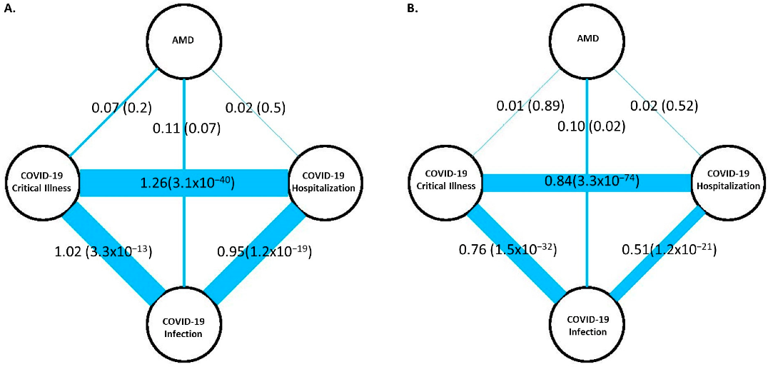
3.1. Genetic Correlation of AMD with COVID-19 Outcomes

LDSC analysis showed that the three COVID-19 outcomes are perfectly genetically correlated (rGs = 1.0, p < 3.3 × 10⁻¹³), indicating that the same genetic factors largely influence these traits. However, the correlations estimated using GNOVA were somewhat lower (0.51 < rGs < 0.84, p < 1.2 × 10⁻²¹). There was a modest genetic correlation between AMD and COVID-19 infection rate (LDSC rG = 0.11, p = 0.07; GNOVA rG = 0.10, p = 0.02; Figure 1) but not with critical illness and hospitalizations, perhaps because the sample size of COVID-19 infections was at least four times larger than the sample of those with severe illness and hospitalization.

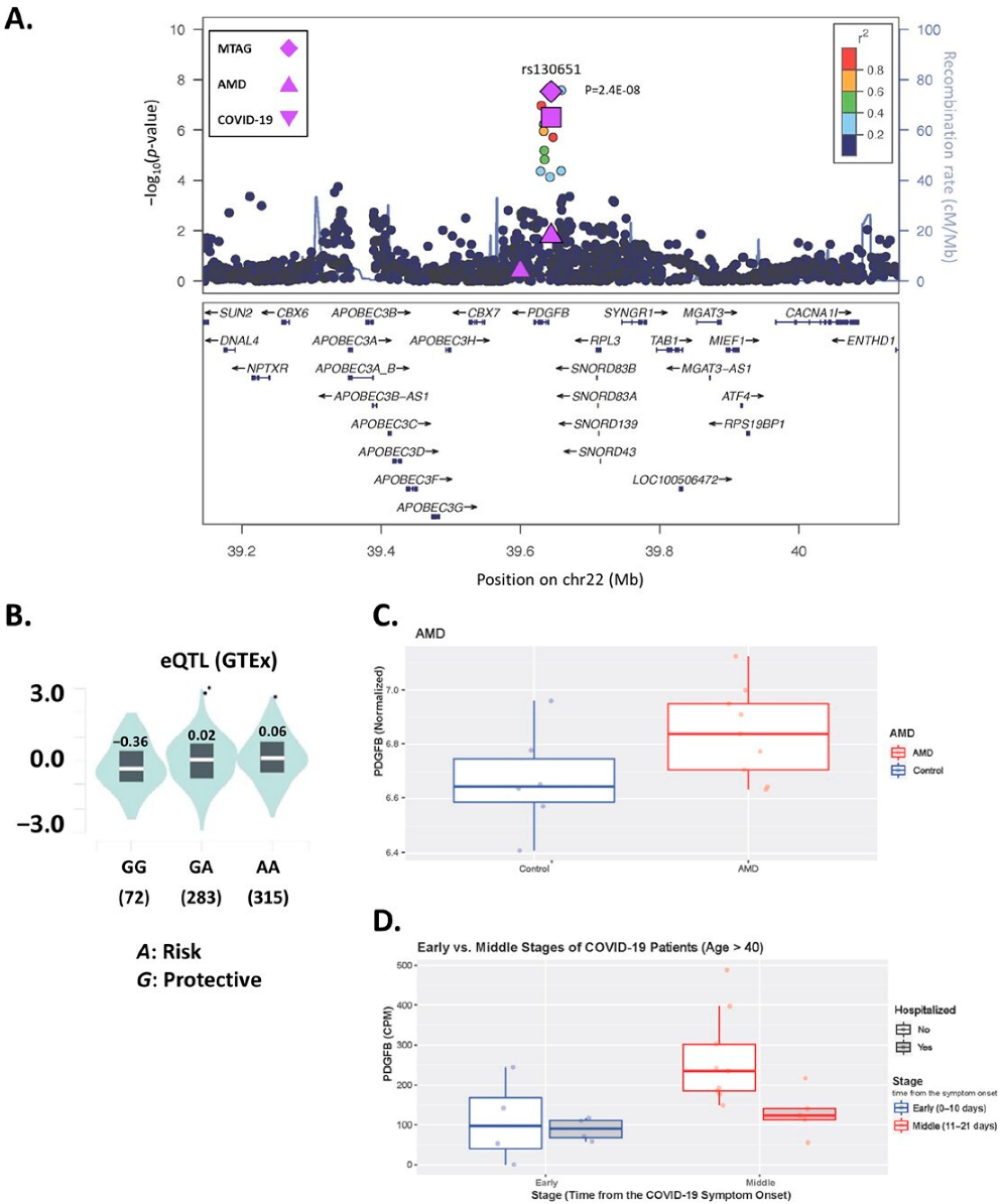
3.2. Multi-Trait Analysis of GWAS for AMD and COVID-19

There was modest evidence for genomic inflations with each of the three COVID-19 outcomes in the MTAG results for AMD (Figure S1; 1.07 < λ ≤ 1.08). Genome-wide significant (GWS; p < 5.0 × 10⁻⁸) associations were observed at 60 loci in the MTAG results for AMD with the three COVID-19 outcomes (Figure S2; Table S6). Notably, 49 of these loci were previously identified as associated with AMD [9] and 10 other loci were previously associated with one of the COVID-19 outcomes [10]. In addition to these previously identified loci, we identified GWS association with two SNPs located 3.2 kb pairs upstream of a novel locus, PDGFB (rs130651, MTAG p = 2.44 × 10⁻⁸ and rs4820371, MTAG p = 2.60 × 10⁻⁸; Table 1; Figure 2A). Both SNPs showed evidence suggestive of association with AMD when analyzed separately (rs130651, p = 1.36 × 10⁻⁷ and rs4820371, p = 1.71 × 10⁻⁷) and were marginally associated with COVID-19 infection (rs130651, p = 0.024 and rs4820371, p = 0.003) (Table 1). In addition, there was GWS evidence of pleiotropy with both SNPs when combining the GWAS results for AMD with those for critical illness (rs130651, p = 3.5 × 10⁻⁸ and rs4820371, p = 6.1 × 10⁻⁸) and hospitalization (rs130651, p = 4.4 × 10⁻⁸ and rs4820371, p = 5.7 × 10⁻⁸), but neither SNP was associated with critical illness (p > 0.27) or hospitalization (p > 0.87; Table 1). Gene-based testing did not identify associations with any novel genes after multiple testing correction (p < 5.88 × 10⁻⁶; Figures S3–S6).

3.3. PDGFB SNP rs130651 Regulates PDGFB Expression

Examination of the association of the two GWS SNPs identified in the MTAG analysis (rs130651 and rs4820371) with expression in blood of 27 genes located within 500 kb of these variants revealed that rs30651 is a study-wide significant cis-eQTL for PDGFB in blood in the GTEx database (p = 1.8 × 10⁻¹¹; Figure 2B) and in data from Lepik et al. (p = 8.15 × 10⁻²¹) and Schmeidel et al. (p = 3.8 × 10⁻⁹) reported in the FIVEx database (Table 2). Specifically, the disease risk rs130651-A allele was significantly associated with increased expression of PDGFB. Rs4820371 was significantly associated with expression of PDGFB in blood, but only in the Lepik et al. study (p = 5.78 × 10⁻⁸). Rs130651 was also significantly associated with PDGFB expression in several immune cell types, most notably T-cells (p = 7.1 × 10⁻²⁰; Table 2; Figure S7). The evidence supporting either SNP as a cis-eQTL with nearby gene CACNA1I was markedly weaker (Table S7). Colocalization analysis of 1761 SNPs in the PDGFB locus demonstrated that the genetic association signal at PDGFB colocalized with the PDGFB eQTL signals in blood (PP = 93.8%).
According to the mQTL GoDMC database, the disease risk alleles of rs130651(A) and rs4820371 (T) are significantly associated with decreased DNA methylation levels of CpG sites located 70–145 kb upstream of PDGFB (Table S8). The most significant cis-mQTL association was observed between rs130651 and cg11247378, which is located 143.9 kb from PDGFB (β = −0.18; p = 9.74 × 10−64).

3.4. PDGFB Is Differentially Expressed in AMD and COVID-19

PDGFB expression in human RPE/choroid tissue was slightly higher in nine AMD cases compared to six controls, although the association was not statistically significant {fold change (FC) = 1.02, p = 0.065; Figure 2C}. There was no difference in PDGFB expression in healthy controls and the three COVID-19 stages (ANOVA p-value = 0.85; Figure S8). However, PDGFB expression was significantly higher among COVID-19 patients in the middle stage compared to those in the early stage who were older than age 40 (FC = 2.17, p = 0.03; Figure 2D) or older than age 50 (FC = 2.15, p = 0.04), but not in the total group of COVID-19 patients in these stages (FC = 0.99, p = 0.61; Table S9 and Figure S9) or among early-stage patients compared to the middle- and late-stage patients combined (p > 0.10; Table S9).

3.5. Pathway Analysis

Pathway analysis including genes located at or near top-ranked SNPs in any of the MTAG analyses revealed four significant (p < 8.45 × 10⁻⁶) GO categories, including high density lipoprotein particle remodeling (p = 1.71 × 10⁻⁸), reverse cholesterol transport (p = 1.53 × 10⁻⁶), humoral immune response mediated by circulating immunoglobulin (p = 4.31 × 10⁻⁶), and extracellular structure organization (p = 3.53 × 10⁻⁶), which includes PDGFB (Table S10).

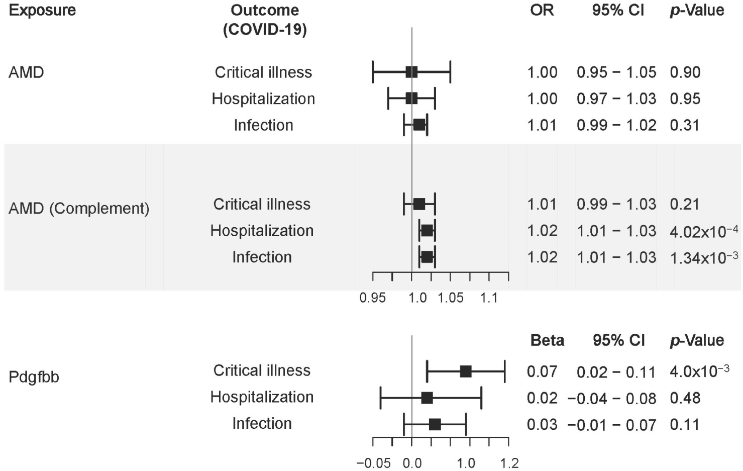
3.6. Mendelian Randomization

No significant causal relationships between the AMD genome-wide risk and any of the COVID-19 outcomes were observed (p > 0.31; Figure 3). However, AMD likely arising from genetic predisposition to dysfunction in the complement system was significantly associated with increased risk for COVID-19 hospitalization (OR = 1.02 [1.01–1.03], p = 4.02 × 10⁻⁴) and infection {OR = 1.02 (1.01–1.03), p = 1.34 × 10⁻³}. In addition, the serum PDGF-BB concentration was significantly associated with increased risk of COVID-19 critical illness {β = 0.07 (0.02–0.11), p = 4.00 × 10⁻³}. Sensitivity analysis showed that none of these results were confounded by directional pleiotropy (Table S11).

4. Discussion

Recent studies have shown an increased risk of infection and mortality from COVID-19 among AMD patients [4,5,6]. We hypothesized that biological mechanisms shared between the two diseases exist and attempted to identify underlying genetic factors shared between them using data from GWAS for AMD and three outcomes related to COVID-19. Analysis of these data using a pleiotropy model revealed one novel genome-wide significant association in the PDGFB locus for AMD and COVID-19 infection.
Considering variation across the genome as a whole, we observed a modest genetic correlation between AMD and COVID-19 infection, which indicates that some genetic factors contribute to risk for both diseases. We did not observe a strong genetic correlation of AMD with COVID-19 critical illness and hospitalization. However, since all three COVID-19 outcomes share strongly genetically correlated influence, it is likely that the sevenfold smaller sample for critical illness and fourfold smaller sample for hospitalization compared to infection rate limited our ability to detect significant genetic correlations between AMD and these two COVID-19 outcomes. Results from MR analysis including only SNPs in the genes for the complement system suggest a causal relationship between complement pathway dysfunction in AMD and subsequent infection by and hospitalization due to COVID-19. In contrast, considering all genes associated with AMD, there was no evidence for causality of AMD on COVID-19 outcomes. Taken together, these MR findings may explain the modest genetic correlations between the two diseases when comparing entire genomes, yet they reinforce the clinical observation of higher risk for infection by and severe illness from COVID-19 among AMD patients [5,6].
We evaluated various omics data to identify possible functional links between PDGFB and AMD and COVID-19 outcomes. The top-ranked SNP from the pleiotropy analysis of AMD and COVID-19 infection risk, rs130651, is a significant cis-acting eQTL and mQTL, in which the disease risk allele A is associated with higher PDGFB expression and lower DNA methylation levels in the PDGFB promoter region. In addition, colocalization analysis confirmed that, among the 28 genes located under the association peak, PDGFB is most likely to be functionally linked to AMD and COVID-19 infection. Taken together, the findings from our pleiotropy and expression/methylation QTL analyses suggest that increased PDGFB expression is associated with increased risk for AMD and COVID-19 infection and severity.
Our DGE analysis of the AMD sample showed that PDGFB tended to be more highly expressed in AMD cases compared to controls in human RPE tissue, although the result was not statistically significant (p = 0.067), perhaps due to the small sample size. However, a recent snRNA-Seq expression study in human retina/RPE tissues showed that PDGFB expression was significantly higher in AMD cases compared to controls, particularly in myeloid immune cells (FC = 6.12, p = 4.89 × 10⁻⁹) [28]. Myeloid cells, together with RPE and glia, have been considered to play important roles in the etiology of AMD [28]. Our expression findings are also consistent with a previous study showing that PDGFB expression in blood was significantly higher in neovascular (wet type) AMD patients than in AMD-free controls, and significantly correlated with the expression of vascular endothelial growth factor (VEGF) [29].
Our finding of significantly higher PDGFB expression among COVID-19 patients ages 40 years and older in the middle stage (11–21 days after the symptom onset) compared to the early stage (0–10 days) is consistent with the observation that COVID-19 symptoms peak 10 days after symptom onset [30]. Although we did not observe significantly different PDGFB expression between COVID-19 patients and COVID-19-free controls, recent studies showed significantly higher serum PDGF-BB concentration in COVID-19 patients compared to the controls (Table S12) [30,31]. Furthermore, Liu et al. found that increased serum PDGF-BB concentrations are significantly associated with increased viral load (p = 0.002) and lung injury (p = 0.001) in COVID-19 patients [32]. These collective findings suggest that lowering PDGFB expression and serum PDGF-BB concentration may reduce the severity of COVID-19, particularly among older people.
PDGFB encodes a member of the protein family comprised of both platelet-derived growth factors (PDGF). PDGF proteins bind and activate PDGC-receptor tyrosine kinases, which have a role in a wide range of developmental processes. PDGFB is involved in the recruitment of pericytes to newly formed blood vessels in the course of physiological angiogenesis during retinal development, as well as in the context of pathological neovascularization that occurs in diabetic retinopathy and AMD [33]. In addition, blocking PDGF signaling is thought to inhibit the pericyte–endothelial process in neovascularization [34]. Therapeutic strategies combining anti-VEGF therapy with antagonists for blocking PDGF signaling have been considered even more effective than the single VEGF treatment and are currently under investigation (e.g., phase III trials—https://clinicaltrials.gov/ identifiers: NCT01944839, NCT01940900, and NCT01940887) [33,35].
A growing body of evidence suggests that the deleterious impact of the exaggerated immune response in COVID-19 patients is a consequence of the SARS-CoV-2 virus activating and exploiting multiple complement cascades, which drive inflammation and thrombosis and result in severe clinical outcomes and poor prognosis [36,37,38]. Interestingly, the Beaver Dam Eye Study [39] and a retrospective cohort study of data in the National Health Insurance Database of Taiwan [40] discovered that subjects with signs and symptoms of obstructive pulmonary diseases such as emphysema and chronic obstructive pulmonary disease had a higher incidence of AMD, presumably due to inflammatory dysregulation. Several AMD studies have identified genetic variants associated with complement dysfunction and its impact on inflammation, oxidative stress, and metabolism, and these pathways have been found to be significantly upregulated and dysfunctional in COVID-19 infection as well [24,25,38,41,42,43,44].
Our study has limitations. First, the patients in the COVID-19 infection cohort were based on self-reporting or confirmed patients at clinics, which indicates that asymptomatic COVID-19 patients may not be included. This may have contributed to the underpowering of the analysis, further confirmed by the imbalance in sample size between AMD and COVID-19 outcomes for critical illness and hospitalization. Second, the unscreened population controls may have included patients with both asymptomatic and symptomatic disease who were unable to be tested or obtain care during the height of the pandemic. A third concern is the lack of genetic correlation between AMD and the COVID-19 outcomes of critical illness and hospitalization. This may be explained in part by the ascertainment of the AMD and COVID-19 GWAS cohorts included in this study. The AMD sample was comprised primarily of older individuals (mean age = 73.7 ± 9.4 years) whereas the COVID-19 sample included persons across a wide age spectrum (mean age ranges from 36 different datasets = 35.6–72.3 years). Genetic correlation in the overall sample would be weakened if it is age-dependent. Furthermore, a study of a large Korean cohort (N = 135,435) showed that only the exudative (wet) form of AMD was significantly associated with severe COVID-19 illness and COVID-19 infection [6]. In addition, Allegrini et al. found a significant loss in visual function among the exudative AMD patients who experienced COVID-19 infection compared to AMD patients before the pandemic [45]. Unfortunately, we were unable to evaluate genetic correlations between COVID-19 outcomes and exudative and non-exudative AMD separately because our analyses were conducted using AMD GWAS summary-level data. Moreover, because the distribution of AMD subtypes in the GWAS dataset is highly imbalanced (67.4% exudative, 19.7% geographic atrophy, 12.9% with each form in one eye), separate analyses for each AMD subtype would not be meaningful. A balanced age distribution between the AMD and COVID-19 cohorts and AMD subtypes might have resulted in a different estimate of the genetic correlation between the two diseases. Although age differences between the individuals in the AMD and COVID-19 datasets may have influenced the genetic correlation between the two disorders, age disparity does not impact the results of pleiotropy analyses, which are conducted separately within datasets for each disease and subsequently pooled by meta-analysis. This conclusion is supported by pleiotropy studies for diseases with very distinct onset age distributions, such as childhood obesity and adult cardiometabolic diseases [46] and autism spectrum disorder and schizophrenia [47]. Finally, our study was conducted using GWAS and gene expression datasets obtained from European ancestry samples, which limits the generalizability of our results to other ancestral groups. Future studies in other populations are needed to replicate and extend our findings.
In summary, our findings add to the body of evidence for an increased risk of infection and mortality from COVID-19 among AMD patients. Our analysis lends credence to previously reported clinical studies that found those with AMD are an at-risk group for COVID-19 infection and severe disease, and that this increased risk may have a genetic basis. The discovery of shared genetic risk factors via GWAS will require a larger sample size for critical illness and hospitalizations to better understand the shared pathology and risk factors that contribute to worsening clinical outcomes in both disease states.

Supplementary Materials

The following are available online at https://www.mdpi.com/article/10.3390/jcm12010109/s1, Table S1: Publicly available summary-level genetic association data used in this study, Table S2: Characteristics of subjects in the gene expression dataset derived from 96 peripheral blood samples obtained from 32 COVID-19 patients and 8 controls at multiple time points, corresponding to different disease stages measured from symptom onset, Table S3: Characteristics of subjects (9 AMD cases and 6 age-matched healthy controls) in the gene expression dataset derived from human retinal pigment epithelium/choroid tissues, Table S4: Tests of causal inference of AMD-associated genetic variants (instrumental variables) with AMD (exposure) and COVID-19 outcomes (critical illness, hospitalization, and infection), Table S5: Tests of causal inference of variants associated with Pdgfbb level (instrument variables) with human cytokine Pdgfbb level (exposure) and COVID-19 outcomes, Table S6: Summary statistics of genome-wide significant (p < 5.0 × 10⁻⁸) associations in the MTAG analysis of AMD and COVID-19 outcomes (critical illness, hospitalization, and infection), Table S7: eQTL associations of rs130651 and rs482037 with CACNA1I in whole blood in the GTEx portal database, Table S8: cis-mQTL associations of PDGFB SNPs rs130651 and rs4820371 with CpGs located upstream of PDGFB (chr22:39619364-39640987) in the GoDMC database (http://www.godmc.org.uk), Table S9: Differential expression of PDGFB in blood from COVID-19 patients in three different stages measured from symptom onset (early ≤ 10 days, middle 11–20 days, late ≥ 21 days), Table S10: Gene ontology (GO) pathways based on MTAG results from GWAS of AMD and COVID-19 infection, Table S11: Mendelian randomization analysis of AMD and Pdgf-bb as exposures and COVID-19 traits (critical illness, hospitalization, and infection) as outcomes, Table S12: Association of human cytokine Pdgfbb level with COVID-19 status. The positive effect direction indicates that the mean cytokine Pdgfbb level was higher in cases compared to controls, Figure S1: QQ plots of the MTAG results for AMD with COVID-19 outcomes: A. critical illness, B. hospitalization, and C. infection, Figure S2: Manhattan plots of the MTAG results for AMD with COVID-19 outcomes: A. critical illness, B. hospitalization, and C. infection, Figure S3: Manhattan plots of S-Predixcan results in blood using the MTAG results for AMD with COVID-19 outcomes. A. critical illness, B. hospitalization, and C. infection, Figure S4: Manhattan plots of S-Predixcan results in A. blood, B. lung, C. aorta and D. coronary artery using the MTAG results for AMD with COVID-19 critical illness, Figure S5: Manhattan plots of S-Predixcan results in A. bloods, B. lung, C. aorta and D. coronary artery using the MTAG results for AMD with COVID-19 hospitalization, Figure S6: Manhattan plots of S-Predixcan results in A. bloods, B. lung, C. aorta (artery) and D. coronary (artery) using the MTAG results for AMD with the COVID-19 infection, Figure S7: eQTL associations for PDFGB SNPs rs130651 (A) and rs4820371(B) with genes located within 500 kilobases, Figure S8: PDGFB expression in COVID-19-free controls and COVID-19 patients according to time after symptom onset, Figure S9: PDGFB expression in COVID-19 patients by age and time after symptom onset.

Author Contributions

Conceptualization, J.C., L.A.F. and M.L.S.; methodology, J.C. and L.A.F.; formal analysis, J.C., X.S. and X.H.; investigation, J.C.; resources, M.M.D. and L.A.F.; data curation, J.C. and X.S.; writing—original draft preparation, J.C.; writing—review and editing, J.C., G.T.O., X.C., M.M.D., L.A.F. and M.L.S.; visualization, J.C., X.S. and X.H.; supervision, L.A.F. and M.L.S.; project administration, J.C., V.V., X.S. and X.H.; funding acquisition, L.A.F. All authors have read and agreed to the published version of the manuscript.

Funding

J.C., X.H. and L.A.F. were supported in this work by National Institutes of Health grants RF1 AG057519, R01 AG069453, R01 AG048927, U19 AG068753, and U01 AG062602.

Institutional Review Board Statement

This study was conducted in accordance with the Declaration of Helsinki and approved by the Boston University Medical Center Institutional Review Board (H-29124).

Informed Consent Statement

Informed consent was obtained from all subjects involved in this study.

Data Availability Statement

All data generated or analyzed in this study are included in this published article.

Conflicts of Interest

The authors declare no conflict of interest.

References

  1. Carter, P.; Anderson, M.; Mossialos, E. Health system, public health, and economic implications of managing COVID-19 from a cardiovascular perspective. Eur. Heart J. 2020, 41, 2516–2518. [Google Scholar] [CrossRef] [Green Version]
  2. Zhou, Y.; Yang, Q.; Chi, J.; Dong, B.; Lv, W.; Shen, L.; Wang, Y. Comorbidities and the risk of severe or fatal outcomes associated with coronavirus disease 2019: A systematic review and meta-analysis. Int. J. Infect. Dis. 2020, 99, 47–56. [Google Scholar] [CrossRef] [PubMed]
  3. The Lancet. Neurology, The neurological impact of COVID-19. Lancet Neurol 2020, 19, 471. [Google Scholar] [CrossRef] [PubMed]
  4. Leung, E.H.; Fan, J.; Flynn, H.W., Jr.; Albini, T.A. Ocular and Systemic Complications of COVID-19: Impact on Patients and Healthcare. Clin. Ophthalmol. 2022, 16, 1–13. [Google Scholar] [CrossRef] [PubMed]
  5. Ramlall, V.; Thangaraj, P.M.; Meydan, C.; Foox, J.; Butler, D.; Kim, J.; May, B.; De Freitas, J.K.; Glicksberg, B.S.; Mason, C.E.; et al. Immune complement and coagulation dysfunction in adverse outcomes of SARS-CoV-2 infection. Nat. Med. 2020, 26, 1609–1615. [Google Scholar] [CrossRef]
  6. Yang, J.M.; Moon, S.Y.; Lee, J.Y.; Agalliu, D.; Yon, D.K.; Lee, S.W. COVID-19 Morbidity and Severity in Patients with Age-Related Macular Degeneration: A Korean Nationwide Cohort Study. Am. J. Ophthalmol. 2021, 239, 159–169. [Google Scholar] [CrossRef]
  7. Smith, W.; Assink, J.; Klein, R.; Mitchell, P.; Klaver, C.C.; Klein, B.E.; Hofman, A.; Jensen, S.; Wang, J.J.; de Jong, P.T. Risk factors for age-related macular degeneration: Pooled findings from three continents. Ophthalmology 2001, 108, 697–704. [Google Scholar] [CrossRef]
  8. Chakravarthy, U.; Wong, T.Y.; Fletcher, A.; Piault, E.; Evans, C.; Zlateva, G.; Buggage, R.; Pleil, A.; Mitchell, P. Clinical risk factors for age-related macular degeneration: A systematic review and meta-analysis. BMC Ophthalmol. 2010, 10, 31. [Google Scholar] [CrossRef] [Green Version]
  9. Fritsche, L.G.; Igl, W.; Bailey, J.N.; Grassmann, F.; Sengupta, S.; Bragg-Gresham, J.L.; Burdon, K.P.; Hebbring, S.J.; Wen, C.; Gorski, M.; et al. A large genome-wide association study of age-related macular degeneration highlights contributions of rare and common variants. Nat. Genet. 2016, 48, 134–143. [Google Scholar] [CrossRef] [Green Version]
  10. COVID-19 Host Genetics Initiative. Mapping the human genetic architecture of COVID-19. Nature 2021, 600, 472–477. [Google Scholar] [CrossRef]
  11. Bulik-Sullivan, B.; Finucane, H.K.; Anttila, V.; Gusev, A.; Day, F.R.; Loh, P.R.; ReproGen Consortium; Psychiatric Genomics Consortium; Genetic Consortium for Anorexia Nervosa of the Wellcome Trust Case Control Consortium 3; Duncan, L.; et al. An atlas of genetic correlations across human diseases and traits. Nat. Genet. 2015, 47, 1236–1241. [Google Scholar] [CrossRef] [Green Version]
  12. Lu, Q.; Li, B.; Ou, D.; Erlendsdottir, M.; Powles, R.L.; Jiang, T.; Hu, Y.; Chang, D.; Jin, C.; Dai, W.; et al. A Powerful Approach to Estimating Annotation-Stratified Genetic Covariance via GWAS Summary Statistics. Am. J. Hum. Genet. 2017, 101, 939–964. [Google Scholar] [CrossRef] [Green Version]
  13. Turley, P.; Walters, R.K.; Maghzian, O.; Okbay, A.; Lee, J.J.; Fontana, M.A.; Nguyen-Viet, T.A.; Wedow, R.; Zacher, M.; Furlotte, N.A.; et al. Multi-trait analysis of genome-wide association summary statistics using MTAG. Nat. Genet. 2018, 50, 229–237. [Google Scholar] [CrossRef]
  14. Chung, J.; Zhang, X.; Allen, M.; Wang, X.; Ma, Y.; Beecham, G.; Montine, T.J.; Younkin, S.G.; Dickson, D.W.; Golde, T.E.; et al. Genome-wide pleiotropy analysis of neuropathological traits related to Alzheimer’s disease. Alzheimer’s Res. Ther. 2018, 10, 22. [Google Scholar] [CrossRef]
  15. Barbeira, A.N.; Dickinson, S.P.; Bonazzola, R.; Zheng, J.; Wheeler, H.E.; Torres, J.M.; Torstenson, E.S.; Shah, K.P.; Garcia, T.; Edwards, T.L.; et al. Exploring the phenotypic consequences of tissue specific gene expression variation inferred from GWAS summary statistics. Nat. Commun. 2018, 9, 1825. [Google Scholar] [CrossRef]
  16. Consortium, G.T. The Genotype-Tissue Expression (GTEx) project. Nat Genet 2013, 45, 580–585. [Google Scholar] [CrossRef]
  17. Kwong, A.; Boughton, A.P.; Wang, M.; VandeHaar, P.; Boehnke, M.; Abecasis, G.; Kang, H.M. FIVEx: An interactive eQTL browser across public datasets. Bioinformatics 2021, 38, 559–561. [Google Scholar] [CrossRef]
  18. Giambartolomei, C.; Vukcevic, D.; Schadt, E.E.; Franke, L.; Hingorani, A.D.; Wallace, C.; Plagnol, V. Bayesian test for colocalisation between pairs of genetic association studies using summary statistics. PLoS Genet. 2014, 10, e1004383. [Google Scholar] [CrossRef] [Green Version]
  19. Bonilla, C.; Bertoni, B.; Min, J.L.; Hemani, G.; Genetics of DNA Methylation Consortium; Elliott, H.R. Investigating DNA methylation as a potential mediator between pigmentation genes, pigmentary traits and skin cancer. Pigment Cell Melanoma Res. 2021, 34, 892–904. [Google Scholar] [CrossRef]
  20. Whitmore, S.S.; Braun, T.A.; Skeie, J.M.; Haas, C.M.; Sohn, E.H.; Stone, E.M.; Scheetz, T.E.; Mullins, R.F. Altered gene expression in dry age-related macular degeneration suggests early loss of choroidal endothelial cells. Mol. Vis. 2013, 19, 2274–2297. [Google Scholar]
  21. McClain, M.T.; Constantine, F.J.; Henao, R.; Liu, Y.; Tsalik, E.L.; Burke, T.W.; Steinbrink, J.M.; Petzold, E.; Nicholson, B.P.; Rolfe, R.; et al. Dysregulated transcriptional responses to SARS-CoV-2 in the periphery. Nat. Commun. 2021, 12, 1079. [Google Scholar] [CrossRef] [PubMed]
  22. Garcia-Layana, A.; Cabrera-Lopez, F.; Garcia-Arumi, J.; Arias-Barquet, L.; Ruiz-Moreno, J.M. Early and intermediate age-related macular degeneration: Update and clinical review. Clin. Interv. Aging 2017, 12, 1579–1587. [Google Scholar] [CrossRef] [PubMed] [Green Version]
  23. Watanabe, K.; Taskesen, E.; van Bochoven, A.; Posthuma, D. Functional mapping and annotation of genetic associations with FUMA. Nat. Commun. 2017, 8, 1826. [Google Scholar] [CrossRef] [Green Version]
  24. Johnson, L.V.; Leitner, W.P.; Staples, M.K.; Anderson, D.H. Complement activation and inflammatory processes in Drusen formation and age related macular degeneration. Exp. Eye Res. 2001, 73, 887–896. [Google Scholar] [CrossRef] [PubMed]
  25. Hageman, G.S.; Luthert, P.J.; Victor Chong, N.H.; Johnson, L.V.; Anderson, D.H.; Mullins, R.F. An integrated hypothesis that considers drusen as biomarkers of immune-mediated processes at the RPE-Bruch’s membrane interface in aging and age-related macular degeneration. Prog. Retin. Eye Res. 2001, 20, 705–732. [Google Scholar] [CrossRef]
  26. Ahola-Olli, A.V.; Wurtz, P.; Havulinna, A.S.; Aalto, K.; Pitkanen, N.; Lehtimaki, T.; Kahonen, M.; Lyytikainen, L.P.; Raitoharju, E.; Seppala, I.; et al. Genome-wide Association Study Identifies 27 Loci Influencing Concentrations of Circulating Cytokines and Growth Factors. Am. J. Hum. Genet. 2017, 100, 40–50. [Google Scholar] [CrossRef] [Green Version]
  27. Yavorska, O.O.; Burgess, S. MendelianRandomization: An R package for performing Mendelian randomization analyses using summarized data. Int. J. Epidemiol. 2017, 46, 1734–1739. [Google Scholar] [CrossRef] [Green Version]
  28. Orozco, L.D.; Chen, H.H.; Cox, C.; Katschke, K.J., Jr.; Arceo, R.; Espiritu, C.; Caplazi, P.; Nghiem, S.S.; Chen, Y.J.; Modrusan, Z.; et al. Integration of eQTL and a Single-Cell Atlas in the Human Eye Identifies Causal Genes for Age-Related Macular Degeneration. Cell Rep. 2020, 30, 1246–1259. [Google Scholar] [CrossRef] [Green Version]
  29. Zehetner, C.; Kirchmair, R.; Neururer, S.B.; Kralinger, M.T.; Bechrakis, N.E.; Kieselbach, G.F. Systemic upregulation of PDGF-B in patients with neovascular AMD. Investig. Ophthalmol. Vis. Sci. 2014, 55, 337–344. [Google Scholar] [CrossRef] [Green Version]
  30. Huang, C.; Wang, Y.; Li, X.; Ren, L.; Zhao, J.; Hu, Y.; Zhang, L.; Fan, G.; Xu, J.; Gu, X.; et al. Clinical features of patients infected with 2019 novel coronavirus in Wuhan, China. Lancet 2020, 395, 497–506. [Google Scholar] [CrossRef] [Green Version]
  31. Petrey, A.C.; Qeadan, F.; Middleton, E.A.; Pinchuk, I.V.; Campbell, R.A.; Beswick, E.J. Cytokine release syndrome in COVID-19: Innate immune, vascular, and platelet pathogenic factors differ in severity of disease and sex. J. Leukoc. Biol. 2021, 109, 55–66. [Google Scholar] [CrossRef] [PubMed]
  32. Liu, Y.; Zhang, C.; Huang, F.; Yang, Y.; Wang, F.; Yuan, J.; Zhang, Z.; Qin, Y.; Li, X.; Zhao, D.; et al. Elevated plasma levels of selective cytokines in COVID-19 patients reflect viral load and lung injury. Natl. Sci. Rev. 2020, 7, 1003–1011. [Google Scholar] [CrossRef] [PubMed] [Green Version]
  33. Diaz-Lezama, N.; Wolf, A.; Koch, S.; Pfaller, A.M.; Biber, J.; Guillonneau, X.; Langmann, T.; Grosche, A. PDGF Receptor Alpha Signaling Is Key for Muller Cell Homeostasis Functions. Int. J. Mol. Sci. 2021, 22, 1174. [Google Scholar] [CrossRef] [PubMed]
  34. Jaffe, G.J.; Ciulla, T.A.; Ciardella, A.P.; Devin, F.; Dugel, P.U.; Eandi, C.M.; Masonson, H.; Mones, J.; Pearlman, J.A.; Quaranta-El Maftouhi, M.; et al. Dual Antagonism of PDGF and VEGF in Neovascular Age-Related Macular Degeneration: A Phase IIb, Multicenter, Randomized Controlled Trial. Ophthalmology 2017, 124, 224–234. [Google Scholar] [CrossRef] [PubMed] [Green Version]
  35. Chen, D.B.; Zheng, J. Regulation of placental angiogenesis. Microcirculation 2014, 21, 15–25. [Google Scholar] [CrossRef] [Green Version]
  36. Skendros, P.; Mitsios, A.; Chrysanthopoulou, A.; Mastellos, D.C.; Metallidis, S.; Rafailidis, P.; Ntinopoulou, M.; Sertaridou, E.; Tsironidou, V.; Tsigalou, C.; et al. Complement and tissue factor-enriched neutrophil extracellular traps are key drivers in COVID-19 immunothrombosis. J. Clin. Investig. 2020, 130, 6151–6157. [Google Scholar] [CrossRef]
  37. Lo, M.W.; Kemper, C.; Woodruff, T.M. COVID-19: Complement, Coagulation, and Collateral Damage. J. Immunol. 2020, 205, 1488–1495. [Google Scholar] [CrossRef]
  38. Java, A.; Apicelli, A.J.; Liszewski, M.K.; Coler-Reilly, A.; Atkinson, J.P.; Kim, A.H.; Kulkarni, H.S. The complement system in COVID-19: Friend and foe? JCI Insight 2020, 5, e140711. [Google Scholar] [CrossRef]
  39. Klein, R.; Moss, S.E.; Meuer, S.M.; Klein, B.E. The 15-year cumulative incidence of retinal vein occlusion: The Beaver Dam Eye Study. Arch. Ophthalmol. 2008, 126, 513–518. [Google Scholar] [CrossRef] [Green Version]
  40. Bair, P.J.; Hsia, N.Y.; Lin, C.L.; Yang, Y.C.; Shen, T.C.; Li, C.Y. Population-based retrospective cohort study on risk of age-related macular degeneration in people with chronic obstructive pulmonary disease. Sci. Rep. 2021, 11, 15079. [Google Scholar] [CrossRef]
  41. Armento, A.; Ueffing, M.; Clark, S.J. The complement system in age-related macular degeneration. Cell. Mol. Life Sci. 2021, 78, 4487–4505. [Google Scholar] [CrossRef]
  42. Holter, J.C.; Pischke, S.E.; de Boer, E.; Lind, A.; Jenum, S.; Holten, A.R.; Tonby, K.; Barratt-Due, A.; Sokolova, M.; Schjalm, C.; et al. Systemic complement activation is associated with respiratory failure in COVID-19 hospitalized patients. Proc. Natl. Acad. Sci. USA 2020, 117, 25018–25025. [Google Scholar] [CrossRef]
  43. Geerlings, M.J.; de Jong, E.K.; den Hollander, A.I. The complement system in age-related macular degeneration: A review of rare genetic variants and implications for personalized treatment. Mol. Immunol. 2017, 84, 65–76. [Google Scholar] [CrossRef]
  44. Lakshmanan, M.; Schaffer, A.; Robbins, J.; Reynolds, J.; Norton, J. A simplified low iodine diet in I-131 scanning and therapy of thyroid cancer. Clin. Nucl. Med. 1988, 13, 866–868. [Google Scholar] [CrossRef]
  45. Allegrini, D.; Raimondi, R.; Montesano, G.; Borgia, A.; Sorrentino, T.; Tsoutsanis, P.; Romano, M.R. Short-Term Outcomes After COVID-19-Related Treatment Interruption Among Patients with Neovascular Age-Related Macular Degeneration Receiving Intravitreal Bevacizumab. Clin. Ophthalmol. 2021, 15, 4073–4079. [Google Scholar] [CrossRef]
  46. Tekola-Ayele, F.; Lee, A.; Workalemahu, T.; Sanchez-Pozos, K. Shared genetic underpinnings of childhood obesity and adult cardiometabolic diseases. Hum. Genom. 2019, 13, 17. [Google Scholar] [CrossRef] [Green Version]
  47. Vorstman, J.A.; Breetvelt, E.J.; Thode, K.I.; Chow, E.W.; Bassett, A.S. Expression of autism spectrum and schizophrenia in patients with a 22q11.2 deletion. Schizophr. Res. 2013, 143, 55–59. [Google Scholar] [CrossRef]
Figure 1. Genetic correlations between AMD and three COVID-19 outcomes, including critical illness, hospitalization, and infections, which were computed by (A) LDSC and (B) GNOVA. Line width is proportional to genetic correlations between the four phenotypes. The numbers show the genetic correlations and p-values amongst the phenotypes.
Jcm 12 00109 g001
Figure 2. (A) Regional association plot for the genome-wide significant associations in the MTAG analysis of AMD and COVID-19 infections. Rs130651 is the most significant SNP in this locus (MTAG p-value = 2.44 × 10⁻⁸). PDGFB gene expression analyses of (B) expression quantitative trait locus (eQTL) with rs130651 and differential gene expression in samples of (C) AMD cases and controls and (D) COVID-19 stages by time from symptom onset (early ≤ 10 days and middle 11–20 days).
Jcm 12 00109 g002
Figure 3. Forest plot illustrating the Mendelian randomization estimates of genetic liability of AMD risk, liability of AMD risk based on genes in the complement pathway, and human serum PDGF-BB levels (exposures) with COVID-19 phenotypes (outcomes) based on inverse-variance weighted Mendelian randomization.
Jcm 12 00109 g003
Table 1. Genome-wide significant associations in the PDGFB locus identified in the MTAG analysis of AMD with three COVID-19 outcomes (critical illness, hospitalization, and infections). Rs130651 and rs4820371 are in a moderate linkage disequilibrium (LD) in the European population of 1000 genome reference (r2 = 0.36).
rs130651
(EA/NEA = G/A; EAF = 0.32)
rs4820371
(EA/NEA = C/T; EAF = 0.23)
Analytical MethodTraitOR95% CIpOR95% CIp
Single GWASAMD0.900.87–0.941.36 × 10−70.900.86–0.931.71 × 10−7
COVID-19Critical Illness0.960.89–1.040.271.010.93–1.090.78
Hospitalization1.000.96–1.040.871.000.94–1.060.93
Infections0.980.96–1.000.0240.960.94–0.980.003
MTAG of AMD
with COVID-19
COVID-19Critical Illness0.960.94–0.973.5 × 10−80.950.93–0.976.1 × 10−8
Hospitalization0.960.94–0.974.4 × 10−80.950.93–0.975.7 × 10−8
Infections0.960.94–0.972.4 × 10−80.950.93–0.972.6 × 10−8
EA/NEA: Effect allele/Non-effect allele. EAF: Effect allele frequency. OR: odds ratio. CI: confidence interval.
Table 2. Summary statistics of eQTL associations between the two SNPs (rs130651 and rs4820371) and the PDGFB gene expression levels from different tissue and cell types in the GTEx and FIVEx databases.
SchemeStudyTissue/Cell TypeEffectSEp-Value
rs130651 (A)GTExWhole blood0.260.041.8 × 10−11
Lepik et al. 2017Whole blood0.310.0318.5 × 10−21
BLUEPRINT (Immune cells)T cell0.870.0837.1 × 10−20
DICE (Immune cells)Th17 cell0.960.0818.7 × 10−19
Tfh cell0.630.0564.7 × 10−18
T cell1.440.141.2 × 10−15
Th2 cell1.110.117.1 × 10−15
CD4 T cell (naïve)1.130.128.1 × 10−14
Th1-17 cell0.540.075.1 × 10−11
T regulatory cell0.610.0879.3 × 10−10
Th1 cell0.510.0753.4 × 10−9
CD8 T cell (naïve)0.850.136.2 × 10−9
TwinsUKWhole blood0.220.0363.8 × 10−9
rs4820371 (T)GTExWhole blood0.130.043.5 × 10−3
Lepik et al. 2017Whole blood0.200.0355.8 × 10−8
EA: effect allele. SE: standard error. PMID: literature ID in the PubMed database. GTEx: genotype-tissue expression. The eQTL results from Lepick et al. 2017, BLUEPRINT, DICE (Database of Immune Cell Expression), and TwinsUK are provided in the FIVEx database.
Disclaimer/Publisher’s Note: The statements, opinions and data contained in all publications are solely those of the individual author(s) and contributor(s) and not of MDPI and/or the editor(s). MDPI and/or the editor(s) disclaim responsibility for any injury to people or property resulting from any ideas, methods, instructions or products referred to in the content.

