Article

Biological Activity of fac-[Re(CO)3(phen)(aspirin)], fac-[Re(CO)3(phen)(indomethacin)] and Their Original Counterparts against Ishikawa and HEC-1A Endometrial Cancer Cells

Department of Molecular Microbiology, Institute of Microbiology, Biotechnology and Immunology, Faculty of Biology and Environmental Protection, University of Lodz, 12/16 Banacha, 90-237 Lodz, Poland
*
Author to whom correspondence should be addressed.
Int. J. Mol. Sci. 2022, 23(19), 11568; https://doi.org/10.3390/ijms231911568
Submission received: 2 September 2022 / Revised: 21 September 2022 / Accepted: 27 September 2022 / Published: 30 September 2022
(This article belongs to the Special Issue State-of-the-Art Biochemistry in Poland)

Abstract

:
Nonsteroidal anti-inflammatory drugs (NSAIDs) are inhibitors of cyclooxygenase enzyme (COX) and were found to have positive effects in reducing the risk of developing gynecological cancers. However, long-term administration of NSAIDs carries the risk of various side effects, including those in the digestive and circulatory systems. Therefore, there is a constant need to develop new NSAID derivatives. In this work, we investigated rhenium NSAIDs, comparing their effects on endometrial cancer cells with original NSAIDs, demonstrating the high activity of aspirin and indomethacin derivatives. The cytotoxic activity of rhenium derivatives against the Ishikawa and HEC-1A cancer cell lines was higher than that of the original NSAIDs. The IC50 after 24-h incubation of Ishikawa and HEC-1A were 188.06 µM and 394.06 µM for rhenium aspirin and 228.6 µM and 1459.3 µM for rhenium indomethacin, respectively. At the same time, IC50 of aspirin and indomethacin were 10,024.42 µM and 3295.3 µM for Ishikawa, and 27,255.8 µM and 5489.3 µM for HEC-1A, respectively. Moreover, these derivatives were found to inhibit the proliferation of both cell lines in a time- and state-dependent manner. The Ishikawa cell proliferation was strongly inhibited by rhenium aspirin and rhenium indomethacin after 72-h incubation (*** = p < 0.001), while the HEC-1A proliferation was inhibited by the same agents already after 24-h incubation (*** = p < 0.001). Furthermore, the ROS level in the mitochondria of the tested cells generated in the presence of rhenium derivatives was higher than the original NSAIDs. That was associated with rhenium indomethacin exclusively, which had a significant effect (*** = p < 0.001) on both Ishikawa and HEC-1A cancer cells. Rhenium aspirin had a significant effect (*** = p < 0.001) on the mitochondrial ROS level of Ishikawa cells only. Overall, the research revealed a high potential of the rhenium derivatives of aspirin and indomethacin against endometrial cancer cells compared with the original NSAIDs.

1. Introduction

Nonsteroidal anti-inflammatory drugs (NSAIDs) are the most commonly used drugs worldwide. Since their first use in the 1960s, the chemical structure and the principal objective of their usage was modified and changed. Along with their intended application as anti-inflammatory agents, these drugs are currently administered as analgesics, antipyretics, and antiplatelet-aggregation agents [1]. The well-known activity of NSAIDs involves the inhibition of isoforms of the cyclooxygenase (COX) enzyme-COX-1, COX-2, or both, depending on the specific drug. In particular, those NSAIDs capable of inhibiting COX-2 (alone or together with COX-1) are of particular interest in the context of cancer prevention and supportive treatment, as this enzyme has been found to be an inducible isoform associated with inflammation and cancer proliferation [2]. Recently, several research groups have focused on the role of inflammatory processes in the development of gynaecologic cancers [3,4,5], as well as the reduction of the risk of developing these types of cancer as a result of long-term NSAIDs administration. The consumption of aspirin in low doses has already been recommended for the prevention of colorectal cancer and cardiovascular diseases. It has also been suggested to be helpful in the case of lung, breast, and prostate cancers [6,7,8,9]. In addition to the anti-inflammatory effect, the cytostatic activity of NSAIDs was noticed. Chiu et al. [10] reported the inhibition of melanoma cell (A375) proliferation after celecoxib and indomethacin treatment. Marinov et al. [11] indicated the high potential of diclofenac as a cytostatic agent against breast (MCF-7) and colorectal (HT-29) cancer cells but not cervical cancer cells (HeLa). Moreover, studies by Buzharevski et al. [12] performed with carboranyl derivatives of rofecoxib against melanoma (A375, 518A2, B16, B16F10) and colon (HCT116, SW480, SW620, CT25CL26) cancer cells highlighted the potential perspectives of modified NSAIDs as anticancer agents with cytostatic properties. Here, the observed activities of agents imply that the anticancer potential of NSAIDs may not be explained only by their COX-inhibitory activities. Still, the major clinical problem with NSAID consumption in anticancer therapy is gastrointestinal complications. The side effects of NSAIDs (especially those that inhibit COX-1) can lead to gastric/duodenal bleeding and ulceration, so modifications to these commonly used drugs have been necessary. Some of these modifications, such as substitution with nitric oxide (NO), hydrogen sulfide (H2S), lipids, and metal complexation, were shown to give a broad spectrum of biological activities. Furthermore, the complexation of NSAIDs with certain metals, such as copper or zinc, reduced the effective dose of the “native” drug [13,14,15]. Complexes of rhenium with aspirin, naproxen, indomethacin, and ibuprofen, were synthesized by Skiba et al. and subsequently tested by our group on HeLa and L929 cell lines [16]. The key aspect of the antitumor activity of these complexes was not the increased inhibition of the COX enzyme but the broadening of the spectrum of activity of the rhenium derivatives of the NSAIDs tested. Indeed, rhenium-aspirin was found to exhibit higher cytotoxicity against cancerous HeLa cells compared with non-cancerous L929 cells. It was also noted that the mechanism inducing cancer cell death was based on an increase in reactive oxygen species (ROS) levels, which was correlated with a cell cycle arrest in sub-G1 and S phases [16]. Here, we report our findings using an in vitro model of the endometrial cancer cell lines Ishikawa and HEC-1-A. Our studies focused on the cytotoxic effects of rhenium-modified NSAIDs, as well as the effects of metal complementation with these agents.

2. Results

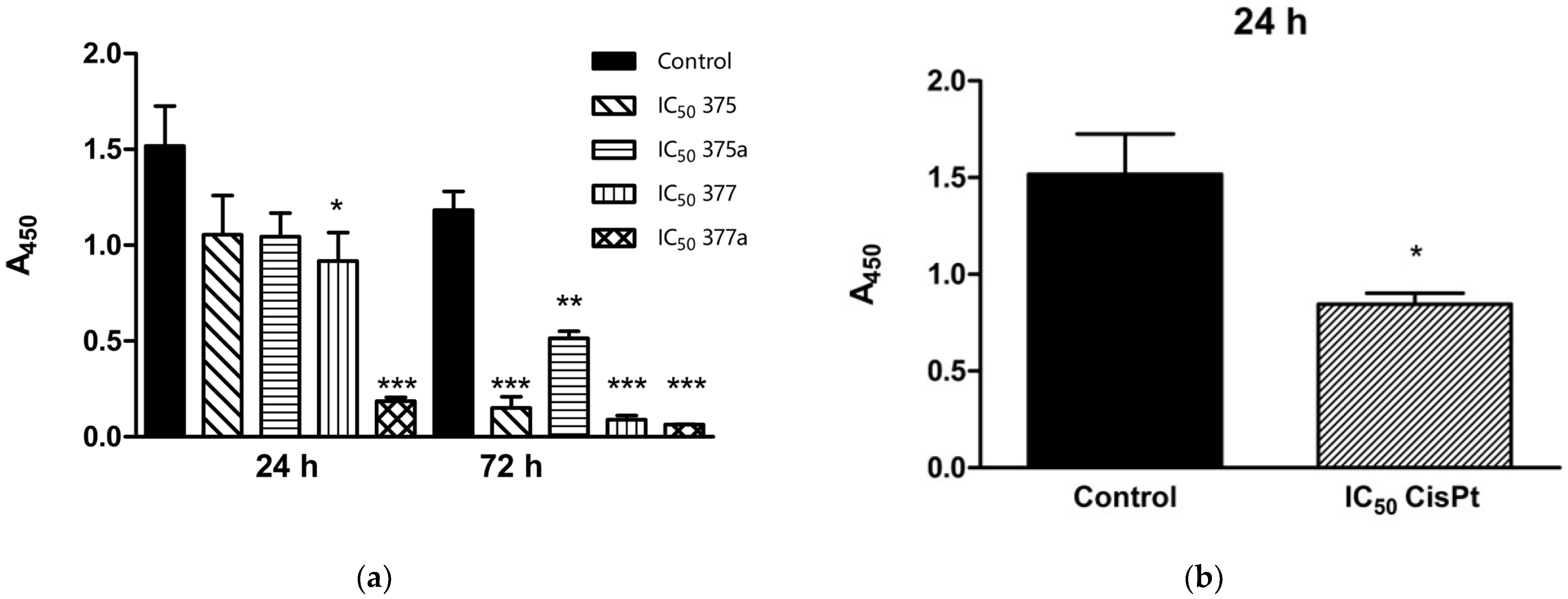
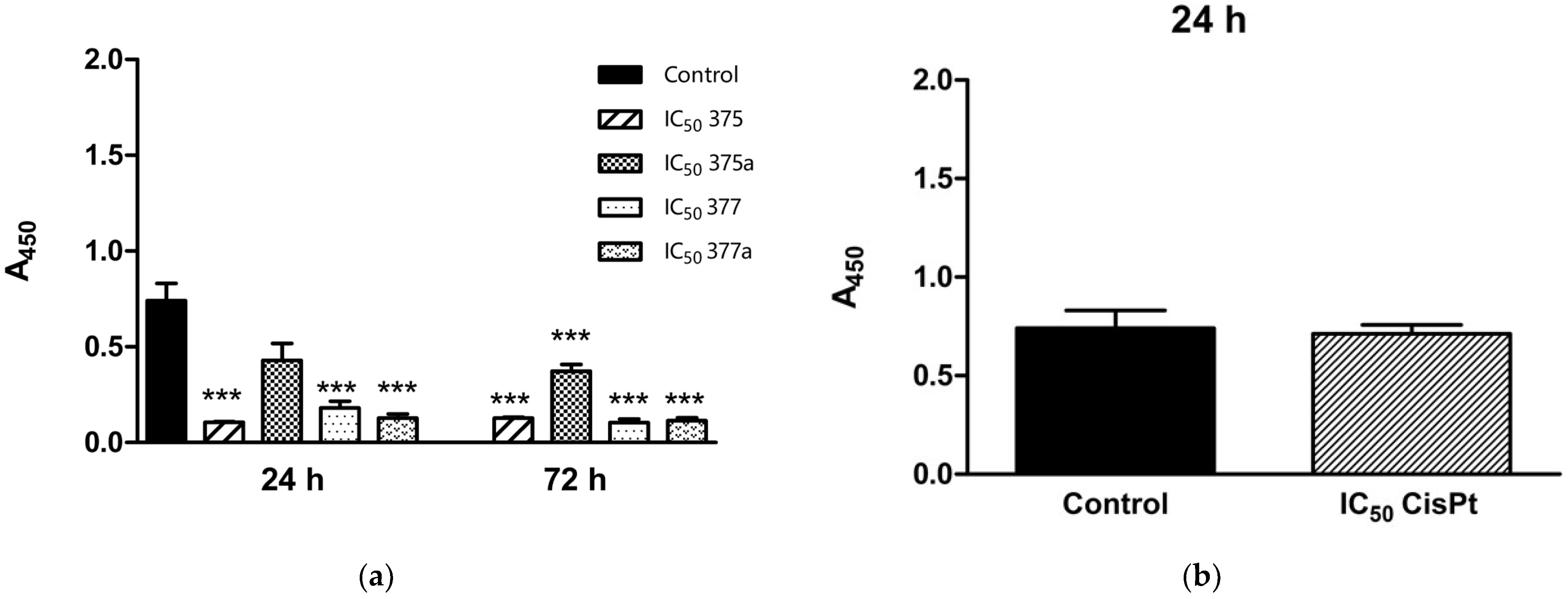
2.1. Effects of Aspirin, Indomethacin and Their Rhenium Derivatives on Endometrial Cancer Cell Viability

The tested compounds, i.e., aspirin, indomethacin, naproxen, ibuprofen, and their rhenium derivatives, were applied at selected concentrations against Ishikawa and HEC-1A cells and their effects on cell viability were evaluated by the MTT assay. Only two rhenium compounds–rhenium aspirin (375) and rhenium-indomethacin (377), and their corresponding NSAIDs, aspirin (375a) and indomethacin (377a), showed an effect on endometrial cancer cell viability. The examination was performed using the same methodology as described in previous studies [16] conducted by our research group.
The IC50 values were determined with a tetrazolium (MTT) assay, and the results are summarized in Table 1 for the Ishikawa cell line and Table 2 for the HEC-1A cell line. After 24-h incubation, the cytotoxic activity of rhenium derivatives 375 and 377 against Ishikawa and HEC-1A cancer cell lines was significantly higher than that of 375a and 377a, indicating that they were more effective than the corresponding original NSAIDs. IC50 for 375 was 188.06 µM and 394.06 µM for Ishikawa and HEC-1A cancer cells, respectively, whereas the IC50 of 375a was several folds higher for both cancer cell lines, reaching 10,024.42 µM and 27,255.8 µM, respectively. The IC50 values of 377 for Ishikawa and HEC-1A were 228.6 µM and 1459.3 µM, respectively, while the IC50 values of 377a for both cancer cell lines were 3295.3 µM and 5489.3 µM, respectively. The cytotoxic effect of the reference agent CisPt, was also studied under the same conditions. The IC50 for Ishikawa and HEC-1A was 13.57 µM and 20.05 µM, respectively. As the results showed, cells of the Ishikawa line were more susceptible to the compounds during the first 24-h incubation period than cells of the HEC-1A line, which could be related to the pre- and postmenopausal nature of the cells.
After a 72-h incubation period, the enhancement of the cytotoxic effect of both rhenium derivatives on Ishikawa and HEC-1A cancer cells was observed. IC50 for 375 in Ishikawa and HEC-1A was 146.3 µM and 112.4 µM, respectively, while the initial NSAID IC50 value of 375a was 17,395.6 µM for Ishikawa and 17,168 µM for HEC-1A cell line. The IC50 value of 377 was 107.7 µM for Ishikawa and 138.5 µM for HEC-1A cells, whereas the IC50 value of 377a was 4737.4 µM and 6375.2 µM, respectively. The IC50 values of CisPt for both Ishikawa and HEC-1A cancer cell lines were 41.03 µM and 10.27 µM, respectively. After 72-h incubation with the studied agents, the discrepancy between IC50 values was lower than that between IC50 values after 24-h incubation. Moreover, the inhibitory effect of the agents against HEC-1A cancer cells was increased.

2.2. Inhibition of Endometrial Cancer Cell Proliferation by Aspirin, Indomethacin, and Their Rhenium Derivatives

Due to the previous results of the MTT test, further research was continued by using 375, 375a, 377, and 377a. To evaluate the effects of NSAIDs and their derivatives on the proliferation of Ishikawa and HEC-1A cells, a BrdU assay was performed, which involves the incorporation of BrdU into newly synthesized DNA followed by its detection. Ishikawa and HEC-1A cells were incubated with each compound for 24 and 72 h.
The changes in the proliferation status of Ishikawa endometrial cancer cells were examined for each of the tested compounds at IC50 (Figure 1), ½ IC50, and ¼ IC50 (Table 3) after 24- and 72-h incubation. After 24-h incubation with 375, 375a, 377, and 377a, only 377 and 377a showed a significant antiproliferative effect on the tested cells. However, the rhenium derivative 377 inhibited proliferation less (* = p < 0.05) than the original NSAID 377a (*** = p < 0.001). Differences in proliferation status of the tested cells were also observed in the case of ½ and ¼ IC50. Here, both 377 and 377a showed higher significance (*** = p < 0.001) in inhibiting cell proliferation, compared with 375 and 375a, where only the effect of 375a was significant (* = p < 0.05). The proliferation inhibitory effect of the reference drug CisPt was tested after 24-h incubation under the same test conditions (* = p < 0.05). All tested compounds significantly inhibited the proliferation of Ishikawa cells after 72-h incubation. Only 375a had a lower proliferation inhibitory effect on cells in IC50 (** = p < 0.01), compared with the other compounds tested (*** = p < 0.001).
The proliferation status of HEC-1A endometrial cancer cells was studied under the same conditions as the Ishikawa cells described above. Studies were performed using the tested compounds at their IC50 (Figure 2), ½ IC50, and ¼ IC50 (Table 4) after 24- and 72-h incubation. After 24-h incubation, only 1 of the tested compounds revealed no significant antiproliferative effect on the studied cells, 375a, while the other compounds showed high inhibitory activity (*** = p < 0.001). This effect was not observed at the lower concentrations of the compounds, where only two, 375 at ½ IC50 and 375a at ¼ IC50, showed significant inhibition of cell proliferation (* = p < 0.05). The reference drug CisPt was tested after 24-h incubation under the same assay conditions. However, no significant effect on cells was observed. The lack of proliferation inhibitory effect of CisPt on HEC-1A after 24-h incubation could be related to the short incubation time with the drug. Proliferation inhibition by the tested derivatives was enhanced after 72-h incubation (*** = p < 0.001), and all agents had a similar effect on HEC-1A cells.

2.3. The Effect of Aspirin, Indomethacin and Their Rhenium Derivatives on ROS Level in the Studied Endometrial Cancer Cells

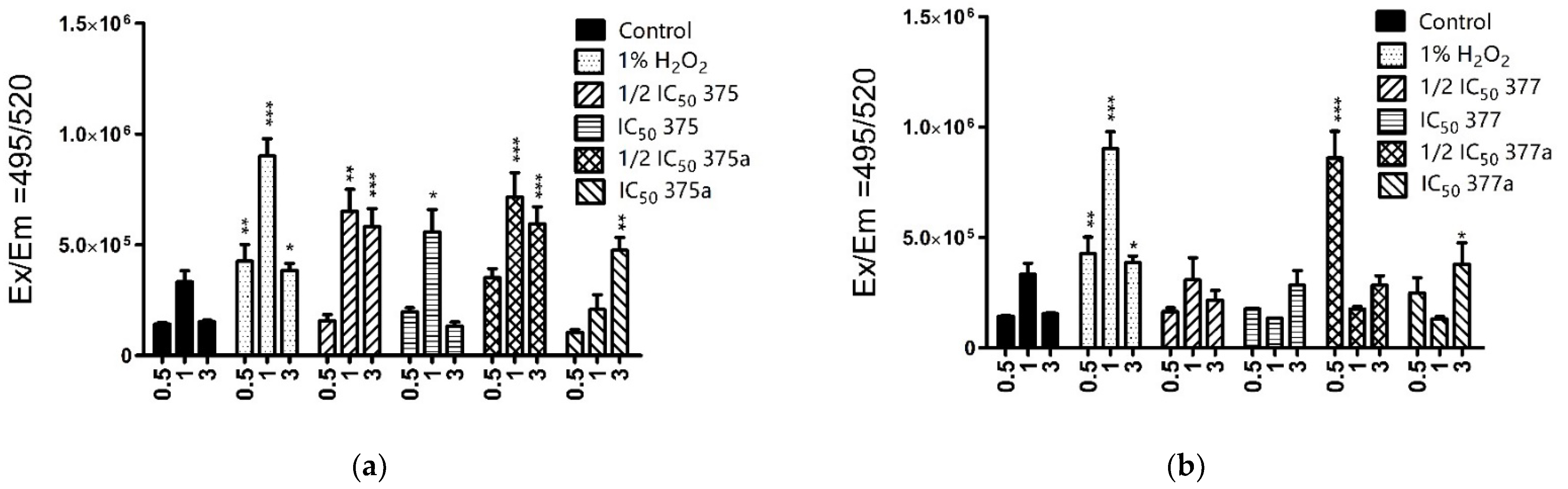
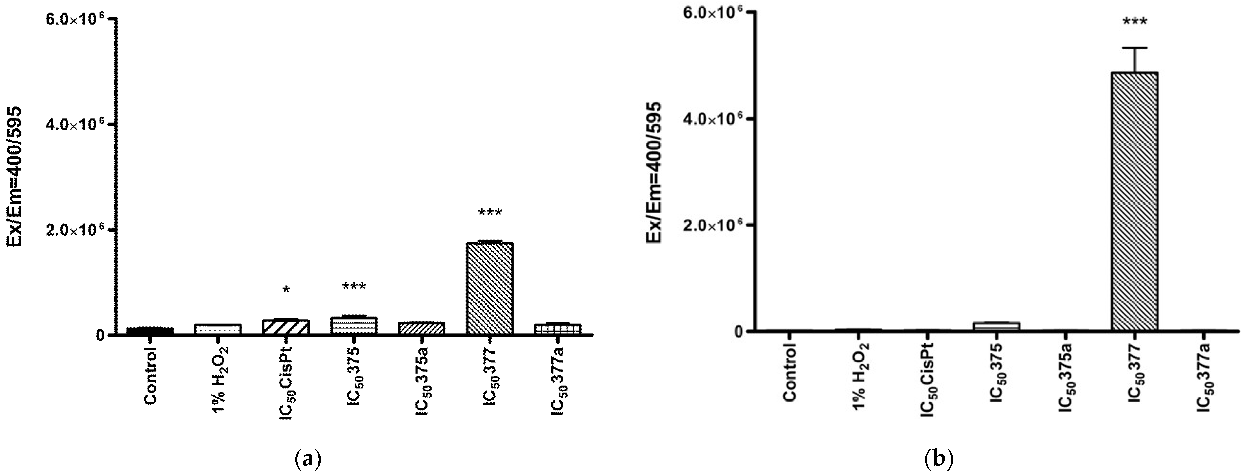
2.3.1. Cytosolic ROS Level Measurement

The ROS are well-known for their signaling capabilities and can trigger oxidative stress-induced cancer cell death. On the other hand, cancer cells can maintain ROS homeostasis and escape the initiation of cell death by the increase of their antioxidant properties [17]. Thus, new drugs that strongly induce the formation of ROS to overcome this defense may prove effective in fighting cancer cells. Therefore, the tested compounds were analyzed in this respect against Ishikawa and HEC-1A cell lines using H2DCF-DA dye (Thermo Fisher, Warsaw, Poland).
The measurements in Ishikawa cells (Figure 3) revealed the high ROS inducing activity of aspirin rhenium derivative 375 at ½ IC50 (*** = p < 0.001), and IC50 (* = p < 0.05), as well as the native NSAID 375a at ½ IC50 (*** = p < 0.001) and IC50 (** = p < 0.01). In Ishikawa cells, 377 was found to be less effective in generating ROS compared to the native NSAID, with 377a at ½ IC50 and IC50 being significant (*** = p < 0.001 and * = p < 0.05, respectively). In the case of the HEC-1A cell line (Figure 4), the level of ROS produced by the rhenium compounds was significant for both 375 and 377 (*** = p < 0.001) but not for corresponding NSAIDs. In addition, both rhenium derivatives induced increased ROS levels at ½ IC50 concentrations (*** = p < 0.001). However, the activity of the drug was not constant at all measured time points (0.5, 1 or 3 h after incubation), indicating a time-dependent effect of the NSAIDs and rhenium-NSAIDs on ROS induction. In addition, lower drug doses (½ IC50) were found to initiate the production of ROS more rapidly, possibly related to the high solubility of the compounds in the culture medium, leading to immediate uptake of the drugs across the cell membrane.

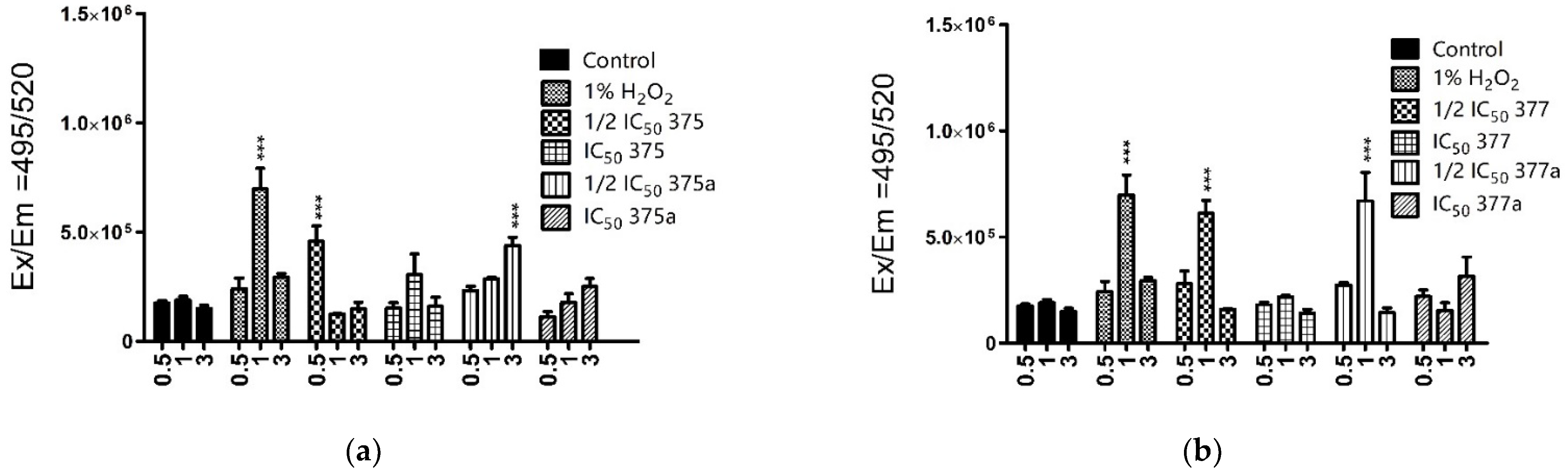
2.3.2. Mitochondrial ROS Level Measurement

It has been found that about 90% of cellular reactive oxygen species is produced by mitochondria. Disruption of this overproduction leads to oxidative stress, which in turn leads to oxidative damage. This damage affects cellular components such as lipids, DNA, and proteins, which is unfavorable for cells [18]. However, the accumulation of rhenium NSAIDs in mitochondria was reported in a previous study [16] and could possibly be considered as an anticancer mechanism of rhenium NSAIDs action. Therefore, mitochondria-specific ROS production was also investigated in endometrial cancer cells.
The mitochondrial ROS changes were observed only after 2-h incubation with studied agents, by using MitoSOX Red dye (Invitrogen, Waltham, MA, USA). These results showed that the rhenium NSAID derivatives produced a significant amount of mitochondrial ROS compared to the original NSAIDs. In addition, differences were found between 375 and 377. In both Ishikawa (Figure 5a) and HEC-1A cells (Figure 5b), 377 generated more mitochondrial ROS than 375 when these drugs were used at their IC50 values. At the same time, no correlation was observed between mitochondrial and cytosolic ROS levels, which is probably related to a different nature of ROS and incubation time. H2O2 is used as a secondary messenger to coordinate the oxidative metabolism of cells. Levels of H2O2 are regulated through its production and degradation, where mitochondria serve as “stabilizing devices” and buffer cellular H2O2 levels. The stabilization of hydrogen peroxide is related to the high concentration of antioxidant defense enzymes in the matrix [19,20]. These properties of H2O2 cleavage by mitochondria can explain the lack of significant mitochondrial ROS production initiated by 1% H2O2.

3. Discussion

Positive anticancer effects of NSAIDs, including specific action against endometrial cancer, have already been studied [21]. It was found that the positive effect of low-dose aspirin and its synergism with some other NSAIDs were not only associated with good patient survival outcomes but also had an anti-proliferative effect on cancer cells [22,23,24]. These effects were also previously noticed in vitro for Ishikawa and HEC-1A endometrial cancer cells. Studies performed by Gao et al. [25] determined the effects of aspirin, indomethacin, and chosen cyclooxygenase (COX-2)–selective inhibitor-NS398. All three NSAIDs significantly inhibited the proliferation of Ishikawa and HEC-1A cancer cells in a dose- and time-dependent manner. In addition, aspirin and indomethacin were involved in cellular apoptosis initiation through a cytochrome c-related pathway. Other studies have shown the inhibition of growth, apoptosis induction, and reduced Bcl-2 expression of Ishikawa cells about 21–88% after aspirin treatment [26,27]. Indomethacin was found to be related to the up-regulation of the PTEN tumor suppressor, which affected the HEC-1B cancer cell line [28]. A selective COX-2 inhibitor-celecoxib, reduced HEC-1A and HEC-1B cell lines growth and assisted in the inhibition of tumor cell proliferation [29]. However, none of these studies used modified NSAIDs. Structural and chemical modifications of NSAIDs may not only enhance the antitumor activity but also reduce the risk of side effects which are common during regular NSAIDs intake [30,31]. For this reason, new NSAID derivatives have been developed and synthesized, such as: NO-NSAIDs (hybrid nitrate NSAIDs), cGMP PDE (cyclic guanosine monophosphate phosphodiesterase), AChE (acetylcholinesterase inhibitors), phospho-NSAIDs, TEMPO (2,2,6,6-tetramethyl-1-piperidinyloxy), TEMPOL (4-hydroxy-TEMPO) and HS-NSAIDs (hydrogen sulfide NSAIDs) [23,24,25,32,33,34]. However, the study omitted endometrial cancer cells, which are known to overexpress COX-2 [35,36]. Because of metal complexation, NSAIDs could also get new anticancer qualities. Some zinc complexes of naproxen had shown high activity against bacterial strains while copper-aspirin complexation had served as superoxide dismutase mimetics and had high cytotoxic activity against selected human cancer cells. These agents were not associated with COX inhibition properties but were involved in DNA binding and cleavage [37,38].
The results of the cell viability assay (MTT) showed the activity of 375a and 377a as well as their rhenium derivatives 375 (Re-aspirin) and 377 (Re-indomethacin) against Ishikawa and HEC-1A cells. Both cell lines responded to the rhenium derivatives, for which the biologically active concentrations were significantly lower than in the case of the original NSAID counterparts. Moreover, the rhenium derivatives showed higher inhibitory activity during prolonged incubation, which was not observed with NSAIDs. These effects were found to be not only time-dependent but also cell line-dependent, as different concentrations were required for 50% inhibition of Ishikawa and HEC-1A viability. The nature of the endometrial cancer cell lines may be the reason for these differences. According to Bokhman’s classification [39], based on different etiopathological, genetic and clinical features, Ishikawa cells belong to type I EC, while HEC-1A cells are representatives of type II. Carcinoma cells of type I had shown high expression of estrogen and progesterone receptors, had low histological differentiation, and are considered typical of early-stage disease. Type II cells represent highly aggressive and invasive carcinoma types, with low levels of expression of estrogen and progesterone receptors and are commonly referred to as typical of the advanced stage of the disease. Moreover, these two cell lines were derived from adenocarcinomas from women at different ages (39-year and 71-year-old for Ishikawa and HEC-1A, respectively), which is associated with the premenopausal and postmenopausal nature of the cells [40]. Ishikawa cell line is a well-differentiated human adenocarcinoma cell line carrying estrogen and progesterone receptors. This cell line is known to produce corticotropin-releasing hormone, placental alkaline phosphatase, and chorionic gonadotropin and is responsive to steroid hormones [41,42]. HEC-1A cancer cells express the wild-type form of the estrogen receptor, and 17-beta-estradiol induces proliferation of these cells. In addition, these cells contain a nonsense mutation in the hpms2 and a splicing mutation in the hmsh6 genes, which play key roles in DNA’s methyl-directed mismatch repair (MMR) pathway [43,44]. These differences in characteristics are probably the main reason for the different responses of the cells to treatment with NSAIDs versus rhenium-NSAIDs., The better activity of rhenium derivatives compared with the original NSAIDs was also observed in previous studies performed by our research group using HeLa and L929 cell lines. Rhenium-aspirin and rhenium-indomethacin were more active against cancerous HeLa cells than non-cancerous L929 cells with IC50 values of 36 µM and 158 µM, respectively. The IC50 values for aspirin and indomethacin against HeLa were much more higher (2819 µM and 326 µM, respectively) [16].
The proliferation assay showed higher sensitivity of HEC-1A cells to original NSAIDs and their rhenium derivatives than Ishikawa cells. In this case, HEC-1A cells were affected by both tested rhenium compounds already after the 24-h incubation period, while Ishikawa cells proved susceptible to 377 and 377a. For both cancer cell lines, the anti-proliferative effect increased after 72-h incubation with tested NSAIDs and their rhenium derivatives. The proliferation rate of cells grown under normal conditions varies and depends on the type of cancer. In the case of Ishikawa cells, their proliferation rate is faster than that of HEC-1A cells and increases with the number of passages [40,45]. That may be a possible reason for the lower inhibitory effect of the tested compounds against Ishikawa cells proliferation. Changes in proliferation rate were studied not only at IC50 but also at concentrations equal to ½ IC50 and ¼ IC50 (Table 3 and Table 4). For the lower concentrations, only 375a showed a significant anti-proliferative effect after 24-h incubation, where ½ IC50 and ¼ IC50 of this compound (* = p < 0.05) inhibited cell proliferation of Ishikawa and HEC-1A cells, respectively. This observation is consistent with previous reports showing that low doses of NSAIDs, especially aspirin, provided good survival outcomes for women with endometrial cancer [46,47]. Cisplatin (CisPt) is a good candidate for comparison because of its widespread use as an anti-EC drug as a so-called first-line chemotherapeutic agent, which is administered to patients in single doses as well as in combination chemotherapy (at a dose and regimen of 50–100 mg per m2 of a body surface area, every 3 weeks) [48]. CisPt was found to inhibit EC cell proliferation by increasing the expression of semaphorin 3B (SEMA3B), which has suppressive effects on cancer cell proliferation and angiogenesis. As our results showed, after 24-h incubation, CisPt had a significant (* = p < 0.05) anti-proliferative effect only on Ishikawa endometrial cancer cells but not on HEC-1A. That could be explained by the overexpression of BCL2, which is associated with the resistance of HEC-1A cancer cells to CisPt and the longer incubation time required to transpose the drug across the cell membrane, as shown by Rouette et al. [49].
It has already been shown that NSAIDs are associated with an increase in the production of reactive oxygen species (ROS). This mechanism of action of NSAIDs is commonly suggested to initiate side effects, particularly within the gastroenterological tract. However, this mechanism also may be used to destroy cancer cells by inhibiting cell growth and metabolism and disrupting the signaling pathways. Increased levels of ROS induce disruption of cellular homeostasis, leading to cellular damage and initiation of apoptosis [50,51,52]. Therefore, the induction of high ROS levels has been suggested by our group as a positive mechanism of rhenium-NSAIDs activity against previously studied cells [16]. In this study, this effect was evident only in HeLa cancer cells, not in non-neoplastic L-929 fibroblast cells. It was observed that cancer cells have a significantly higher ability to accumulate rhenium derivatives in mitochondria, which could also be related to an increase in mitochondria-specific ROS. As shown in the present study, there was increased production of cytosolic ROS in Ishikawa cells following treatment with both NSAIDs and their rhenium derivatives. Moreover, both NSAIDs and their rhenium derivatives, had a significant impact on ROS levels in HEC-1A, but at ½ IC50 concentrations, which may be related to better penetration of compounds across cell membranes. Furthermore, the ½ IC50 concentrations of rhenium derivatives 375 and 377 were lower than ½ IC50 of original NSAIDS 375a and 377a, corresponding to 197.03 µM and 729.65 µM and to 13,627.9 µM and 2744.65 µM, respectively. These results indicate a more effective induction of ROS by rhenium compounds than by their original NSAID counterparts because of the higher activity of the former at lower concentrations than the latter. The result of measuring mitochondrial-specific ROS levels did not match the effects of the agents on cytosolic ROS levels. In this case, the highest level (which is considered to be a 100% increase in ROS amount) for both cell lines appeared to be associated with 377. In the case of Ishikawa cells, this effect was additionally evident for 375, but not to the same extent as for 377. That may be related to the cell-dependent mode of interaction of the compounds with mitochondria. It should additionally be noted that the incubation time of the compounds with cells was only two hours, which is related to the limitations of an assay.
The investigated rhenium derivatives have potential as agents against uterine-related cancers, but the study of the biological activity of these compounds should be further pursued. The next steps should include investigating the mechanisms of rhenium-NSAIDs impact at deeper molecular levels.

4. Materials and Methods

4.1. NSAIDs and Rhenium-NSAIDs Complexes

NSAIDs and their complexes with rhenium were synthesized in the Department of Organic Chemistry, Faculty of Chemistry, University of Lodz, by K. Kowalski research group [16]. Compounds tested in this study are listed in Table 5.

4.2. Human Cell Culture

Adherent human endometrial cancer cell lines Ishikawa (ECACC 99040201) and HEC-1-A (ATTC® HTB-112) were obtained from the European Collection of Authenticated Cell Cultures (ECACC, Porton Down, UK) and the American Type Culture Collection (ATCC®, Manassas, VA, USA), respectively. Both cell lines were grown in DMEM-F12 medium with a stable L-glutamine, 15 mM HEPES, and supplemented with 10% Ultra-low Endotoxin Fetal Bovine Serum (FBS) (Biowest, Nuaille, France), and 100 U/mL of penicillin and 100 mg/mL of streptomycin (Biowest, Nuaille, France), under standard conditions (37 °C, 5% CO2). Cells were passaged every 5–6 days; the growth medium was changed every 2–3 days after a passage. For microplate assays, cells were seeded at the optimal density of 10,000 cells/well.

4.3. Cell Viability Assay

The effects of individual NSAIDs and their rhenium derivatives on the endometrial cancer cell viability were evaluated using the MTT (3-(4,5-dimethylthiazol-2-yl)-2,5-diphenyltetrazolium bromide) assay. Ishikawa and HEC-1A cells were initially seeded into 96-well microplates at optimal density (10,000 cells/well) and cultured for the next 24 h under standard conditions. After pre-cultivation, cells were treated with compounds that were added in two-fold dilutions ranging from 10 to 500 µM. All stock solutions of the tested compounds were prepared in DMSO. 1% DMSO was used as a negative control because the concentration of the solvent under experimental conditions never exceeded this value; cisplatin, a well-known anticancer drug was used as a positive control and dissolved in 150 mM saline. Cells were incubated in the presence of the test compounds for 24 and 72 h. After incubation, MTT was dissolved in a culture medium and added to the cells in the final concentration of 0.5 mg/mL. The plates were then incubated for 2 h under standard conditions. Formazan crystals formed during the MTT reaction were solubilized in 150 µL of DMSO. Absorbance was measured at 570 nm using a SpectraMax i3 microplate reader (Molecular Devices, Silicon Valley, CA, USA). The assay was performed in three independent experiments. The IC50 (compound concentration inhibiting cell proliferation by 50%) values were determined using GraphPad Prism 7 Software.

4.4. BrdU Assay

The effects of the compounds on cell proliferation were examined using a BrdU cell proliferation assay kit (Biovision, Milpitas, CA, USA). BrdU (5-bromo-2-deoxyuridine) is a pyrimidine analog that is incorporated into the newly synthesized DNA of proliferating cells by replacing thymine. The incorporated BrdU was quantified using mouse anti-BrdU primary antibodies, HRP-linked anti-mouse IgG antibodies, and 3,3′,5,5′-tetramethylbenzidine (TMB) as an HRP substrate, according to the manufacturer’s instructions. Endometrial cancer cells were seeded into 96-well plates at optimal density and precultured overnight under standard conditions. Cells were then treated with the selected compounds at IC50, ½ IC50, and ¼ IC50 concentrations established previously in the MTT assay and incubated under standard conditions for 24 and 72 h. Absorbance was measured at 450 nm using a SpectraMax i3 plate reader (Molecular Devices, Silicon Valley, CA, USA). The assay was performed in three independent experiments. The results were calculated using GraphPad Prism 7 software.

4.5. Cellular ROS Level Measurement

Reactive oxygen species (ROS) levels were measured using H2DCFDA (ThermoFisher) fluorescent dye. Fluorescence measurement is made by converting H2DCF-DA to 2′,7′-dichlorofluorescein (DCF), the amount of which is directly dependent on the amount of ROS produced in the cells. Thus, ROS detection was performed by measuring the fluorescence of H2DCF-DA-treated Ishikawa and HEC-1A cancer cells at Ex/Em = 495/520 nm. Ishikawa and HEC-1A cells were seeded into black bottom 96-well cell culture plates. Once cells reached approximately 80–90% confluence, 100 µL of DMEM-F12 medium w/o FBS supplemented with H2DCF-DA dye at a final concentration of 5 µM was added. Cells were then incubated for 40 min under standard conditions, protected from light. After incubation, cells were washed twice with DPBS, treated with the selected compound at IC50 and ½ IC50 concentrations, obtained in the MTT assay, and then incubated for 0.5, 1, and 3 h. After each time point, cells were washed twice with DPBS and suspended in 100 µL of DPBS. Fluorescence was measured at Ex/Em = 495/520 nm using a SpectraMax i3 plate reader (Molecular Devices, Silicon Valley, CA, USA). The assay was performed in three independent experiments, and the results were calculated using GraphPad Prism 7 Software.

4.6. Mitochondrial ROS Level Measurement

The assay was performed using the MitoSOX Red dye (Invitrogen, Waltham, MA, USA). When incorporated into mitochondria, this dye is rapidly oxidized by peroxide and converted into the fluorescent product 2-hydroxymitoethidium. Initially, cells were seeded into a black bottom 96-well plate. Once approximately 80–90% cell confluence was achieved, the culture medium was replaced with the medium w/o FBS containing 5 µM MitoSOX Red dye and incubated for 10 min under standard conditions. After incubation, cells were washed twice with DPBS and treated with NSAIDs and rhenium-NSAIDs at IC50 concentrations. 1% H2O2 and cisplatin were used as controls. Cells were incubated with the compounds for 2 h under standard conditions, protected from light. Cells were then washed twice with DPBS and resuspended in 100 µL of DPBS. Fluorescence was measured at Ex/Em = 400/595 nm using SpectraMax i3 plate reader (Molecular Devices, Silicon Valley, CA, USA). The assay was performed in three independent replicates and the results were calculated using GraphPad Prism 7 software.

Author Contributions

Conceptualization, O.K., A.K. and P.S.; methodology, O.K.; investigation, O. K.; writing—original draft preparation, O.K.; writing—review and editing, P.S. and A.K.; All authors have read and agreed to the published version of the manuscript.

Funding

This research was funded in part by the National Science Centre, Poland (grant SONATA 17 UMO-2021/43/D/NZ7/01386), and by a subsidy for scientific activity provided to the Department of Molecular Microbiology by the University of Lodz (B2111000000038.01). For the purpose of Open Access, the author has applied a CC-BY public copyright license to any Author Accepted Manuscript (AAM) version arising from this submission.

Institutional Review Board Statement

Not applicable.

Informed Consent Statement

Not applicable.

Data Availability Statement

Not applicable.

Acknowledgments

Some of the biological experiments were performed in part using equipment of the Laboratory of Microscopic Imaging and Specialized Biological Techniques, Faculty of Biology and Environmental Protection, University of Lodz, Poland.

Conflicts of Interest

The authors declare no conflict of interest.

References

  1. Smith, W.L.; Dewitt, D.L.; Garavito, R.M. Cyclooxygenases: Structural, Cellular, and Molecular Biology. Annu. Rev. Biochem. 2000, 69, 145–182. [Google Scholar] [CrossRef]
  2. De Leval, X.; Delarge, J.; Somers, F.; De Tullio, P.; Henrotin, Y.; Pirotte, B.; Dogne, J.-M. Recent advances in inducible cyclooxygenase (COX-2) inhibition. Curr. Med. Chem. 2000, 7, 1041–1062. [Google Scholar] [CrossRef]
  3. Macciò, A.; Madeddu, C. Inflammation and ovarian cancer. Cytokine 2012, 58, 133–147. [Google Scholar] [CrossRef]
  4. Deivendran, S.; Marzook, K.H.; Pillai, M.R. The Role of Inflammation in Cervical Cancer. Adv. Exp. Med. Biol. 2014, 816, 377–399. [Google Scholar] [CrossRef]
  5. Szewczyk, G.; Maciejewski, T.M.; Szukiewicz, D. Current progress in the inflammatory background of angiogenesis in gynecological cancers. Inflamm. Res. 2019, 68, 247–260. [Google Scholar] [CrossRef]
  6. Chubak, J.; Whitlock, E.P.; Williams, S.B.; Kamineni, A.; Burda, B.U.; Buist, D.S.; Anderson, M.L. Aspirin for the Prevention of Cancer Incidence and Mortality: Systematic Evidence Reviews for the U.S. Preventive Services Task Force. Ann. Intern. Med. 2016, 164, 814–825. [Google Scholar] [CrossRef]
  7. Friis, S.; Riis, A.H.; Erichsen, R.; Baron, J.A.; Sørensen, H.T. Low-Dose Aspirin or Nonsteroidal Anti-inflammatory Drug Use and Colorectal Cancer Risk. Ann. Intern. Med. 2015, 163, 347–355. [Google Scholar] [CrossRef]
  8. Kehm, R.D.; Hopper, J.L.; John, E.M.; Phillips, K.-A.; MacInnis, R.J.; Dite, G.S.; Milne, R.L.; Liao, Y.; Zeinomar, N.; Knight, J.A.; et al. Regular use of aspirin and other non-steroidal anti-inflammatory drugs and breast cancer risk for women at familial or genetic risk: A cohort study. Breast Cancer Res. 2019, 21, 52. [Google Scholar] [CrossRef]
  9. Doat, S.; Cénée, S.; Trétarre, B.; Rebillard, X.; Lamy, P.-J.; Bringer, J.-P.; Iborra, F.; Murez, T.; Sanchez, M.; Menegaux, F. Nonsteroidal anti-inflammatory drugs (NSAIDs) and prostate cancer risk: Results from the EPICAP study. Cancer Med. 2016, 6, 2461–2470. [Google Scholar] [CrossRef]
  10. Chiu, L.; Tong, K.; Ooi, V. Cytostatic and cytotoxic effects of cyclooxygenase inhibitors and their synergy with docosahexaenoic acid on the growth of human skin melanoma A-375 cells. Biomed. Pharmacother. 2005, 59, S293–S297. [Google Scholar] [CrossRef]
  11. Marinov, L.; Georgieva, A.; Voynikov, Y.; Toshkova, R.; Nikolova, I.; Malchev, M. Cytotoxic and antiproliferative effects of the nonsteroidal anti-inflammatory drug diclofenac in human tumour cell lines. Biotechnol. Biotechnol. Equip. 2021, 35, 1118–1126. [Google Scholar] [CrossRef]
  12. Buzharevski, A.; Paskaš, S.; Sárosi, M.-B.; Laube, M.; Lönnecke, P.; Neumann, W.; Murganić, B.; Mijatović, S.; Maksimović-Ivanić, D.; Pietzsch, J.; et al. Carboranyl Derivatives of Rofecoxib with Cytostatic Activity against Human Melanoma and Colon Cancer Cells. Sci. Rep. 2020, 10, 4827. [Google Scholar] [CrossRef]
  13. Sostres, C.; Gargallo, C.J.; Lanas, A. Nonsteroidal anti-inflammatory drugs and upper and lower gastrointestinal mucosal damage. Arthritis Res. Ther. 2013, 15, 3. [Google Scholar] [CrossRef]
  14. Lim, Y.J.; Lee, J.S.; Ku, Y.S.; Hahm, K.-B. Rescue strategies against non-steroidal anti-inflammatory drug-induced gastroduodenal damage. J. Gastroenterol. Hepatol. 2009, 24, 1169–1178. [Google Scholar] [CrossRef]
  15. Dillon, C.T.; Hambley, T.W.; Kennedy, B.J.; Lay, P.A.; Zhou, Q.; Davies, N.M.; Biffin, J.R.; Regtop, H.L. Gastrointestinal Toxicity, Antiinflammatory Activity, and Superoxide Dismutase Activity of Copper and Zinc Complexes of the Antiinflammatory Drug Indomethacin. Chem. Res. Toxicol. 2003, 16, 28–37. [Google Scholar] [CrossRef]
  16. Skiba, J.; Kowalczyk, A.; Stączek, P.; Bernaś, T.; Trzybiński, D.; Woźniak, K.; Schatzschneider, U.; Czerwieniec, R.; Kowalski, K. Luminescent fac-[Re(CO)3(phen)] carboxylato complexes with non-steroidal anti-inflammatory drugs: Synthesis and mechanistic insights into the in vitro anticancer activity of fac-[Re(CO)3(phen)(aspirin)]. New J. Chem. 2019, 43, 573–583. [Google Scholar] [CrossRef]
  17. Reczek, C.R.; Chandel, N.S. The Two Faces of Reactive Oxygen Species in Cancer. Annu. Rev. Cancer Biol. 2017, 1, 79–98. [Google Scholar] [CrossRef]
  18. Tirichen, H.; Yaigoub, H.; Xu, W.; Wu, C.; Li, R.; Li, Y. Mitochondrial Reactive Oxygen Species and Their Contribution in Chronic Kidney Disease Progression Through Oxidative Stress. Front. Physiol. 2021, 12, 627837. [Google Scholar] [CrossRef]
  19. Mailloux, R.J. Mitochondrial Antioxidants and the Maintenance of Cellular Hydrogen Peroxide Levels. Oxidative Med. Cell. Longev. 2018, 2018, 7857251. [Google Scholar] [CrossRef]
  20. Murphy, M.P. How mitochondria produce reactive oxygen species. Biochem. J. 2009, 417, 1. [Google Scholar] [CrossRef] [Green Version]
  21. Brasky, T.M.; Felix, A.S.; Cohn, D.E.; McMeekin, D.S.; Mutch, D.G.; Creasman, W.T.; Thaker, P.H.; Walker, J.L.; Moore, R.G.; Lele, S.B.; et al. Nonsteroidal Anti-inflammatory Drugs and Endometrial Carcinoma Mortality and Recurrence. J. Natl. Cancer Inst. 2017, 109, djw251. [Google Scholar] [CrossRef]
  22. Matsuo, K.; Cahoon, S.S.; Yoshihara, K.; Shida, M.; Kakuda, M.; Adachi, S.; Moeini, A.; Machida, H.; Garcia-Sayre, J.; Ueda, Y.; et al. Association of Low-Dose Aspirin and Survival of Women With Endometrial Cancer. Obstet. Gynecol. 2016, 128, 127–137. [Google Scholar] [CrossRef]
  23. Verdoodt, F.; Friis, S.; Dehlendorff, C.; Albieri, V.; Kjaer, S.K. Non-steroidal anti-inflammatory drug use and risk of endometrial cancer: A systematic review and meta-analysis of observational studies. Gynecol. Oncol. 2016, 140, 352–358. [Google Scholar] [CrossRef]
  24. Wang, Y.; Zhao, J.; Chen, X.; Zhang, F.; Li, X. Aspirin use and endometrial cancer risk: A meta-analysis and systematic review. Ann. Transl. Med. 2020, 8, 461. [Google Scholar] [CrossRef]
  25. Gao, J.; Niwa, K.; Sun, W.; Takemura, M.; Lian, Z.; Onogi, K.; Seishima, M.; Mori, H.; Tamaya, T. Non-steroidal anti-inflammatory drugs inhibit cellular proliferation and upregulate cyclooxygenase-2 protein expression in endometrial cancer cells. Cancer Sci. 2004, 95, 901–907. [Google Scholar] [CrossRef]
  26. Arango, H. Aspirin effects on endometrial cancer cell growth. Obstet. Gynecol. 2001, 97, 423–427. [Google Scholar] [CrossRef]
  27. Webb, P.M.; Na, R.; Weiderpass, E.; Adami, H.O.; Anderson, K.E.; Bertrand, K.A.; Botteri, E.; Brasky, T.M.; Brinton, L.A.; Chen, C.; et al. Use of aspirin, other nonsteroidal anti-inflammatory drugs and acetaminophen and risk of endometrial cancer: The Epidemiology of Endometrial Cancer Consortium. Ann. Oncol. 2019, 30, 310–316. [Google Scholar] [CrossRef]
  28. Abdulkareem, I.; Blair, M. Effects of indomethacin on expression of PTEN tumour suppressor in human cancers. Niger. Med. J. 2013, 54, 100–106. [Google Scholar] [CrossRef]
  29. Xiao, Y.; Teng, Y.; Zhang, R.; Luo, L. Antitumor effect of the selective COX-2 inhibitor celecoxib on endometrial adenocarcinoma in vitro and in vivo. Oncol. Lett. 2012, 4, 1219–1224. [Google Scholar] [CrossRef]
  30. Wongrakpanich, S.; Wongrakpanich, A.; Melhado, K.; Rangaswami, J. A Comprehensive Review of Non-Steroidal Anti-Inflammatory Drug Use in The Elderly. Aging Dis. 2018, 9, 143–150. [Google Scholar] [CrossRef] [Green Version]
  31. Henry, D.A. Side-effects of non-steroidal anti-inflammatory drugs. Baillière’s Clin. Rheumatol. 1988, 2, 425–454. [Google Scholar] [CrossRef]
  32. Dunlap, T.; Abdul-Hay, S.O.; Chandrasena, R.E.P.; Hagos, G.K.; Sinha, V.; Wang, Z.; Wang, H.; Thatcher, G.R.J. Nitrates and NO-NSAIDs in cancer chemoprevention and therapy: In vitro evidence querying the NO donor functionality. Nitric Oxide 2008, 19, 115–124. [Google Scholar] [CrossRef] [PubMed]
  33. Tinsley, H.N.; Piazza, G.A. Novel Therapeutics: NSAIDs, Derivatives, and Phosphodiesterases. Curr. Color. Cancer Rep. 2012, 8, 325–330. [Google Scholar] [CrossRef]
  34. Qandil, A.M. Prodrugs of Nonsteroidal Anti-Inflammatory Drugs (NSAIDs), More Than Meets the Eye: A Critical Review. Int. J. Mol. Sci. 2012, 13, 17244–17274. [Google Scholar] [CrossRef]
  35. Hasegawa, K.; Ohashi, Y.; Ishikawa, K.; Yasue, A.; Kato, R.; Achiwa, Y.; Nishio, E.; Udagawa, Y. Expression of cyclooxygenase-2 in uterine endometrial cancer and anti-tumor effects of a selective COX-2 inhibitor. Int. J. Oncol. 2005, 26, 1419–1428. [Google Scholar] [CrossRef] [PubMed]
  36. Jeon, Y.-T.; Kang, S.; Kang, D.-H.; Yoo, K.-Y.; Park, I.-A.; Bang, Y.-J.; Kim, J.W.; Park, N.-H.; Kang, S.-B.; Lee, H.-P.; et al. Cyclooxygenase-2 and p53 Expressions in Endometrial Cancer. Cancer Epidemiol. Biomark. Prev. 2004, 13, 1538–1542. [Google Scholar] [CrossRef]
  37. Abu Ali, H.; Fares, H.; Darawsheh, M.; Rappocciolo, E.; Akkawi, M.; Jaber, S. Synthesis, characterization and biological activity of new mixed ligand complexes of Zn(II) naproxen with nitrogen based ligands. Eur. J. Med. Chem. 2015, 89, 67–76. [Google Scholar] [CrossRef]
  38. O’Connor, M.; Kellett, A.; McCann, M.; Rosair, G.; McNamara, M.; Howe, O.; Creaven, B.S.; McClean, S.; Kia, A.F.-A.; O’Shea, D.; et al. Copper(II) Complexes of Salicylic Acid Combining Superoxide Dismutase Mimetic Properties with DNA Binding and Cleaving Capabilities Display Promising Chemotherapeutic Potential with Fast Acting in Vitro Cytotoxicity against Cisplatin Sensitive and Resistant Cancer Cell Lines. J. Med. Chem. 2012, 55, 1957–1968. [Google Scholar] [CrossRef]
  39. Wilczyński, M.; Danielska, J.; Wilczyński, J. An update of the classical Bokhman’s dualistic model of endometrial cancer. Menopausal Rev. 2016, 15, 63–68. [Google Scholar] [CrossRef]
  40. Kozak, J.; Wdowiak, P.; Maciejewski, R.; Torres, A. A guide for endometrial cancer cell lines functional assays using the measurements of electronic impedance. Cytotechnology 2018, 70, 339–350. [Google Scholar] [CrossRef] [PubMed] [Green Version]
  41. Nishida, M.; Kasahara, K.; Kaneko, M.; Iwasaki, H.; Hayashi, K. Establishment of a new human endometrial adenocarcinoma cell line, Ishikawa cells, containing estrogen and progesterone receptors. Nihon Sanka Fujinka Gakkai Zasshi 1985, 37, 1103–1111. [Google Scholar] [PubMed]
  42. Nishida, M. Ishikawa Cells: Opening of In Vitro Hormone Research on Endometrial Carcinoma. In Cell and Molecular Biology of Endometrial Carcinoma; Springer: Tokyo, Japan, 2003; pp. 35–38. [Google Scholar] [CrossRef]
  43. Castro-Rivera, E.; Safe, S. Estrogen- and antiestrogen-responsiveness of HEC1A endometrial adenocarcinoma cells in culture. J. Steroid Biochem. Mol. Biol. 1998, 64, 287–295. [Google Scholar] [CrossRef]
  44. Glaab, W.E.; Risinger, J.I.; Umar, A.; Kunkel, T.A.; Barrett, J.C.; Tindall, K.R. Characterization of Distinct Human Endometrial Carcinoma Cell Lines Deficient in Mismatch Repair That Originated from a Single Tumor. J. Biol. Chem. 1998, 273, 26662–26669. [Google Scholar] [CrossRef] [PubMed]
  45. Nishida, M. The Ishikawa cells from birth to the present. Hum. Cell 2002, 15, 104–117. [Google Scholar] [CrossRef]
  46. Kuźmycz, O.; Stączek, P. Prospects of NSAIDs administration as double-edged agents against endometrial cancer and pathological species of the uterine microbiome. Cancer Biol. Ther. 2020, 21, 486–494. [Google Scholar] [CrossRef]
  47. Nevadunsky, N.S.; Van Arsdale, A.; Strickler, H.D.; Spoozak, L.A.; Moadel, A.; Kaur, G.; Girda, E.; Goldberg, G.L.; Einstein, M.H. Association Between Statin Use and Endometrial Cancer Survival. Obstet. Gynecol. 2015, 126, 144–150. [Google Scholar] [CrossRef]
  48. Moxley, K.M.; McMeekin, D.S. Endometrial Carcinoma: A Review of Chemotherapy, Drug Resistance, and the Search for New Agents. Oncologist 2010, 15, 1026–1033. [Google Scholar] [CrossRef]
  49. Rouette, A.; Parent, S.; Girouard, J.; Leblanc, V.; Asselin, E. Cisplatin increases B-cell-lymphoma-2 expression via activation of protein kinase C and Akt2 in endometrial cancer cells. Int. J. Cancer 2012, 130, 1755–1767. [Google Scholar] [CrossRef]
  50. Ghosh, R.; Alajbegovic, A.; Gomes, A.V. NSAIDs and Cardiovascular Diseases: Role of Reactive Oxygen Species. Oxidative Med. Cell. Longev. 2015, 2015, 536962. [Google Scholar] [CrossRef] [Green Version]
  51. Kusuhara, H.; Komatsu, H.; Sumichika, H.; Sugahara, K. Reactive oxygen species are involved in the apoptosis induced by nonsteroidal anti-inflammatory drugs in cultured gastric cells. Eur. J. Pharmacol. 1999, 383, 331–337. [Google Scholar] [CrossRef]
  52. Adachi, M.; Sakamoto, H.; Kawamura, R.; Wang, W.; Imai, K.; Shinomura, Y. Nonsteroidal anti-inflammatory drugs and oxidative stress in cancer cells. Histol. Histopathol. 2007, 22, 437–442. [Google Scholar] [PubMed]
Figure 1. Changes in proliferation status of Ishikawa endometrial cancer cells after 24- and 72-h incubation with 375 (rhenium aspirin), 375a (aspirin), 377 (rhenium indomethacin) and 377a (indomethacin) at IC50 concentrations (a). The assay was performed using the BrdU proliferation kit (Biovision). Cisplatin (CisPt) was used as a positive control (b). Data were obtained by measuring absorbance at 450 nm in three independent experiments. Results were statistically compared with the negative control (untreated cells with 1% DMSO dissolved in cell culture medium) using a Two-way ANOVA with Bonferroni multiple comparison test; * = p < 0.05, ** = p < 0.01, *** = p < 0.001.
Ijms 23 11568 g001
Figure 2. Changes in proliferation status of HEC-1A endometrial cancer cells after 24- and 72-h incubation with 375 (rhenium aspirin), 375a (aspirin), 377 (rhenium indomethacin) and 377a (indomethacin) at IC50 concentrations (a). The assay was performed using the BrdU proliferation kit (Biovision). Cisplatin (CisPt) was used as a positive control (b). Data were obtained by measuring absorbance at 450 nm in three independent experiments. Results were statistically compared with the negative control (untreated cells with 1% DMSO dissolved in cell culture medium) using a Two-way ANOVA with Bonferroni multiple comparison test; *** = p < 0.001.
Ijms 23 11568 g002
Figure 3. Cytosolic levels of reactive oxygen species in Ishikawa cells after 0.5, 1 and 3-h treatment with 375 (rhenium aspirin), 375a (aspirin) (a), 377 (rhenium indomethacin) and 377a (indomethacin) (b). Cells were preincubated with H2DCF-DA dye and then treated with the tested drugs. Data were obtained by measuring the fluorescence at Ex/Em = 495/520 nm. 1% H2O2 was used as a positive control. Results were statistically compared with the negative control (untreated cells with 1% DMSO dissolved in cell culture medium). Two-way ANOVA with Bonferroni multiple comparison test was used for statistical analyses; * = p < 0.05, ** = p < 0.01, *** = p < 0.001.
Ijms 23 11568 g003
Figure 4. Cytosolic levels of reactive oxygen species in HEC-1A cells after 0.5, 1 and 3 h-treatment with 375 (rhenium aspirin), 375a (aspirin) (a), 377 (rhenium indomethacin) and 377a (indomethacin) (b). Cells were preincubated with H2DCF-DA dye and then treated with the tested drugs. Data were obtained by measuring the fluorescence at Ex/Em = 495/520 nm. 1% H2O2 was used as a positive control. Results were statistically compared with the negative control (untreated cells with 1% DMSO dissolved in cell culture medium). Two-way ANOVA with Bonferroni multiple comparison test was used for statistical analyses; *** = p < 0.001.
Ijms 23 11568 g004
Figure 5. (a). Mitochondrial levels of reactive oxygen species in Ishikawa (a) and HEC-1A (b) cells after 2-h treatment with 375 (rhenium aspirin), 375a (aspirin), 377 (rhenium indomethacin) and 377a (indomethacin). Cells were preincubated with MitoSOX Red dye and then treated with the tested drugs. Data were obtained by measuring the fluorescence at Ex/Em = 400/595 nm. 1% H2O2 was used as a positive control. Results were statistically compared with the negative control (untreated cells with 1% DMSO dissolved in cell culture medium). Two-way ANOVA with Bonferroni multiple comparison test was used for statistical analyses; * = p < 0.05, *** = p < 0.001.
Ijms 23 11568 g005
Table 1. Effect of studied NSAIDs and their rhenium derivatives on the viability of Ishikawa cell line after 24- and 72-h treatment; the results were expressed as the mean IC50 (µM; µg/mL) from three independent experiments; CisPt–cisplatin.
24 h
IC50373373a375375a376376a377377aCisPt
µg/mLna *na11.83180.6nana18.45117.94.072
µMnana188.0610,024.42nana228.63295.313.57
72 h
IC50373373a375375a376376a377377aCisPt
µg/mLnana9.208313.4nana8.695169.512.31
µMnana146.317,395.6nana107.74737.441.03
* na—no activity against studied cells; 373a—naproxen; 373—rhenium naproxen; 375a—aspirin; 375—rhenium aspirin; 376a—ibuprofen; 376—rhenium ibuprofen; 377a—indomethacin; 377—rhenium indomethacin.
Table 2. Effect of studied NSAIDs and their rhenium derivatives on the viability of HEC-1A cell line after 24- and 72-h treatment; the results were expressed as the mean IC50 (µM; µg/mL) from three independent experiments; CisPt–cisplatin.
24 h
IC50373373a375375a376376a377377aCisPt
µg/mLna *na24.81490.5nana117.8196.46.016
µMnana394.0627,255.8nana1459.35489.320.05
72 h
IC50373373a375375a376376a377377aCisPt
µg/mLnana7.702309.3nana11.18228.13.083
µMnana112.417,168nana138.56375.210.27
* na—no activity against studied cells; 373a—naproxen; 373—rhenium naproxen; 375a—aspirin; 375—rhenium aspirin; 376a—ibuprofen; 376—rhenium ibuprofen; 377a—indomethacin; 377—rhenium indomethacin.
Table 3. Effect of 375, 375a, 377, and 377a at IC50, ½ IC50, and ¼ IC50 concentrations on Ishikawa cells proliferation. Results are presented for 24- and 72-h incubation periods. One-way ANOVA with Bonferroni’s comparison test was chosen for statistical analysis.
Compoundp < 0.05SDCompoundp < 0.05SD
24 h375 at ½ IC50no *±0.005102375a at ½ IC50*±0.00035
375 at ¼ IC50no±0.0002375a at ¼ IC50no±0.00015
377 at ½ IC50***±0.0003377a at ½ IC50***±0.02975
377 at ¼ IC50***±0.1768377a at ¼ IC50***±0.1933
72 h375 at ½ IC50***±0.0308375a at ½ IC50***±0.1401
375 at ¼ IC50***±0.01290375a at ¼ IC50***±0.01455
377 at ½ IC50***±0.0432377a at ½ IC50***±0.07570
377 at ¼ IC50***±0.0914377a at ¼ IC50***±0.1038
* no—no significant changes; 373a—naproxen; 373—rhenium naproxen; 375a—aspirin; 375—rhenium aspirin; 376a—ibuprofen; 376—rhenium ibuprofen; 377a—indomethacin; 377—rhenium indomethacin; SD—standard deviation; * = p < 0.05, *** = p< 0.001.
Table 4. Effect of 375, 375a, 377, and 377a at IC50, ½ IC50, and ¼ IC50 concentrations on HEC-1A cells proliferation. Results are presented for 24- and 72-h incubation periods. One-way ANOVA with Bonferroni’s comparison test was chosen for statistical analysis.
Compoundp < 0.05SDCompoundp < 0.05SD
24 h375 at ½ IC50*±0.0600375a at ½ IC50no *±0.04030
375 at ¼ IC50no±0.3835375a at ¼ IC50*±0.4275
377 at ½ IC50no±0.1122377a at ½ IC50no±0.1232
377 at ¼ IC50no±0.1461377a at ¼ IC50no±0.1284
72 h375 at ½ IC50***±0.0115375a at ½ IC50***±0.0886
375 at ¼ IC50***±0.1675375a at ¼ IC50***±0.1061
377 at ½ IC50***±0.04905377a at ½ IC50***±0.05215
377 at ¼ IC50***±0.0171377a at ¼ IC50**±0.3529
* no—no significant changes; 373a—naproxen; 373—rhenium naproxen; 375a—aspirin; 375—rhenium aspirin; 376a—ibuprofen; 376—rhenium ibuprofen; 377a—indomethacin; 377—rhenium indomethacin; SD—standard deviation; * = p < 0.05, ** = p< 0.01, *** = p< 0.001.
Table 5. NSAIDs and their corresponding rhenium complexes used in cell viability assay of Ishikawa and HEC-1A cell lines.
NSAIDs and Rhenium-NSAIDs Agents
StructureName/FormulaMolecular Weight [g/mol]
Ijms 23 11568 i001Naproxen (373a)
C14H14O3
230.26
Ijms 23 11568 i002Rhenium-naproxen (373)
C29H21N2O6Re1
679.69
Ijms 23 11568 i003Aspirin (375a)
C9H8O4
180.16
Ijms 23 11568 i004Rhenium-aspirin (375) C24H15N2O7Re1629.59
Ijms 23 11568 i005Ibuprofen (376a)
C14H22O2
206.28
Ijms 23 11568 i006Rhenium-ibuprofen (376)
C28H25N2O5Re1
655.72
Ijms 23 11568 i007Indomethacin (377a)
C19H16Cl1N1O4
357.79
Ijms 23 11568 i008Rhenium-indomethacin (377); C34H24Cl1N3O7Re1807.22
Publisher’s Note: MDPI stays neutral with regard to jurisdictional claims in published maps and institutional affiliations.

