Review

Immune Signaling Kinases in Amyotrophic Lateral Sclerosis (ALS) and Frontotemporal Dementia (FTD)

by
Raquel García-García
1,2,†,
Laura Martín-Herrero
1,2,†,
Laura Blanca-Pariente
1,2,
Jesús Pérez-Cabello
1,2 and
Cintia Roodveldt
1,2,*
1
CABIMER-Andalusian Center for Molecular Biology & Regenerative Medicine, University Seville-CSIC-UPO, 41092 Seville, Spain
2
Department of Medical Biochemistry, Molecular Biology and Immunology, University of Seville, 41009 Seville, Spain
*
Author to whom correspondence should be addressed.
These authors contributed equally to this work.
Int. J. Mol. Sci. 2021, 22(24), 13280; https://doi.org/10.3390/ijms222413280
Submission received: 23 October 2021 / Revised: 6 December 2021 / Accepted: 7 December 2021 / Published: 10 December 2021
(This article belongs to the Special Issue Protein Kinases and Neurodegenerative Diseases)

Abstract

:
Amyotrophic lateral sclerosis (ALS) is the most common neurodegenerative disorder of motor neurons in adults, with a median survival of 3–5 years after appearance of symptoms, and with no curative treatment currently available. Frontotemporal dementia (FTD) is also an adult-onset neurodegenerative disease, displaying not only clinical overlap with ALS, but also significant similarities at genetic and pathologic levels. Apart from the progressive loss of neurons and the accumulation of protein inclusions in certain cells and tissues, both disorders are characterized by chronic inflammation mediated by activated microglia and astrocytes, with an early and critical impact of neurodegeneration along the disease course. Despite the progress made in the last two decades in our knowledge around these disorders, the underlying molecular mechanisms of such non-cell autonomous neuronal loss still need to be clarified. In particular, immune signaling kinases are currently thought to have a key role in determining the neuroprotective or neurodegenerative nature of the central and peripheral immune states in health and disease. This review provides a comprehensive and updated view of the proposed mechanisms, therapeutic potential, and ongoing clinical trials of immune-related kinases that have been linked to ALS and/or FTD, by covering the more established TBK1, RIPK1/3, RACK I, and EPHA4 kinases, as well as other emerging players in ALS and FTD immune signaling.

1. Pathological and Molecular Features of ALS and FTD

Amyotrophic lateral sclerosis (ALS) is an adult-onset neurodegenerative disease characterized by the selective and progressive degeneration and loss of motor neurons in spinal cord, brain and brainstem, controlling voluntary muscles [1]. With an incidence of 1–2:100,000, ALS is currently incurable and ultimately fatal, and once symptoms arise it is typically lethal within the next 5 years [2,3]. There is substantial heterogeneity in clinical manifestations, and most cases are considered sporadic as they occur without a known cause or a known family history. However, even though clinically indistinguishable from sporadic ALS, about 5–10% of cases are caused by genetic mutations and referred to as familiar ALS [3]. The pathophysiological processes underlying ALS are currently believed to be multifactorial, reflecting a complex interaction between genetic and environmental factors [4]. Multiple mutations have been located within four major ALS-associated genes, namely C9orf72, SOD1, TARDBP (encoding the TDP-43 protein) and FUS, in addition to at least 40 other genes [5].
A key pathological hallmark of ALS is the accumulation of misfolded TDP-43 and, in some cases, SOD1 or FUS proteins. These aberrant protein aggregates are currently thought to cause neuronal damage due to a loss of the native protein’s function and/or by a gain of toxic function exerted through various possible mechanisms [6]. Indeed, different pathogenic processes are supported by the accumulated data from in vitro, ex vivo and in vivo studies: mitochondrial dysfunction, reactive oxygen species (ROS) associated oxidative stress, dysregulated proteostasis, impaired autophagy, activation of ER-induced cell death, impaired stress granule formation, and altered RNA metabolism and/or processing [3,6]. Apart from these general cytotoxic mechanisms, neuroinflammation—a non-cell autonomous mechanism of neurodegeneration and neuronal loss—is currently considered a key player in the onset and progression of ALS and other neurodegenerative diseases [7].
Frontotemporal degeneration (FTD) is a fatal type of dementia characterized by atrophy of the frontal and temporal brain lobes, and is associated with behavioral and personality changes [3,8]. Mutations in three genes, namely MAPT (encoding tau), GRN and C9orf72, agglutinate the majority of familial FTD cases. Remarkably, approximately 15% of patients display motor symptoms and up to 50% of ALS patients develop frontal lobe dysfunction that may be associated with cognitive and behavioral impairment [9]. Even though ALS and FTD are thought to be at different ends of a pathological spectrum and have traditionally been considered two independent clinical entities, many patients present both conditions (ALS-FTD) [3,8]. Moreover, it is increasingly recognized that they partially overlap in their clinical manifestations and display certain common genetic origins and pathophysiological mechanisms [8,10]. For example, a number of genes have been identified as causes or risk factors of both ALS and FTD, such as TARDBP, C9orf72, VCP, UBQLN2, SQSTM1 and TBK1, among others. Interestingly, several of these genes are implicated in specific molecular pathways, namely RNA regulation, autophagy, vesicle and inclusion formation, and immunity [5,10]. Furthermore, the accumulation of ubiquitinated protein aggregates in degenerating neurons and other CNS cell types, particularly cytoplasmic TDP-43 species and C9orf72 dipeptide-repeat protein (DPR) aggregates, are also a common denominator in tissue samples from ALS and FTD patients, both familial and sporadic [10].
Another common feature of ALS and FTD, which is shared with virtually all neurodegenerative diseases, is the build-up of sustained neuroinflammation in affected regions of the CNS [3]. Peripheral immune cells, particularly T cells and other non-CNS-residing immune cells, are known to contribute to disease progression in some neurodegenerative diseases, and we have recently found that disease-like protein aggregates are capable of eliciting responses both from innate and peripheral immune cells [11,12]. However, the involvement of peripheral immunity in ALS or FTD is still poorly understood, and the bulk of evidence points to activation of innate immune signaling pathways in microglia and astrocytes resident in the CNS as a very early event in the pathological process [3,13]. In ALS, even though both neuroinflammation and systemic inflammation are consistently present in ALS patients—and, according to animal models, since the early stages of disease—whether these phenomena are an outcome of disease, or instead play a contributory or even causative role in pathogenesis, is still under debate. Importantly, activated microglia and astrocytes in animal models of ALS at the initial stages show anti-inflammatory and neuroprotective profiles, whereas in the late stages they display proinflammatory and neurodegenerative features [4], supporting the notion that neuroinflammation has a central role in the onset, exacerbation, progression, and stages of the disease [3,13].

2. Importance of Immune-Related Kinases in ALS and FTD

Remarkably, the accumulated evidence for ALS indicates that, regardless of the molecular mechanisms of injury operating on neuronal cells, different cellular pathways that may be triggered converge, leading to a neurodegenerative inflammatory cascade [4]. At the same time, cascades of different processes could result in motor neuron death, including cell-autonomous and non-cell autonomous mechanisms, regardless of the exact cause of each ALS case [3]. Either way, neuroinflammation is currently recognized as a major contributor to neurodegeneration in ALS and other neurodegenerative disorders [4,7]. One of the mechanisms that could have a major impact in driving uncontrolled neuroinflammation and, ultimately, neuronal cell death in ALS and FTD, is the dysfunctional regulation of certain kinases involved in immune signaling (Figure 1).
Indeed, apart from the various studies revealing the dysregulated expression of inflammatory-related genes and proteins in ALS patient tissues [3], and the implication of several signaling kinases in the pathophysiology of ALS [14], the recent discovery of TBK1 as an ALS-causing gene [15,16] suggests a direct link between neuroinflammation and signaling kinases in ALS and other related diseases.

3. Kinases and Immune Signaling in ALS and FTD Pathogenesis

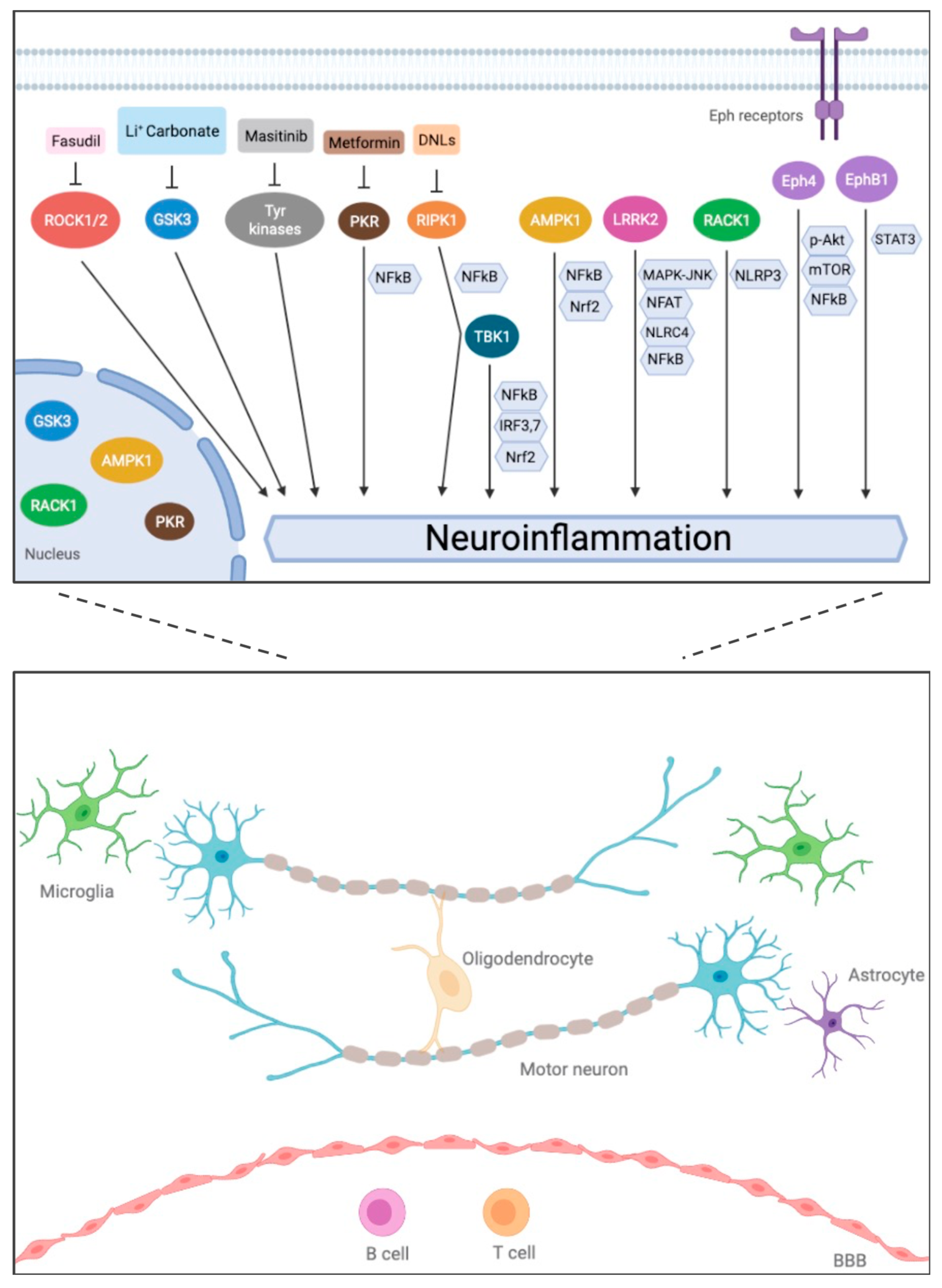
3.1. TBK1 (TANK-Binding Kinase 1)

TBK1, a pleiotropic serine/threonine kinase, is involved in the regulation of selective autophagy and inflammatory responses via type-I interferon (IFN) signaling [17]. Two independent human sequencing studies were the first to link different mutations in the TBK1 gene to ALS [15,16]. At the same time, three pioneering studies originally connected TBK1 mutations with FTD [15,18,19]. Thus far, several studies have identified a large number of TBK1 variations dispersed throughout the protein sequence to be a major cause of both familial and sporadic, ALS, FTD and ALS-FTD [17]. Some of them have been shown to cause a loss of kinase function [20], which resulted in rather evident abnormalities including dendritic swelling, abnormally shaped astrocytes, and p62-and ubiquitin-positive aggregates in the cerebellum, as observed in TBK1-KO mice [21]. However, the majority of ALS-associated TBK1 variants are missense mutations and their contribution to ALS pathogenesis still needs to be elucidated [17]. Interestingly, post-mortem brain analyses from ALS/FTD patients carrying TBK1 mutations have revealed cytoplasmic or perinuclear TDP-43-positive and p62-positive inclusions [16,19,22]. These findings may not seem surprising as TBK1 has been implicated in autophagy [17], while an ALS-associated TBK1 variant was unable to promote TDP-43 autophagic degradation dependent on p62 phosphorylation [23].
TBK1 is highly expressed in neurons among other cell types, and its expression and activation has also been observed in microglia [17,24]. TBK1 participates in immune signaling pathways by acting as an essential component of the type I IFN response (reviewed in [25]). Indeed, during the antiviral innate immune response, TBK1—which belongs to the IKK-kinase family—becomes activated and phosphorylates the downstream transcription factors IRF3 and IRF7, which dimerize, translocate to the nucleus, and initiate gene expression of IFNα and IFNβ cytokines. Recent studies in vitro have shown that ALS-associated TBK1 variants retain different levels of kinase activity and display a range of signaling efficiencies leading to IRF3 phosphorylation [26]. In addition, results from another in vitro study indicate that, whereas wild-type TBK1 promotes the NF-kB pathway and suppresses Nrf2 signaling, the ALS-associated p.G175S kinase-deficient, TBK1 mutant does not [23].
In addition, an essential role of TBK1 in inhibiting RIPK1-dependent apoptosis and inflammation, via phosphorylation of a particular Thr residue downstream of TNF receptor 1 (TNFR1), was demonstrated [27]. Remarkably, the study further showed that, in TBK1 deficient mice, the reduced myeloid expression of TAK1—an endogenous RIPK1 inhibitor—promoted the key ALS/FTD phenotype hallmarks, including microgliosis, TDP-43 aggregation, neuronal loss, and behavioral deficits [27]. Moreover, global heterozygous deletion of TBK1 modelling the TBK1 loss-of-function mutations found in ALS/FTD in humans, was seen to exert a dual and stage-specific effect in SOD1G93A mice, by initially preponing muscular denervation and clinical onset, but reducing microglial neuroinflammation and decelerating disease progression at later stages [28]. Interestingly, a recent study based both on general knockout and motor-neuron-selective deletion of TBK1 in SOD1G93A mice proposed that the observed reduction in gliosis in spinal cord is actually a non-cell autonomous result of selective loss of TBK1 in motor neurons [29], whereas decreased TBK1 kinase activity in all cells reduces the phosphorylation of IRF3 in SOD1G93A mice, resulting in suppression of IFN-stimulated genes in glial cells and leading to slowing of neurodegeneration [29].

3.2. RIPK1 (Receptor-Interacting Kinase 1)

RIPK1 is a death-domain containing Ser/Thr kinase which functions as a key regulator of cell death—including necroptosis and RIPK1-dependent apoptosis—and inflammatory responses, and has been implicated in a number of CNS diseases [30,31]. In particular, RIPK1 kinase activity has also been shown to promote inflammatory responses under caspase-deficient conditions [32]. Even though there is no causal evidence linking RIPK1 mutations to ALS, this gene was shown to mediate axonal degeneration by promoting inflammation and subsequent oligodendrocytes necroptosis in ALS [33], and a proinflammatory role of RIPK1 kinase activity in the CNS in association with TBK1 was demonstrated, as mentioned above [27]. Importantly, a recent study based on single-cell RNA sequencing has identified a RIPK1-regulated inflammatory microglial (RRIM) subset in early-stage SOD1G93A mice [34]. Differently from Disease-Associated Microglia (DAM), the RRIM state was characterized by up-regulation of classic proinflammatory pathways, and was found to peak at an earlier time point in the ALS model, compared to the DAM phenotype [34].

3.3. Eph Receptors (Ephrin Receptor Family)

The Eph receptors of tyrosine kinases, in combination with their ephrin ligands, constitute a bidirectional signaling mechanism known to mediate neuron–glia crosstalk and to undergo expression alterations following neuronal injury and neurodegeneration [35]. A link for these receptors with ALS was originally established when Epha4 was identified as a modulator of motor neuron degeneration and disease progression in fish and mouse disease models [36]. In addition to its role in the regulation of axonal growth and remodeling, Epha4 was found to function as a mediator of inflammation in spinal cord and traumatic brain injuries [37,38], potentially via the mTOR, p-Akt and NF-kB pathways [38].
Epha4-specific nanobodies [39], an EphA4 receptor soluble antagonist [40] and selective Epha4-binding compounds [41], were developed to treat ALS and delivered encouraging results in delaying disease progression in the SOD1G93A model. However, pre-symptomatic lowering of Epha4 levels in ALS mice, ubiquitously or specifically in CNS, by genetic abrogation or by administration of antisense oligonucleotides (ASOs) did not show a significant improvement in motor function, disease onset, or survival [42,43]. This probably reflects a highly complex mechanism of Epha4, or the ephrin system in general, in ALS pathogenic processes [14]. On the other hand, a study based on the SOD1G93A mouse model and on human iPSC-derived astrocytes provided evidence that EphB1-mediated signaling can induce astrocytic STAT3 activation, invoking a protective transcriptional profile [44]. Importantly, the authors also demonstrated that such EphB1-induced pathways are disrupted in ALS, resulting in impaired astrocyte activation and failure in response to neuronal injury [44].

3.4. RACK1 (Receptor of Activated Protein C Kinase 1)

RACK1, an intracellular protein receptor for protein kinase C (PKC), has been associated with ALS as it was found to co-localize with cytoplasmic TDP-43 in spinal motor neurons in patients [45]. Additionally, the same study revealed that RACK1—which functions as a ribosomal scaffold for several proteins—mediates TDP-43 binding to ribosomes and polysomes and recruitment of TDP-43 to stress granules [45]. Interestingly, RACK1 was also shown to modulate microglial resistance against LPS-induced inflammatory injury [46]. Moreover, non-ribosomal RACK1 was recently shown to mediate the activation of the inflammasome system—intracellular multiprotein complexes belonging to innate immunity that enable a rapid and powerful inflammatory response against multiple stimuli—by serving as a key NLRP3 interacting protein that promotes the NLRP3 active conformation and inflammasome assembly [47].

3.5. AMPK (AMP-Activated Protein Kinase) and LRRK2 (Leucine-Rich-Repeat Kinase 2), New Immune Signaling Kinases in ALS/FTD

Certain kinases with known functions in immune signaling have been recently involved in ALS or FTD based on studies using preclinical models and samples from patients. AMPK, a Ser/Thr kinase which is ubiquitously expressed in mammalian cells, including neurons and immune cells in the brain, is one such case. Intriguingly, studies have reported enhanced AMPK activation in spinal cord from SOD1G93A mice and spinal motor neurons from ALS patients, but diminished AMPK activation in spinal cord and brain from TDP-43 mutant mice (reviewed in [48]). AMPK functions as a master energy sensor for regulating cellular metabolism and maintaining energy homeostasis, and metabolic and energetic dysfunction are well-documented features in ALS [48]. In addition, AMPK plays a prominent role—shared with the mTOR complex 1 (mTORC1) kinase—in initiation of autophagy by activating the ULK1 complex. Indeed, there is mounting evidence that the autophagy-lysosome pathway is compromised, via multiple proposed mechanisms, of C9orf72 and TDP-43 ALS-FTD models (reviewed in [49,50]). Importantly, accumulating data based on cell culture studies and rodent animal models, also implicate AMPK in the regulation of neuroinflammation by promoting the shift of microglial activation from pro-inflammatory to anti-inflammatory states, via inhibition of NF-kB signaling and enhancement of the Nrf2 pathway (reviewed in [51]).
A recent genome-wide association study assessing the genetic overlap between FTD-related disorders and inflammatory diseases identified LRRK2 and TBK-binding protein 1 (TBKBP1) as FTD association candidates [52]. Two other genetic studies had previously proposed an association between LRRK2 mutations—well known to be a cause of Parkinson’s and Crohn’s diseases—and FTD [53,54] and a recent NGS-bioinformatics-based analysis of a sporadic FTD cohort, further supported this association [55]. LRRK2 is a multi-domain protein bearing GTPase and Ser/Thr kinase catalytic activities, as well as diverse protein interacting regions. LRRK2 is highly expressed in immune cells and has been widely implicated in diverse immune-related pathways in peripheral and central immune cells, including microglia (reviewed in [56,57]). In particular, results from studies based on inflammatory and neurodegenerative disease models indicate that LRRK2 promotes neuroinflammation by a number of mechanisms that may be dysregulated in microglia and other immune cells, including the activation of MAPK-JNK signaling [58,59,60], suppression of the NFAT cytokine-regulating transcription factor [61,62], dectin-1-mediated C-type lectin signaling [63], phosphorylation and activation of NLRC4-inflammasome [64], and promoting the NF-κB pathway by controlling the NF-κB p50 inhibitory signal and by negatively regulating the activation of protein kinase A (PKA), an inhibitor of NF-κB signaling [65,66].
Apart from its prominent role in immune signaling, accumulating evidence also supports a role of LRRK2 in autophagy and the lysosome pathway in immune cells, particularly in the context of inflammatory responses in monocytes, astrocytes and macrophages (reviewed in [57]). Interestingly, LRRK2 was found to interact with RIPK1 and to enable the formation of a distinct and essential ubiquitinated RIPK1 intermediate along the RIPK1-dependent apoptosis pathway [67,68]. However, possibly owing to the very recent connection established between LRRK2 and ALS/FTD, no study has thus far interrogated the possible role of LRRK2 in ALS/FTD immune dysregulation.

4. Other Emerging Signaling Kinases in ALS/FTD

Protein kinase RNA-activated (PKR) is a central intracellular stress sensor that can be activated by a variety of stimuli, particularly dsRNA, leading to autophosphorylation and resulting in proinflammatory responses via NF-kB, p38 and c-Jun N-terminal kinase (JNK) signaling, as part of the classical innate immunity to viral infection [69]. Repetitive element (RE) transcripts, which are prone to form dsRNA species, were found at higher levels in ALS patients’ brains [70], while mechanistic relationships between dysregulated TDP-43 function or C9orf72-repeat expansion, and increased transposable RE transcripts, have been suggested [70,71]. Interestingly, a recent study based on astrocyte primary cell cultures demonstrated that knocked-down TDP-43 leads to higher RE transcript and dsRNA levels [72]. Moreover, results from double knockdown assays support a mechanism by which PKR—which had been detected at higher levels in spinal cord samples from ALS patients [73]—mediates astrocyte activation and the ‘reactive’ proinflammatory phenotype resulting from TDP-43 downregulation [72]. On the other hand, a recent study demonstrated that PKR highly regulates repeat-associated non-AUG (RAN) translation [74], an aberrant mechanism reported in various ‘repeat expansion diseases’ such as C9orf72 ALS and FTD [75,76,77]. Furthermore, the study showed that repeat expansion RNAs, including ALS/FTD ‘C9 repeats, increase active/phosphorylated PKR (p-PKR) levels in vitro and in vivo in mice, while higher p-PKR levels were detected in hippocampal tissue from C9orf72 ALS patients [74].
Recently, as a result of our study of the inflammatory response in microglial cells exposed to TDP-43 aggregates, we identified MAPK/MAK/MRK overlapping kinase (MOK) as a protein that strongly interacts with cytoplasmic TDP-43 aggregates [78]. Moreover, MOK was found to alter its phosphorylation state in critical residue Tyr161, both in primary microglia and organotypic spinal cord cultures [78]. MOK is an atypical Ser/Thr signaling kinase that belongs to the MAPK superfamily [79], and which has been predicted to play an important role in diverse physiological and pathological processes [80].

5. Immune Kinase Modulation for ALS and FTD in Disease Managing and in Clinical Trials

Thus far, only two drugs have been approved for therapeutic treatment of ALS: Riluzole, by both the FDA and EMA agencies and also approved for FTD [81], and Edaravone, authorized more recently by the FDA [82]. Riluzole—via poorly understood mechanisms—displays activity against glutamatergic excitotoxic and non-excitotoxic oxidative neuronal injury, and has been used extensively in the clinic for 25 years, prolonging median survival by only two to three months in ALS patients [83]. Interestingly, Riluzole was later found to be a direct inhibitor of protein kinase C (PKC) [84], a kinase family well-known to play a key role in the development and effector functions of major immune cell types, and implicated in both innate and adaptive responses [85]. Because of the limited therapeutic power of these drugs, there is a clear need to identify novel molecular targets that could be potentially modulated in a specific and effective manner for the treatment of ALS and FTD. Currently, a few immune kinase inhibitors are being tested in clinical trials (Table 1).

5.1. Masitinib: A Receptor Tyr Kinase Inhibitor

Masitinib is a selective tyrosine kinase inhibitor [86] with neuroprotective and anti-inflammatory capability demonstrated in a number of preclinical studies [86,87,88,89]. Masitinib was shown to target a limited number of kinases known to be involved in cancer and inflammatory diseases, at safe doses [86] and was later demonstrated to reduce neuroinflammation by controlling microgliosis and counteracting mast cell degranulation [87,90]. The compound is thought to exert neuroprotection via its immunomodulatory properties through targeting microglia, macrophages, mast cells and neutrophils, and implicating both central and peripheral nervous systems [91,92]. Importantly, oral administration of this compound showed a 25% delay in disease progression in a recent phase II/III clinical as an add-on therapy to Riluzole in ALS patients [92]. Masitinib has now entered a phase III clinical trial, which is currently ongoing.

5.2. Fasudil: A ROCK1/2 (Rho-Kinase 1/2) Inhibitor

ROCK is a Ser/Thr kinase that is expressed as two homologues, ROCK1 and ROCK2, and whose activity is modulated by Ras-superfamily upstream regulators, namely the Rho GTPases RhoA and RhoC. ROCK phosphorylates a number of targets involved in regulation of a variety of crucial cellular processes including cell motility, apoptosis and cell survival in different cell types, including neurons and microglia (reviewed in [93,94]). In the latter, the ROCK pathway contributes to defining microglial phenotype by regulating microglial migration, phagocytosis and the release of inflammatory cytokines [95,96]. While abnormally high levels of ROCK were detected in skeletal muscle biopsies from sporadic ALS patients [97], pre-symptomatic administration of Fasudil to SOD1G93A mice improved survival and motor performance [98].
At a pathological level, motor neuron degeneration was shown to be partially suppressed and neuromuscular junction remodeling, enhanced. Interestingly, while the number of astroglial cells was reduced, Fasudil treatment led to increased microglia numbers and shifted activation profile from the neurotoxic M1 to the protective M2 phenotype [98], in line with previous in vitro and in vivo studies in other pathophysiological contexts [99,100]. Furthermore, oral administration of Fasudil to symptomatic SOD1G93A mice resulted in improved motor behavior in males [101]. Still, there is currently no sufficient evidence for a direct immune-based mechanism of ROCK in ALS pathology. Instead, recent pre-clinical studies with Fasudil in SOD1 mice provide evidence for participation of ROCK dysregulation in ALS pathogenesis caused by imbalance of pro-survival Rac/pro-apoptotic Rho-ROCK signaling in motor neurons [102] and by impaired ROCK-signaling controlling axonal regeneration [103]. This compound, applied intravenously, is currently undergoing a phase II clinical trial.

5.3. DNL Compounds: RIPK1 Inhibitors

Based on the functions recently described for RIPK1 kinase in regulating inflammation and necroptotic cell death, specific small-molecule inhibitors have been developed as treatment candidates for chronic inflammatory diseases [104,105]. In 2017 and shortly after its initiation, a phase Ia clinical study of DNL104—a brain-penetrant RIPK1 inhibitor—was discontinued due to liver toxicity effects. A selective and brain-permeable RIPK1 inhibitor DNL747 was designed subsequently and announced in 2020 for a phase Ib/II clinical trial in ALS [105], but the study was paused when it was concluded that higher doses—which were likely to cause adverse effects according to studies with cynomolgus monkeys [106]—would be needed to improve efficacy. Importantly, the alternative compound DNL788, which appears to display a superior profile and is expected to avoid the side effects seen at high doses for DNL747, is due to enter phase II clinical testing in 2022 [107].

5.4. Lithium Carbonate: A GSK3α/β (Glycogen Synthase Kinase 3) Inhibitor

GSK3 has been found to be a point of convergence of many signaling pathways and to regulate, in a highly complex manner, many cellular functions through its capacity to phosphorylate a wide variety of substrates [108]. Remarkably, studies in the last two decades have revealed that GSK3 participates in several signaling pathways and is an important regulator of innate immune responses by enabling cytokine production upon TLR stimulation in both the periphery and the CNS, and of adaptive immunity by modulating the specificity and clonal expansion of T and B cells [108,109]. Based on its role as a major tau kinase, inhibition of GSK3 with lithium—the gold-standard treatment for bipolar disorder—has been tested for the treatment of tauopathies, including AD and subtypes of FTD. In cultured cells and in transgenic mice, lithium-induced GSK3β inhibition was seen to reduce tau phosphorylation, increase the binding of tau to microtubules and promote microtubule assembly [110,111]. Interestingly, Li+ has been shown to rescue autophagy failure observed in ALS/FTD and, in recent years, lithium administration has been assessed in clinical trials in ALS, thus far with debatable results [112]. Of note, a phase II clinical trial with low-dose lithium (against placebo) for the treatment of behavioral symptoms in FTD is currently ongoing [113].

6. Immune Kinase Modulators in the Pipeline towards ALS/FTD Therapy

6.1. Metformin: A PKR Inhibitor

Based on results obtained in vitro which showed that metformin—a widely prescribed drug for type 2 diabetes treatment, known to activate the AMPK pathway [114]—inhibits PKR and reduces the levels of several RAN proteins in mammalian cells, the effect of this drug was tested in vivo by using C9-BAC mice, a C9orf72-based pre-clinical model of ALS [74]. Administration of metformin at the pre-symptomatic stage resulted in lowered DPR aggregates and p-PKR levels. Moreover, as opposed to a previous study using the SOD1G93A ALS model [115], treatment of C9-BAC mice with the drug up until the advanced stage led to substantially reduced astrogliosis in the motor cortex, decreased motor neuron degeneration in the spinal cord, and improved locomotive and behavioral parameters, compared to non-treated animals [74].

6.2. C4: An Epha4 Ligand

Following encouraging results by a study showing significant delay in disease progression in the SOD1G93A mouse model [41], the brain-penetrant, high-selectivity and high-affinity small molecule EPHa4 ligand 123C4—presumably a receptor agonist—is currently being evaluated in preclinical models for ALS by Iron Horse Therapeutics (San Diego, CA, USA) [88].

7. Concluding Remarks

In the last few years, it has become increasingly evident that ALS and FTD share clinical, genetic, and mechanistic cues, a feature which may provide hints on their elusive pathogenic mechanisms. A number of signaling pathways and their executing kinases—many of which are known to mediate immune functions, particularly, inflammatory responses—have been recently linked to ALS or FTD. Some of them have been associated genetically to these disorders, such as TBK1; others, including e.g., RIPK1, do not seem to have a direct genetic link with these disorders, but are known to participate in their pathogenic mechanisms.
An increasing number of studies in recent years point to a mechanistic link between mitochondrial and autophagy disruption in neurodegenerative disorders, including ALS and FTD. Interestingly, neuroinflammation, which has emerged as a principal determinant of neurodegeneration in these disorders, has been recently linked to both dysregulated processes [17,116], adding a possible new layer of complexity to the neuroinflammation–neurodegeneration interaction network.
Based on the growing body of knowledge generated in the last two decades in ALS and especially in AD and other related neurodegenerative diseases, a number of immune kinases have emerged as highly promising molecular targets for therapeutic intervention. Indeed, a short list of immune kinase inhibitors are currently being tested in clinical trials or are undergoing pre-clinical studies. Surely, due to a virtual lack of an effective therapy for ALS and FTD patients, further investigation towards the identification of novel therapeutic targets is warranted.
From the reviewed data, it seems obvious that, despite the progress made over the last two decades, substantial effort must still be made in the field to improve our understanding of the intricate mechanisms underlying ALS and FTD pathogenesis, including immune signaling pathways, which would prompt the identification of novel kinase therapeutic targets. In addition to a better understanding of these highly complex pathogenic processes, better stratification of patients would improve the design and outcome of clinical trials and, thereby, boost therapeutic development.

Author Contributions

Conceptualization, R.G.-G., L.M.-H. and C.R.; investigation and writing, R.G.-G., L.M.-H., L.B.-P., J.P.-C. and C.R.; review and editing, R.G.-G., L.M.-H. and C.R.; supervision, C.R. All authors have read and agreed to the published version of the manuscript.

Funding

Financial support to C.R. has been granted by the Spanish Ministry of Economy (RTI2018-098432-B-I00), Fundación ‘Ramón Areces’ (CIVP19A5938), Andalusian Regional Government-FEDER (US-1265227) and I+D PAIDI (PY20_01097), and ‘Programa Ramón y Cajal’ of the Spanish Ministry of Economy (RYC-2017-23127).

Institutional Review Board Statement

Not applicable.

Informed Consent Statement

Not applicable.

Data Availability Statement

Not applicable.

Acknowledgments

Research on immune signaling and neurodegenerative proteinopathies in our laboratory has been supported by the Spanish Ministry of Economy, Fundación Ramón Areces, the Andalusian Regional Government, and FEDER (European Regional Development Fund).

Conflicts of Interest

The authors declare no conflict of interest.

References

  1. Caballero-Hernandez, D.; Toscano, M.G.; Cejudo-Guillen, M.; Garcia-Martin, M.L.; Lopez, S.; Franco, J.M.; Quintana, F.J.; Roodveldt, C.; Pozo, D. The “Omics” of Amyotrophic Lateral Sclerosis. Trends Mol. Med. 2016, 22, 53–67. [Google Scholar] [CrossRef]
  2. Jimenez-Pacheco, A.; Franco, J.M.; Lopez, S.; Gomez-Zumaquero, J.M.; Magdalena Leal-Lasarte, M.; Caballero-Hernandez, D.E.; Cejudo-Guillén, M.; Pozo, D. Epigenetic Mechanisms of Gene Regulation in Amyotrophic Lateral Sclerosis. Adv. Exp. Med. Biol. 2017, 978, 255–275. [Google Scholar] [CrossRef] [PubMed]
  3. McCauley, M.E.; Baloh, R.H. Inflammation in ALS/FTD pathogenesis. Acta Neuropathol. 2019, 137, 715–730. [Google Scholar] [CrossRef] [PubMed] [Green Version]
  4. Beers, D.R.; Appel, S.H. Immune dysregulation in amyotrophic lateral sclerosis: Mechanisms and emerging therapies. Lancet Neurol. 2019, 18, 211–220. [Google Scholar] [CrossRef]
  5. Mejzini, R.; Flynn, L.L.; Pitout, I.L.; Fletcher, S.; Wilton, S.D.; Akkari, P.A. ALS Genetics, Mechanisms, and Therapeutics: Where Are We Now? Front. Neurosci. 2019, 13, 1310. [Google Scholar] [CrossRef] [Green Version]
  6. Wood, A.; Gurfinkel, Y.; Polain, N.; Lamont, W.; Lyn Rea, S. Molecular Mechanisms Underlying TDP-43 Pathology in Cellular and Animal Models of ALS and FTLD. Int. J. Mol. Sci. 2021, 22, 4705. [Google Scholar] [CrossRef]
  7. Ransohoff, R.M. How neuroinflammation contributes to neurodegeneration. Science 2016, 353, 777–783. [Google Scholar] [CrossRef]
  8. Lomen-Hoerth, C.; Anderson, T.; Miller, B. The overlap of amyotrophic lateral sclerosis and frontotemporal dementia. Neurology 2002, 59, 1077–1079. [Google Scholar] [CrossRef]
  9. Strong, M.J.; Grace, G.M.; Freedman, M.; Lomen-Hoerth, C.; Woolley, S.; Goldstein, L.H.; Murphy, J.; Shoesmith, C.; Rosenfeld, J.; Leigh, P.N.; et al. Consensus criteria for the diagnosis of frontotemporal cognitive and behavioural syndromes in amyotrophic lateral sclerosis. Amyotroph. Lateral Scler. 2009, 10, 131–146. [Google Scholar] [CrossRef] [PubMed]
  10. Abramzon, Y.A.; Fratta, P.; Traynor, B.J.; Chia, R. The Overlapping Genetics of Amyotrophic Lateral Sclerosis and Frontotemporal Dementia. Front. Neurosci. 2020, 14, 42. [Google Scholar] [CrossRef] [Green Version]
  11. Mannini, B.; Vecchi, G.; Labrador-Garrido, A.; Fabre, B.; Fani, G.; Franco, J.M.; Lilley, K.; Pozo, D.; Vendruscolo, M.; Chiti, F.; et al. Differential Interactome and Innate Immune Response Activation of Two Structurally Distinct Misfolded Protein Oligomers. ACS Chem. Neurosci. 2019, 10, 3464–3478. [Google Scholar] [CrossRef] [PubMed]
  12. Leal-Lasarte, M.; Mannini, B.; Chiti, F.; Vendruscolo, M.; Dobson, C.M.; Roodveldt, C.; Pozo, D. Distinct responses of human peripheral blood cells to different misfolded protein oligomers. Immunology 2021, 64, 358–371. [Google Scholar] [CrossRef] [PubMed]
  13. Haukedal, H.; Freude, K. Implications of Microglia in Amyotrophic Lateral Sclerosis and Frontotemporal Dementia. J. Mol. Biol. 2019, 431, 1818–1829. [Google Scholar] [CrossRef] [PubMed]
  14. Guo, W.; Vandoorne, T.; Steyaert, J.; Staats, K.A.; Van Den Bosch, L. The multifaceted role of kinases in amyotrophic lateral sclerosis: Genetic, pathological and therapeutic implications. Brain 2020, 143, 1651–1673. [Google Scholar] [CrossRef] [PubMed]
  15. Cirulli, E.T.; Lasseigne, B.N.; Petrovski, S.; Sapp, P.C.; Dion, P.A.; Leblond, C.S.; Couthouis, J.; Lu, Y.F.; Wang, Q.; Krueger, B.J.; et al. Exome sequencing in amyotrophic lateral sclerosis identifies risk genes and pathways. Science 2015, 347, 1436–1441. [Google Scholar] [CrossRef] [Green Version]
  16. Freischmidt, A.; Wieland, T.; Richter, B.; Ruf, W.; Schaeffer, V.; Müller, K.; Marroquin, N.; Nordin, F.; Hübers, A.; Weydt, P.; et al. Haploinsufficiency of TBK1 causes familial ALS and fronto-temporal dementia. Nat. Neurosci. 2015, 18, 631–636. [Google Scholar] [CrossRef]
  17. Oakes, J.A.; Davies, M.C.; Collins, M.O. TBK1: A new player in ALS linking autophagy and neuroinflammation. Mol. Brain 2017, 10, 5. [Google Scholar] [CrossRef] [Green Version]
  18. Gijselinck, I.; Van Mossevelde, S.; van der Zee, J.; Sieben, A.; Philtjens, S.; Heeman, B.; Engelborghs, S.; Vandenbulcke, M.; De Baets, G.; Bäumer, V.; et al. Loss of TBK1 is a frequent cause of frontotemporal dementia in a Belgian cohort. Neurology 2015, 85, 2116–2125. [Google Scholar] [CrossRef] [Green Version]
  19. Pottier, C.; Bieniek, K.F.; Finch, N.; van de Vorst, M.; Baker, M.; Perkersen, R.; Brown, P.; Ravenscroft, T.; van Blitterswijk, M.; Nicholson, A.M.; et al. Whole-genome sequencing reveals important role for TBK1 and OPTN mutations in frontotemporal lobar degeneration without motor neuron disease. Acta Neuropathol. 2015, 130, 77–92. [Google Scholar] [CrossRef]
  20. de Majo, M.; Topp, S.D.; Smith, B.N.; Nishimura, A.L.; Chen, H.J.; Gkazi, A.S.; Miller, J.; Wong, C.H.; Vance, C.; Baas, F.; et al. ALS-associated missense and nonsense TBK1 mutations can both cause loss of kinase function. Neurobiol. Aging 2018, 71, 266.e1–266.e10. [Google Scholar] [CrossRef]
  21. Duan, W.; Guo, M.; Yi, L.; Zhang, J.; Bi, Y.; Liu, Y.; Li, Y.; Li, Z.; Ma, Y.; Zhang, G.; et al. Deletion of Tbk1 disrupts autophagy and reproduces behavioral and locomotor symptoms of FTD-ALS in mice. Aging 2019, 11, 2457–2476. [Google Scholar] [CrossRef]
  22. van der Zee, J.; Gijselinck, I.; Van Mossevelde, S.; Perrone, F.; Dillen, L.; Heeman, B.; Bäumer, V.; Engelborghs, S.; De Bleecker, J.; Baets, J.; et al. TBK1 Mutation Spectrum in an Extended European Patient Cohort with Frontotemporal Dementia and Amyotrophic Lateral Sclerosis. Hum. Mutat. 2017, 38, 297–309. [Google Scholar] [CrossRef] [Green Version]
  23. Foster, A.D.; Downing, P.; Figredo, E.; Polain, N.; Stott, A.; Layfield, R.; Rea, S.L. ALS-associated TBK1 variant p.G175S is defective in phosphorylation of p62 and impacts TBK1-mediated signalling and TDP-43 autophagic degradation. Mol. Cell. Neurosci. 2020, 108, 103539. [Google Scholar] [CrossRef]
  24. Moore, A.S.; Holzbaur, E.L. Dynamic recruitment and activation of ALS-associated TBK1 with its target optineurin are required for efficient mitophagy. Proc. Natl. Acad. Sci. USA 2016, 113, E3349–E3358. [Google Scholar] [CrossRef] [PubMed] [Green Version]
  25. Ahmad, L.; Zhang, S.Y.; Casanova, J.L.; Sancho-Shimizu, V. Human TBK1: A Gatekeeper of Neuroinflammation. Trends Mol. Med. 2016, 22, 511–527. [Google Scholar] [CrossRef] [PubMed]
  26. Ye, J.; Cheung, J.; Gerbino, V.; Ahlsén, G.; Zimanyi, C.; Hirsh, D.; Maniatis, T. Effects of ALS-associated TANK binding kinase 1 mutations on protein-protein interactions and kinase activity. Proc. Natl. Acad. Sci. USA 2019, 116, 24517–24526. [Google Scholar] [CrossRef] [PubMed]
  27. Xu, D.; Jin, T.; Zhu, H.; Chen, H.; Ofengeim, D.; Zou, C.; Mifflin, L.; Pan, L.; Amin, P.; Li, W.; et al. TBK1 Suppresses RIPK1-Driven Apoptosis and Inflammation during Development and in Aging. Cell 2018, 174, 1477–1491.e19. [Google Scholar] [CrossRef] [Green Version]
  28. Brenner, D.; Sieverding, K.; Bruno, C.; Lüningschrör, P.; Buck, E.; Mungwa, S.; Fischer, L.; Brockmann, S.J.; Ulmer, J.; Bliederhäuser, C.; et al. Heterozygous Tbk1 loss has opposing effects in early and late stages of ALS in mice. J. Exp. Med. 2019, 216, 267–278. [Google Scholar] [CrossRef] [Green Version]
  29. Gerbino, V.; Kaunga, E.; Ye, J.; Canzio, D.; O’Keeffe, S.; Rudnick, N.D.; Guarnieri, P.; Lutz, C.M.; Maniatis, T. The Loss of TBK1 Kinase Activity in Motor Neurons or in All Cell Types Differentially Impacts ALS Disease Progression in SOD1 Mice. Neuron 2020, 106, 789–805.e5. [Google Scholar] [CrossRef]
  30. Yuan, J.; Amin, P.; Ofengeim, D. Necroptosis and RIPK1-mediated neuroinflammation in CNS diseases. Nat. Rev. Neurosci. 2019, 20, 19–33. [Google Scholar] [CrossRef]
  31. Humphries, F.; Yang, S.; Wang, B.; Moynagh, P.N. RIP kinases: Key decision makers in cell death and innate immunity. Cell Death Differ. 2015, 22, 225–236. [Google Scholar] [CrossRef] [Green Version]
  32. Christofferson, D.E.; Li, Y.; Hitomi, J.; Zhou, W.; Upperman, C.; Zhu, H.; Gerber, S.A.; Gygi, S.; Yuan, J. A novel role for RIP1 kinase in mediating TNFα production. Cell Death Dis. 2012, 3, e320. [Google Scholar] [CrossRef]
  33. Ito, Y.; Ofengeim, D.; Najafov, A.; Das, S.; Saberi, S.; Li, Y.; Hitomi, J.; Zhu, H.; Chen, H.; Mayo, L.; et al. RIPK1 mediates axonal degeneration by promoting inflammation and necroptosis in ALS. Science 2016, 353, 603–608. [Google Scholar] [CrossRef] [PubMed] [Green Version]
  34. Mifflin, L.; Hu, Z.; Dufort, C.; Hession, C.C.; Walker, A.J.; Niu, K.; Zhu, H.; Liu, N.; Liu, J.S.; Levin, J.Z.; et al. A RIPK1-regulated inflammatory microglial state in amyotrophic lateral sclerosis. Proc. Natl. Acad. Sci. USA 2021, 118, e2025102118. [Google Scholar] [CrossRef]
  35. Kania, A.; Klein, R. Mechanisms of ephrin-Eph signalling in development, physiology and disease. Nat. Rev. Mol. Cell Biol. 2016, 17, 240–256. [Google Scholar] [CrossRef] [PubMed]
  36. Van Hoecke, A.; Schoonaert, L.; Lemmens, R.; Timmers, M.; Staats, K.A.; Laird, A.S.; Peeters, E.; Philips, T.; Goris, A.; Dubois, B.; et al. EPHA4 is a disease modifier of amyotrophic lateral sclerosis in animal models and in humans. Nat. Med. 2012, 18, 1418–1422. [Google Scholar] [CrossRef] [PubMed]
  37. Goldshmit, Y.; Galea, M.P.; Wise, G.; Bartlett, P.F.; Turnley, A.M. Axonal regeneration and lack of astrocytic gliosis in EphA4-deficient mice. J. Neurosci. 2004, 24, 10064–10073. [Google Scholar] [CrossRef] [Green Version]
  38. Kowalski, E.A.; Chen, J.; Hazy, A.; Fritsch, L.E.; Gudenschwager-Basso, E.K.; Chen, M.; Wang, X.; Qian, Y.; Zhou, M.; Byerly, M.; et al. Peripheral loss of EphA4 ameliorates TBI-induced neuroinflammation and tissue damage. J. Neuroinflamm. 2019, 16, 210. [Google Scholar] [CrossRef]
  39. Schoonaert, L.; Rué, L.; Roucourt, B.; Timmers, M.; Little, S.; Chávez-Gutiérrez, L.; Dewilde, M.; Joyce, P.; Curnock, A.; Weber, P.; et al. Identification and characterization of Nanobodies targeting the EphA4 receptor. J. Biol. Chem. 2017, 292, 11452–11465. [Google Scholar] [CrossRef] [Green Version]
  40. Zhao, J.; Cooper, L.T.; Boyd, A.W.; Bartlett, P.F. Decreased signalling of EphA4 improves functional performance and motor neuron survival in the SOD1 G93A ALS mouse model. Sci. Rep. 2018, 8, 11393. [Google Scholar] [CrossRef] [Green Version]
  41. Wu, B.; De, S.K.; Kulinich, A.; Salem, A.F.; Koeppen; Wang, R.; Barile, E.; Wang, S.; Zhang, D.; Ethell, I.; et al. Potent and Selective EphA4 Agonists for the Treatment of ALS. Cell Chem. Biol. 2017, 24, 293–305. [Google Scholar] [CrossRef] [Green Version]
  42. Ling, K.K.; Jackson, M.; Alkam, D.; Liu, D.; Allaire, N.; Sun, C.; Kiaei, M.; McCampbell, A.; Rigo, F. Antisense-mediated reduction of EphA4 in the adult CNS does not improve the function of mice with amyotrophic lateral sclerosis. Neurobiol. Dis. 2018, 114, 174–183. [Google Scholar] [CrossRef] [PubMed]
  43. Rué, L.; Timmers, M.; Lenaerts, A.; Smolders, S.; Poppe, L.; de Boer, A.; Van Den Bosch, L.; Van Damme, P.; Robberecht, W.; Lemmens, R. Reducing EphA4 before disease onset does not affect survival in a mouse model of Amyotrophic Lateral Sclerosis. Sci. Rep. 2019, 9, 14112. [Google Scholar] [CrossRef] [Green Version]
  44. Tyzack, G.E.; Hall, C.E.; Sibley; Cymes, T.; Forostyak, S.; Carlino, G.; Meyer, I.F.; Schiavo, G.; Zhang, S.C.; Gibbons, G.M.; et al. A neuroprotective astrocyte state is induced by neuronal signal EphB1 but fails in ALS models. Nat. Commun. 2017, 8, 1164. [Google Scholar] [CrossRef] [PubMed]
  45. Russo, A.; Scardigli, R.; La Regina, F.; Murray, M.E.; Romano, N.; Dickson, D.W.; Wolozin, B.; Cattaneo, A.; Ceci, M. Increased cytoplasmic TDP-43 reduces global protein synthesis by interacting with RACK1 on polyribosomes. Hum. Mol. Genet. 2017, 26, 1407–1418. [Google Scholar] [CrossRef] [Green Version]
  46. Yin, H.; Song, S.; Pan, X. Knockdown of miR-155 protects microglia against LPS-induced inflammatory injury via targeting RACK1: A novel research for intracranial infection. J. Inflamm. 2017, 14, 17. [Google Scholar] [CrossRef] [PubMed] [Green Version]
  47. Duan, Y.; Zhang, L.; Angosto-Bazarra, D.; Pelegrín, P.; Núñez, G.; He, Y. RACK1 Mediates NLRP3 Inflammasome Activation by Promoting NLRP3 Active Conformation and Inflammasome Assembly. Cell Rep. 2020, 33, 108405. [Google Scholar] [CrossRef]
  48. Perera, N.D.; Turner, B.J. AMPK Signalling and Defective Energy Metabolism in Amyotrophic Lateral Sclerosis. Neurochem. Res. 2016, 41, 544–553. [Google Scholar] [CrossRef]
  49. Beckers, J.; Tharkeshwar, A.K.; Van Damme, P. C9orf72 ALS-FTD: Recent evidence for dysregulation of the autophagy-lysosome pathway at multiple levels. Autophagy 2021, 17, 3306–3322. [Google Scholar] [CrossRef]
  50. Root, J.; Merino, P.; Nuckols, A.; Johnson, M.; Kukar, T. Lysosome dysfunction as a cause of neurodegenerative diseases: Lessons from frontotemporal dementia and amyotrophic lateral sclerosis. Neurobiol. Dis. 2021, 154, 105360. [Google Scholar] [CrossRef]
  51. Saito, M.; Saito, M.; Das, B.C. Involvement of AMP-activated protein kinase in neuroinflammation and neurodegeneration in the adult and developing brain. Int. J. Dev. Neurosci. 2019, 77, 48–59. [Google Scholar] [CrossRef]
  52. Broce, I.; Karch, C.M.; Wen, N.; Fan, C.C.; Wang, Y.; Tan, C.H.; Kouri, N.; Ross, O.A.; Höglinger, G.U.; Muller, U.; et al. Immune-related genetic enrichment in frontotemporal dementia: An analysis of genome-wide association studies. PLoS Med. 2018, 15, e1002487. [Google Scholar] [CrossRef]
  53. Dächsel, J.C.; Ross, O.A.; Mata, I.F.; Kachergus, J.; Toft, M.; Cannon, A.; Baker, M.; Adamson, J.; Hutton, M.; Dickson, D.W.; et al. Lrrk2 G2019S substitution in frontotemporal lobar degeneration with ubiquitin-immunoreactive neuronal inclusions. Acta Neuropathol. 2007, 113, 601–606. [Google Scholar] [CrossRef]
  54. Ferrari, R.; Wang, Y.; Vandrovcova, J.; Guelfi, S.; Witeolar, A.; Karch, C.M.; Schork, A.J.; Fan, C.C.; Brewer, J.B.; Momeni, P.; et al. Genetic architecture of sporadic frontotemporal dementia and overlap with Alzheimer’s and Parkinson’s diseases. J. Neurol. Neurosurg. Psychiatry 2017, 88, 152–164. [Google Scholar] [CrossRef]
  55. Ciani, M.; Bonvicini, C.; Scassellati, C.; Carrara, M.; Maj, C.; Fostinelli, S.; Binetti, G.; Ghidoni, R.; Benussi, L. The Missing Heritability of Sporadic Frontotemporal Dementia: New Insights from Rare Variants in Neurodegenerative Candidate Genes. Int. J. Mol. Sci. 2019, 20, 3903. [Google Scholar] [CrossRef] [Green Version]
  56. Ahmadi Rastegar, D.; Dzamko, N. Leucine Rich Repeat Kinase 2 and Innate Immunity. Front. Neurosci. 2020, 14, 193. [Google Scholar] [CrossRef] [Green Version]
  57. Wallings, R.L.; Tansey, M.G. LRRK2 regulation of immune-pathways and inflammatory disease. Biochem. Soc. Trans. 2019, 47, 1581–1595. [Google Scholar] [CrossRef] [PubMed]
  58. Gloeckner, C.J.; Schumacher, A.; Boldt, K.; Ueffing, M. The Parkinson disease-associated protein kinase LRRK2 exhibits MAPKKK activity and phosphorylates MKK3/6 and MKK4/7, in vitro. J. Neurochem. 2009, 109, 959–968. [Google Scholar] [CrossRef] [PubMed]
  59. Hsu, C.H.; Chan, D.; Wolozin, B. LRRK2 and the stress response: Interaction with MKKs and JNK-interacting proteins. Neurodegener. Dis. 2010, 7, 68–75. [Google Scholar] [CrossRef]
  60. Chen, C.Y.; Weng, Y.H.; Chien, K.Y.; Lin, K.J.; Yeh, T.H.; Cheng, Y.P.; Lu, C.S.; Wang, H.L. (G2019S) LRRK2 activates MKK4-JNK pathway and causes degeneration of SN dopaminergic neurons in a transgenic mouse model of PD. Cell Death Differ. 2012, 19, 1623–1633. [Google Scholar] [CrossRef] [Green Version]
  61. Liu, Z.; Lee, J.; Krummey, S.; Lu, W.; Cai, H.; Lenardo, M.J. The kinase LRRK2 is a regulator of the transcription factor NFAT that modulates the severity of inflammatory bowel disease. Nat. Immunol. 2011, 12, 1063–1070. [Google Scholar] [CrossRef] [Green Version]
  62. Panagiotakopoulou, V.; Ivanyuk, D.; De Cicco, S.; Haq, W.; Arsić, A.; Yu, C.; Messelodi, D.; Oldrati, M.; Schöndorf, D.C.; Perez, M.J.; et al. Interferon-γ signaling synergizes with LRRK2 in neurons and microglia derived from human induced pluripotent stem cells. Nat. Commun. 2020, 11, 5163. [Google Scholar] [CrossRef] [PubMed]
  63. Takagawa, T.; Kitani, A.; Fuss, I.; Levine, B.; Brant, S.R.; Peter, I.; Tajima, M.; Nakamura, S.; Strober, W. An increase in LRRK2 suppresses autophagy and enhances Dectin-1-induced immunity in a mouse model of colitis. Sci. Transl. Med. 2018, 10, eaan8162. [Google Scholar] [CrossRef] [PubMed] [Green Version]
  64. Liu, W.; Liu, X.; Li, Y.; Zhao, J.; Liu, Z.; Hu, Z.; Wang, Y.; Yao, Y.; Miller, A.W.; Su, B.; et al. LRRK2 promotes the activation of NLRC4 inflammasome during Salmonella Typhimurium infection. J. Exp. Med. 2017, 214, 3051–3066. [Google Scholar] [CrossRef] [PubMed]
  65. Russo, I.; Berti, G.; Plotegher, N.; Bernardo, G.; Filograna, R.; Bubacco, L.; Greggio, E. Leucine-rich repeat kinase 2 positively regulates inflammation and down-regulates NF-κB p50 signaling in cultured microglia cells. J. Neuroinflamm. 2015, 12, 230. [Google Scholar] [CrossRef] [PubMed] [Green Version]
  66. Russo, I.; Di Benedetto, G.; Kaganovich, A.; Ding, J.; Mercatelli, D.; Morari, M.; Cookson, M.R.; Bubacco, L.; Greggio, E. Leucine-rich repeat kinase 2 controls protein kinase A activation state through phosphodiesterase 4. J. Neuroinflamm. 2018, 15, 297. [Google Scholar] [CrossRef]
  67. Ho, C.C.-Y.; Rideout, H.J.; Ribe, E.; Troy, C.M.; Dauer, W.T. The Parkinson Disease Protein Leucine-Rich Repeat Kinase 2 Transduces Death Signals via Fas-Associated Protein with Death Domain and Caspase-8 in a Cellular Model of Neurodegeneration. J. Neurosci. 2009, 29, 1011. [Google Scholar] [CrossRef]
  68. Amin, P.; Florez, M.; Najafov, A.; Pan, H.; Geng, J.; Ofengeim, D.; Dziedzic, S.A.; Wang, H.; Barrett, V.J.; Ito, Y.; et al. Regulation of a distinct activated RIPK1 intermediate bridging complex I and complex II in TNFα-mediated apoptosis. Proc. Natl. Acad. Sci. USA 2018, 115, E5944. [Google Scholar] [CrossRef] [Green Version]
  69. Balachandran, S.; Roberts, P.C.; Brown, L.E.; Truong, H.; Pattnaik, A.K.; Archer, D.R.; Barber, G.N. Essential role for the dsRNA-dependent protein kinase PKR in innate immunity to viral infection. Immunity 2000, 13, 129–141. [Google Scholar] [CrossRef] [Green Version]
  70. Prudencio, M.; Gonzales, P.K.; Cook, C.N.; Gendron, T.F.; Daughrity, L.M.; Song, Y.; Ebbert, M.T.W.; van Blitterswijk, M.; Zhang, Y.J.; Jansen-West, K.; et al. Repetitive element transcripts are elevated in the brain of C9orf72 ALS/FTLD patients. Hum. Mol. Genet. 2017, 26, 3421–3431. [Google Scholar] [CrossRef]
  71. Li, W.; Jin, Y.; Prazak, L.; Hammell, M.; Dubnau, J. Transposable elements in TDP-43-mediated neurodegenerative disorders. PLoS ONE 2012, 7, e44099. [Google Scholar] [CrossRef] [PubMed] [Green Version]
  72. LaRocca, T.J.; Mariani, A.; Watkins, L.R.; Link, C.D. TDP-43 knockdown causes innate immune activation via protein kinase R in astrocytes. Neurobiol. Dis. 2019, 132, 104514. [Google Scholar] [CrossRef]
  73. Hu, J.H.; Zhang, H.; Wagey, R.; Krieger, C.; Pelech, S.L. Protein kinase and protein phosphatase expression in amyotrophic lateral sclerosis spinal cord. J. Neurochem. 2003, 85, 432–442. [Google Scholar] [CrossRef] [PubMed]
  74. Zu, T.; Guo, S.; Bardhi, O.; Ryskamp, D.A.; Li, J.; Khoramian Tusi, S.; Engelbrecht, A.; Klippel, K.; Chakrabarty, P.; Nguyen, L.; et al. Metformin inhibits RAN translation through PKR pathway and mitigates disease in C9orf72 ALS/FTD mice. Proc. Natl. Acad. Sci. USA 2020, 117, 18591–18599. [Google Scholar] [CrossRef]
  75. Ash, P.E.; Bieniek, K.F.; Gendron, T.F.; Caulfield, T.; Lin, W.L.; Dejesus-Hernandez, M.; van Blitterswijk, M.M.; Jansen-West, K.; Paul, J.W.; Rademakers, R.; et al. Unconventional translation of C9ORF72 GGGGCC expansion generates insoluble polypeptides specific to c9FTD/ALS. Neuron 2013, 77, 639–646. [Google Scholar] [CrossRef] [Green Version]
  76. Mori, K.; Weng, S.M.; Arzberger, T.; May, S.; Rentzsch, K.; Kremmer, E.; Schmid, B.; Kretzschmar, H.A.; Cruts, M.; Van Broeckhoven, C.; et al. The C9orf72 GGGGCC repeat is translated into aggregating dipeptide-repeat proteins in FTLD/ALS. Science 2013, 339, 1335–1338. [Google Scholar] [CrossRef]
  77. Zu, T.; Liu, Y.; Bañez-Coronel, M.; Reid, T.; Pletnikova, O.; Lewis, J.; Miller, T.M.; Harms, M.B.; Falchook, A.E.; Subramony, S.H.; et al. RAN proteins and RNA foci from antisense transcripts in C9ORF72 ALS and frontotemporal dementia. Proc. Natl. Acad. Sci. USA 2013, 110, E4968–E4977. [Google Scholar] [CrossRef] [Green Version]
  78. Leal-Lasarte, M.M.; Franco, J.M.; Labrador-Garrido, A.; Pozo, D.; Roodveldt, C. Extracellular TDP-43 aggregates target MAPK/MAK/MRK overlapping kinase (MOK) and trigger caspase-3/IL-18 signaling in microglia. FASEB J. 2017, 31, 2797–2816. [Google Scholar] [CrossRef] [PubMed] [Green Version]
  79. Miyata, Y.; Akashi, M.; Nishida, E. Molecular cloning and characterization of a novel member of the MAP kinase superfamily. Genes Cells 1999, 4, 299–309. [Google Scholar] [CrossRef]
  80. Fu, Z. Regulations and Functions of ICK/MAK/MOK—A Novel MAPK-Related Kinase Family Linked to Human Diseases. In Protein Kinases; InTech: London, UK, 2012. [Google Scholar]
  81. Panza, F.; Lozupone, M.; Seripa, D.; Daniele, A.; Watling, M.; Giannelli, G.; Imbimbo, B.P. Development of disease-modifying drugs for frontotemporal dementia spectrum disorders. Nat. Rev. Neurol. 2020, 16, 213–228. [Google Scholar] [CrossRef] [PubMed]
  82. Jaiswalv, M.K. Riluzole and edaravone: A tale of two amyotrophic lateral sclerosis drugs. Med. Res. Rev. 2019, 39, 733–748. [Google Scholar] [CrossRef] [PubMed]
  83. Cheah, B.C.; Vucic, S.; Krishnan, A.V.; Kiernan, K. Riluzole, neuroprotection and amyotrophic lateral sclerosis. Curr. Med. Chem. 2010, 17, 1942–1959. [Google Scholar] [CrossRef]
  84. Noh, K.M.; Hwang, J.Y.; Shin, H.C.; Koh, J.Y. A novel neuroprotective mechanism of riluzole: Direct inhibition of protein kinase C. Neurobiol. Dis. 2000, 7, 375–383. [Google Scholar] [CrossRef] [Green Version]
  85. Altman, A.; Kong, K.F. Protein Kinase C Enzymes in the Hematopoietic and Immune Systems. Annu. Rev. Immunol. 2016, 34, 511–538. [Google Scholar] [CrossRef] [PubMed]
  86. Dubreuil, P.; Letard, S.; Ciufolini, M.; Gros, L.; Humbert, M.; Castéran, N.; Borge, L.; Hajem, B.; Lermet, A.; Sippl, W.; et al. Masitinib (AB1010), a potent and selective tyrosine kinase inhibitor targeting KIT. PLoS ONE 2009, 4, e7258. [Google Scholar] [CrossRef]
  87. Trias, E.; Ibarburu, S.; Barreto-Núñez, R.; Babdor, J.; Maciel, T.T.; Guillo, M.; Gros, L.; Dubreuil, P.; Díaz-Amarilla, P.; Cassina, P.; et al. Post-paralysis tyrosine kinase inhibition with masitinib abrogates neuroinflammation and slows disease progression in inherited amyotrophic lateral sclerosis. J. Neuroinflamm. 2016, 13, 177. [Google Scholar] [CrossRef]
  88. Palomo, V.; Nozal, N.; Rojas-Prats, E.; Gil, C.; Martinez, A. Protein kinase inhibitors for amyotrophic lateral sclerosis therapy. Br. J. Pharmacol. 2021, 178, 1316–1335. [Google Scholar] [CrossRef] [PubMed]
  89. Liscic, R.M.; Alberici, A.; Cairns, N.J.; Romano, M.; Buratti, E. From basic research to the clinic: Innovative therapies for ALS and FTD in the pipeline. Mol. Neurodegener. 2020, 15, 31. [Google Scholar] [CrossRef]
  90. Trias, E.; Ibarburu, S.; Barreto-Núñez, R.; Varela, V.; Moura, I.C.; Dubreuil, P.; Hermine, O.; Beckman, J.S.; Barbeito, L. Evidence for mast cells contributing to neuromuscular pathology in an inherited model of ALS. JCI Insight 2017, 2, e95934. [Google Scholar] [CrossRef] [Green Version]
  91. Trias, E.; King, P.H.; Si, Y.; Kwon, Y.; Varela, V.; Ibarburu, S.; Kovacs, M.; Moura, I.C.; Beckman, J.S.; Hermine, O.; et al. Mast cells and neutrophils mediate peripheral motor pathway degeneration in ALS. JCI Insight 2018, 3, 3249. [Google Scholar] [CrossRef]
  92. Mora, J.S.; Genge, A.; Chio, A.; Estol, C.J.; Chaverri, D.; Hernández, M.; Marín, S.; Mascias, J.; Rodriguez, G.E.; Povedano, M.; et al. Masitinib as an add-on therapy to riluzole in patients with amyotrophic lateral sclerosis: A randomized clinical trial. Amyotroph. Lateral Scler. Frontotemporal. Degener. 2020, 21, 5–14. [Google Scholar] [CrossRef] [Green Version]
  93. Roser, A.E.; Tönges, L.; Lingor, P. Modulation of Microglial Activity by Rho-Kinase (ROCK) Inhibition as Therapeutic Strategy in Parkinson’s Disease and Amyotrophic Lateral Sclerosis. Front. Aging Neurosci. 2017, 9, 94. [Google Scholar] [CrossRef]
  94. Hensel, N.; Rademacher, S.; Claus, P. Chatting with the neighbors: Crosstalk between Rho-kinase (ROCK) and other signaling pathways for treatment of neurological disorders. Front. Neurosci. 2015, 9, 198. [Google Scholar] [CrossRef] [Green Version]
  95. Yan, J.; Zhou, X.; Guo, J.J.; Mao, L.; Wang, Y.J.; Sun, J.; Sun, L.X.; Zhang, L.Y.; Zhou, X.F.; Liao, H. Nogo-66 inhibits adhesion and migration of microglia via GTPase Rho pathway in vitro. J. Neurochem. 2012, 120, 721–731. [Google Scholar] [CrossRef]
  96. Borrajo, A.; Rodriguez-Perez, A.I.; Villar-Cheda, B.; Guerra, M.J.; Labandeira-Garcia, J.L. Inhibition of the microglial response is essential for the neuroprotective effects of Rho-kinase inhibitors on MPTP-induced dopaminergic cell death. Neuropharmacology 2014, 85, 1–8. [Google Scholar] [CrossRef] [PubMed]
  97. Conti, A.; Riva, N.; Pesca, M.; Iannaccone, S.; Cannistraci, C.V.; Corbo, M.; Previtali, S.C.; Quattrini, A.; Alessio, M. Increased expression of Myosin binding protein H in the skeletal muscle of amyotrophic lateral sclerosis patients. Biochim. Biophys. Acta 2014, 1842, 99–106. [Google Scholar] [CrossRef] [PubMed] [Green Version]
  98. Tönges, L.; Günther, R.; Suhr, M.; Jansen, J.; Balck, A.; Saal, K.A.; Barski, E.; Nientied, T.; Götz, A.A.; Koch, J.C.; et al. Rho kinase inhibition modulates microglia activation and improves survival in a model of amyotrophic lateral sclerosis. Glia 2014, 62, 217–232. [Google Scholar] [CrossRef]
  99. Ding, J.; Li, Q.Y.; Wang, X.; Sun, C.H.; Lu, C.Z.; Xiao, B.G. Fasudil protects hippocampal neurons against hypoxia-reoxygenation injury by suppressing microglial inflammatory responses in mice. J. Neurochem. 2010, 114, 1619–1629. [Google Scholar] [CrossRef] [PubMed]
  100. Zhang, H.; Li, Y.; Yu, J.; Guo, M.; Meng, J.; Liu, C.; Xie, Y.; Feng, L.; Xiao, B.; Ma, C. Rho kinase inhibitor fasudil regulates microglia polarization and function. Neuroimmunomodulation 2013, 20, 313–322. [Google Scholar] [CrossRef]
  101. Günther, R.; Balckv, A.; Koch, J.C.; Nientiedt, T.; Sereda, M.; Bähr, M.; Lingor, P.; Tönges, L. Rho Kinase Inhibition with Fasudil in the SOD1 G93A Mouse Model of Amyotrophic Lateral Sclerosis-Symptomatic Treatment Potential after Disease Onset. Front. Pharmacol. 2017, 8, 17. [Google Scholar] [CrossRef] [PubMed] [Green Version]
  102. Stankiewicz, T.R.; Pena, C.; Bouchard, R.; Linseman, D.A. Dysregulation of Rac or Rho elicits death of motor neurons and activation of these GTPases is altered in the G93A mutant hSOD1 mouse model of amyotrophic lateral sclerosis. Neurobiol. Dis. 2020, 136, 104743. [Google Scholar] [CrossRef]
  103. Joshi, A.R.; Muke, I.; Bobylev, I.; Lehmann, H.C. ROCK inhibition improves axonal regeneration in a preclinical model of amyotrophic lateral sclerosis. J. Comp. Neurol. 2019, 527, 2334–2340. [Google Scholar] [CrossRef] [PubMed]
  104. Harris, P.A.; Berger, S.B.; Jeong, J.U.; Nagilla, R.; Bandyopadhyay, D.; Campobasso, N.; Capriotti, C.A.; Cox, J.A.; Dare, L.; Dong, X.; et al. Discovery of a First-in-Class Receptor Interacting Protein 1 (RIP1) Kinase Specific Clinical Candidate (GSK2982772) for the Treatment of Inflammatory Diseases. J. Med. Chem. 2017, 60, 1247–1261. [Google Scholar] [CrossRef]
  105. Mifflin, L.; Ofengeim, D.; Yuan, J. Receptor-interacting protein kinase 1 (RIPK1) as a therapeutic target. Nat. Rev. Drug Discov. 2020, 19, 553–571. [Google Scholar] [CrossRef] [PubMed]
  106. Globe Newswire by Notified. Available online: https://www.globenewswire.com/news-release/2020/06/09/2045940/0/en/Denali-Therapeutics-Provides-Broad-Update-on-Its-RIPK1-Program-Partnered-With-Sanofi.html (accessed on 20 September 2021).
  107. ALS New Today. Available online: https://alsnewstoday.com/news-posts/2020/06/15/denali-sanofi-pause-dnl747-studies-favor-advancing-dnl788/ (accessed on 20 October 2021).
  108. Beurel, E. Regulation by glycogen synthase kinase-3 of inflammation and T cells in CNS diseases. Front. Mol. Neurosci. 2011, 4, 18. [Google Scholar] [CrossRef] [Green Version]
  109. Hoffmeister, L.; Diekmann, M.; Brand, K.; Huber, R. GSK3: A Kinase Balancing Promotion and Resolution of Inflammation. Cells 2020, 9, 820. [Google Scholar] [CrossRef] [Green Version]
  110. Hong, M.; Chen, D.C.; Klein, P.S.; Lee, V.M. Lithium reduces tau phosphorylation by inhibition of glycogen synthase kinase-3. J. Biol. Chem. 1997, 272, 25326–25332. [Google Scholar] [CrossRef] [Green Version]
  111. Noble, W.; Planel, E.; Zehr, C.; Olm, V.; Meyerson, J.; Suleman, F.; Gaynor, K.; Wang, L.; LaFrancois, J.; Feinstein, B.; et al. Inhibition of glycogen synthase kinase-3 by lithium correlates with reduced tauopathy and degeneration in vivo. Proc. Natl. Acad. Sci. USA 2005, 102, 6990–6995. [Google Scholar] [CrossRef] [Green Version]
  112. Puglisi-Allegra, S.; Ruggieri, S.; Fornai, F. Translational evidence for lithium-induced brain plasticity and neuroprotection in the treatment of neuropsychiatric disorders. Transl. Psychiatry 2021, 11, 366. [Google Scholar] [CrossRef]
  113. NIH US Library of Medicine. Available online: https://clinicaltrials.gov/ct2/show/NCT02862210 (accessed on 20 October 2021).
  114. Lv, Z.; Guo, Y. Metformin and Its Benefits for Various Diseases. Front. Endocrinol. 2020, 11, 191. [Google Scholar] [CrossRef] [PubMed]
  115. Kaneb, H.M.; Sharp, P.S.; Rahmani-Kondori, N.; Wells, D.J. Metformin treatment has no beneficial effect in a dose-response survival study in the SOD1(G93A) mouse model of ALS and is harmful in female mice. PLoS ONE 2011, 6, e24189. [Google Scholar] [CrossRef]
  116. Peters, O.M.; Ghasemi, M.; Brown, R.H., Jr. Emerging mechanisms of molecular pathology in ALS. J. Clin. Investig. 2015, 125, 2548. [Google Scholar] [CrossRef] [Green Version]
Figure 1. Immune-related kinases involved in ALS/FTD physiopathology. Schematic overview of immune-related kinases that have been implicated in in ALS or FTD, based on in vitro, ex vivo, in vivo or genetic studies. (Top): Signaling kinases known to display immune-related functions (ovals) and the identified signaling pathways in which they participate (hexagons) from various inflammation-based disease models. Specific kinase inhibitors (rectangles) that have been used to modulate some of these kinases are also shown. (Bottom): Different cell types located within the CNS or in the periphery, through which immune-related kinases may play a role in ALS/FTD pathophysiological processes.
Ijms 22 13280 g001
Table 1. Clinical trials based on immune kinase inhibitors currently underway for ALS or FTD treatment.
DrugKinase TargetStudy PhaseYear Launched 1Code NumberDisease
MasitinibTyrosine kinase receptorsIII2020–2022NCT03127267ALS
FasudilROCKII2020NCT03792490ALS
DNL747RIPK1Ib
(paused)
2020NCT03757351ALS
DNL788/
SAR443820
RIPK1II
(announced)
Q1 2022N/AALS
Lithium
carbonate
GSK3II
(recruiting)
2021NCT02862210FTD
1 Phase starting year (according to latest update).
Publisher’s Note: MDPI stays neutral with regard to jurisdictional claims in published maps and institutional affiliations.

