
How to Improve the Biocompatibility of Peritoneal Dialysis Solutions (without Jeopardizing the Patient’s Health)

 ,
,  , ,
, ,
Abstract
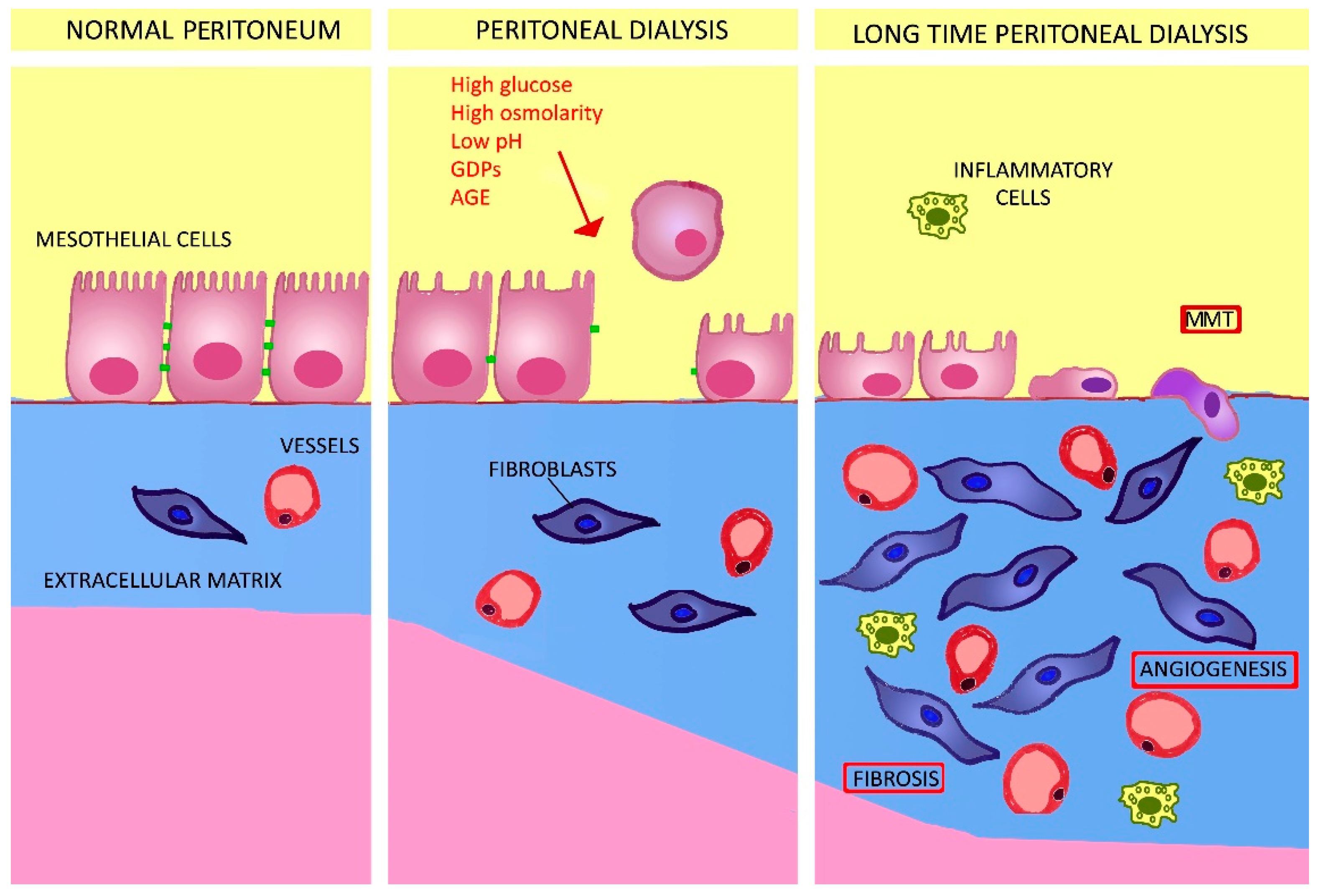
:1. Introduction
2. Long-Term Changes to the Peritoneum and Related Consequences in PD
2.1. The Role of Growth Factors and Cytokynes
2.2. The Role of Glycolysis, Glutaminolysis, and Fatty Acid Oxidation
2.3. The Positive Role of L-Carnitine
2.4. Glycolytic, Fatty Acid, and Pyruvate Metabolism as Targets to Control Peritoneal Fibrosis
3. Strategies Devised to Improve the Biocompatibility of PD Solution
3.1. Neutral-pH, Low-GDP Solutions
3.2. Glucose-Free Approaches to Peritoneal Dialysis Solutions
3.3. Addition of Membrane-Protective Compounds to the Peritoneal Dialysis Solution
3.4. Use of Metabolically Active Osmolytes (Osmo-Metabolites) in the Peritoneal Dialysis Solution
4. Conclusions
Author Contributions
Funding
Conflicts of Interest
References
- Howell, M.; Walker, R.C.; Howard, K. Cost effectiveness of dialysis modalities: A systemic review of 723 economic evaluations. Appl. Health Econ. Health Policy 2019, 17, 315–330. [Google Scholar] [CrossRef]
- Mehrotra, R.; Devuyst, O.; Davies, S.J.; Johnson, D.W. The Current State of Peritoneal Dialysis. J. Am. Soc. Nephrol. 2016, 27, 3238–3252. [Google Scholar] [CrossRef] [PubMed] [Green Version]
- Bartosova, M.; Schmitt, C.P. Biocompatible peritoneal dialysis: The target is still way off. Front. Physiol. 2019, 9, 9. [Google Scholar] [CrossRef]
- Li, P.K.; Szeto, C.C.; Piraino, B.; De Arteaga, J.; Fan, S.; Figueiredo, A.E.; Fish, D.N.; Goffin, E.; Kim, Y.-L.; Salzer, W.; et al. ISPD Peritonitis Recommendations: 2016 Update on Prevention and Treatment. Perit. Dial. Int. 2016, 36, 481–508. [Google Scholar] [CrossRef] [Green Version]
- Hayat, A.; Collins, J.; Saweirs, W. Study of early complications associated with peritoneal dialysis catheters: An analysis of the New Zealand Peritoneal Dialysis Registry data. Int. Urol. Nephrol. 2021, 53, 1705–1711. [Google Scholar] [CrossRef] [PubMed]
- Bajo, M.A.; Del Peso, G.; Teitelbaum, I. Peritoneal Membrane Preservation. Semin. Nephrol. 2017, 37, 77–92. [Google Scholar] [CrossRef]
- Balzer, M.S. Molecular pathways in peritoneal fibrosis. Cell. Signal. 2020, 75, 109778. [Google Scholar] [CrossRef]
- Davies, S.J.; Phillips, L.; Griffiths, A.M.; Russell, L.H.; Naish, P.F.; Russell, G.I. What really happens to people on long-term peritoneal dialysis? Kidney Int. 1998, 54, 2207–2217. [Google Scholar] [CrossRef] [Green Version]
- Schmitt, C.P.; Aufricht, C. Is there such a thing as biocompatible peritoneal dialysis fluid? Pediatric Nephrol. 2016, 32, 1835–1843. [Google Scholar] [CrossRef] [Green Version]
- Numata, M.; Nakayama, M.; Nimura, S.; Kawakami, M.; Lindholm, B.; Kawaguchi, Y. Association between an Increased Surface Area of Peritoneal Microvessels and a High Peritoneal Solute Transport Rate. Perit. Dial. Int. 2003, 23, 116–122. [Google Scholar] [CrossRef] [PubMed]
- Tawada, M.; Ito, Y.; Hamada, C.; Honda, K.; Mizuno, M.; Suzuki, Y.; Sakata, F.; Terabayashi, T.; Matsukawa, Y.; Maruyama, S.; et al. Vascular endothelial cell injury is an important factor in the development of encapsulating peritoneal sclerosis in long-term peritoneal dialysis patients. PLoS ONE 2016, 11, e0154644. [Google Scholar] [CrossRef] [PubMed] [Green Version]
- Garosi, G.; Di Paolo, N. Peritoneal Sclerosis: One or Two Nosological Entities? Semin. Dial. 2000, 13, 297–308. [Google Scholar] [CrossRef] [PubMed]
- Mutsaers, S.E.; Wilkosz, S. Structure and function of mesothelial cells. Cancer Treat. Res. 2007, 134, 1–19. [Google Scholar] [CrossRef]
- Lambie, M.L.; John, B.; Mushahar, L.; Huckvale, C.; Davies, S.J. The peritoneal osmotic conductance is low well before the diagnosis of encapsulating peritoneal sclerosis is made. Kidney Int. 2010, 78, 611–618. [Google Scholar] [CrossRef] [Green Version]
- Zhou, Q.; Bajo, M.A.; Del Peso, G.; Yu, X.; Selgas, R. Preventing peritoneal membrane fibrosis in peritoneal dialysis patients. Kidney Int. 2016, 90, 515–524. [Google Scholar] [CrossRef]
- Yáñez-Mó, M.; Lara-Pezzi, E.; Selgas, R.; Ramírez-Huesca, M.; Domínguez-Jiménez, C.; Jiménez-Heffernan, J.A.; Aguilera, A.; Sánchez-Tomero, J.A.; Bajo, M.A.; Álvarez, V.; et al. Peritoneal Dialysis and Epithelial-to-Mesenchymal Transition of Mesothelial Cells. N. Engl. J. Med. 2003, 348, 403–413. [Google Scholar] [CrossRef] [PubMed]
- Aroeira, L.S.; Aguilera, A.; Sanchez-Tomero, J.A.; Bajo, M.A.; del Peso, G.; Jimenez-Heffernan, J.A.; Selgas, R.; Lopez-Cabrera, M. Epithelial to mesenchymal transition and peritoneal membrane failure in peritoneal dialysis patients: Pathologic significance and potential therapeutic interventions. J. Am. Soc. Nephrol. 2007, 18, 2004–2013. [Google Scholar] [CrossRef] [Green Version]
- Masola, V.; Granata, S.; Bellin, G.; Gambaro, G.; Onisto, M.; Rugiu, C.; Lupo, A.; Zaza, G. Specific heparanase inhibition reverses glucose-induced mesothelial-to-mesenchymal transition. Nephrol. Dial. Transplant. 2017, 32, 1145–1154. [Google Scholar] [CrossRef]
- Kang, D.H.; Hong, Y.S.; Lim, H.J.; Choi, J.H.; Han, D.S.; Yoon, K.I. High glucose solution and spent dialysate stimulate the synthesis of transforming growth factor-β1 of human peritoneal mesothelial cells: Effect of cytokine costimulation. Perit. Dial. Int. 1999, 19, 221–230. [Google Scholar] [CrossRef]
- Yao, Q.; Pawlaczyk, K.; Ayala, E.R.; Styszynski, A.; Breborowicz, A.; Heimburger, O.; Qian, J.Q.; Stenvinkel, P.; Lindholm, B.; Axelsson, J. The Role of the TGF/Smad Signaling Pathway in Peritoneal Fibrosis Induced by Peritoneal Dialysis Solutions. Nephron Exp. Nephrol. 2008, 109, e71–e78. [Google Scholar] [CrossRef]
- Derynck, R.; Zhang, Y.E. Smad-dependent and Smad-independent pathways in TGF-beta family signaling. Nature 2003, 425, 577–584. [Google Scholar] [CrossRef]
- Margetts, P.J.; Kolb, M.; Galt, T.; Hoff, C.M.; Shockley, T.R.; Gauldie, J. Gene Transfer of Transforming Growth Factor-β1 to the Rat Peritoneum: Effects on Membrane Function. J. Am. Soc. Nephrol. 2001, 12, 2029–2039. [Google Scholar] [CrossRef]
- Zhang, J.; Oh, K.H.; Xu, H.; Margetts, P.J. Vascular Endothelial Growth Factor Expression in Peritoneal Mesothelial Cells Undergoing Transdifferentiation. Perit. Dial. Int. 2008, 28, 497–504. [Google Scholar] [CrossRef] [PubMed]
- Sekiguchi, Y.; Zhang, J.; Patterson, S.; Liu, L.; Hamada, C.; Tomino, Y.; Margetts, P.J. Rapamycin inhibits transforming growth factor β-induced peritoneal angiogenesis by blocking the secondary hypoxic response. J. Cell. Mol. Med. 2011, 16, 1934–1945. [Google Scholar] [CrossRef] [PubMed]
- Ueno, T.; Nakashima, A.; Doi, S.; Kawamoto, T.; Honda, K.; Yokoyama, Y.; Doi, T.; Higashi, Y.; Yorioka, N.; Kato, Y.; et al. Mesenchymal stem cells ameliorate experimental peritoneal fibrosis by suppressing inflammation and inhibiting TGF-beta1 signaling. Kidney Int. 2013, 84, 297–307. [Google Scholar] [CrossRef] [Green Version]
- Zhao, J.; Shi, J.; Shan, Y.; Yu, M.; Zhu, X.; Zhu, Y.; Liu, L.; Sheng, M. Asiaticoside inhibits TGF-β1-induced mesothelial-mesenchymal transition and oxidative stress via the Nrf2/HO-1 signaling pathway in the human peritoneal mesothelial cell line HMrSV5. Cell. Mol. Biol. Lett. 2020, 25, 33. [Google Scholar] [CrossRef]
- Guo, Y.; Wang, L.; Gou, R.; Wang, Y.; Shi, X.; Pang, X.; Tang, L. SIRT1-modified human umbilical cord mesenchymal stem cells ameliorate experimental peritoneal fibrosis by inhibiting the TGF-β/Smad3 pathway. Stem Cell Res. Ther. 2020, 11, 1–12. [Google Scholar] [CrossRef] [PubMed]
- Guo, Y.; Wang, L.; Gou, R.; Wang, Y.; Shi, X.; Zhang, Y.; Pang, X.; Tang, L. Ameliorative role of SIRT1 in peritoneal fibrosis: An in vivo and in vitro study. Cell Biosci. 2021, 11, 1–11. [Google Scholar] [CrossRef]
- Lho, Y.; Do, J.-Y.; Heo, J.-Y.; Kim, A.-Y.; Kim, S.-W.; Kang, S.-H. Effects of TGF-β1 Receptor Inhibitor GW788388 on the Epithelial to Mesenchymal Transition of Peritoneal Mesothelial Cells. Int. J. Mol. Sci. 2021, 22, 4739. [Google Scholar] [CrossRef]
- Topley, N.; Jörres, A.; Luttmann, W.; Petersen, M.M.; Lang, M.J.; Thierauch, K.H.; Müller, C.; Coles, G.A.; Davies, M.; Williams, J.D. Human peritoneal mesothelial cells synthesize interleukin-6: Induction by IL-1 beta and TNF alpha. Kidney Int. 1993, 43, 226–233. [Google Scholar] [CrossRef] [Green Version]
- Yang, X.; Zhang, H.; Hang, Y.; Yan, H.; Lin, A.; Huang, J.; Ni, Z.; Qian, J.; Fang, W. Intraperitoneal Interleukin-6 Levels Predict Peritoneal Solute Transport Rate: A Prospective Cohort Study. Am. J. Nephrol. 2014, 39, 459–465. [Google Scholar] [CrossRef]
- Li, X.Y.; Wu, J.; Luo, D.; Chen, W.X.; Zhu, G.L.; Zhang, Y.X.; Bi, Z.M.; Feng, B.H. Effect of high glucose-based peritoneal dialysis fluids on NLRP3-IL-1beta in human peritoneal mesothelial cells. Beijing Da Xue Xue Bao. Yi Xue Ban J. Peking University Health Sci. 2017, 49, 954–960. [Google Scholar]
- Krediet, R.T.; Zweers, M.M.; van der Wal, A.C.; Struijk, D.G. Neoangiogenesis in the Peritoneal Membrane. Perit. Dial. Int. 2000, 20, 19–25. [Google Scholar] [CrossRef]
- Raby, A.-C.; González-Mateo, G.T.; Williams, A.; Topley, N.; Fraser, D.; López-Cabrera, M.; Labéta, M.O. Targeting Toll-like receptors with soluble Toll-like receptor 2 prevents peritoneal dialysis solution–induced fibrosis. Kidney Int. 2018, 94, 346–362. [Google Scholar] [CrossRef] [PubMed]
- Strippoli, R.; Sandoval, P.; Moreno-Vicente, R.; Rossi, L.; Battistelli, C.; Terri, M.; Pascual-Antón, L.; Loureiro, M.; Matteini, F.; Calvo, E.; et al. Caveolin1 and YAP drive mechanically induced mesothelial to mesenchymal transition and fibrosis. Cell Death Dis. 2020, 11, 1–19. [Google Scholar] [CrossRef]
- Namvar, S.; Woolf, A.S.; Zeef, L.A.; Wilm, T.; Wilm, B.; Herrick, S.E. Functional molecules in mesothelial-to-mesenchymal transition revealed by transcriptome analyses. J. Pathol. 2018, 245, 491–501. [Google Scholar] [CrossRef] [PubMed] [Green Version]
- Ruiz-Carpio, V.; Sandoval, P.; Aguilera, A.; Albar-Vizcaíno, P.; Perez-Lozano, M.L.; González-Mateo, G.T.; Acuña-Ruiz, A.; Garcia-Cantalejo, J.; Botías, P.; Bajo, M.A.; et al. Genomic reprograming analysis of the Mesothelial to Mesenchymal Transition identifies biomarkers in peritoneal dialysis patients. Sci. Rep. 2017, 7, 1–15. [Google Scholar] [CrossRef] [PubMed] [Green Version]
- Lambie, M.; Chess, J.; Donovan, K.L.; Kim, Y.L.; Do, J.Y.; Lee, H.B.; Noh, H.; Williams, P.F.; Williams, A.J.; Davison, S.; et al. Independent Effects of Systemic and Peritoneal Inflammation on Peritoneal Dialysis Survival. J. Am. Soc. Nephrol. 2013, 24, 2071–2080. [Google Scholar] [CrossRef] [PubMed] [Green Version]
- Henderson, J.; O’Reilly, S. The emerging role of metabolism in fibrosis. Trends Endocrinol. Metab. 2021, 32, 639–653. [Google Scholar] [CrossRef]
- Jiang, L.; Xiao, L.; Sugiura, H.; Huang, X.; Ali, A.; Kuro-o, M.; Deberardinis, R.J.; Boothman, D.A. Metabolic reprogramming during TGFβ1-induced epithelial-to-mesenchymal. Oncogene 2015, 34, 3908–3916. [Google Scholar] [CrossRef] [Green Version]
- Vander Heiden, M.G.; Cantley, L.C.; Thompson, C.B. Understanding the Warburg effect: The metabolic requirements of cell proliferation. Science 2009, 324, 1029–1033. [Google Scholar] [CrossRef] [Green Version]
- Zhang, D.; Tang, Z.; Huang, H.; Zhou, G.; Cui, C.; Weng, Y.; Liu, W.; Kim, S.; Lee, S.; Perez-Neut, M.; et al. Metabolic regulation of gene expression by histone lactylation. Nat. Cell Biol. 2019, 574, 575–580. [Google Scholar] [CrossRef]
- Yang, L.; Venneti, S.; Nagrath, D. Glutaminolysis: A Hallmark of Cancer Metabolism. Annu. Rev. Biomed. Eng. 2017, 19, 163–194. [Google Scholar] [CrossRef] [PubMed]
- Cheng, S.C.; Quintin, J.; Cramer, R.A.; Shepardson, K.M.; Saeed, S.; Kumar, V.; Giamarellos-Bourboulis, E.; Martens, J.; Rao, N.A.; Aghajanirefah, A.; et al. mTOR- and HIF-1α mediated aerobic glycolysis as metabolic basis for trained immunity. Science 2014, 345, 1250684. [Google Scholar] [CrossRef] [Green Version]
- Tannahill, G.M.; Curtis, A.M.; Adamik, J.; Palsson-McDermott, E.M.; McGettrick, A.F.; Goel, G.; Frezza, C.; Bernard, N.J.; Kelly, B.; Foley, N.H.; et al. Succinate is an inflammatory signal that induces IL-1β through HIF-1α. Nature 2013, 496, 238–242. [Google Scholar] [CrossRef]
- Zammit, V.A.; Ramsay, R.R.; Bonomini, M.; Arduini, A. Carnitine, mitochondrial function and therapy. Adv. Drug Deliv. Rev. 2009, 61, 1353–1362. [Google Scholar] [CrossRef]
- Zammit, V.A. The malonyl-CoA-long-chain acyl-CoA axis in the maintenance of mammalian cell function. Biochem. J. 1999, 343, 505–515. [Google Scholar] [CrossRef]
- Arduini, A.; Bonomini, M.; Savica, V.; Amato, A.; Zammit, V. Carnitine in metabolic disease: Potential for pharmacological intervention. Pharmacol. Ther. 2008, 120, 149–156. [Google Scholar] [CrossRef] [PubMed]
- Bonomini, M.; Zammit, V.; Divino-Filho, J.C.; Davies, S.J.; Di Liberato, L.; Arduini, A.; Lambie, M. The osmo-metabolic approach: A novel and tantalizing glucose-sparing strategy in peritoneal dialysis. J. Nephrol. 2021, 34, 503–519. [Google Scholar] [CrossRef] [PubMed]
- Xiao, W.; Ren, M.; Zhang, C.; Li, S.; An, W. Amelioration of nonalcoholic fatty liver disease by hepatic stimulator substance via preservation of carnitine palmitoyl transferase-1 activity. Am. J. Physiol. Cell Physiol. 2015, 309, C215–C227. [Google Scholar] [CrossRef] [Green Version]
- Wang, W.; Zhang, L.; Battiprolu, P.K.; Fukushima, A.; Nguyen, K.; Milner, K.; Gupta, A.; Altamimi, T.; Byrne, N.; Mori, J.; et al. Malonyl CoA Decarboxylase Inhibition Improves Cardiac Function Post-Myocardial Infarction. JACC Basic Transl. Sci. 2019, 4, 385–400. [Google Scholar] [CrossRef]
- Hu, H.J.; Zhang, C.; Tang, Z.H.; Qu, S.L.; Jiang, Z.S. Regulating the Warburg effect on metabolic stress and myocardi-al fibrosis remodeling and atrial intracardiac waveform activity induced by atrial fibrillation. Biochem. Biophys. Res. Commun. 2019, 516, 653–660. [Google Scholar] [CrossRef]
- Pajak, B.; Siwiak, E.; Sołtyka, M.; Priebe, A.; Zieliński, R.; Fokt, I.; Ziemniak, M.; Jaśkiewicz, A.; Borowski, R.; Domoradzki, T.; et al. 2-Deoxy-d-Glucose and Its Analogs: From Diagnostic to Therapeutic Agents. Int. J. Mol. Sci. 2019, 21, 234. [Google Scholar] [CrossRef] [Green Version]
- Wilson, R.B. Hypoxia, cytokines and stromal recruitment: Parallels between pathophysiology of encapsulating peritoneal sclerosis, endometriosis and peritoneal metastasis. Pleura Peritoneum 2018, 3, 20180103. [Google Scholar] [CrossRef]
- Si, M.; Wang, Q.; Li, Y.; Lin, H.; Luo, D.; Zhao, W.; Dou, X.; Liu, J.; Zhang, H.; Huang, Y.; et al. Inhibition of hyperglycolysis in mesothelial cells prevents peritoneal fibrosis. Sci. Transl. Med. 2019, 11, eaav5341. [Google Scholar] [CrossRef]
- Laussel, C.; Léon, S. Cellular toxicity of the metabolic inhibitor 2-deoxyglucose and associated resistance mechanisms. Biochem. Pharmacol. 2020, 182, 114213. [Google Scholar] [CrossRef] [PubMed]
- Szeto, C.C.; Johnson, D.W. Low GDP Solution and Glucose-Sparing Strategies for Peritoneal Dialysis. Semin. Nephrol. 2017, 37, 30–42. [Google Scholar] [CrossRef] [PubMed]
- Nataatmadja, M.S.; Johnson, D.W.; Pascoe, E.M.; Darssan, D.; Hawley, C.M.; Cho, Y. Associations between Peritoneal Glucose Exposure, Glucose Degradation Product Exposure, and Peritoneal Membrane Transport Characteristics in Peritoneal Dialysis Patients: Secondary Analysis of the balANZ Trial. Perit. Dial. Int. 2018, 38, 349–355. [Google Scholar] [CrossRef] [PubMed]
- Stinghen, A.E.; Massy, Z.A.; Vlassara, H.; Striker, G.E.; Boullier, A. Uremic Toxicity of Advanced Glycation End Products in CKD. J. Am. Soc. Nephrol. 2015, 27, 354–370. [Google Scholar] [CrossRef] [Green Version]
- Jiang, J.; Chen, P.; Chen, J.; Yu, X.; Xie, D.; Mei, C.; Xiong, F.; Shi, W.; Zhou, W.; Liu, X.; et al. Accumulation of tissue advanced glycation end products correlated with glucose exposure dose and associated with cardiovascular morbidity in patients on peritoneal dialysis. Atheroscler. 2012, 224, 187–194. [Google Scholar] [CrossRef]
- Htay, H.; Johnson, D.W.; Wiggins, K.J.; Badve, S.V.; Craig, J.C.; Strippoli, G.F.M.; Cho, Y. Biocompatible dialysis fluids for peritoneal dialysis. Cochrane Database Syst. Rev. 2018, 10, CD007554. [Google Scholar] [CrossRef] [PubMed]
- Lee, H.Y.; Choi, H.Y.; Park, H.C.; Seo, B.J.; Do, J.Y.; Yun, S.R.; Song, H.Y.; Kim, Y.H.; Kim, Y.-R.; Kim, D.J.; et al. Changing prescribing practice in CAPD patients in Korea: Increased utilization of low GDP solutions improves patient outcome. Nephrol. Dial. Transplant. 2006, 21, 2893–2899. [Google Scholar] [CrossRef] [PubMed] [Green Version]
- Lichodziejewska-Niemierko, M.; Chmielewski, M.; Dudziak, M.; Ryta, A.; Rutkowski, B. Hydration Status of Patients Dialyzed with Biocompatible Peritoneal Dialysis Fluids. Perit. Dial. Int. 2016, 36, 257–261. [Google Scholar] [CrossRef]
- Blake, P.G. Is the peritoneal dialysis biocompatibility hypothesis dead? Kidney Int. 2018, 94, 246–248. [Google Scholar] [CrossRef]
- Schaefer, B.; Bartosova, M.; Macher-Goeppinger, S.; Sallay, P.; Vörös, P.; Ranchin, B.; Vondrak, K.; Ariceta, G.; Zaloszyc, A.; Bayazit, A.K.; et al. Neutral pH and low–glucose degradation product dialysis fluids induce major early alterations of the peritoneal membrane in children on peritoneal dialysis. Kidney Int. 2018, 94, 419–429. [Google Scholar] [CrossRef]
- Sugiyama, N.; Tawada, M.; Sun, T.; Suzuki, Y.; Kinashi, H.; Yamaguchi, M.; Katsuno, T.; Aten, J.; Vlahu, C.A.; van Kuppevelt, T.H.; et al. Low-GDP, pH-neutral solutions preserve peritoneal endothelial glycocalyx during long-term peritoneal dialysis. Clin. Exp. Nephrol. 2021. [Google Scholar] [CrossRef]
- Butler, M.J.; Down, C.J.; Foster, R.R.; Satchell, S.C. The pathological relevance of increased endothelial glycocalyx permeability. Am. J. Pathol. 2020, 190, 742–751. [Google Scholar] [CrossRef] [PubMed]
- Liu, H.Q.; Li, J.; Xuan, C.L.; Ma, H.C. A review on the physiological and pathophysiological role of endothelial glycocalyx. J. Biochem. Mol. Toxicol. 2020, 34, e22571. [Google Scholar] [CrossRef]
- Ushiyama, A.; Kataoka, H.; Iijima, T. Glycocalyx and its involvement in clinical pathophysiologies. J. Intensive Care 2016, 4, 1–11. [Google Scholar] [CrossRef] [Green Version]
- Sieve, I.; Münster-Kühnel, A.K.; Hilfiker-Kleiner, D. Regulation and function of endothelial glycocalyx layer in vascular diseases. Vasc. Pharmacol. 2018, 100, 26–33. [Google Scholar] [CrossRef] [PubMed]
- Burkart, J.M. Poor Nutritional Status and Inflammation: Metabolic Consequences of Peritoneal Dialysis. Semin. Dial. 2004, 17, 498–504. [Google Scholar] [CrossRef]
- Wang, I.K.; Lin, C.L.; Chen, H.C.; Lin, S.Y.; Chang, C.T.; Yen, T.H.; Sung, F.C. Risk of new-onset diabetes in end-stage renal disease patients undergoing dialysis: Analysis from registry data of Taiwan. Nephrol. Dial. Transplant. 2017, 33, 670–675. [Google Scholar] [CrossRef]
- Wang, Z.; Yu, D.; Cai, Y.; Ma, S.; Zhao, B.; Zhao, Z.; Simmons, D. Dialysate glucose response phenotypes during peritoneal equilibration test and their association with cardiovascular death: A cohort study. Medicine 2020, 99, e20447. [Google Scholar] [CrossRef] [PubMed]
- Holmes, C.J. Glucotoxicity in peritoneal dialysis—Solutions for the solution! Adv. Chronic Kidney Dis. 2007, 14, 269–278. [Google Scholar] [CrossRef]
- Jones, M.; Hagen, T.; Boyle, C.A.; Vonesh, E.; Hamburger, R.; Charytan, C.; Sandroni, S.; Bernard, D.; Piraino, B.; Schreiber, M.; et al. Treatment of malnutrition with 1.1% amino acid peritoneal dialysis solution: Results of a multicenter outpatient study. Am. J. Kidney Dis. 1998, 32, 761–769. [Google Scholar] [CrossRef]
- Johnson, D.W.; Agar, J.; Collins, J.; Disney, A.; Harris, D.C.; Ibels, L.; Irish, A.; Saltissi, D.; Suranyi, M. Recommendations for the use of icodextrin in peritoneal dialysis patients. Review Article. Nephrology 2003, 8, 1–7. [Google Scholar] [CrossRef]
- Dousdampanis, P.; Musso, C.; Trigka, K. Icodextrin and peritoneal dialysis: Advantages and new applications. Int. Urol. Nephrol. 2017, 50, 495–500. [Google Scholar] [CrossRef]
- Morelle, J.; Sow, A.; Fustin, C.A.; Fillée, C.; Garcia-Lopez, E.; Lindholm, B.; Goffin, E.; Vandemaele, F.; Rippe, B.; Öberg, C.M.; et al. Mechanisms of Crystalloid versus Colloid Osmosis across the Peritoneal Membrane. J. Am. Soc. Nephrol. 2018, 29, 1875–1886. [Google Scholar] [CrossRef] [PubMed]
- Olszowska, A.; Waniewski, J.; Stachowska-Pietka, J.; Garcia-Lopez, E.; Lindholm, B.; Wańkowicz, Z. Long Peritoneal Dialysis Dwells with Icodextrin: Kinetics of Transperitoneal Fluid and Polyglucose Transport. Front. Physiol. 2019, 10, 1326. [Google Scholar] [CrossRef]
- Goossen, K.; Becker, M.; Marshall, M.R.; Bühn, S.; Breuing, J.; Firanek, C.A.; Hess, S.; Nariai, H.; Sloand, J.A.; Yao, Q.; et al. Icodextrin Versus Glucose Solutions for the Once-Daily Long Dwell in Peritoneal Dialysis: An Enriched Systematic Review and Meta-analysis of Randomized Controlled Trials. Am. J. Kidney Dis. 2020, 75, 830–846. [Google Scholar] [CrossRef] [Green Version]
- Papasotiriou, M.; Liakopoulos, V.; Kehagias, I.; Vareta, G.; Ntrinias, T.; Papachristou, E.; Goumenos, D.S. Favorable effects of peritoneal dialysis in patients with refractory heart failure and overhydration. Perit. Dial. Int. 2020, 28, 896860820970097. [Google Scholar] [CrossRef]
- Wojtaszek, E.; Grzejszczak, A.; Niemczyk, S.; Malyszko, J.; Matuszkiewicz-Rowińska, J. Peritoneal Ultrafiltration in the Long-Term Treatment of Chronic Heart Failure Refractory to Pharmacological Therapy. Front. Physiol. 2019, 10, 310. [Google Scholar] [CrossRef] [PubMed]
- Han, S.H.; Ahn, S.V.; Yun, J.Y.; Tranaeus, A.; Han, D.S. Effects of icodextrin on patient survival and technique success in pa-tients undergoing peritoneal dialysis. Nephrol. Dial. Transplant. 2012, 27, 2044–2050. [Google Scholar] [CrossRef] [PubMed] [Green Version]
- Yang, J.-Y.; Chen, L.; Peng, Y.-S.; Chen, Y.-Y.; Huang, J.-W.; Hung, K.-Y. Icodextrin is Associated with a Lower Mortality Rate in Peritoneal Dialysis Patients. Perit. Dial. Int. 2019, 39, 252–260. [Google Scholar] [CrossRef] [PubMed]
- Freida, P.; Issad, B.; Dratwa, M.; Lobbedez, T.; Wu, L.; Leypoldt, J.K.; Divino-Filho, J.C. A Combined Crystalloid and Colloid PD Solution as a Glucose-Sparing Strategy for Volume Control in High-Transport APD Patients: A Prospective Multicenter Study. Perit. Dial. Int. 2009, 29, 433–442. [Google Scholar] [CrossRef]
- Moriishi, M.; Kawanishi, H. Icodextrin and Intraperitoneal Inflammation. Perit. Dial. Int. 2008, 28, 96–100. [Google Scholar] [CrossRef]
- Velloso, M.S.S.; Otoni, A.; Sabino, A.; de Castro, W.V.; Pinto, S.W.L.; Marinho, M.A.S.; Rios, D.R.A. Peritoneal dialysis and inflammation. Clin. Chim. Acta 2014, 430, 109–114. [Google Scholar] [CrossRef]
- Yamaguchi, N.; Miyamoto, K.; Murata, T.; Ishikawa, E.; Horiuchi, T. Newly Developed Neutralized pH Icodextrin Dialysis Fluid: Nonclinical Evaluation. Artif. Organs 2016, 40, E158–E166. [Google Scholar] [CrossRef]
- Higuchi, C.; Kuriyma, J.; Sakura, H. Effect of Neutral pH Icodextrin Peritoneal Dialysis Fluid on Mesothelial Cells. Ther. Apher. Dial. 2018, 22, 656–661. [Google Scholar] [CrossRef]
- Asola, M.; Virtanen, K.; Någren, K.; Helin, S.; Taittonen, M.; Kastarinen, H.; Anderstam, B.; Knuuti, J.; Metsärinne, K.; Nuutila, P. Amino-acid-based peritoneal dialysis solution improves amino-acid transport into skeletal muscle. Kidney Int. 2008, 73, S131–S136. [Google Scholar] [CrossRef] [Green Version]
- Canepa, A.; Verrina, E.; Perfumo, F.; Carrea, A.; Menoni, S.; Delucchi, P.; Gusmano, R. Value of Intraperitoneal Amino Acids in Children Treated with Chronic Peritoneal Dialysis. Perit. Dial. Int. 1999, 19, 435–440. [Google Scholar] [CrossRef]
- Canepa, A.; Carrea, A.; Menoni, S.; Verrina, E.; Trivelli, A.; Gusmano, R.; Perfumo, F. Acute effects of simultaneous intraperitoneal infusion of glucose and amino acids. Kidney Int. 2001, 59, 1967–1973. [Google Scholar] [CrossRef] [Green Version]
- Mortier, S.; Faict, D.; Schalkwijk, C.G.; Lameire, N.H.; De Vriese, A. Long-term exposure to new peritoneal dialysis solutions: Effects on the peritoneal membrane. Kidney Int. 2004, 66, 1257–1265. [Google Scholar] [CrossRef] [PubMed] [Green Version]
- Reimann, D.; Dachs, D.; Meye, C.; Gross, P. Amino acid-based peritoneal dialysis solution stimulates mesothelial nitric oxide production. Perit. Dial. Int. 2004, 24, 378–384. [Google Scholar] [CrossRef]
- Combet, S.; Miyata, T.; Moulin, P.; Pouthier, D.; Goffin, E.; Devuyst, O. Vascular Proliferation and Enhanced Expression of Endothelial Nitric Oxide Synthase in Human Peritoneum Exposed to Long-Term Peritoneal Dialysis. J. Am. Soc. Nephrol. 2000, 11, 717–728. [Google Scholar] [CrossRef]
- Li, P.K.; Culleton, B.F.; Ariza, A.; Do, J.Y.; Johnson, D.W.; Sanabria, M.; Shockley, T.R.; Story, K.; Vatazin, A.; Verrelli, M.; et al. Randomized, controlled trial of glucose-sparing peritoneal dialysis in diabetic patients. J. Am. Soc. Nephrol. 2013, 24, 1889–1900. [Google Scholar] [CrossRef] [PubMed] [Green Version]
- Trottier, C.; Perl, J.; Freeman, M.; Thadhani, R.; Berg, A.; Kalim, S. Protein Carbamylation in Peritoneal Dialysis and the Effect of Low Glucose plus Amino Acid Solutions. Perit. Dial. Int. 2018, 38, 149–152. [Google Scholar] [CrossRef] [PubMed]
- Lebeck, J. Metabolic impact of the glycerol channels AQP7 and AQP9 in adipose tissue and liver. J. Mol. Endocrinol. 2014, 52, R165–R178. [Google Scholar] [CrossRef] [PubMed] [Green Version]
- Matthys, E.; Dolkart, R.; Lameire, N. Potential Hazards of Glycerol Dialysate in Diabetic CAPD Patients. Perit. Dial. Int. 1987, 7, 16–19. [Google Scholar] [CrossRef]
- Van Biesen, W.; Boer, W.; De Greve, B.; Dequidt, C.; Vijt, D.; Faict, D.; Lameire, N. A Randomized Clinical Trial with a 0.6% Amino Acid/1.4% Glycerol Peritoneal Dialysis Solution. Perit. Dial. Int. 2004, 24, 222–230. [Google Scholar] [CrossRef]
- Smit, W.; Ho-Dac-Pannekeet, M.M.; Krediet, R.T. Treatment of severe ultrafiltration failure with nonglucose dialysis solutions in patients with and without peritoneal sclerosis. NDT Plus 2008, 1, iv63–iv70. [Google Scholar] [CrossRef] [Green Version]
- Aufricht, C.; Beelen, R.; Eberl, M.; Fischbach, M.; Fraser, D.; Jörres, A.; Kratochwill, K.; Lópezcabrera, M.; Rutherford, P.; Schmitt, C.-P.; et al. Biomarker research to improve clinical outcomes of peritoneal dialysis: Consensus of the European Training and Research in Peritoneal Dialysis (EuTRiPD) network. Kidney Int. 2017, 92, 824–835. [Google Scholar] [CrossRef] [Green Version]
- Bonomini, M.; Borras, F.E.; Troya-Saborido, M.; Carreras-Planella, L.; Di Liberato, L.; Arduini, A. Proteomic Research in Peritoneal Dialysis. Int. J. Mol. Sci. 2020, 21, 5489. [Google Scholar] [CrossRef]
- de Graaff, M.; Zegwaard, A.H.; Zweers, M.M.; Vlijm, A.; de Waart, D.R.; Vandemaele, F.; Struijk, D.G.; Krediet, R.T. The effects of a dialysis solution with a combination of glycerol/amino acids/dextrose on the peritoneal membrane in chronic renal failure. Perit. Dial. Int. 2010, 30, 192–200. [Google Scholar] [CrossRef]
- Van Westrhenen, R.; Vlijm, A.; Hiralall, J.K.; Krediet, R.T. Experimental study on long-term exposure to a biocompatible, hypertonic, pyruvate-buffered dialysis solution. Perit. Dial. Int. 2008, 28, S43–S47. [Google Scholar] [CrossRef] [PubMed]
- Van Westrhenen, R.; Zweers, M.M.; Kunne, C.; de Waart, D.R.; van der Wal, A.C.; Krediet, R.T. A pyruvate-buffered dialysis fluid induces less peritoneal angiogenesis and fibrosis than a conventional solution. Perit. Dial. Int. 2008, 28, 487–496. [Google Scholar] [CrossRef] [PubMed]
- Kao, K.K.; Fink, M.P. The biochemical basis for the anti-inflammatory and cytoprotective actions of ethyl pyruvate and related compounds. Biochem. Pharmacol. 2010, 80, 151–159. [Google Scholar] [CrossRef] [PubMed]
- Nishimura, H.; Ikehara, O.; Naito, T.; Higuchi, C.; Sanaka, T. Evaluation of taurine as an osmotic agent for peritoneal dialysis solution. Perit. Dial. Int. 2009, 29, 204–216. [Google Scholar] [CrossRef]
- Suliman, M.E.; Barany, P.; Filho, J.C.D.; Lindholm, B.; Bergström, J. Accumulation of taurine in patients with renal failure. Nephrol. Dial. Transplant. 2002, 17, 528–529. [Google Scholar] [CrossRef] [Green Version]
- Du, C.; Mendelson, A.A.; Guan, Q.; Dairi, G.; Chafeeva, I.; Da Roza, G.; Kizhakkedathu, J.N. Hyperbranched polyglycerol is superior to glucose for long-term preservation of peritoneal membrane in a rat model of chronic peritoneal dialysis. J. Transl. Med. 2016, 14, 1–17. [Google Scholar] [CrossRef] [PubMed] [Green Version]
- La Han, B.; Guan, Q.; Chafeeva, I.; Mendelson, A.A.; Da Roza, G.; Liggins, R.; Kizhakkedathu, J.N.; Du, C. Peritoneal and Systemic Responses of Obese Type II Diabetic Rats to Chronic Exposure to a Hyperbranched Polyglycerol-Based Dialysis Solution. Basic Clin. Pharmacol. Toxicol. 2018, 123, 494–503. [Google Scholar] [CrossRef] [Green Version]
- Lo, W.K. Metabolic syndrome and obesity in peritoneal dialysis. Kidney Res. Clin. Pract. 2016, 35, 10–14. [Google Scholar] [CrossRef] [Green Version]
- Sanguankeo, A.; Upala, S. Metabolic syndrome increases mortality risk in dialysis patients: A systematic review and meta-analysis. Int. J. Endocrinol. Metab. 2018, 16, e61201. [Google Scholar] [PubMed] [Green Version]
- Bazzato, G.; Fracasso, A.; Gambaro, G.; Baggio, B. Use of glycosaminoglycans to increase efficiency of long-term continuous peritoneal dialysis. Lancet 1995, 346, 740–741. [Google Scholar] [PubMed]
- Rodrigues, A.; Cabrita, A.; Maia, P.; Guimarães, S. Peritoneal rest may successfully recover ultrafiltration in patients who develop peritoneal hyperpermeability with time on continuous ambulatory peritoneal dialysis. Adv. Perit. Dial. 2002, 18, 78–80. [Google Scholar]
- Sjøland, J.A.; Pedersen, R.S.; Jespersen, J.; Gram, J. Intraperitoneal heparin reduces peritoneal permeability and increases ultrafiltration in peritoneal dialysis patients. Nephrol. Dial. Transplant. 2004, 19, 1264–1268. [Google Scholar] [CrossRef] [Green Version]
- Del Peso, G.; Bajo, M.A.; Perez Fontán, M.; Martinez, J.; Marrón, B.; Selgas, R. on behalf of the Group of Study on “Bemidextrin”. Effect of self-administered intraperitoneal Bemiparin on peritoneal transport and ultrafiltration capacity in peritoneal dialysis patients with membrane dysfunction. A randomized, multi-centre open clinical trial. Nephrol. Dial. Transplant. 2012, 27, 2051–2058. [Google Scholar]
- Bazargani, F.; Albrektsson, A.; Yahyapour, N.; Braide, M. Low Molecular Weight Heparin Improves Peritoneal Ultrafiltration and Blocks Complement and Coagulation. Perit. Dial. Int. 2005, 25, 394–404. [Google Scholar] [CrossRef]
- Braide, M.; Haraldsson, B.; Persson, U. Citrate supplementation of PD fluid: Effects on net ultrafiltration and clearance of small molecules in single dwells. Nephrol. Dial. Transplant. 2009, 24, 286–292. [Google Scholar] [CrossRef] [Green Version]
- Alhamdani, M.S.S.; Al-Azzawie, H.F.; Abbas, F.K. Decreased formation of advanced glycation end-products in peritoneal fluid by carnosine and related peptides. Perit. Dial. Int. 2007, 27, 86–89. [Google Scholar] [CrossRef]
- Werynski, A.; Waniewski, J.; Wang, T.; Anderstam, B.; Lindholm, B.; Bergström, J. Kinetic studies of dipeptide-based and amino acid-based peritoneal dialysis solutions. Kidney Int. 2001, 59, 363–371. [Google Scholar] [CrossRef] [Green Version]
- Ferrantelli, E.; Liappas, G.; Cuenca, M.C.; Keuning, E.D.; Foster, T.L.; Vervloet, M.G.; Lopéz-Cabrera, M.; Beelen, R.H.J. The dipeptide alanyl-glutamine ameliorates peritoneal fibrosis and attenuates IL-17 dependent pathways during peritoneal dialysis. Kidney Int. 2016, 89, 625–635. [Google Scholar] [CrossRef] [PubMed]
- Herzog, R.; Bartosova, M.; Tarantino, S.; Wagner, A.; Unterwurzacher, M.; Sacnun, J.M.; Lichtenauer, A.M.; Kuster, L.; Schaefer, B.; Alper, S.L.; et al. Peritoneal Dialysis Fluid Supplementation with Alanyl-Glutamine Attenuates Conventional Dialysis Fluid-Mediated Endothelial Cell Injury by Restoring Perturbed Cytoprotective Responses. Biomolecules 2020, 10, 1678. [Google Scholar] [CrossRef] [PubMed]
- Kratochwill, K.; Boehm, M.; Herzog, R.; Gruber, K.; Lichtenauer, A.M.; Kuster, L.; Csaicsich, D.; Gleiss, A.; Alper, S.L.; Aufricht, C.; et al. Addition of Alanyl-Glutamine to Dialysis Fluid Restores Peritoneal Cellular Stress Responses—A First-In-Man Trial. PLoS ONE 2016, 11, e0165045. [Google Scholar] [CrossRef] [PubMed] [Green Version]
- Herzog, R.; Kuster, L.; Becker, J.; Gluexam, T.; Pils, D.; Spittler, A.; Bhasin, M.K.; Alper, S.L.; Vychytil, A.; Aufricht, C.; et al. Functional and transcriptomic characterization of peritoneal immune-modulation by addition of alanyl-glutamine to dialysis fluid. Sci. Rep. 2017, 7, 1–15. [Google Scholar] [CrossRef] [Green Version]
- Vychytil, A.; Herzog, R.; Probst, P.; Ribitsch, W.; Lhotta, K.; Machold-Fabrizii, V.; Wiesholzer, M.; Kaufmann, M.; Salmhofer, H.; Windpessl, M.; et al. A randomized controlled trial of alanyl-glutamine supplementation in peritoneal dialysis fluid to assess impact on biomarkers of peritoneal health. Kidney Int. 2018, 94, 1227–1237. [Google Scholar] [CrossRef] [PubMed] [Green Version]
- Wiesenhofer, F.M.; Herzog, R.; Boehm, M.; Wagner, A.; Unterwurzacher, M.; Kasper, D.C.; Alper, S.L.; Vychytil, A.; Aufricht, C.; Kratochwill, K. Targeted Metabolomic Profiling of Peritoneal Dialysis Effluents Shows Anti-oxidative Capacity of Alanyl-Glutamine. Front. Physiol. 2019, 9, 1961. [Google Scholar] [CrossRef] [Green Version]
- Ohsawa, I.; Ishikawa, M.; Takahashi, K.; Watanabe, M.; Nishimaki, K.; Yamagata, K.; Katsura, K.-I.; Katayama, Y.; Asoh, S.; Ohta, S. Hydrogen acts as a therapeutic antioxidant by selectively reducing cytotoxic oxygen radicals. Nat. Med. 2007, 13, 688–694. [Google Scholar] [CrossRef]
- Nakayama, M.; Zhu, W.J.; Watanabe, K.; Gibo, A.; Sherif, A.M.; Kabayama, S.; Ito, S. Dissolved molecular hydrogen (H2) in Peritoneal Dialysis (PD) solutions preserves mesothelial cells and peritoneal membrane integrity. BMC Nephrol. 2017, 18, 1–9. [Google Scholar] [CrossRef] [Green Version]
- Ichihara, M.; Sobue, S.; Ito, M.; Hirayama, M.; Ohno, K. Beneficial biological effects and the underlying mechanisms of mo-lecular hydrogen-comprehensive review of 321 original articles. Med. Gas. Res. 2015, 5, 1–21. [Google Scholar] [CrossRef] [Green Version]
- Terawaki, H.; Hayashi, Y.; Zhu, W.J.; Matsuyama, Y.; Terada, T.; Kabayama, S.; Watanabe, T.; Era, S.; Sato, B.; Nakayama, M. Transperitoneal administration of dissolved hydrogen for peritoneal dialysis patients: A novel approach to suppress oxidative stress in the peritoneal cavity. Med. Gas. Res. 2013, 3, 1–7. [Google Scholar] [CrossRef] [PubMed] [Green Version]
- Lu, H.; Chen, W.; Liu, W.; Si, Y.; Zhao, T.; Lai, X.; Kang, Z.; Sun, X.; Guo, Z. Molecular hydrogen regulates PTEN-AKT-mTOR signalling via ROS to alleviate peritoneal dialysis-related peritoneal fibrosis. FASEB J. 2020, 34, 4134–4146. [Google Scholar] [CrossRef] [Green Version]
- Bonomini, M.; Di Silvestre, S.; Di Tomo, P.; Di Pietro, N.; Mandatori, D.; Di Liberato, L.; Sirolli, V.; Chiarelli, F.; Indiveri, C.; Pandolfi, A.; et al. Effect of peritoneal dialysis fluid containing osmo-metabolic agents on human endothelial cells. Drug Des. Dev. Ther. 2016, 10, 3925–3932. [Google Scholar] [CrossRef] [Green Version]
- Bonomini, M.; Di Liberato, L.; Zammit, V.; Arduini, A. Current Opinion on Usage of L-Carnitine in End-Stage Renal Disease Patients on Peritoneal Dialysis. Molecules 2019, 24, 3449. [Google Scholar] [CrossRef] [PubMed] [Green Version]
- Longo, N.; Frigeni, M.; Pasquali, M. Carnitine transport and fatty acid oxidation. Biochim. Biophys. Acta (BBA)-Mol. Cell Res. 2016, 1863, 2422–2435. [Google Scholar] [CrossRef]
- Wang, Y.M.; Van Eys, J. Nutritional Significance of Fructose and Sugar Alcohols. Annu. Rev. Nutr. 1981, 1, 437–475. [Google Scholar] [CrossRef]
- Gaggiotti, E.; Arduini, A.; Bonomini, M.; Valentini, G.; Sacchi, G.; Sansoni, E.; Salvo, D.; Di Paolo, N. Prevention of peritone-al sclerosis: A new proposal to substitute glucose with carnitine dialysis solution (biocompatibility testing in vitro and in rabbits). Int. J. Artif. Organs 2005, 28, 177–187. [Google Scholar] [CrossRef]
- Bonomini, M.; Pandolfi, A.; Di Liberato, L.; Di Silvestre, S.; Cnops, Y.; Di Tomo, P.; D’arezzo, M.; Monaco, M.P.; Giardinelli, A.; Di Pietro, N.; et al. L-carnitine is an osmotic agent suitable for peritoneal dialysis. Kidney Int. 2011, 80, 645–654. [Google Scholar] [CrossRef] [Green Version]
- Bazzato, G.; Coli, U.; Landini, S.; Fracasso, A.; Morachiello, P.; Righetto, F.; Scanferla, F.; Onesti, G. Xylitol as osmotic agent in CAPD: An alternative to glucose for uremic diabetic patients? Trans. Am. Soc. Artif. Intern. Organs 1982, 28, 280–286. [Google Scholar]
- Bonomini, M.; Di Liberato, L.; Del Rosso, G.; Stingone, A.; Marinangeli, G.; Consoli, A.; Bertoli, S.; De Vecchi, A.; Bosi, E.; Russo, R.; et al. Effect of an l-Carnitine–Containing Peritoneal Dialysate on Insulin Sensitivity in Patients Treated with CAPD: A 4-Month, Prospective, Multicenter Randomized Trial. Am. J. Kidney Dis. 2013, 62, 929–938. [Google Scholar] [CrossRef] [PubMed]
- Wang, A.Y.-M.; Dong, J.; Xu, X.; Davies, S. Volume management as a key dimension of a high-quality PD prescription. Perit. Dial. Int. 2020, 40, 282–292. [Google Scholar] [CrossRef] [PubMed] [Green Version]
- Brown, E.A.; Blake, P.G.; Boudville, N.; Davies, S.; De Arteaga, J.; Dong, J.; Finkelstein, F.; Foo, M.; Hurst, H.; Johnson, D.W.; et al. International Society for Peritoneal Dialysis practice recommendations: Prescribing high-quality goal-directed peritoneal dialysis. Perit. Dial. Int. 2020, 40, 244–253. [Google Scholar] [CrossRef] [Green Version]
- Kishore, P.; Kehlenbrink, S.; Hu, M.; Zhang, K.; Gutierrez-Juarez, R.; Koppaka, S.; El-Maghrabi, M.R.; Hawkins, M. Xylitol prevents NEFA-induced insulin resistance in rats. Diabetologia 2012, 55, 1808–1812. [Google Scholar] [CrossRef] [PubMed] [Green Version]
- Woelnerhanssen, B.K.; Cajacob, L.; Keller, N.; Doody, A.; Rehfeld, J.F.; Drewe, J.; Peterli, R.; Beglinger, C.; Meyer-Gerspach, A.C. Gut hormone secretion, gastric emptying, and glycemic responses to erythritol and xylitol in lean and obese subjects. Am. J. Physiol.-Endocrinol. Metab. 2016, 310, E1053–E1061. [Google Scholar] [CrossRef] [Green Version]
- Adeva-Andany, M.M.; Martínez-Rodríguez, J.; González-Lucán, M.; Fernández-Fernández, C.; Castro-Quintela, E. Insulin resistance is a cardiovascular risk factor in humans. Diabetes Metab. Syndr. Clin. Res. Rev. 2019, 13, 1449–1455. [Google Scholar] [CrossRef] [PubMed]
- Spoto, B.; Pisano, A.; Zoccali, C. Insulin resistance in chronic kidney disease: A systematic review. Am. J. Physiol.-Ren. Physiol. 2016, 311, F1087–F1108. [Google Scholar] [CrossRef] [Green Version]
- Piccapane, F.; Bonomini, M.; Castellano, G.; Gerbino, A.; Carmosino, M.; Svelto, M.; Arduini, A.; Procino, G. A novel for-mulation of glucose-sparing peritoneal dialysis solutions with L-carnitine improves biocompatibility on human mesothelial cells. Int. J. Mol. Sci. 2020, 22, 123. [Google Scholar] [CrossRef]
- Selgas, R.; Bajo, A.; Jiménez-Heffernan, J.A.; Sánchez-Tomero, J.A.; Del Peso, G.; Aguilera, A.; López-Cabrera, M. Epithelial-to-mesenchymal transition of the mesothelial cell—its role in the response of the peritoneum to dialysis. Nephrol. Dial. Transplant. 2006, 21, ii2–ii7. [Google Scholar] [CrossRef]
- Pochini, L.; Scalise, M.; Di Silvestre, S.; Belviso, S.; Pandolfi, A.; Arduini, A.; Bonomini, M.; Indiveri, C. Acetylcholine and acetylcarnitine transport in peritoneum: Role of the SLC22A4 (OCTN1) transporter. Biochim. Biophys. Acta (BBA)-Biomembr. 2016, 1858, 653–660. [Google Scholar] [CrossRef] [PubMed]
- Mihara, T.; Otsubo, W.; Horiguchi, K.; Mikawa, S.; Kaji, N.; Iino, S.; Ozaki, H.; Hori, M.J. The anti-inflammatory pathway regulated via nicotinic acetylcholine receptors in rat intestinal mesothelial cells. J. Veter. Med. Sci. 2017, 79, 1795–1802. [Google Scholar] [CrossRef] [PubMed] [Green Version]
- Masola, V.; Bonomini, M.; Onisto, M.; Ferraro, P.M.; Arduini, A.; Gambaro, G. Biological Effects of XyloCore, a Glucose Sparing PD Solution, on Mesothelial Cells: Focus on Mesothelial-Mesenchymal Transition, Inflammation and Angiogenesis. Nutrients 2021, 13, 2282. [Google Scholar] [CrossRef] [PubMed]
- Rago, C.; Lombardi, T.; Di Fulvio, G.; Di Liberato, L.; Arduini, A.; Divino-Filho, J.C.; Bonomini, M. A New Peritoneal Dialysis Solution Containing L-Carnitine and Xylitol for Patients on Continuous Ambulatory Peritoneal Dialysis: First Clinical Experience. Toxins 2021, 13, 174. [Google Scholar] [CrossRef] [PubMed]


| - Low/absent formation of GDP and neutral pH |
| Lactate buffer |
| Bicarbonate buffer |
| Lactate and bicarbonate buffer |
| - Replacement of glucose with other osmotic agent(s) |
| Icodextrin |
| Amino acids |
| Glycerol |
| Taurine |
| Hyperbranched polyglycerol |
| - Addition of cytoprotective agents |
| Sulodexide |
| Heparin |
| Sodium citrate |
| Carnosine |
| Alanyl-glutamine |
| Molecular hydrogen |
| - Use of osmo-metabolic agents |
| L-carnitine |
| Xylitol |
| L-carnitine and xylitol |
| Glucose-Based Lactate Buffer | Biocompatible Glucose-Based Lactate and/or Bicarbonate Buffer | Icodextrin | Aminoacids | Glycerol and Aminoacids | Xylitol–Carnitine-Glucose | Glucose and Carnitine | Glucose and Alanyl-Glutamine | Glucose and Sulodexide | |
|---|---|---|---|---|---|---|---|---|---|
| Glucose load | Max exposure | Max exposure | None | None | None | Less exposure | Exposure | Exposure | Exposure |
| Glucose sparing | No | No | Yes | Yes | Yes | Yes | No | No | No |
| GDP formation | High formation | Less formation | Less formation | None | None | Less formation | Yes | Yes | Yes |
| Potential advantage (systemic) | Nutritional | Nutritional | Volemia | Protein synthesis | Nutritional | Antidiabetic | Carnitine deficiency | Anti-inflammatory | Less protein loss |
| Potential advantage (peritoneal) | Osmotic | Osmotic and pH | Long-dwell UF | Osmotic | Osmotic | Osmotic, antifibrotic, and antiangiogenic | Osmotic and membrane preservation | Osmotic and membrane preservation | Osmotic and dialysis efficiency |
| Osmo-metabolic effects * | No | No | No | Yes | Yes | Yes | Yes | No | No |
Publisher’s Note: MDPI stays neutral with regard to jurisdictional claims in published maps and institutional affiliations. |
© 2021 by the authors. Licensee MDPI, Basel, Switzerland. This article is an open access article distributed under the terms and conditions of the Creative Commons Attribution (CC BY) license (https://creativecommons.org/licenses/by/4.0/).
Share and Cite
Bonomini, M.; Masola, V.; Procino, G.; Zammit, V.; Divino-Filho, J.C.; Arduini, A.; Gambaro, G. How to Improve the Biocompatibility of Peritoneal Dialysis Solutions (without Jeopardizing the Patient’s Health). Int. J. Mol. Sci. 2021, 22, 7955. https://doi.org/10.3390/ijms22157955
Bonomini M, Masola V, Procino G, Zammit V, Divino-Filho JC, Arduini A, Gambaro G. How to Improve the Biocompatibility of Peritoneal Dialysis Solutions (without Jeopardizing the Patient’s Health). International Journal of Molecular Sciences. 2021; 22(15):7955. https://doi.org/10.3390/ijms22157955
Chicago/Turabian StyleBonomini, Mario, Valentina Masola, Giuseppe Procino, Victor Zammit, José C. Divino-Filho, Arduino Arduini, and Giovanni Gambaro. 2021. "How to Improve the Biocompatibility of Peritoneal Dialysis Solutions (without Jeopardizing the Patient’s Health)" International Journal of Molecular Sciences 22, no. 15: 7955. https://doi.org/10.3390/ijms22157955