Article

Plastid Anionic Lipids Are Essential for the Development of Both Photosynthetic and Non-Photosynthetic Organs in Arabidopsis thaliana

1
Department of Biological Science, Graduate School of Science, Osaka Prefecture University, 1-1 Gakuencho, Naka-ku, Sakai 599-8531, Japan
2
Department of Chemical and Biological Sciences, Faculty of Science, Japan Women’s University, 2-8-1 Mejirodai, Bunkyo-ku, Tokyo 112-8681, Japan
3
Department of Life Sciences, Graduate School of Arts and Sciences, The University of Tokyo, 3-8-1 Komaba, Meguro-ku, Tokyo 153-8902, Japan
4
Faculty of Liberal Arts and Sciences, Osaka Prefecture University, 1-1 Gakuen-cho, Naka-ku, Sakai, Osaka 599-8531, Japan
*
Author to whom correspondence should be addressed.
Int. J. Mol. Sci. 2021, 22(9), 4860; https://doi.org/10.3390/ijms22094860
Submission received: 31 March 2021 / Revised: 1 May 2021 / Accepted: 2 May 2021 / Published: 4 May 2021
(This article belongs to the Special Issue Chloroplast Development and Function)

Abstract

:
The lipid bilayer matrix of the thylakoid membrane of cyanobacteria and chloroplasts of plants and algae is mainly composed of uncharged galactolipids, but also contains anionic lipids sulfoquinovosyldiacylglycerol (SQDG) and phosphatidylglycerol (PG) as major constituents. The necessity of PG for photosynthesis is evident in all photosynthetic organisms examined to date, whereas the requirement of SQDG varies with species. In plants, although PG and SQDG are also found in non-photosynthetic plastids, their importance for the growth and functions of non-photosynthetic organs remains unclear. In addition, plants synthesize another anionic lipid glucuronosyldiacylglycerol (GlcADG) during phosphorus starvation, but its role in plant cells is not elucidated yet. To understand the functional relationships among PG, SQDG, and GlcADG, we characterized several Arabidopsis thaliana mutants defective in biosynthesis of these lipids. The mutants completely lacking both PG and SQDG biosynthesis in plastids showed developmental defects of roots, hypocotyls, and embryos in addition to leaves, which suggests that these lipids are pleiotropically required for the development of both photosynthetic and non-photosynthetic organs. Furthermore, our analysis revealed that SQDG, but not GlcADG, is essential for complementing the role of PG, particularly in photosynthesis under PG-deficient conditions such as phosphorus starvation.

1. Introduction

Thylakoid membranes of both cyanobacteria and plant chloroplasts contain four major lipids, monogalactosyldiacylglycerol (MGDG), digalactosyldiacylglycerol (DGDG), sulfoquinovosyldiacylglycerol (SQDG), and phosphatidylglycerol (PG). Uncharged galactolipids (MGDG and DGDG) consist ~80 mol% of total thylakoid glycerolipids, and SQDG and PG, which are anionic lipids with a negative charge in polar head groups, account for the rest ~20 mol% [1]. In plants, a few percent of phosphatidylinositol is also found in the thylakoid membrane [2].
PG is essential for photosynthesis and growth of all oxygenic phototrophs examined to date [3]. In both cyanobacteria and plants, phosphatidylglycerophosphate (PGP) synthase catalyzes the conversion of cytidine diphosphate-diacylglycerol (CDP-DAG) and glycerol-3-phosphate to PGP, which is subsequently dephosphorylated to PG by PGP phosphatase. Genes for PGP synthase have been well characterized particularly in model organisms Arabidopsis thaliana and Synechocystis sp. PCC 6803 [3], although genes responsible for the PGP phosphatase activity are still not fully revealed in both plants and cyanobacteria [4,5,6]. Knockout mutations of the PGP synthase gene (pgsA) in cyanobacteria severely impaired formation and functions of photosystems, particularly photosystem II (PSII) and cell growth [7,8,9]. In A. thaliana, two homologs for PGP synthase have been identified and designated as PGP1 and PGP2 [10]. PGP1 is dually targeted to plastids and mitochondria [11], whereas PGP2 is targeted to the endoplasmic reticulum [12]. A single amino acid substitution (P170S) of PGP1 by a point mutation (pgp1-1) reduces its activity by 80%, which causes a 30% reduction of overall PG content and a slight decrease in photosynthetic activity in A. thaliana [13]. A knockout mutation of the PGP1 gene by a T-DNA insertion (pgp1-2) decreased PG content in leaves by 88% of the wild-type level, which resulted in severe impairments of thylakoid membrane development, chlorophyll accumulation, photochemical reactions of PSII, and the photosynthetic electron transport [14,15,16]. The pgp1-2 mutant absolutely requires supplementation of sugar for growth, and even in the presence of sugar, its growth is stunted at seedling stages, resulting in seedling lethality. In contrast to severely disrupted chloroplasts, mitochondria are functional in PGP1 knockout mutants [11,14]. Because mitochondria of the PGP1 knockout mutant accumulated cardiolipin, the mitochondrion-specific lipid synthesized from PG, to the wild-type level [11], ER-localized PGP2 would function in PG biosynthesis to support the activity of mitochondria. In fact, although the single pgp2 knockout mutant showed no obvious defective phenotype, the pgp1-2 pgp2 double mutant could not produce PG and cardiolipin and caused embryonic lethality [12]. These data suggest a subsidiary role of the ER pathway by PGP2 in A. thaliana.
In contrast to the necessity of PG in plants and cyanobacteria, the requirement of SQDG for photosynthesis and growth varies with species [1]. SQDG is synthesized in two steps: first, sulfite and the uridine diphosphate (UDP)-glucose are converted into UDP-sulfoquinovose (UDP-SQ), and next, the SQ moiety of UDP-SQ is transferred to DAG to yield SQDG. In A. thaliana, the first and second reactions are catalyzed by SQD1 and SQD2, respectively, each of which is encoded by a single gene. The knockout mutants of SQD1 and SQD2 both completely lacked SQDG but showed wild type-like phenotypes under nutrient-sufficient conditions [17,18], indicating that SQDG is not essential for growth of A. thaliana under optimal conditions. In these mutants, loss of SQDG was accompanied by increased PG content. The complementary increase in PG in response to loss of SQDG was also observed in cyanobacteria [9,19,20] and green algae Chlamydomonas reinhardtii [21,22], which implies that PG substitutes for the decreased SQDG, as they share common features of anionic lipids. Consistent with these observations, when the pgp1-1 mutation was introduced into the sqd2 mutant, the resulting sqd2 pgp1-1 double mutant showed stronger impairments in photosynthesis and growth than each single mutant with decreased total anionic lipid content [23]. The data further confirm a complementary relationship between SQDG and PG and suggest an importance of maintaining total amount of anionic lipids in membranes.
Under phosphorus (P)-starved conditions, the PG content decreases and conversely the SQDG content increases in various photosynthetic organisms, including A. thaliana. In C. reinhardtii [22] and cyanobacteria [9,20], a lack of SQDG biosynthesis by genetic disruptions enhances the growth impairment under P-starved conditions, suggesting that the compensation for decreased PG by increased SQDG is important for the growth under P starvation. The sqd2 mutants of A. thaliana also showed stronger growth retardation than wild type under P starvation [17,18]. However, the growth of the sqd1 mutant under P starvation was similar to that of wild type [18], although this mutant lacks SQDG as does sqd2. Okazaki et al. (2013) revealed that A. thaliana wild type and sqd1, but not sqd2, accumulate another anionic lipid glucuronosyldiacylglycerol (GlcADG) under P-starved conditions [18]. From these findings, the authors proposed that SQD2 catalyze the biosynthesis of GlcADG in addition to SQDG, presumably by using DAG and UDP-glucuronate as substrates. They speculated that GlcADG has an important role in plant growth under P-starved conditions, and, thus, the lack of GlcADG causes growth impairment in P-starved sqd2.
The much stronger growth impairment of the sqd2 pgp1-1 double mutant than each single mutant suggests an importance of compensation between SQDG and PG in A. thaliana [23]. However, SQD2 has been implicated to be responsible for the biosynthesis of GlcADG in addition to SQDG, and what extent GlcADG can contribute to the complementary relationship between SQDG and PG was unknown. Moreover, because pgp1-1 is a leaky mutant having 30% PGP synthesizing activity compared with wild type [13], the sqd2 pgp1-1 mutant still retains the ability to synthesize PG in plastids. Therefore, the effects of the lack of SQDG with complete loss of plastid PG biosynthesis on plant growth remain undetermined. To understand the functions of anionic lipids synthesized in plastids in plant growth, we generated double mutants of sqd1 pgp1-1, sqd2 pgp1-1, sqd1 pgp1-2, and sqd2 pgp1-2 (Table 1) and compared the phenotypes of these mutants, including those in the development of non-photosynthetic organs.

2. Results

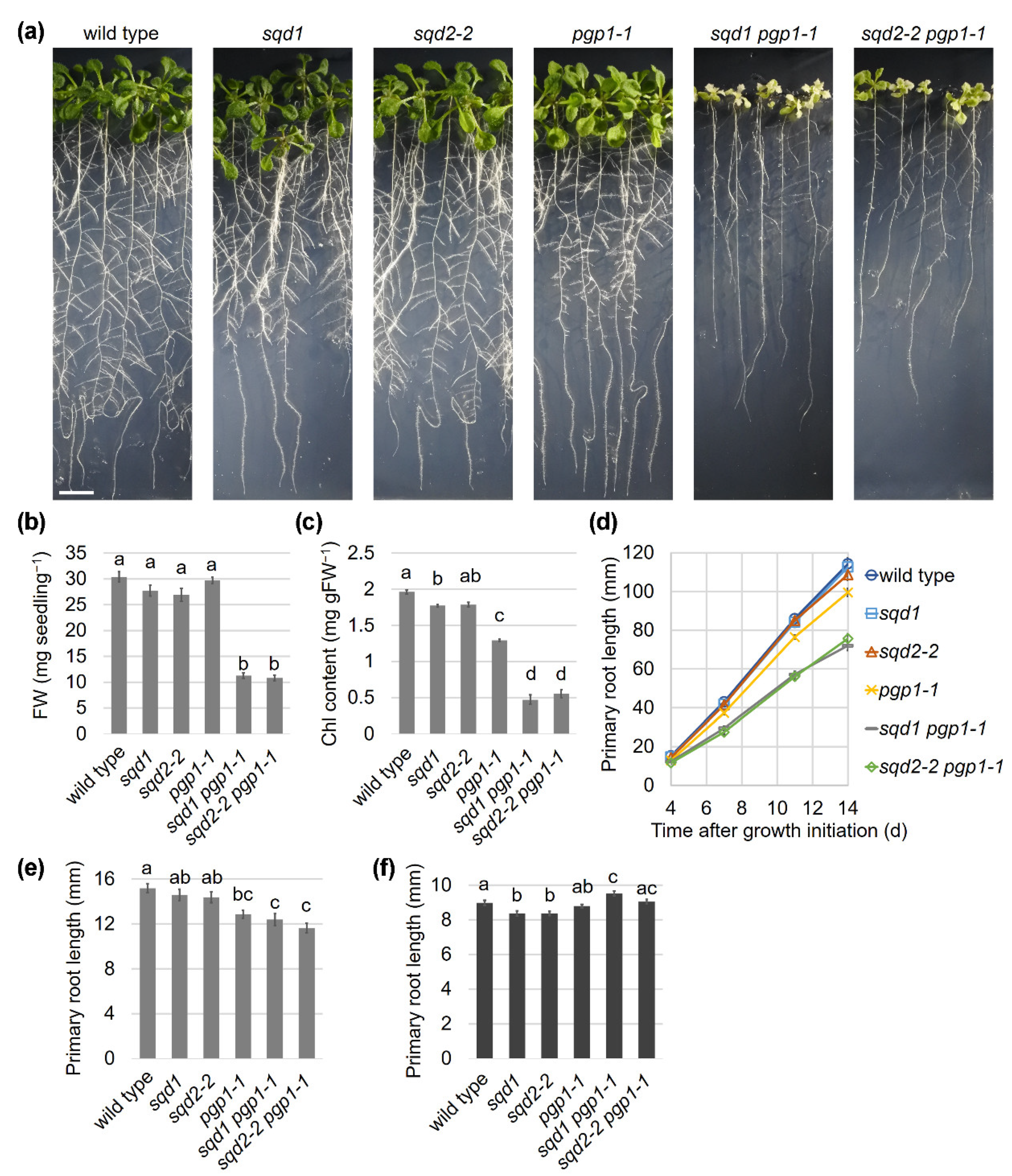
2.1. Loss of SQD1 and SQD2 in the pgp1-1 Background Causes the Same Growth Defects under P-sufficient Conditions

Although Yu and Benning (2003) reported that the sqd2 pgp1-1 double mutant showed more severe growth defects than each single mutant [23], to what extent the loss of GlcADG biosynthesis contributed to the defective phenotype of the double mutant was unknown. To assess whether GlcADG has any functions under P-sufficient conditions, we compared growth phenotypes of 14-d-old seedlings of pgp1-1, sqd1, sqd2-2, and their double mutants with wild type. Visible phenotypes of sqd1 and sqd2-2, which carry a T-DNA insertion at the first exon of SQD1 and SQD2, respectively [18,24], were almost comparable to that of wild type (Figure 1a). Fresh weight (FW) of the above-ground part of sqd1 and sqd2-2 appeared slightly lower than that of wild type, but with a significant difference only between sqd2-2 and wild type (Figure 1b). Chlorophyll (Chl) content in sqd1 and sqd2-2 was slightly but significantly lower than that in wild type (Figure 1c). Measurements of primary root length indicate no difference among wild type, sqd1, and sqd2-2 in root growth (Figure 1d). The pgp1-1 mutant showed a ~30% reduction in Chl content compared to wild type (Figure 1c), although its FW was not significantly different from the wild-type level (Figure 1b). Elongation of the primary root slightly decreased in pgp1-1 compared with wild type (Figure 1d). The double homozygous mutants of sqd1 pgp1-1 and sqd2-2 pgp1-1, which were generated by crossing sqd1 or sqd2-2 with pgp1-1, showed a pale yellow-green leaf phenotype (Figure 1a), with their FW and Chl content reduced by ~70% and ~75%, respectively, from the wild-type level (Figure 1b,c). Primary root growth of sqd1 pgp1-1 and sqd2-2 pgp1-1 further decreased from the pgp1-1 single mutant (Figure 1d). Meanwhile, no significant differences were observed between sqd1 pgp1-1 and sqd2-2 pgp1-1 in FW, Chl content, and primary root length.
The pgp1-1, sqd1 pgp1-1, and sqd2-2 pgp1-1 mutants showed shorter primary root length than wild type already at 4 d after growth initiation (Figure 1e). To eliminate the effect of photosynthesis on root growth, we grew wild type and the mutants in complete darkness for 4 d. In the dark condition, both sqd1 and sqd2-2 showed a statistically significant reduction of the primary root length compared with wild type, although the difference was marginal (~95% of the wild-type level) (Figure 1f). In contrast, pgp1-1, sqd1 pgp1-1, and sqd2-2 pgp1-1 mutants showed no reduced elongation of the primary root compared with wild type. Thus, the reduced primary root growth of these mutants was dependent on light.

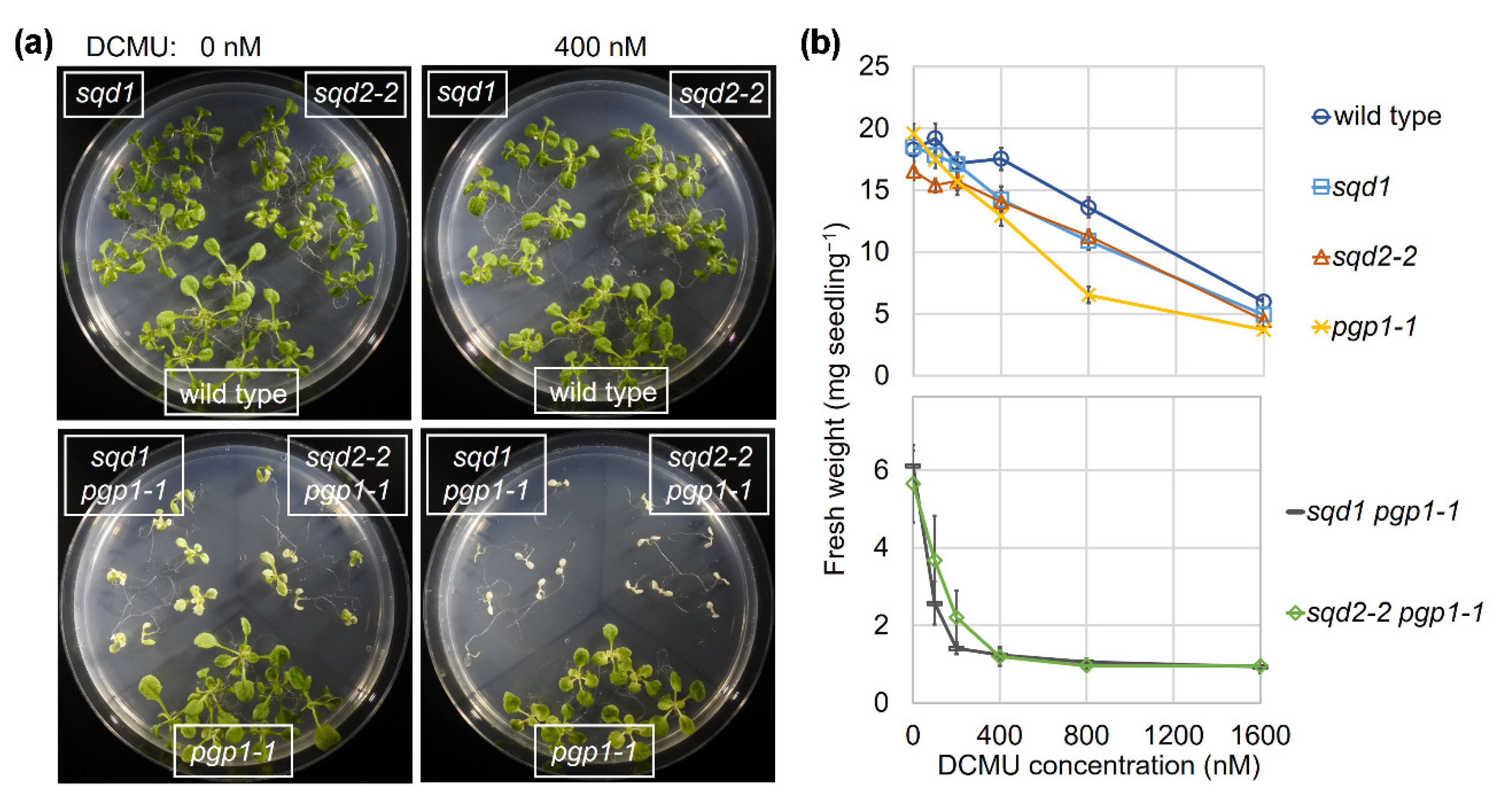
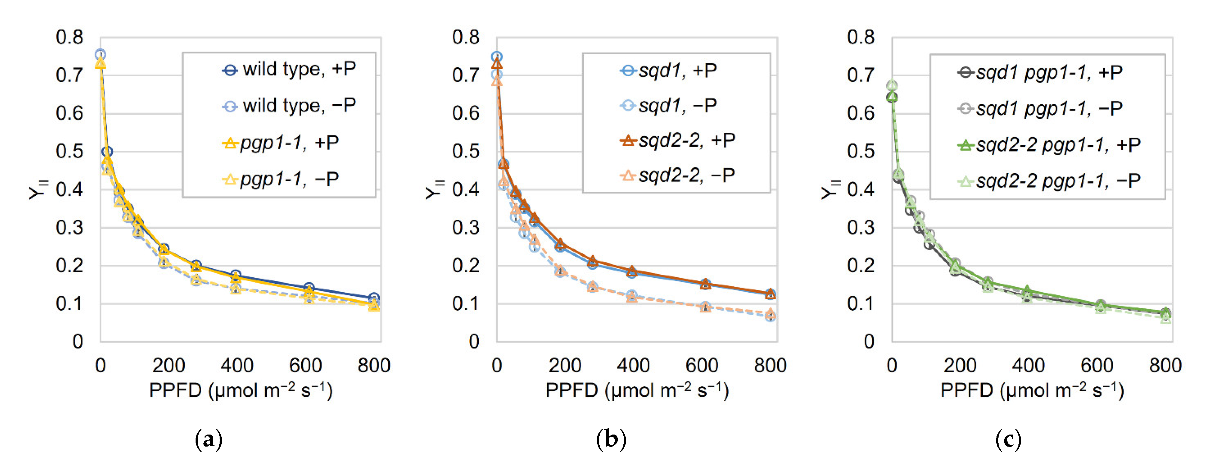
2.2. Loss of SQD1 and SQD2 Causes Similar Photosynthetic Defects

Yu and Benning (2003) reported that the pgp1-1 and sqd2 mutants were slightly more susceptible to 3(3,4-dichlorophenyl)-1,1-dimethylurea (DCMU), a photosynthesis electron transport inhibitor, than wild type in terms of growth impairment [23]. Moreover, the growth of the sqd2 pgp1-1 double mutant was severely impaired by DCMU. To assess whether the lack of GlcADG biosynthesis in sqd2 mutants is associated with the growth impairment by DCMU, we analyzed the effects of several concentrations of DCMU on the growth of wild type and the mutants (Figure 2). The sqd1 and sqd2-2 mutants showed a stronger growth impairment by DCMU than wild type, and the growth impairment of pgp1-1 was more severe than sqd1 and sqd2-2. Moreover, both sqd1 pgp1-1 and sqd2-2 pgp1-1 showed an intensive growth impairment even under low DCMU concentrations.
To further dissect the effect of loss of SQDG and GlcADG biosynthesis on photosynthesis, we grew plants under P-sufficient and -starved conditions and analyzed the light–response curves of effective quantum yield of PSII (YII) with the imaging pulse amplitude modulation Chl fluorometer (imaging-PAM) (Figure 3). In wild type, the YII levels were slightly decreased by P starvation compared with those under the P-sufficient condition. The light–response curves of YII in pgp1-1 were almost the same as those in wild type under each P condition. The sqd1 and sqd2-2 mutants also showed the YII curves similar to those in wild type and pgp1-1 under the P-sufficient condition. However, under the P-starved condition, the YII levels in sqd1 and sqd2-2 decreased more strongly than wild type and pgp1-1. Between sqd1 and sqd2-2, no remarkable difference was observed under both P conditions. In the sqd1 pgp1-1 and sqd2-2 pgp1-1 double mutants, the YII levels were lower than those in wild type and single mutants under the P-sufficient condition. In these double mutants, no further decrease in YII was observed in response to P starvation. In addition, no noticeable difference was observed between sqd1 pgp1-1 and sqd2-2 pgp1-1.

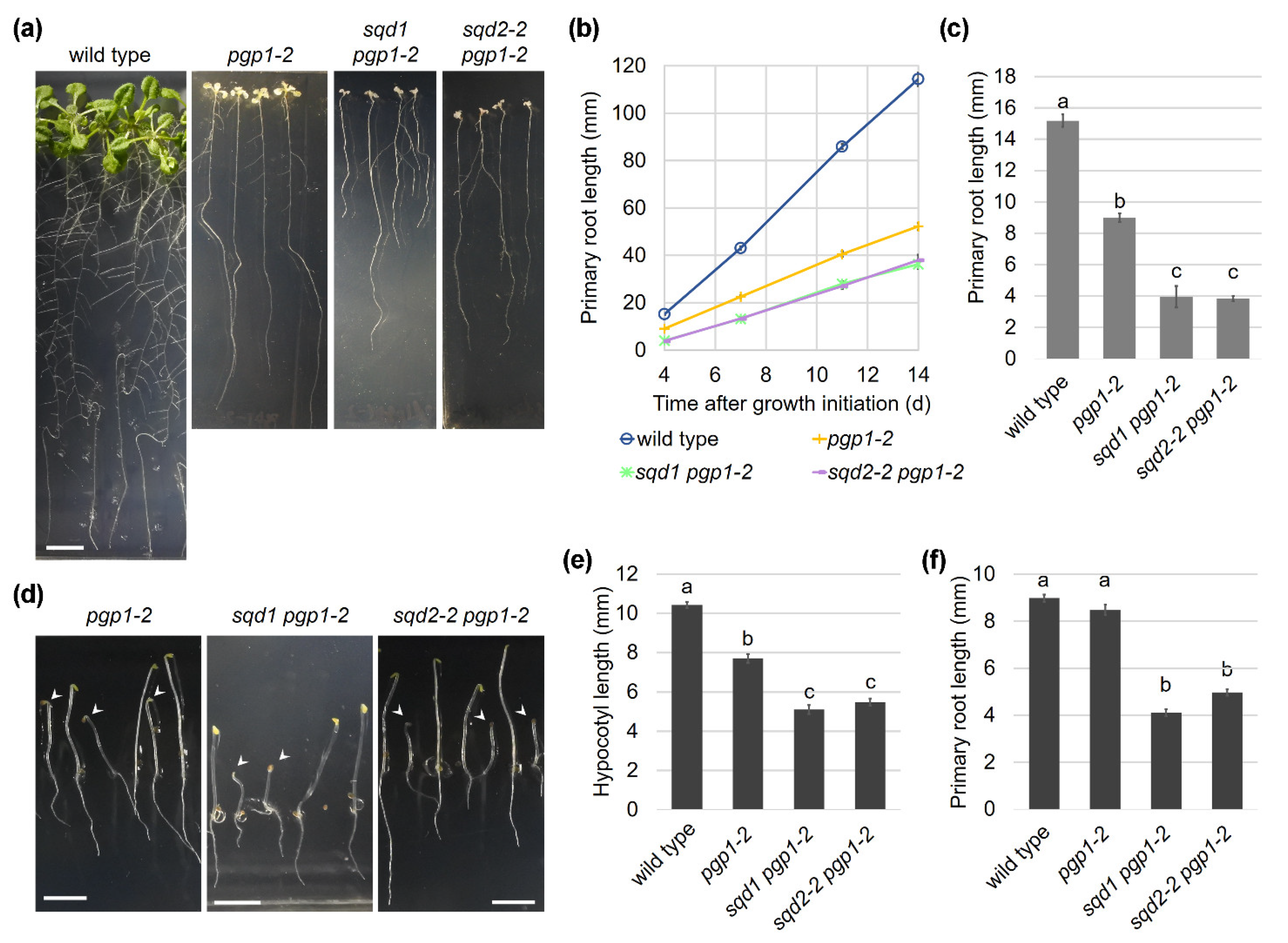
2.3. Complete Loss of Anionic Lipid Biosynthesis in Plastids Substantially Impairs Plant Development

The comparisons of sqd1 pgp1-1 and sqd2-2 pgp1-1 revealed that SQDG, but not GlcADG, plays a pivotal role in compensating the decrease in plastid PG biosynthesis in the pgp1-1 mutant. However, pgp1-1 retains 30% activity of PGP1; thus, the effect of complete lack of anionic lipid biosynthesis in plastids was undetermined. To further understand the roles of plastid anionic lipids in plant growth, we introduced the knockout pgp1-2 mutation into sqd1 and sqd2-2 and characterized the resulting double mutants.
Because the homozygous pgp1-2 mutant is seedling lethal, this allele is maintained in the heterozygous state. Consistent with previous reports [14,15,16], the homozygous pgp1-2 mutants, which were obtained from self-pollinated heterozygous parents, showed a severe impairment of growth and Chl accumulation even in the presence of 1% sucrose in the growth medium (Figure 4a–c).
By crossing the heterozygous pgp1-2 plants with the homozygous sqd1 and sqd2-2 mutants and subsequent segregation, we obtained heterozygous pgp1-2 plants with homozygous sqd1 or sqd2-2 mutation (sqd1/sqd1 PGP1/pgp1-2 and sqd2-2/sqd2-2 PGP1/pgp1-2). In the progeny of these plants, we identified double homozygous mutants of sqd1 pgp1-2 (sqd1/sqd1 pgp1-2/pgp1-2) and sqd2-2 pgp1-2 (sqd2-2/sqd2-2 pgp1-2/pgp1-2), both of which showed a severe growth impairment and an albino phenotype (Figure 4a). The pgp1-2 single mutant developed only few small leaves even at 24 d after growth initiation and the sqd1 pgp1-2 and sqd2-2 pgp1-2 double mutants showed stronger growth defects than pgp1-2. The leaves of both double mutants were severely underdeveloped with irregular shapes and rough outlines. FW of these double mutants was substantially low and Chl content was almost negligible (Figure 4b,c).

2.4. The Photosystem II Function Is Completely Abolished by Loss of Anionic Lipid Biosynthesis in Plastids

We previously reported that loss of plastid PG biosynthesis causes a severe but not complete dysfunction of PSII photochemistry [15,16], as shown in a decrease in the maximum PSII quantum efficiency (Fv/Fm) in this mutant under the P-sufficient condition (Figure 5a). As observed previously [15], the pgp1-2 mutant showed high minimum Chl fluorescence (Fo) because of a severe inhibition of energy transfer from the antenna system to the reaction center in photosystems and consequent dissipation of absorbed light energy as fluorescence. Under the P-starved condition, some pgp1-2 plants showed complete loss of the PSII photochemical activity, although there were large individual differences. In both sqd1 pgp1-2 and sqd2-2 pgp1-2 double mutants, no PSII activity was observed regardless of P conditions, suggesting that SQDG is essential for the remaining PSII activity in the pgp1-2 mutant.
Our previous study revealed that P starvation activates biosynthesis of glycolipids including SQDG and enhances thylakoid formation and Chl accumulation in pgp1-2 leaves [16]. To ascertain whether SQDG is required for the activation of chloroplast development in pgp1-2 under P starvation, we analyzed Chl content in the sqd1 pgp1-2 and sqd2-2 pgp1-2 mutants under P-sufficient and -starved conditions (Figure 5b,c). No difference in Chl content between the two P conditions in these mutants demonstrated that SQDG, but not GlcADG, is important for the complementation of decreased PG content under P-starved conditions.

2.5. Anionic Lipids Are Essential for Thylakoid Membrane Biogenesis

We previously reported that plastids in the pgp1-2 leaves are unable to form thylakoid membrane networks and only partially develop immature internal membrane structures [14,16]. By contrast, the morphology of mitochondria appeared normal in PGP1 knockout mutants [11,14]. To understand how the lack of SQDG in addition to PG affects the morphology of plastids and mitochondria in leaf cells, we examined the ultrastructure of these organelles in leaves of sqd1 pgp1-2 and sqd2-2 pgp1-2 by transmission electron microscopy. In both mutants, leaf plastids developed no thylakoid membranes and instead accumulated many small membrane fragments (Figure 6). Some plastids showed clusters of high electron-dense particles, which resembled plastoglobuli. In the leaf plastids of these double mutants, vacuole-like large electron transparent areas were often observed in addition to vesicle-like small structures. Some plastids also showed cytosol-like inclusions inside. These structures may reflect intrusions of membranous tubes of cytosol or vacuole compartment into plastids, which were not observed in wild-type chloroplasts. In contrast to the abnormal plastid structures, the morphology of mitochondria in these double mutants was similar to that in wild type.

2.6. Root Growth Is Impaired in Anionic Lipid Mutants

In addition to the shoot growth, the primary root growth was severely impaired in pgp1-2, and this phenotype was further enhanced in the sqd1 pgp1-2 and sqd2-2 pgp1-2 double mutants (Figure 7a,b). In these mutants, root elongation was strongly impaired from the early stage of growth under light (Figure 7c). To eliminate the effect of light on plant growth, we grew these mutants in complete darkness for 4 d and analyzed the phenotypes. The development of the mutants was impaired even in the dark (Figure 7d). The hypocotyl length of the pgp1-2 mutant was 75% of the wild-type level and those of the double knockout mutants further reduced by half (Figure 7e). Moreover, the double knockout mutants also showed ~50% reduction in root length, whereas pgp1-2 elongated the primary root to the wild-type level in the dark (Figure 7f).

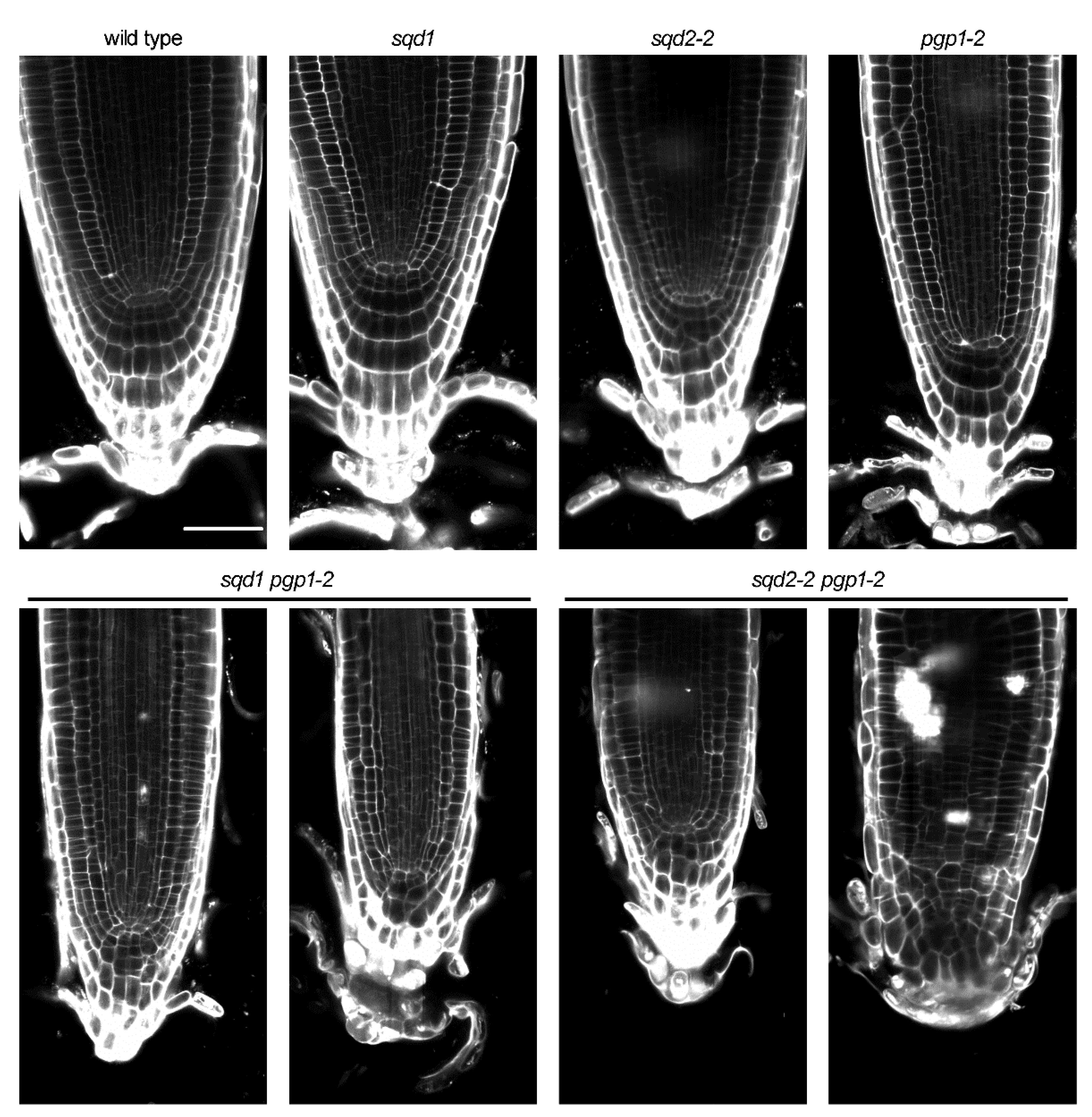
2.7. Cellular Architecture of the Root Tip Was Disrupted by Loss of Anionic Lipids

To examine whether the loss of anionic lipids affects the cellular organization of the root tip, the primary roots of 7-d-old plants were stained with propidium iodide and observed under a confocal fluorescence microscope (Figure 8). No visible difference of cellular architecture at the root tip region was observed among wild type, sqd1, and sqd2-2. Moreover, the cell organization of the root tip was not notably disrupted in pgp1-2, although the primary root elongation was severely decreased in this mutant in the light (Figure 7a–c). By contrast, both the sqd1 pgp1-2 and sqd2-2 pgp1-2 double mutants showed irregular cell arrangement at the root cap and the meristematic zone.

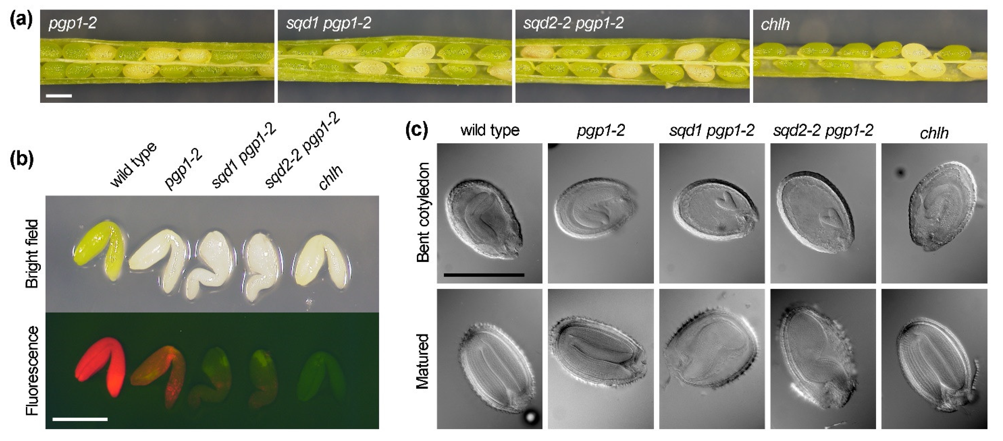
2.8. Loss of Plastid Anionic Lipids Impairs Embryo Development and Decreases Germination Capacity

Because seedling development was severely impaired in the sqd1 pgp1-2 and sqd2-2 pgp1-2 double homozygous mutants immediately after germination, we examined whether embryo development was perturbed in these mutants. As reported previously [25], pale yellow seeds, which correspond to the pgp1-2 homozygote, were detected within the fully developed but still green siliques of the self-pollinated heterozygous pgp1-2 mutant at a frequency of 25.0% (Figure 9a, Table 2). Similarly, pale yellow seeds were observed in the siliques of the self-pollinated sqd1/sqd1 PGP1/pgp1-2 and sqd2-2/sqd2-2 PGP1/pgp1-2 plants at a frequency of 24.9% and 24.8%, respectively. To test whether embryo development is arrested in these pale yellow seeds, embryos were taken out from the seeds in fully developed green siliques and observed under a fluorescent stereomicroscope (Figure 9b). Consistent with the previous report [25], the homozygous pgp1-2 embryos fully developed as did wild type, although they showed an albino phenotype with substantially reduced Chl fluorescence. Embryos from pale yellow seeds in sqd1/sqd1 PGP1/pgp1-2 and sqd2-2/sqd2-2 PGP1/pgp1-2 siliques were also albino and showed only a faint Chl fluorescence. In contrast to the normal morphology of pgp1-2 embryos, the albino embryos of these double mutants showed a developmental arrest with their hypocotyl irregularly bending. Differential interference contrast microscopy revealed that, unlike in the wild-type and the albino pgp1-2 seeds, the growth of embryos in the albino seeds of the double mutants was retarded and terminated incompletely (Figure 9c). To assess whether the lack of Chl causes the arrest of embryo development, we observed albino embryos of the chlh mutant, which has a knockout mutation in the CHLH gene encoding the Mg-chelatase H subunit and completely lacks the activity to synthesize Mg-protoporphyrin IX, an essential intermediate of the Chl biosynthesis pathway [26]. Although the albino chlh embryos accumulated no Chl, their morphology was similar to the wild-type embryos (Figure 3b,c), which indicates that Chl accumulation is not essential for embryo development.
Then, we investigated whether the impaired embryo development in the sqd1 pgp1-2 and sqd2-2 pgp1-2 double mutants decreases germination capacity of seeds. In wild type, sqd1 and sqd2-2, the percentage of germinated seeds were ≥95% of total seeds sowed on plates. However, the germination of seeds from self-pollinated heterozygous pgp1-2 decreased to 92%. Moreover, in the seeds from self-pollinated sqd1/sqd1 PGP1/pgp1-2 and sqd2-2/sqd2-2 PGP1/pgp1-2 plants, the germination percentage further decreased to 79% and 84%, respectively. We observed that self-pollinated heterozygous parents produced albino seeds at ~25% frequency (Table 2). However, the emergence of albino seedlings after seed germination was 18.4 % for pgp1-2, and it further decreased to 10.2% and 12.0% for sqd1 pgp1-2 and sqd2-2- pgp1-2, respectively. From the ~25% frequency of albino seeds in the parent siliques, the germination percentage of homozygous pgp1-2, sqd1 pgp1-2, and sqd2-2 pgp1-2 genotypes was estimated as 73.6%, 41.0%, and 48.4%, respectively, which are notably low as compared with wild-type seeds.

3. Discussion

This is the first study to characterize the mutants completely lacking both SQDG biosynthesis and PG biosynthesis in plastids. The mutants, sqd1 pgp1-2 and sqd2-2 pgp1-2, showed a developmental arrest during embryogenesis (Figure 9), decreased germinability (Table 2), severe growth impairments, and cell abnormalities in the shoot and root (Figure 4, Figure 7 and Figure 8). Chloroplast development was completely abolished in these mutants (Figure 6) with only faint Chl accumulation (Figure 4) and no PSII activity (Figure 5). All these data demonstrate indispensable roles of plastid anionic lipids in various processes of plant growth. We also compared sqd1 and sqd2-2 in the pgp1-1 background (Figure 1, Figure 2 and Figure 3) in which the PGP1 activity decreases to 30% of the wild-type level [13]. Our analysis allows for distinguishing the role of SQDG and GlcADG and revealed that SQDG, but not GlcADG, is essential for the partial compensation of decreased PG biosynthesis in plastids.

3.1. Role of SQDG in Complementing the Function of PG in Chloroplast Development and Photosynthesis

In contrast to the severe impairment of leaf development in the sqd1 pgp1-2 and sqd2-2 pgp1-2 double mutants, the relatively milder growth defect of the sqd1 pgp1-1 and sqd2-2 pgp1-1 mutants allowed for detailed photosynthetic characterization under P-sufficient and -starved conditions. Under the P-sufficient condition, both sqd1 pgp1-1 and sqd2-2 pgp1-1 showed lower YII levels than pgp1-1 (Figure 3), which suggests that, in pgp1-1, SQDG, but not GlcADG, maintains the photosynthetic electron transport activity to the wild-type levels despite a partial loss of PG. The similar results were observed in the sqd1 and sqd2-2 single mutants under the P-starved condition. Although sqd1 and sqd2-2 showed wild-type levels of YII under the P-sufficient condition, these mutants decreased YII more strongly than wild type and pgp1-1 with P starvation. In sqd1 and sqd2-2, a decrease in PG by P starvation in addition to the genetic loss of SQDG biosynthesis would cause a crucial lack of anionic lipids in chloroplasts and thereby impair photosynthetic reactions under P starvation. By contrast, in sqd1 pgp1-1 and sqd2-2 pgp1-1, YII levels were not altered by P starvation because PG content, and thus YII levels, were low even under the P-sufficient condition. No differences between sqd1 and sqd2-2 and between sqd1 pgp1-1 and sqd2-2 pgp1-1 under P starvation confirm no contribution of GlcADG to complementing PG functions in photosynthesis. GlcADG would play an important role in cellular processes other than photosynthesis under P starvation.
Although the pgp1-2 mutant showed much stronger impairments of photosynthetic reactions and chloroplast development than pgp1-1, this mutant still contains a small amount of Chl and partially maintained the photosynthetic electron transport activity [15]. However, the additional mutation of either sqd1 or sqd2-2 in pgp1-2 remarkably impaired Chl accumulation and completely abolished the PSII activity (Figure 4 and Figure 5). These data indicate that SQDG functions to maintain the partial Chl accumulation and photosynthetic activity in pgp1-2. We previously reported that P starvation, which activated the biosynthesis of glycolipids including SQDG, slightly increased Chl content and thylakoid-like membrane formation in the pgp1-2 plastids [16]. In this study, P starvation did not increase Chl content in the sqd1 pgp1-2 and sqd2-2 pgp1-2 double mutants. Therefore, the increased Chl content in pgp1-2 under the P-starved condition requires SQDG accumulated in response to P starvation. In contrast to increased Chl content and partial thylakoid development, Fv/Fm in pgp1-2 did not increase and even decreased under the P-starved condition [16], which may be due to excessive accumulation of Chl over functional PSII with a further decrease in PG content by P starvation. Similarly, in this study, no improvement and, in some pgp1-2 plants, complete abolishment of Fv/Fm were observed under the P-starved condition (Figure 5). Therefore, SQDG increased during P starvation can partially compensate the loss of PG in terms of Chl accumulation but is unable to maintain the PSII functionality without PG. Essential roles of specific PG molecules in the PSII complex have been revealed in a cyanobacterium Synechocystis sp. PCC 6803 [27,28]. The specific functions of PG in PSII may not be replaced by SQDG in plants as well as cyanobacteria. A similar speculation can be made for the PSI complex, which also contains PG molecules as structural components [29,30], although the particular function of each PG molecule in the PSI complex remains unclear. P starvation, which induces GlcADG accumulation in A. thaliana, did not positively affect the PSII photochemistry and Chl accumulation in sqd1 pgp1-2 as compared with sqd2-2 pgp1-2, so GlcADG has no capacity to replace the PG molecules in photosynthetic complexes.
Although sqd1 and sqd2-2 showed no noticeable difference in the YII levels compared with wild type under the P-sufficient condition, these mutants showed a slight decrease in Chl content (Figure 1) and increased susceptibility to DCMU (Figure 2). Therefore, although PG is the main anionic lipid for chloroplast development and photosynthesis, SQDG also contributes to these processes along with PG even under P-sufficient conditions. Of note, SQDG molecules are found in PSII of plants and cyanobacteria as structural components [31,32]. Characterization of PSII from a SQDG-deficient mutant of Thermosynechococcus elongatus revealed that the loss of SQDG partially impaired the PSII activity by inhibiting electron transfer at the acceptor site [33]. In the SQDG-lacking PSII, other lipids, most likely PG molecules, occupied the SQDG-binding sites. The data in T. elongatus suggest that although SQDG molecules in the PSII-binding sites are necessary to fully maintain the activity and stability of PSII, replacement of SQDGs by other lipids, probably PG, can partially complement for their functions, which would lead to only a partial impairment of PSII by lack of SQDG. Because T. elongatus and C. reinhardtii showed stronger defects in the PSII function and growth by loss of SQDG biosynthesis than A. thaliana under P-sufficient conditions [9,21,34], the replaceability of SQDG by PG or other lipids may partially vary with species.

3.2. Role of Plastid Anionic Lipids in Root Growth and Embryo Development

The complementary relationship between PG and SQDG has been mainly discussed in relation to their functions in photosynthesis and thylakoid membrane formation. In this study, we revealed that loss of these anionic lipids impaired the primary root growth and embryo development. The pgp1-1 mutant showed a slight decrease in the primary root growth in the light and the decreased root growth was further enhanced by the additional mutation of sqd1 or sqd2-2 with pgp1-1 (Figure 1). However, these mutants showed no growth impairment of the primary root in the dark, which implies that a light-dependent pathway such as photosynthesis causes the impaired growth of the primary root in these mutants, although the plants were grown in the presence of 1% sucrose. Decreased primary root growth was also reported in the pgpp1-1 mutant lacking PGP phosphatase 1 (PGPP1), which catalyzes the final step of PG biosynthesis [5]. Because the pgpp1-1 mutant showed reduced Chl content and slightly impaired chloroplast development [35], decreased photosynthesis may explain in part the decreased root growth. However, in pgpp1-1, the impaired root growth was stronger than pgp1-1, although the chloroplast dysfunctions were similar to or milder than those in pgp1-1. Moreover, a weaker PGPP1 mutant pgpp1-2 also had a shorter root phenotype, although this mutant had no photosynthetic impairments [5]. Therefore, the disruption of PGPP1 is likely to cause the root growth impairment independently of photosynthesis. Because PGPP1 is targeted to mitochondria in roots [5], PG biosynthesis by PGPP1 in mitochondria may be required for the root growth. As is PGPP1, PGP1 is also targeted to mitochondria in addition to plastids [11]. However, for the PGP biosynthesis, PGP2 localized to ER can complement the role of PGP1 in mitochondria [11,12], which may explain why pgp1-1 showed a weaker impairment of the primary root growth than pgpp1-1. Why disrupted PG biosynthesis, presumably in mitochondria, causes the short root phenotype remains unknown. Because cardiolipin, which is formed from PG in mitochondria, is required for mitochondrial functions and loss of CL impairs root growth in A. thaliana mutants [36], PG may be involved in root growth as a substrate of CL biosynthesis. However, we cannot exclude PG-specific functions in root growth, which should be addressed in future studies. The pgp1-2 mutant also showed a shorter primary root than wild type in the light, but not in the dark (Figure 7). Babiychuk et al. (2003) showed that the PGP1 knockout mutant accumulated cardiolipin to the wild-type level and developed mitochondria with normal structure and respiratory functions [11]. Therefore, the complete loss of PGP1 would not remarkably affect mitochondrial functions in roots.
In contrast to the pgp1-2 single mutant, the sqd1 pgp1-2 and sqd2-2 pgp1-2 double mutants showed decreased primary root growth even in the dark (Figure 7). Because SQDG is not included in mitochondria at least under P-sufficient conditions [37], the decreased root growth of these double mutants may occur independently of mitochondrial activity. The double mutants showed severe disruption of root tip architecture (Figure 8), which would lead to the shorter primary root of the mutants. Of note, the embryo development is severely retarded in the sqd1 pgp1-2 and sqd2-2 pgp1-2 double mutants, but not in the pgp1-2 single mutant (Figure 9). Therefore, the incomplete plant body development during embryogenesis might be associated with the impaired primary root growth after germination. Photosynthetic dysfunctions would be unrelated to the impaired embryo development in the double mutants because Chl-deficient embryos of pgp1-2 and chlh could fully develop during seed maturation. Abnormal embryo development was reported in several A. thaliana mutants related to lipid metabolism in plastids [38,39,40], which includes a knockout mutant for MGDG biosynthesis [41]. Therefore, plastid lipid metabolism, in which anionic lipid biosynthesis is involved, may be crucial for the normal development of embryos. The severe disruption of leaf formation, root tip architecture, and embryo development in sqd1 pgp1-2 and sqd2-2 pgp1-2 suggests that anionic lipids synthesized in plastids are essential for cellular organization in the meristematic tissues. Because pgp1-2 as well as sqd1 and sqd2-2 single mutants did not show such severe developmental defects in the meristematic tissues, PG and SQDG can complement each other in these tissues. Considering that PG and SQDG are major glycerolipid constituents in envelope membranes of non-photosynthetic plastids in addition to chloroplasts [42], lack of anionic lipids in the plastid envelope may disrupt plastid functions and/or interorganellar associations required for cellular development and organization. In the dark condition, both sqd1 and sqd2-2 mutant showed slightly reduced primary root growth compared with wild type (Figure 1f). Although it might reflect an unknown function of SQDG in the primary root growth in the dark, we cannot make any conclusion because the sqd1 pgp1-1 and sqd2-2 pgp1-1 double mutants showed no reduction in the primary root growth in the dark. Again, no differences between sqd1 pgp1-2 and sqd2-2 pgp1-2 during embryo and seedling development demonstrate that GlcADG does not have important roles in these processes under P-sufficient conditions. How PG and SQDG are involved in the cellular organization in addition to photosynthetic machinery awaits future investigations.

4. Materials and Methods

4.1. Plant Materials and Growth Conditions

The wild type, sqd1 [24], sqd2-2 [18], pgp1-1 [13], and pgp1-2 [14] were the Columbia ecotype of A. thaliana (Table 1). The sqd1 pgp1-1, sqd2-2 pgp1-1, sqd1 pgp1-2, and sqd2-2 pgp1-2 double mutants were generated by crossing each single mutant line. The genotypes for each allele were confirmed by PCR analysis as described [12,13,18,24]. Because the homozygous pgp1-2 mutation causes a seedling lethal phenotype, the pgp1-2 allele was maintained in the heterozygous state for the pgp1-2 single mutant and sqd1 pgp1-2 and sqd2-2 pgp1-2 double mutants. In these mutant lines, plants in the homozygous pgp1-2 state were identified with their albino or pale yellow-green leaf phenotypes in the progeny of self-pollinated parents under the light condition. For etiolated seedlings germinated in the dark, mutants in the homozygous pgp1-2 state were almost indistinguishable from the heterozygous and wild-type seedlings in their cotyledon color phenotype. Therefore, in the analysis of etiolated seedlings of pgp1-2, sqd1 pgp1-2, and sqd2-2 pgp1-2 lines, we first took photographs of all etiolated seedlings for size measurements and then illuminated the seedlings for 2 d to identify albino individuals with homozygous pgp1-2 allele.
Surface-sterilized seeds on Murashige and Skoog (MS) medium (adjusted to pH 5.7 with KOH) containing 1% (w/v) sucrose were cold-treated at 4 °C for 3 days in the dark. Then, plants were germinated and grown in a chamber maintained at 23 °C under continuous white light (~80 μmol photons m−2 s−1). For the growth phenotype analysis, plants were vertically grown on the MS medium solidified with 0.6% (w/v) Gelrite (Wako, Tokyo, Japan). The length of the primary root and the hypocotyl was determined by using ImageJ software (https://imagej.nih.gov/ij/, accessed on 4 May 2021). For the other analysis, plants were horizontally grown on the MS medium solidified with 0.8% (w/v) agar. For P-starvation analysis, 7-d-old seedlings of wild type, pgp1-1, sqd1, and sqd2-2, 11-d-old seedlings of sqd1 pgp1-1 and sqd2-2 pgp1-1, and 14-d-old seedlings of pgp1-2, sqd1 pgp1-2, and sqd2-2 pgp1-2, which were roughly at a similar developmental stage with four true leaves, were transferred to the MS medium lacking KH2PO4 and further grown for 7 d. For DCMU treatment, 7-d-old seedlings were transferred to the MS medium containing several concentrations of DCMU and grown for another 7 d.
To determine germination ability, seeds were collected from adult A. thaliana plants grown for ~7 weeks in Giffy 7 pots. After desiccation of collected seeds for a week in a dry container at room temperature, surface-sterilized seeds were incubated on the MS medium in a growth chamber for 4 d, and germination percentage was determined under a stereomicroscope.

4.2. Measurement of FW and Chl Content

The shoot of the 14-day-old seedling was cut at the hypocotyl and weighed to determine FW. Then, the shoot was incubated in 80% (v/v) acetone at 4 °C in darkness for two days to extract lipophilic pigments. The absorbance of the acetone extract at 720, 663.2, and 646.8 nm was measured with a spectrometer (V-730 BIO, JASCO, Tokyo, Japan) to determine the total content (Chl a + Chl b) as described [43]. For pgp1-2, sqd1 pgp1-2, and sqd2-2 pgp1-2, the Chl content in a seedling was below a detection limit in the spectrometric analysis. Therefore, for these mutants, intensity of Chl fluorescence at 666 nm under 435 nm excitation was measured with a fluorospectrometer (RF5300, Shimadzu, Kyoto, Japan) and total Chl content was determined by using a known concentration of Chl as a standard.

4.3. Analysis of Photosynthetic Efficiency

Chl fluorescence parameters were analyzed by using an imaging PAM fluorometer (IMAGING-PAM MAXI, Walz, Effeltrich, Germany) and ImagingWin software. Samples incubated under dim light (~2 μmol photons m−2 s−1) for ~30 min were dark-treated for 15 min in the device. After determination of minimal Chl fluorescence (Fo) with the lowest measuring light intensity (~0.2 μmol photons m−2 s−1 at a frequency of 2 Hz), samples were illuminated with a saturating pulse flash (~3400 μmol photons m−2 s−1 for 720 ms) to determine maximal Chl fluorescence (Fm). The maximum quantum yield of PSII (Fv/Fm) was calculated as (Fm-Fo)/Fm. For the analysis of light–response curves of effective quantum yield of PSII (YII), plants were illuminated with actinic light of given intensity for 3 min and a second saturating pulse to determine stationary (F) and maximal fluorescence under light (Fm’), respectively, which were used to calculate YII as (Fm’-F)/Fm’. Photosynthetic photon flux density of actinic light was successively increased every 3 min to 20, 55, 80, 110, 185, 280, 395, 610, and 800 μmol photons m−2 s−1.

4.4. Transmission Electron Microscopic Analysis

The A. thaliana leaves were fixed in 4% (w/v) glutaraldehyde and 4% (w/v) paraformaldehyde, buffered with 50 mM sodium cacodylate at pH 7.0 overnight at 4 °C, and washed with the same buffer at 4 °C for 4 h. Subsequently, they were post-fixed in 2% (w/v) OsO4 in 50 mM cacodylate buffer at 4 °C for 2 h. The fixed samples were run through an alcohol series and embedded in Spurr’s resin (Polysciences Inc., Warrington, PA, USA). Ultra-thin sections (80 nm thick) were cut with a diamond knife (Diatome; Biel, Switzerland) on an ultra-microtome (Ultracut S; Leica, Vienna, Austria). The floating sections were transferred to formvar-coated grids. They were double stained with 1% (v/v) uranyl acetate for 20 min and with lead citrate solution for 10 min. After washing with distilled water, the samples were observed with a JEM-1400 transmission electron microscope (JEOL, Tokyo, Japan).

4.5. Root Tip Observation

For the observation of root tip cells, the primary roots of the 7-day-old seedlings were stained with 10 mg ml−1 propidium iodide (PI) on microscope slides for ~3 min and observed under a confocal laser scanning microscope (LSM700, Zeiss, Jena, Germany), with double-sided tape used as a spacer between a microscope slide and a coverslip. Root tip cells were visualized with PI fluorescence at 600–640 nm under 555 nm excitation.

4.6. Embryo Observation

For the observation of Chl accumulation in embryos, embryos were taken out from seeds that were fully developed but not yet degreened and dehydrated. Chl autofluorescence was observed under a stereomicroscope (MZ 16FA; Leica, Wetzlar, Germany) with a long-pass filter set (TXR, Leica) and a color CCD camera (VB-7010; Keyence, Osaka, Japan) in darkness. For the observation of embryo development in seeds, seeds were collected from siliques at different stages after self-pollination and treated with a clearing solution of chloral hydrate, water, and glycerol (8:2:1, v/v/v) for 1 h. Then, the embryos in seeds were observed under a differential interference contrast microscope (BX60-34 FL, Olympus, Tokyo, Japan).

Author Contributions

Conceptualization, K.K. and H.W.; investigation, A.Y., N.N., and K.K.; writing—original draft preparation, A.Y. and K.K.; writing—review and editing, A.Y., K.K., N.N., and H.W.; project administration, K.K.; funding acquisition, K.K. and H.W. All authors have read and agreed to the published version of the manuscript.

Funding

This research was funded by JSPS KAKENHI Grant Number 17H03692, 17K19342, 18H03941, and 20K06691.

Institutional Review Board Statement

Not applicable.

Informed Consent Statement

Not applicable.

Acknowledgments

We thank Yozo Okazaki (Mie University) for supplying the sqd1 and sqd2-2 mutants and Christoph Benning (Michigan State University) for the pgp1-1 mutant. We thank Keiko Kobayashi and Megumi Kobayashi (Japan Women’s University) for the work on transmission electron microscopy and Haruka Hori (The University of Tokyo) for mutant analysis.

Conflicts of Interest

The authors declare no conflict of interest.

References

  1. Kobayashi, K. Role of membrane glycerolipids in photosynthesis, thylakoid biogenesis and chloroplast development. J. Plant Res. 2016, 129, 565–580. [Google Scholar] [CrossRef] [Green Version]
  2. Dorne, A.J.; Joyard, J.; Douce, R. Do thylakoids really contain phosphatidylcholine? Proc. Natl. Acad. Sci. USA 1990, 87, 71–74. [Google Scholar] [CrossRef] [PubMed] [Green Version]
  3. Kobayashi, K.; Endo, K.; Wada, H. Roles of lipids in photosynthesis. In Lipids in Plant and Algae Development. Subcellular Biochemistry; Nakamura, Y., Li-Beisson, Y., Eds.; Springer: New York, NY, USA, 2016; Volume 86, pp. 21–49. [Google Scholar]
  4. Heinz, S.; Rast, A.; Shao, L.; Gutu, A.; Gügel, I.L.; Heyno, E.; Labs, M.; Rengstl, B.; Viola, S.; Nowaczyk, M.M.; et al. Thylakoid membrane architecture in Synechocystis depends on CurT, a homolog of the granal CURVATURE THYLAKOID1 proteins. Plant Cell 2016, 28, 2238–2260. [Google Scholar] [CrossRef] [PubMed] [Green Version]
  5. Lin, Y.-C.C.; Kobayashi, K.; Wada, H.; Nakamura, Y. Phosphatidylglycerophosphate phosphatase is required for root growth in Arabidopsis. Biochim. Biophys. Acta Mol. Cell Biol. Lipids 2018, 1863, 563–575. [Google Scholar] [CrossRef] [PubMed]
  6. Wu, F.; Yang, Z.; Kuang, T.; Physiology, E.M. Impaired photosynthesis in phosphatidylglycerol-deficient mutant of cyanobacterium Anabaena sp. PCC7120 with a disrupted gene encoding a putative phosphatidylglycerophosphatase. Plant Physiol. 2006, 141, 1274–1283. [Google Scholar] [CrossRef] [Green Version]
  7. Hagio, M.; Gombos, Z.; Várkonyi, Z.; Masamoto, K.; Sato, N.; Tsuzuki, M.; Wada, H. Direct evidence for requirement of phosphatidylglycerol in photosystem II of photosynthesis. Plant Physiol. 2000, 124, 795–804. [Google Scholar] [CrossRef] [Green Version]
  8. Bogos, B.; Ughy, B.; Domonkos, I.; Laczko-Dobos, H.; Komenda, J.; Abasova, L.; Cser, K.; Vass, I.; Sallai, A.; Wada, H.; et al. Phosphatidylglycerol depletion affects photosystem II activity in Synechococcus sp. PCC 7942 cells. Photosynth. Res. 2010, 103, 19–30. [Google Scholar] [CrossRef]
  9. Endo, K.; Kobayashi, K.; Wada, H. Sulfoquinovosyldiacylglycerol has an essential role in Thermosynechococcus elongatus BP-1 under phosphate-deficient conditions. Plant Cell Physiol. 2016, 57, pcw159. [Google Scholar] [CrossRef] [Green Version]
  10. Müller, F.; Frentzen, M. Phosphatidylglycerophosphate synthases from Arabidopsis thaliana. FEBS Lett. 2001, 509, 298–302. [Google Scholar] [CrossRef] [Green Version]
  11. Babiychuk, E.; Müller, F.; Eubel, H.; Braun, H.-P.; Frentzen, M.; Kushnir, S. Arabidopsis phosphatidylglycerophosphate synthase 1 is essential for chloroplast differentiation, but is dispensable for mitochondrial function. Plant J. 2003, 33, 899–909. [Google Scholar] [CrossRef]
  12. Tanoue, R.; Kobayashi, M.; Katayama, K.; Nagata, N.; Wada, H. Phosphatidylglycerol biosynthesis is required for the development of embryos and normal membrane structures of chloroplasts and mitochondria in Arabidopsis. FEBS Lett. 2014, 588, 1680–1685. [Google Scholar] [CrossRef] [Green Version]
  13. Xu, C.; Härtel, H.; Wada, H.; Hagio, M.; Yu, B.; Eakin, C.; Benning, C. The pgp1 mutant locus of Arabidopsis encodes a phosphatidylglycerolphosphate synthase with impaired activity. Plant Physiol. 2002, 129, 594–604. [Google Scholar] [CrossRef] [PubMed] [Green Version]
  14. Hagio, M.; Sakurai, I.; Sato, S.; Kato, T.; Tabata, S.; Wada, H. Phosphatidylglycerol is essential for the development of thylakoid membranes in Arabidopsis thaliana. Plant Cell Physiol. 2002, 43, 1456–1464. [Google Scholar] [CrossRef] [PubMed] [Green Version]
  15. Kobayashi, K.; Endo, K.; Wada, H. Multiple impacts of loss of plastidic phosphatidylglycerol biosynthesis on photosynthesis during seedling growth of Arabidopsis. Front. Plant Sci. 2016, 7, 336. [Google Scholar] [CrossRef] [Green Version]
  16. Kobayashi, K.; Fujii, S.; Sato, M.; Toyooka, K.; Wada, H. Specific role of phosphatidylglycerol and functional overlaps with other thylakoid lipids in Arabidopsis chloroplast biogenesis. Plant Cell Rep. 2015, 34, 631–642. [Google Scholar] [CrossRef] [PubMed]
  17. Yu, B.; Xu, C.; Benning, C. Arabidopsis disrupted in SQD2 encoding sulfolipid synthase is impaired in phosphate-limited growth. Proc. Natl. Acad. Sci. USA 2002, 99, 5732–5737. [Google Scholar] [CrossRef] [PubMed] [Green Version]
  18. Okazaki, Y.; Otsuki, H.; Narisawa, T.; Kobayashi, M.; Sawai, S.; Kamide, Y.; Kusano, M.; Aoki, T.; Hirai, M.Y.; Saito, K. A new class of plant lipid is essential for protection against phosphorus depletion. Nat. Commun. 2013, 4, 1510. [Google Scholar] [CrossRef] [Green Version]
  19. Aoki, M.; Sato, N.; Meguro, A.; Tsuzuki, M. Differing involvement of sulfoquinovosyl diacylglycerol in photosystem II in two species of unicellular cyanobacteria. Eur. J. Biochem. 2004, 271, 685–693. [Google Scholar] [CrossRef]
  20. Güler, S.; Seeliger, A.; Härtel, H.; Renger, G.; Benning, C. A null mutant of Synechococcus sp. PCC7942 deficient in the sulfolipid sulfoquinovosyl diacylglycerol. J. Biol. Chem. 1996, 271, 7501–7507. [Google Scholar] [CrossRef] [Green Version]
  21. Sato, N.; Tsuzuki, M.; Matsuda, Y.; Ehara, T.; Osafune, T.; Kawaguchi, A. Isolation and characterization of mutants affected in Lipid metabolism of Chlamydomonas reinhardtii. Eur. J. Biochem. 1995, 230, 987–993. [Google Scholar] [CrossRef]
  22. Riekhof, W.R.; Ruckle, M.E.; Lydic, T.A.; Sears, B.B.; Benning, C. The sulfolipids 2′-O-acyl-sulfoquinovosyldiacylglycerol and sulfoquinovosyldiacylglycerol are absent from a Chlamydomonas reinhardtii mutant deleted in SQD1. Plant Physiol. 2003, 133, 864–874. [Google Scholar] [CrossRef] [Green Version]
  23. Yu, B.; Benning, C. Anionic lipids are required for chloroplast structure and function in Arabidopsis. Plant J. 2003, 36, 762–770. [Google Scholar] [CrossRef]
  24. Okazaki, Y.; Shimojima, M.; Sawada, Y.; Toyooka, K.; Narisawa, T.; Mochida, K.; Tanaka, H.; Matsuda, F.; Hirai, A.; Hirai, M.Y.; et al. A chloroplastic UDP-glucose pyrophosphorylase from Arabidopsis is the committed enzyme for the first step of sulfolipid biosynthesis. Plant Cell 2009, 21, 892–909. [Google Scholar] [CrossRef] [PubMed] [Green Version]
  25. Kobayashi, K.; Ohta, H. Possible requirement of galactolipids for embryogenesis. In Photosynthesis. Energy from the Sun; Allen, J.F., Gantt, E., Golbeck, J.H., Osmond, B., Eds.; Springer: Heidelberg, Germany, 2008; pp. 783–786. [Google Scholar]
  26. Huang, Y.-S.; Li, H.-M. Arabidopsis CHLI2 can substitute for CHLI1. Plant Physiol. 2009, 150, 636–645. [Google Scholar] [CrossRef] [PubMed] [Green Version]
  27. Endo, K.; Mizusawa, N.; Shen, J.-R.; Yamada, M.; Tomo, T.; Komatsu, H.; Kobayashi, M.; Kobayashi, K.; Wada, H. Site-directed mutagenesis of amino acid residues of D1 protein interacting with phosphatidylglycerol affects the function of plastoquinone QB in photosystem II. Photosynth. Res. 2015, 126, 385–397. [Google Scholar] [CrossRef] [PubMed]
  28. Endo, K.; Kobayashi, K.; Wang, H.T.; Chu, H.A.; Shen, J.R.; Wada, H. Site-directed mutagenesis of two amino acid residues in cytochrome b 559 α subunit that interact with a phosphatidylglycerol molecule (PG772) induces quinone-dependent inhibition of photosystem II activity. Photosynth. Res. 2019, 139, 267–279. [Google Scholar] [CrossRef] [PubMed]
  29. Jordan, P.; Fromme, P.; Witt, H.T.; Klukas, O.; Saenger, W.; Krauss, N. Three-dimensional structure of cyanobacterial photosystem I at 2.5 Å resolution. Nature 2001, 411, 909–917. [Google Scholar] [CrossRef] [PubMed]
  30. Mazor, Y.; Borovikova, A.; Caspy, I.; Nelson, N. Structure of the plant photosystem I supercomplex at 2.6 Å resolution. Nat. Plants 2017, 3, 1–9. [Google Scholar] [CrossRef] [Green Version]
  31. Umena, Y.; Kawakami, K.; Shen, J.-R.; Kamiya, N. Crystal structure of oxygen-evolving photosystem II at a resolution of 1.9 Å. Nature 2011, 473, 55–60. [Google Scholar] [CrossRef] [PubMed]
  32. Wei, X.; Su, X.; Cao, P.; Liu, X.; Chang, W.; Li, M.; Zhang, X.; Liu, Z. Structure of spinach photosystem II-LHCII supercomplex at 3.2 Å resolution. Nature 2016, 534, 69–74. [Google Scholar] [CrossRef]
  33. Nakajima, Y.; Umena, Y.; Nagao, R.; Endo, K.; Kobayashi, K.; Akita, F.; Suga, M.; Wada, H.; Noguchi, T.; Shen, J.R. Thylakoid membrane lipid sulfoquinovosyl-diacylglycerol (SQDG) is required for full functioning of photosystem II in Thermosynechococcus elongatus. J. Biol. Chem. 2018, 293, 14786–14797. [Google Scholar] [CrossRef] [Green Version]
  34. Sato, N.; Sonoike, K.; Tsuzuk, M.; Kawaguchi, A. Impaired photosystem II in a mutant of Chlamydomonas reinhardtii defective in sulfoquinovosyl diacylglycerol. Eur. J. Biochem. 1995, 234, 16–23. [Google Scholar] [CrossRef]
  35. Lin, Y.-C.C.; Kobayashi, K.; Hung, C.-H.H.; Wada, H.; Nakamura, Y. Arabidopsis phosphatidylglycerophosphate phosphatase 1 involved in phosphatidylglycerol biosynthesis and photosynthetic function. Plant J. 2016, 88, 1022–1037. [Google Scholar] [CrossRef] [PubMed]
  36. Katayama, K.; Wada, H. T-DNA insertion in the CLS gene for cardiolipin synthase affects development of Arabidopsis thaliana. Cytologia (Tokyo) 2012, 77, 123–129. [Google Scholar] [CrossRef] [Green Version]
  37. Jouhet, J.; Maréchal, E.; Baldan, B.; Bligny, R.; Joyard, J.; Block, M.A. Phosphate deprivation induces transfer of DGDG galactolipid from chloroplast to mitochondria. J. Cell Biol. 2004, 167, 863–874. [Google Scholar] [CrossRef]
  38. Kim, H.U.; Huang, A.H.C. Plastid lysophosphatidyl acyltransferase is essential for embryo development in Arabidopsis. Plant Physiol. 2004, 134, 1206–1216. [Google Scholar] [CrossRef] [PubMed] [Green Version]
  39. Kazaz, S.; Barthole, G.; Domergue, F.; Ettaki, H.; To, A.; Vasselon, D.; de Vos, D.; Belcram, K.; Lepiniec, L.; Baud, S. Differential activation of partially redundant Δ9 stearoyl-ACP desaturase genes is critical for omega-9 monounsaturated fatty acid biosynthesis during seed development in arabidopsis. Plant Cell 2020, 32, 3613–3637. [Google Scholar] [CrossRef]
  40. Xu, C.; Fan, J.; Froehlich, J.E.; Awai, K.; Benning, C. Mutation of the TGD1 chloroplast envelope protein affects phosphatidate metabolism in Arabidopsis. Plant Cell 2005, 17, 3094–3110. [Google Scholar] [CrossRef] [Green Version]
  41. Kobayashi, K.; Kondo, M.; Fukuda, H.; Nishimura, M.; Ohta, H. Galactolipid synthesis in chloroplast inner envelope is essential for proper thylakoid biogenesis, photosynthesis, and embryogenesis. Proc. Natl. Acad. Sci. USA 2007, 104, 17216–17221. [Google Scholar] [CrossRef] [PubMed] [Green Version]
  42. Alban, C.; Joyard, J.; Douce, R. Preparation and characterization of envelope membranes from nongreen plastids. Plant Physiol. 1988, 88, 709–717. [Google Scholar] [CrossRef] [PubMed] [Green Version]
  43. Lichtenthaler, H.K. Chlorophylls and Carotenoids: Pigments of photosynthetic biomembranes. Methods Enzymol. 1987, 148, 350–382. [Google Scholar] [CrossRef]
Figure 1. Comparison of growth characteristics of wild type and anionic lipid mutants. (a) Visible phenotypes of 14-day-old seedlings. All photographs are in the same scale (bar = 10 mm). (b) Fresh weight (FW) and (c) chlorophyll (Chl) content of the shoot of 14-day-old seedlings. Data are means ± SE of 24 independent samples. (d) Changes of primary root length during seedling growth under light. (e) Primary root length of 4-d-old seedlings grown under light. In (d) and (e), data are means ± SE of 36 seedlings. (f) Primary root length of 4-d-old seedlings grown under the dark condition. Data are means ± SE of 299, 290, 337, 359, 321, and 298 seedlings for wild type, sqd1, sqd2-2, pgp1-1, sqd1 pgp1-1, and sqd2-2 pgp1-1, respectively. In (b,c,e,f), the lowercase letters indicate statistically significant differences between each line (p < 0.05, Tukey’s post hoc honestly significant difference test).
Ijms 22 04860 g001
Figure 2. Growth impairment in wild type and anionic lipid mutants by a photosynthesis electron transport inhibitor 3(3,4-dichlorophenyl)-1,1-dimethylurea (DCMU). Seven-d-old seedlings were transferred to DCMU-containing media and grown for another 7 d. (a) Growth phenotype of each line in the absence (0 nM) and presence (400 nM) of DCMU. (b) Fresh weight of each line under different DCMU concentrations. Data are means ± SE of 14 seedlings for each line from two independent experiments.
Ijms 22 04860 g002
Figure 3. Light–response curves of effective photosystem II quantum yield (YII) in wild type and anionic lipid mutants grown under P-sufficient (+P) and -starved (−P) conditions. (a,b) Wild type, pgp1-1, sqd1, and sqd2-2 were grown for 7 d and (c) sqd1 pgp1-1 and sqd2-2 pgp1-1 were grown for 11 d under the +P condition, followed by the growth under the +P or −P condition for 7 d. Data are means ± SE of 13, 15, 21, 19, 20, and 22 seedlings for the +P condition and 6, 8, 20, 17, 22, and 26 seedlings for the −P condition of wild type, pgp1-1, sqd1, sqd2-2, sqd1 pgp1-1, and sqd2-2 pgp1-1, respectively. PPFD, photosynthetic photon flux density.
Ijms 22 04860 g003
Figure 4. Growth defects of knockout mutants for anionic lipid biosynthesis in plastids. (a) Visible phenotypes of 24-d-old seedlings of mutants. Seedlings of 14-d-old wild type were shown for comparison. Two representative individuals were displayed for each line. Scale bar = 2 mm. (b) Fresh weight (FW) and (c) chlorophyll (Chl) content of the shoot of 14-d-old seedlings. Data are means ± SE of 24 independent samples. Insets focus the data for the sqd1 pgp1-2 and sqd2-2 pgp1-2 mutants. The data of wild type in Figure 1 was shown for comparison.
Ijms 22 04860 g004
Figure 5. Photosynthetic activity and chlorophyll (Chl) accumulation in the knockout mutants of anionic lipid biosynthesis in plastids. Wild-type seedlings were grown for 7 d and pgp1-2, sqd1 pgp1-2, and sqd2-2 pgp1-2 mutants were grown for 14 d under the P-sufficient (+P) condition, followed by the growth under the +P or the P-starved (−P) condition for 7 d. (a) Minimum Chl fluorescence (Fo) and maximum PSII quantum yield (Fv/Fm) in seedlings grown under +P or −P conditions. No Fv/Fm signal was observed in the sqd1 pgp1-2 and sqd2-2 pgp1-2 seedlings. (b) Chl content in the sqd1 pgp1-2 and sqd2-2 pgp1-2 seedlings grown under +P or −P conditions. Data are means ± SE of 5 seedlings for each experiment. No statistical difference was observed (p < 0.05, Tukey’s post hoc honestly significant difference test).
Ijms 22 04860 g005
Figure 6. Ultrastructures of plastids and mitochondria in leaves of sqd1 pgp1-2 and sqd2-2 pgp1-2 mutants grown for 21 d. The structure of these organelles in wild-type leaves was shown for reference. Cp, chloroplast; Pt, plastid; Mt, mitochondrion; c, cytosol-like structure; p, plastoglobule; v, vacuole-like structure. Scale bars = 500 nm.
Ijms 22 04860 g006
Figure 7. Impaired root growth of knockout mutants of anionic lipid biosynthesis in plastids. (a) Root phenotype of each line grown under light for 14 d. All photographs are in the same scale (bar = 10 mm). (b) Changes in primary root length during seedling growth under light. (c) Primary root length of 4-d-old seedlings grown under light. In (b,c), data are means ± SE of 194, 80, and 84 seedlings for pgp1-2, sqd1 pgp1-2, and sqd2-2 pgp1-2, respectively. (d) Phenotype of etiolated seedlings of each line grown in the dark for 4 d. Arrowheads indicate seedlings with the homozygous pgp1-2 allele for each line. Scale bar = 5 mm. (e,f) length of primary root (e) and hypocotyl (f) of 4-d-old seedlings grown under dark. Data are means ± SE of 299, 205, 81, and 112 seedlings for wild type, pgp1-2, sqd1 pgp1-2, and sqd2-2 pgp1-2, respectively. In (b,c,f), the data for wild type in Figure 1 were shown for reference.
Ijms 22 04860 g007
Figure 8. Cellular architecture of root tips of 7-d-old seedlings in wild type and knockout mutants of anionic lipid biosynthesis in plastids. Two different individuals were shown for the sqd1 pgp1-2 and sqd2-2 pgp1-2 double mutants. Scale bar = 50 μm.
Ijms 22 04860 g008
Figure 9. Embryo development in knockout mutants of anionic lipid biosynthesis in plastids. (a) Seeds of pgp1-2, sqd1 pgp1-2, and sqd2-2 pgp1-2 in the silique at fully developed but before dehydration stages were compared with those of the chlh mutant lacking chlorophyll (Chl) biosynthesis. Plants in the heterozygous state for pgp1-2 or chlh were observed. Scale bar = 0.5 mm. (b) Embryos were taken out from albino seeds of each mutant line and observed under a fluorescence stereomicroscope along with wild-type embryos. Images of bright field (upper panel) and Chl autofluorescence (lower panel) were shown. Scale bar = 0.5 mm. (c) Embryos in albino seeds of each mutant line at the bent cotyledon or matured stage were compared with those of wild type under a differential interference contrast microscope. Scale bar = 0.5 mm.
Ijms 22 04860 g009
Table 1. Arabidopsis mutants deficient in plastid anionic lipid biosynthesis used in this study.
LineMutationLipidic PhenotypeGrowth PhenotypeReference
sqd1T-DNA insertion in the first exon of SQD1Complete lack of SQDGWild type-like phenotype under both P-sufficient and -starved conditions[18]
sqd2-2T-DNA insertion in the first exon of SQD2Complete lack of SQDG and GlcADGWild type-like phenotype under a P-sufficient condition and growth defects under a P-starved condition[17,18]
pgp1-1Single amino acid substitution (P170S) of PGP180% reduction of PGP1 activity and 30% reduction of PG contentSlight decrease in chlorophyll content and phtoosynthetic activity[13]
sqd1 pgp1-1Double mutations of sqd1 and pgp1-1Complete lack of SQDG and decreased PGP1 activityStronger defects in growth, chlorophyll accumulation, and photosynthetic activity than pgp1-1This study (Figure 1, Figure 2 and Figure 3)
sqd2-2 pgp1-1Double mutations of sqd2-2 and pgp1-1Complete lack of SQDG and GlcADG and decreased PGP1 activityAlmost the same as sqd1 pgp1-1 in growth and photosynthetic characteristicsThis study (Figure 1, Figure 2 and Figure 3)
pgp1-2T-DNA insertion in the first exon of PGP1 with a start codon deletion88% reduction of PG content by loss of PG biosynthesis in plastidsSeedling lethal phenotype with severe impairments of chlorophyll accumulation, thyalkoid formation, and photosynthesis[14,15,16]
sqd1 pgp1-2Double mutations of sqd1 and pgp1-2Complete lack of SQDG and loss of PG biosynthesis in plastidsStronger defects in growth, chlorophyll accumulation, thylakoid formation, and photosynthesis than pgp1-2This study (Figure 4, Figure 5, Figure 6, Figure 7, Figure 8 and Figure 9)
sqd2-2 pgp1-2Double mutations of sqd2-2 and pgp1-2Complete lack of SQDG and GlcADG and loss of PG biosynthesis in plastidsAlmost the same as sqd1 pgp1-2 in growth and photosynthetic characteristicsThis study (Figure 4, Figure 5, Figure 6, Figure 7, Figure 8 and Figure 9)
Table 2. Percentage of albino seeds, ungerminated seeds, and albino seedlings of wild type and anionic lipid mutants.
PhenotypeWild Typesqd1sqd2-2pgp1-2sqd1 pgp1-2sqd2-2 pgp1-2
albino seed0.0%0.0%0.0%25.0%24.9%24.8%
(0/562)(0/669)(0/626)(199/797)(237/716)(274/1105)
ungerminated seed2.9%5.3%2.5%7.9%21.1%16.1%
(8/280)(17/320)(7/280)(110/1370)(304/1400)(229/1391)
albino seedling0.0%0.0%0.0%18.4%10.2%12.0%
(0/280)(0/320)(0/280)(250/1370)(133/1400)(166/1391)
Seeds from self-pollinated wild type, homozygous sqd1, homozygous sqd2-2, heterozygous pgp1-2 (PGP1/pgp1-2), and the double mutants in the heterozygous pgp1-2 state (sqd1/sqd1 PGP1/pgp1-2 and sqd2-2/sqd2-2 PGP1/pgp1-2) were investigated. Numbers in parentheses indicate the number of albino seeds, ungerminated seeds, or albino seedlings/total in each line.
Publisher’s Note: MDPI stays neutral with regard to jurisdictional claims in published maps and institutional affiliations.

