
The PI3Kδ Inhibitor Idelalisib Diminishes Platelet Function and Shows Antithrombotic Potential

 , , , , , , and
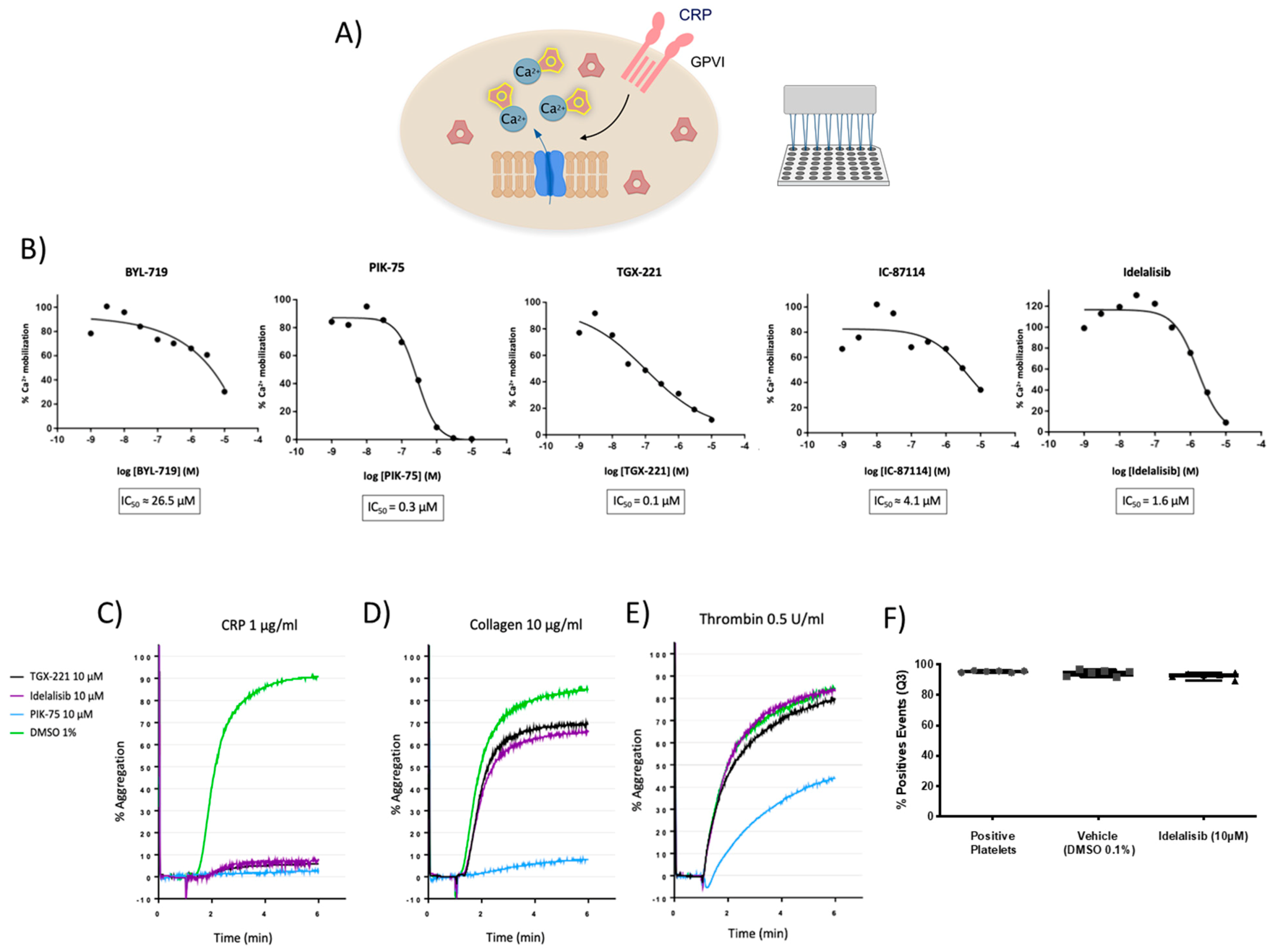
Abstract
:1. Introduction
2. Results
2.1. Prioritization of PI3K-Related Inhibitors as Antiplatelet Agents by Using an Intracellular Ca2+ Mobilization Phenotypic Assay
2.2. GPVI-Mediated Platelet Aggregation Is Preferentially Blocked through p110β and p110δ Inhibition with No Toxic Effects
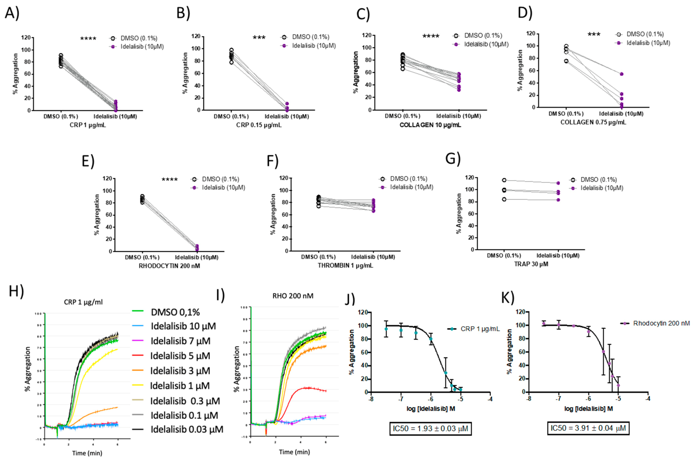
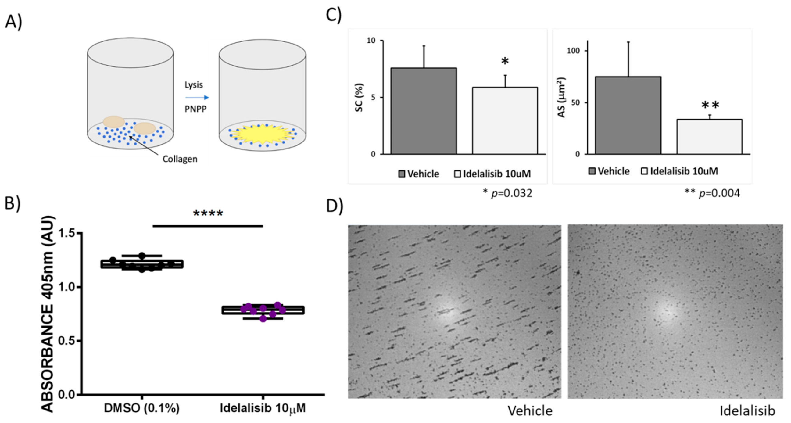
2.3. Idelalisib Inhibits Platelet Aggregation and Adhesion by Favourably Blocking ITAM Signalling Pathways
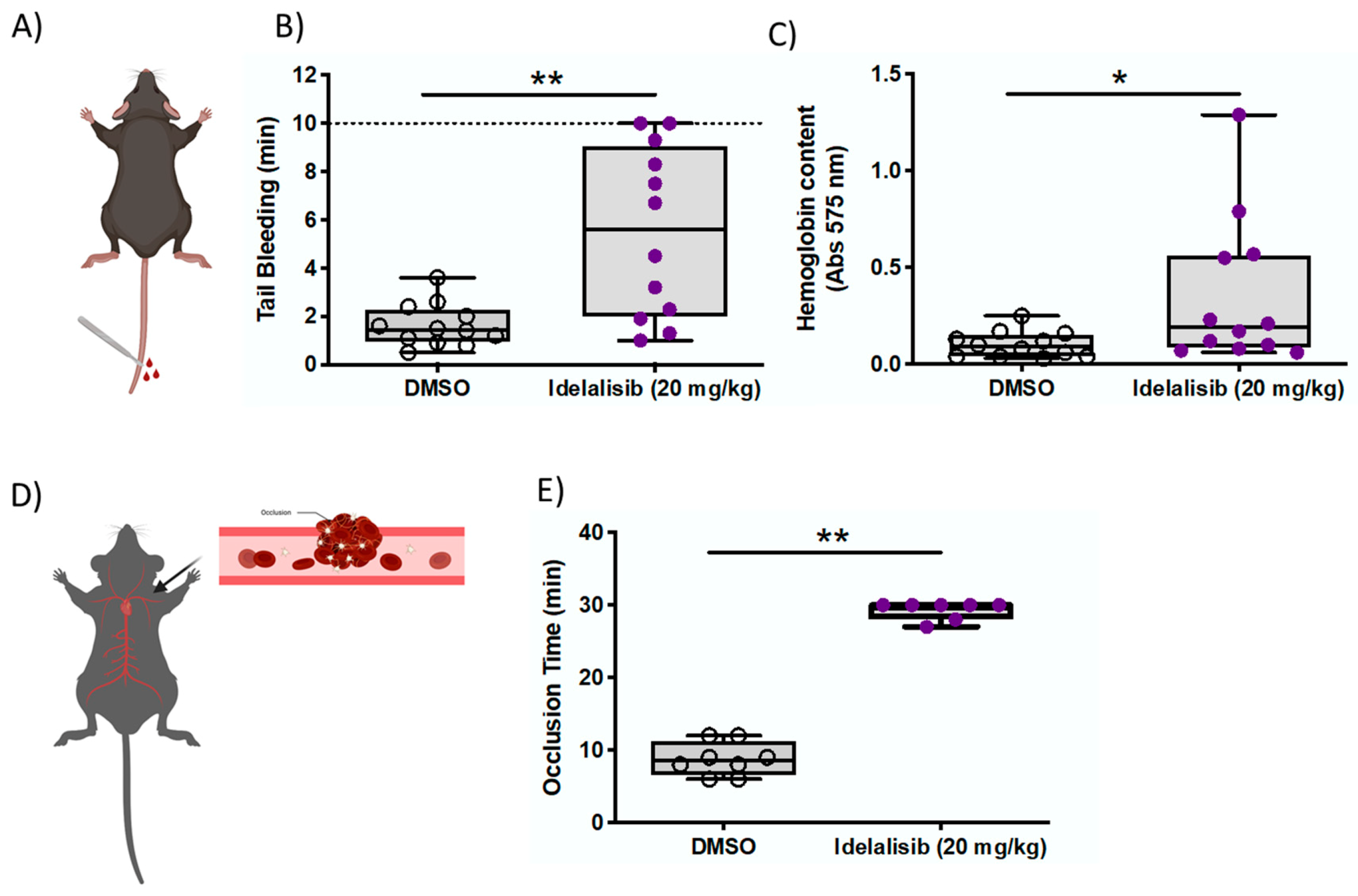
2.4. Idelalisib Reduces Thrombus Formation with Minor Bleeding Effects in Mice
3. Discussion
4. Materials and Methods
4.1. Human Blood Collection and Platelet Isolation
4.2. Intracellular Ca+2 Mobilization Assays
4.3. Platelet Aggregation-Based Selectivity Assays
4.4. Viability-Related Calcein-AM Flow Cytometry Assay
4.5. Platelet Static Adhesion Assays
4.6. Evaluation of Platelet Adhesion and Aggregation by the Impact-R Test
4.7. Animal Studies
4.8. Tail-Bleeding Time
4.9. Chemically Induced Arterial Thrombosis In Vivo
4.10. Analysis of Idelalisib in Mice Plasma
4.11. Statistical Analysis
Supplementary Materials
Author Contributions
Funding
Institutional Review Board Statement
Informed Consent Statement
Data Availability Statement
Acknowledgments
Conflicts of Interest
References
- Wendelboe, A.M.; Raskob, G.E. Global burden of thrombosis: Epidemiologic aspects. Circ. Res. 2016, 118, 1340–1347. [Google Scholar] [CrossRef]
- Berndt, M.C.; Metharom, P.; Andrews, R.K. Primary haemostasis: Newer insights. Haemophilia 2014, 20 (Suppl. 4), 15–22. [Google Scholar] [CrossRef] [PubMed]
- Mackman, N.; Bergmeier, W.; Stouffer, G.A.; Weitz, J.I. Therapeutic strategies for thrombosis: New targets and approaches. Nat. Rev. Drug Discov. 2020, 19, 333–352. [Google Scholar] [CrossRef] [PubMed]
- Gratacap, M.-P.; Guillermet-Guibert, J.; Martin, V.; Chicanne, G.; Tronchère, H.; Gaits-Iacovoni, F.; Payrastre, B. Regulation and roles of PI3Kβ, a major actor in platelet signaling and functions. Adv. Enzym. Regul. 2011, 51, 106–116. [Google Scholar] [CrossRef] [PubMed]
- Laurent, P.-A.; Hechler, B.; Solinhac, R.; Ragab, A.; Cabou, C.; Anquetil, T.; Severin, S.; Denis, C.V.; Mangin, P.H.; Vanhaesebroeck, B.; et al. Impact of PI3Kα (phosphoinositide 3-kinase alpha) inhibition on hemostasis and thrombosis. Arterioscler. Thromb. Vasc. Biol. 2018, 38, 2041–2053. [Google Scholar] [CrossRef]
- Durrant, T.N.; Hers, I. PI3K inhibitors in thrombosis and cardiovascular disease. Clin. Transl. Med. 2020, 9, 8. [Google Scholar] [CrossRef] [PubMed]
- Zheng, Z.; Pinson, J.-A.; Mountford, S.J.; Orive, S.; Schoenwaelder, S.M.; Shackleford, D.; Powell, A.; Nelson, E.M.; Hamilton, J.R.; Jackson, S.P.; et al. Discovery and antiplatelet activity of a selective PI3Kβ inhibitor (MIPS-9922). Eur. J. Med. Chem. 2016, 122, 339–351. [Google Scholar] [CrossRef]
- Nylander, S.; Wågberg, F.; Andersson, M.; Skärby, T.; Gustafsson, D. Exploration of efficacy and bleeding with combined phosphoinositide 3-kinaseβ inhibition and aspirin in man. J. Thromb. Haemost. 2015, 13, 1494–1502. [Google Scholar] [CrossRef]
- Jackson, S.P.; Schoenwaelder, S.M.; Goncalves, I.; Nesbitt, W.S.; Yap, C.L.; Wright, C.E.; Kenche, V.; Anderson, K.E.; Dopheide, S.M.; Yuan, Y.; et al. PI 3-kinase p110β: A new target for antithrombotic therapy. Nat. Med. 2005, 11, 507–514. [Google Scholar] [CrossRef]
- Benimana, O.; Zhao, L.; Kong, Y.; Li, Z.; Xie, Z. The progress in the research of antiplatelet agents (1995–2017). Future Med. Chem. 2017, 9, 1087–1110. [Google Scholar] [CrossRef]
- Senis, Y.A.; Atkinson, B.T.; Pearce, A.C.; Wonerow, P.; Auger, J.M.; Okkenhaug, K.; Pearce, W.; Vigorito, E.; Vanhaesebroeck, B.; Turner, M.; et al. Role of the p110δ PI 3-kinase in integrin and ITAM receptor signalling in platelets. Platelets 2005, 16, 191–202. [Google Scholar] [CrossRef]
- Martin, V.; Guillermet-Guibert, J.; Chicanne, G.; Cabou, C.; Jandrot-Perrus, M.; Plantavid, M.; Vanhaesebroeck, B.; Payrastre, B.; Gratacap, M.-P. Deletion of the p110β isoform of phosphoinositide 3-kinase in platelets reveals its central role in Akt activation and thrombus formation in vitro and in vivo. Blood 2010, 115, 2008–2013. [Google Scholar] [CrossRef] [PubMed]
- Laurent, P.-A.; Severin, S.; Hechler, B.; Vanhaesebroeck, B.; Payrastre, B.; Gratacap, M.-P. Platelet PI3Kβ and GSK3 regulate thrombus stability at a high shear rate. Blood 2015, 125, 881–888. [Google Scholar] [CrossRef]
- Canobbio, I.; Stefanini, L.; Cipolla, L.; Ciraolo, E.; Gruppi, C.; Balduini, C.; Hirsch, E.; Torti, M. Genetic evidence for a predominant role of PI3Kβ catalytic activity in ITAM- and integrin-mediated signaling in platelets. Blood 2009, 114, 2193–2196. [Google Scholar] [CrossRef] [PubMed]
- Flinn, I.W.; Kahl, B.S.; Leonard, J.P.; Furman, R.R.; Brown, J.R.; Byrd, J.C.; Wagner-Johnston, N.D.; Coutre, S.E.; Benson, D.M.; Peterman, S.; et al. Idelalisib, a selective inhibitor of phosphatidylinositol 3-kinase-δ, as therapy for previously treated indolent non-Hodgkin lymphoma. Blood 2014, 123, 3406–3413. [Google Scholar] [CrossRef]
- Brown, J.R.; Byrd, J.C.; Coutre, S.E.; Benson, D.M.; Flinn, I.W.; Wagner-Johnston, N.D.; Spurgeon, S.E.; Kahl, B.S.; Bello, C.; Webb, H.K.; et al. Idelalisib, an inhibitor of phosphatidylinositol 3-kinase p110δ, for relapsed/refractory chronic lymphocytic leukemia. Blood 2014, 123, 3390–3397. [Google Scholar] [CrossRef] [PubMed]
- De Vos, S.; Wagner-Johnston, N.D.; Coutre, S.E.; Flinn, I.W.; Schreeder, M.T.; Fowler, N.H.; Sharman, J.P.; Boccia, R.V.; Barrientos, J.C.; Rai, K.R.; et al. Combinations of idelalisib with rituximab and/or bendamustine in patients with recurrent indolent non-Hodgkin lymphoma. Blood Adv. 2016, 1, 122–131. [Google Scholar] [CrossRef]
- Jin, F.; Robeson, M.; Zhou, H.; Moyer, C.; Wilbert, S.; Murray, B.; Ramanathan, S. Clinical drug interaction profile of idelalisib in healthy subjects. J. Clin. Pharmacol. 2015, 55, 909–919. [Google Scholar] [CrossRef]
- Barrientos, J.C. Idelalisib for the treatment of indolent non-Hodgkin lymphoma: A review of its clinical potential. OncoTargets Ther. 2016, 9, 2945–2953. [Google Scholar] [CrossRef]
- Reda, G.; Cassin, R.; Artoni, A.; Fattizzo, B.; Lecchi, A.; La Marca, S.; Bucciarelli, P.; Levati, G.V.; Peyvandi, F.; Cortelezzi, A. Idelalisib rapidly improves platelet function tests in patients with chronic lymphocytic leukaemia. Br. J. Haematol. 2018, 183, 825–828. [Google Scholar] [CrossRef]
- Ware, J. Dysfunctional platelet membrane receptors: From humans to mice. Thromb. Haemost. 2004, 92, 478–485. [Google Scholar] [CrossRef]
- Schmitt, A.; Guichard, J.; Massé, J.-M.; Debili, N.; Cramer, E.M. Of mice and men: Comparison of the ultrastructure of megakaryocytes and platelets. Exp. Hematol. 2001, 29, 1295–1302. [Google Scholar] [CrossRef]
- Rayes, J.; Watson, S.P.; Nieswandt, B. Functional significance of the platelet immune receptors GPVI and CLEC-2. J. Clin. Investig. 2019, 129, 12–23. [Google Scholar] [CrossRef]
- Nieswandt, B.; Watson, S.P. Platelet-collagen interaction: Is GPVI the central receptor? Blood 2003, 102, 449–461. [Google Scholar] [CrossRef]
- Clemetson, K.J. Platelets and primary haemostasis. Thromb. Res. 2012, 129, 220–224. [Google Scholar] [CrossRef] [PubMed]
- Gardiner, E.E.; Arthur, J.F.; Andrews, R.K. Targeting GPVI as a novel antithrombotic strategy. J. Blood Med. 2014, 5, 59–68. [Google Scholar] [CrossRef]
- Bender, M.; May, F.; Lorenz, V.; Thielmann, I.; Hagedorn, I.; Finney, B.A.; Vögtle, T.; Remer, K.; Braun, A.; Bösl, M.; et al. Combined in vivo depletion of glycoprotein VI and C-type lectin-like receptor 2 severely compromises hemostasis and abrogates arterial thrombosis in mice. Arterioscler. Thromb. Vasc. Biol. 2013, 33, 926–934. [Google Scholar] [CrossRef] [PubMed]
- Foster, H.; Wilson, C.; Philippou, H.; Foster, R. Progress toward a glycoprotein VI modulator for the treatment of thrombosis. J. Med. Chem. 2020, 63, 12213–12242. [Google Scholar] [CrossRef]
- Gresele, P. Antiplatelet agents in clinical practice and their haemorrhagic risk. Blood Transfus. 2013, 11, 349–356. [Google Scholar] [PubMed]
- Labarthe, B.; Idee, J.-M.; Burnett, R.; Corot, C. In vivo comparative antithrombotic effects of ioxaglate and iohexol and interaction with the platelet antiaggregant clopidogrel. Investig. Radiol. 2003, 38, 34–43. [Google Scholar] [CrossRef]
- García, A. Two-Dimensional Gel Electrophoresis in Platelet Proteomics Research. In Vascular Biology Protocols. Methods in Molecular Medicine™; Sreejayan, N., Ren, J., Eds.; Humana Press: Totowa, NJ, USA, 2007; Volume 139, pp. 339–353. [Google Scholar] [CrossRef]
- Shenkman, B.; Savion, N.; Dardik, R.; Tamarin, I.; Varon, D. Testing of platelet deposition on polystyrene surface under flow conditions by the cone and plate(let) analyzer: Role of platelet activation, fibrinogen and von Willebrand factor. Thromb. Res. 2000, 99, 353–361. [Google Scholar] [CrossRef]
- Wang, X.; Cheng, Q.; Xu, L.; Feuerstein, G.Z.; Hsu, M.-Y.; Smith, P.L.; Seiffert, D.A.; Schumacher, W.A.; Ogletree, M.L.; Gailani, D. Effects of factor IX or factor XI deficiency on ferric chloride-induced carotid artery occlusion in mice. J. Thromb. Haemost. 2005, 3, 695–702. [Google Scholar] [CrossRef] [PubMed]


Publisher’s Note: MDPI stays neutral with regard to jurisdictional claims in published maps and institutional affiliations. |
© 2021 by the authors. Licensee MDPI, Basel, Switzerland. This article is an open access article distributed under the terms and conditions of the Creative Commons Attribution (CC BY) license (http://creativecommons.org/licenses/by/4.0/).
Share and Cite
Barrachina, M.N.; Izquierdo, I.; Hermida-Nogueira, L.; Morán, L.A.; Pérez, A.; Arroyo, A.B.; García-Barberá, N.; González-Conejero, R.; Troitiño, S.; Eble, J.A.; et al. The PI3Kδ Inhibitor Idelalisib Diminishes Platelet Function and Shows Antithrombotic Potential. Int. J. Mol. Sci. 2021, 22, 3304. https://doi.org/10.3390/ijms22073304
Barrachina MN, Izquierdo I, Hermida-Nogueira L, Morán LA, Pérez A, Arroyo AB, García-Barberá N, González-Conejero R, Troitiño S, Eble JA, et al. The PI3Kδ Inhibitor Idelalisib Diminishes Platelet Function and Shows Antithrombotic Potential. International Journal of Molecular Sciences. 2021; 22(7):3304. https://doi.org/10.3390/ijms22073304
Chicago/Turabian StyleBarrachina, María N., Irene Izquierdo, Lidia Hermida-Nogueira, Luis A. Morán, Amparo Pérez, Ana B. Arroyo, Nuria García-Barberá, Rocío González-Conejero, Sara Troitiño, Johannes A. Eble, and et al. 2021. "The PI3Kδ Inhibitor Idelalisib Diminishes Platelet Function and Shows Antithrombotic Potential" International Journal of Molecular Sciences 22, no. 7: 3304. https://doi.org/10.3390/ijms22073304