Review

Effects of Glycosylation on the Enzymatic Activity and Mechanisms of Proteases

Structural Biology Group, Faculty of Molecular Biology, University of Salzburg, Billrothstrasse 11, 5020 Salzburg, Austria
Int. J. Mol. Sci. 2016, 17(12), 1969; https://doi.org/10.3390/ijms17121969
Submission received: 30 July 2016 / Revised: 7 November 2016 / Accepted: 10 November 2016 / Published: 25 November 2016
(This article belongs to the Special Issue Glycan–Receptor Interaction)

Abstract

:
Posttranslational modifications are an important feature of most proteases in higher organisms, such as the conversion of inactive zymogens into active proteases. To date, little information is available on the role of glycosylation and functional implications for secreted proteases. Besides a stabilizing effect and protection against proteolysis, several proteases show a significant influence of glycosylation on the catalytic activity. Glycans can alter the substrate recognition, the specificity and binding affinity, as well as the turnover rates. However, there is currently no known general pattern, since glycosylation can have both stimulating and inhibiting effects on activity. Thus, a comparative analysis of individual cases with sufficient enzyme kinetic and structural data is a first approach to describe mechanistic principles that govern the effects of glycosylation on the function of proteases. The understanding of glycan functions becomes highly significant in proteomic and glycomic studies, which demonstrated that cancer-associated proteases, such as kallikrein-related peptidase 3, exhibit strongly altered glycosylation patterns in pathological cases. Such findings can contribute to a variety of future biomedical applications.

Graphical Abstract

1. Introduction

Glycosylation is a posttranslational modification that is found on about 50% of all proteins, in particular on secreted and transmembrane proteins of eukaryotes, archaea and to a lesser extent in prokaryotes [1,2,3]. Eukaryotic proteins require glycosylation for proper folding, oligomerization and solubility, while glycans significantly prolong the stability and half-life time in many cases by protection against proteolysis [4,5]. Although N-glycosylation is more frequent, O-glycosylation can similarly protect against general and specific proteolysis [6,7,8]. Protein trafficking, i.e., the sending of proteins to cellular compartments or to the extracellular matrix, depends on specific, covalently linked glycans [9]. In addition, glycans play an important role in the interaction and recognition of proteins, such as in the context of immunity and cell adhesion [10,11,12]. Glycosylation may even protect against molecular damage by free radicals [13]. In recent years, increasing evidence was found that glycans have distinct effects on the activity of many enzymes, in particular as regulatory modules for substrate binding and turnover. This study gives an overview on the most relevant types of glycosylation of proteases, regarding the structural knowledge and the functions of glycans. The importance of this little investigated field lies in the enormous diversity of possible glycosylation variants and the altered functionality of proteins under healthy or disease conditions.
N-glycosylation at sequons of the Asn-Xaa-Ser/Thr type is widespread in proteins of archaea and eukaryotes, whereby proline is largely excluded as Xaa and disfavored as residue following Ser/Thr [14,15]. Some rare sequons are Asn-Xaa-Cys (1%), Asn-Gly (0.5%) and Asn-Xaa-Val (<0.5%) [16]. The process of N-glycosylation is extensively described in the literature on glycobiology [17]. Essentially, a newly synthesized polypeptide emerging from a ribosome binds with a signal peptide to a signal recognition particle, which docks to a receptor in the endoplasmic reticulum (ER) membrane and forms a complex with the Sec machinery, which transfers the polypeptide through a transmembrane channel into the lumen of the ER [18,19]. A signal peptidase cleaves the N-terminal signal peptide and the oligosaccharyltransferase complex attaches a GlcNAc2Man9Glc3 precursor at a suitable sequon of the Asn-Xaa-Ser/Thr type [20]. Subsequently, the N-glycosylated polypeptide folds in the oxidizing environment of the ER, supported by protein disulfide isomerase for disulfide formation and by various chaperones [21,22]. Afterwards, glucosidases and mannosidases trim the N-glycan precursor to Man5GlcNAc2 or GlcNAcMan3GlcNAc2 core glycans, which are extended by glucosyltransferases, accompanied by protein quality control and followed by sorting and further processing on their way through the ER Golgi intermediate compartment into the Golgi [23,24]. Final modifications of the N-glycans in the Golgi comprise extensions by transferases that attach N-acetyl-glucosamine (GlcNAc), fucose, galactose, mannose, and sialic acid sugars, before sorting to secretory vesicles [25]. Variations of branching generate a large diversity of N-glycans with distinct composition under physiological and pathological conditions (Figure 1A) [26].
Earlier structural database analyses reported a relatively low percentage of only 27% N-glycosylation of all sequons in human proteins, of which 96% belonged to secreted and membrane proteins and 4% to cytoplasmic and nuclear proteins [28,29]. More recent data suggested around 85% occupancy of sequons, with 50% glycosylation of all proteins in the SWISS-PROT sequence data bank [2]. Thorough mass spectrometric analyses demonstrated that in the mouse N-glycoproteome the majority of identified sequons is occupied, e.g., 99% of predicted membrane protein N-glycosylation sites [16]. Glycosylated Asn residues in human proteins are preferentially located in turns (78%) compared to β-sheets (12%) and α-helices (10%), which resembles the situation in murine proteins [16,28]. The strict structural constraints for N-glycosylation are reflected by a similar localization in proteins of fish, insects, plants and lower eukaryotes [28,30].
About 12% of all glycosylated proteins are exclusively O-glycosylated, while about 10% of them are both N- and O-glycosylated [2]. Seven types of O-glycosylation have been found in humans (Figure 1B). The mucin-type with N-acetylgalactosamine (GalNAc) linked to Ser or Thr in membrane or secreted proteins is more common than O-glycosylation with xylose, galactose, glucose, fucose, and mannose, whereas mostly proteins with O-linked GlcNAc localize to the cytoplasm and the nucleus [27]. Although no distinct consensus sequence is known, Pro-rich sequences are favored, such as Pro-Ser/Thr-Pro-Xaa-Pro [31,32]. Usually, the first step of O-glycosylation of the mucin-type is performed in the Golgi by a GalNAc transferase, followed by extensions and branching, which results in eight core glycan subtypes [27,33]. O-glycosylation of the mucin-type is important for tissue development and immune reactions [34].
Among the rarer types of glycosylation is O-mannosylation for quality control and protection against proteolysis [35]. Similarly, O-glycosylation of hydroxylysine can occur in mammals, in contrast to O-glycosylation of hydroxyproline, which is found in other eukaryotes [36,37]. The uncommon C-mannosylation of Trp residues plays a role in protein folding, secretion and signaling [38,39]. Glycosylphosphatidyl-inositol-anchored proteins (GPI-APs) are linked to glycolipids as membrane anchors, which is sometimes termed glypation [9,40]. By contrast, glycation is the uncatalyzed covalent linkage of glucose or fructose with amino groups, which is involved in diabetes and ageing related diseases [41,42]. Apart from this unregulated process, most glycans play very well defined roles in physiology, such as in the regulation of enzymatic activity.

2. Glycosylated Proteases

Currently, the UniProtKB database lists about 250 glycosylated human proteases out of more than 700, with changing numbers due to putative or ambiguous classification [43]. They exhibit either experimentally identified or predicted N-, O-, and C-glycosylation sites, using the NetNGlyc and NetOGlyc tools, which are currently applicable to mammalian entries and the mucin-type of O-glycans [29,44]. The MEROPS database of proteolytic enzymes includes about 1200 known and putative human proteases, including inactive protease homologs [45].

2.1. Aspartic Proteases

An overview of the glycosylated human aspartic proteases shows the 11 confirmed or potentially glycosylated members out of altogether 28, which agrees with their prevalent extracellular localization (Table 1). The prototypic aspartic protease pepsin is not glycosylated, despite its secretion into the stomach, where it cleaves proteins between hydrophobic residues, such as P1-Leu and P1’-Leu according to the Schechter–Berger nomenclature [46]. The related, brain expressed β-secretase 1 (BACE1) is a key player in neuronal regulation and a drug target in Alzheimer´s disease [47]. Although the UniProtKB database annotates BACE1 only as N-glycosylated according to manual assertion, a recombinant variant carries N-glycans according to mass spectrometry and PNGase F treatment [48].

2.2. Cysteine Proteases

Another important enzyme class is formed by the cysteine proteases, of which only 12 out of 158 human members exhibit glycosylation sites, among them several cathepsins (Table 1). This phenomenon can be easily explained by the predominant intracellular location with reducing conditions that are required for the activity of many cysteine proteases. Cysteine cathepsins occur in endolysosomes and under pathological conditions even more often outside cells, where they can be regulated by glycosaminoglycans [49]. For example, the important protease families of the apoptosis- and inflammation-related caspases and of the Ca2+-signal mediating calpains do not possess any ascertained glycosylation site [50,51]. However, legumain, a monomeric caspase-like cysteine protease, is found in the acidic lysosomes and outside cells, carrying at least three N-glycans in its recombinant form, which was generated by Leishmania expression [52,53].

2.3. Metalloproteases

The mostly Zn2+-dependent metalloproteases are secreted from cells to a large extent, with 108 of 163 members carrying at least one glycan (Table 2). Among them are the extracellularly located, soluble or membrane-anchored matrix metalloproteinases (MMPs), which play important roles in development, wound healing, brain processes, and cancer [54]. Their major task is the degradation of matrix proteins by cleaving at larger hydrophobic P1′ residues, which can be supported by collagen-binding hemopexin domains [55]. The majority of MMPs is N-glycosylated, although six of them appear to be non-glycosylated, while MMP-25 is potentially GPI-anchored [56]. Several MMPs possess N-glycosylation sites, which are required for protein interaction and secretion as observed for MMP-9 [57]. MMP-9 is the remarkable example of a protease with a heavily O-glycosylated, Pro-rich linker domain, containing more than a dozen branched mucin-type core 2 glycans [58]. Intriguingly, tumor cells produce MMP-9 mainly with O-glycans of mucin core 1, which reduces the binding affinity to its ligand galectin-3 [59]. The two families of transmembrane and GPI-anchored ADAM and ADAMTS proteases, which cleave extracellular portions of other membrane proteins and matrix proteins, are involved in cell adhesion, growth factor shedding, cell migration, morpho-genesis and cancer [60,61]. An outstanding example is ADAM17 or tumor necrosis factor α converting enzyme (TACE), which sheds ectodomains of numerous substrates depending on O-glycosylation of cleavage sites [62]. Similarly, the heavily N-glycosylated meprin-α and -β act as sheddases and cleave pro-inflammatory cytokines [63,64]. The membrane-bound meprin-β subunits tend to form dimers, whereas α and β subunits can form heterodimers and α subunits may form even higher oligomers [65]. Metallocarboxypeptidases are another relevant subgroup of secreted neurohormone and cytokine processing enzymes, which have been in implicated in diseases of the pancreas, diabetes and cancer and are considered nowadays as promising drug targets [66,67]. To date, the developmentally and pathophysiologically relevant Wnt-signal regulating Tiki 1 and 2 proteases belong to a superfamily of hardly characterized, unique Co2+/Mn2+-dependent enzymes [68].

2.4. Serine Proteases

Serine proteases constitute another large group with about 106 glycosylated members out of 144 according to UniProtKB, with family S1 being the largest one (Table 3). The prototypic digestive proteases trypsin, chymotrypsin and elastase are produced in the pancreas and define the corresponding major substrate specificities, referring to the S1 subsites. Among the digestive proteases, only the membrane-anchored enteropeptidase, also known as enterokinase, is heavily N-glycosylated and activates trypsinogen, which in turn activates other digestive enzymes [69]. The blood coagulation cascade involves several trypsin-like proteases, which leads to the activation of prothrombin by factor Xa, resulting eventually in thrombin cleavage of fibrinogen and formation of fibrin clots, which stop bleeding [70]. N-glycosylated variants of factor VII propeptides prolong its half-life significantly, while corresponding variants of factor IX can also play a role in the treatment of hemophilia [71,72]. Regulation of the fibrinolytic plasmin depends on differential N- and O-glycosylation, which alters the structure significantly [73,74]. In addition, both the tissue-type plasminogen activator (tPA) and urokinase-type plasminogen activator (uPA) possess N- and O-glycans, whose roles have been partially elucidated [75,76]. Kallikrein 1 (KLK1) and the other fourteen kallikrein-related peptidases (KLKs) fulfill numerous crucial tasks in tissue development, reproduction, wound healing or neuronal processes [77]. Since several KLKs exhibit altered expression in various cancers, they are used as biomarkers, e.g., KLK3/PSA (prostate specific antigen), and investigated as drug targets [78,79]. Moreover, most human KLKs are N-glycosylated proteins (except KLK14), for which potential functional roles have been described [80,81].
Secretory proprotein convertases (PPCs), among them the N-glycosylated furin and PPCs 4, 5, 6, 7, and 9, process many substrates upon secretion (Table 4) [82]. Up to now, no distinct functions of their N-glycans were reported, whereas O-glycosylation near the cleavage sites of their substrates contributes to the regulation of turnover [83]. As observed for many glycoproteins, N-glycosylation is critical for folding, secretion and stability of tripeptidyl peptidase I [84]. Interestingly, the hereditary disease late infantile neuronal ceroid lipofuscinosis is caused by a mutation resulting in the loss of an N-glycan and all enzymatic activity [85]. The α/β-hydrolases dipeptidyl peptidase-4 (DPP4) and prolyl endopeptidase (FAP) are multifunctional transmembrane enzymes, which cleave substrates after P1-Pro residues and serve as cell surface receptors [86,87]. Apparently, the heavy N-glycosylation of DPP4 does not influence dimerization, activity, and T-cell protection [88].

2.5. Threonine Proteases

Threonine proteases are both rare and widespread, due to the central role of the ubiquitin-proteasome system for eukaryotic protein degradation, which is highly conserved from yeast to mammals (Table 4) [89]. Besides 14 inactive α-subunits, the proteasome comprises 14 β-subunits in the two inner rings, of which only six are active and cleave after hydrophobic (β1), acidic (β2), and basic (β5) S1 side chains, which is slightly modified in immunoproteasomes [90]. Proteasomes generate peptides that are presented by most cells as epitopes on MHC class I molecules to cytolytic T-cells [91]. Seemingly, O-glycosylation is present in mammalian, but not in yeast 26 proteasomes, while the role of the mucin-type O-glycans on four α-subunits and the β1 and probably the β6 subunit remains unclear [92,93].

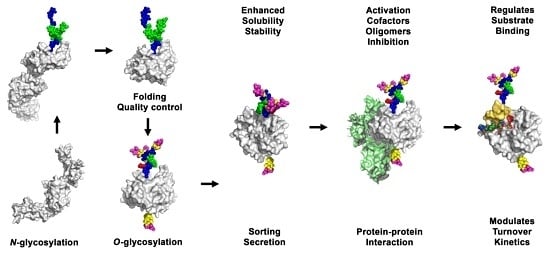
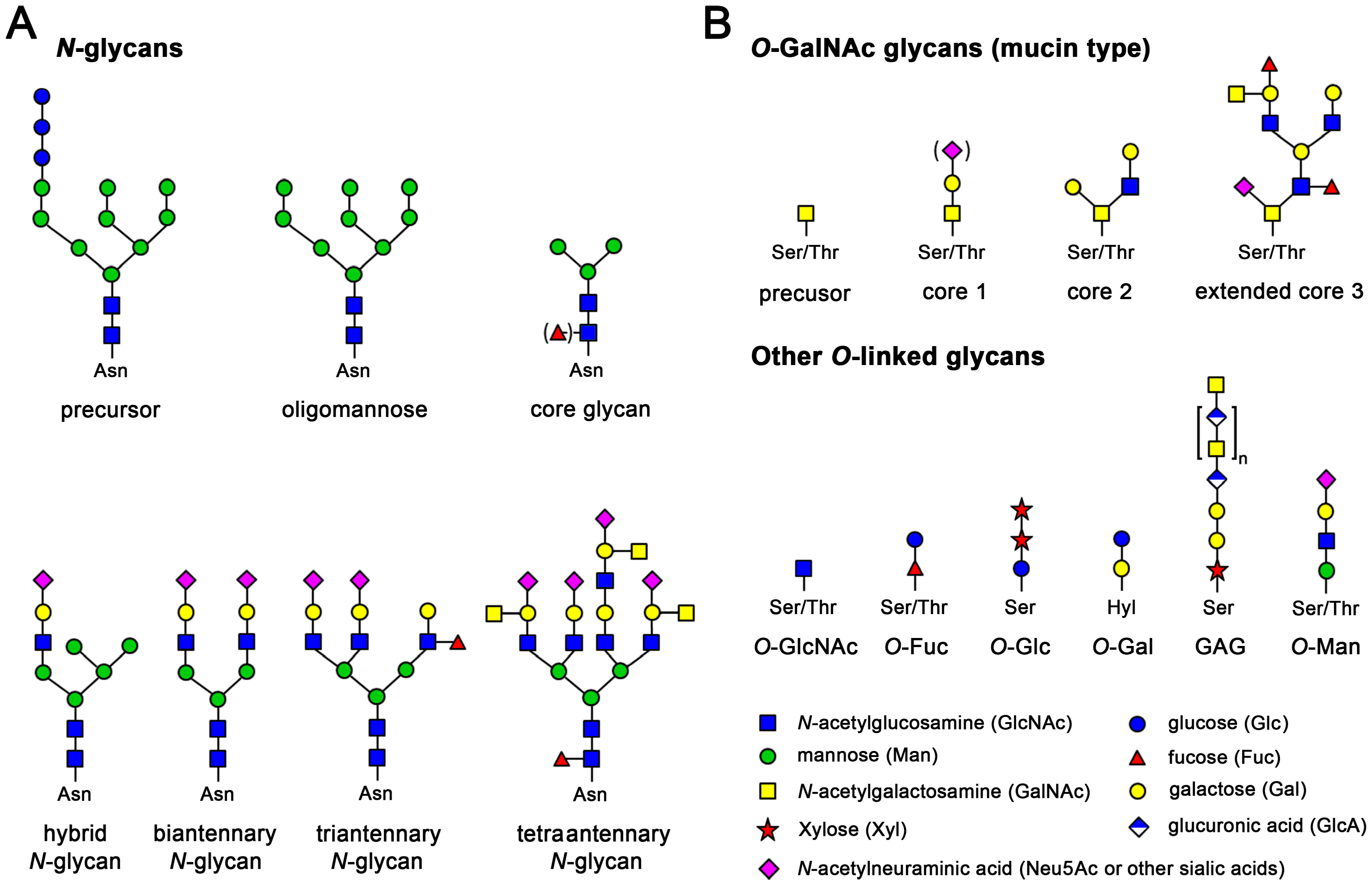
3. Effects of Glycosylation on Proteases

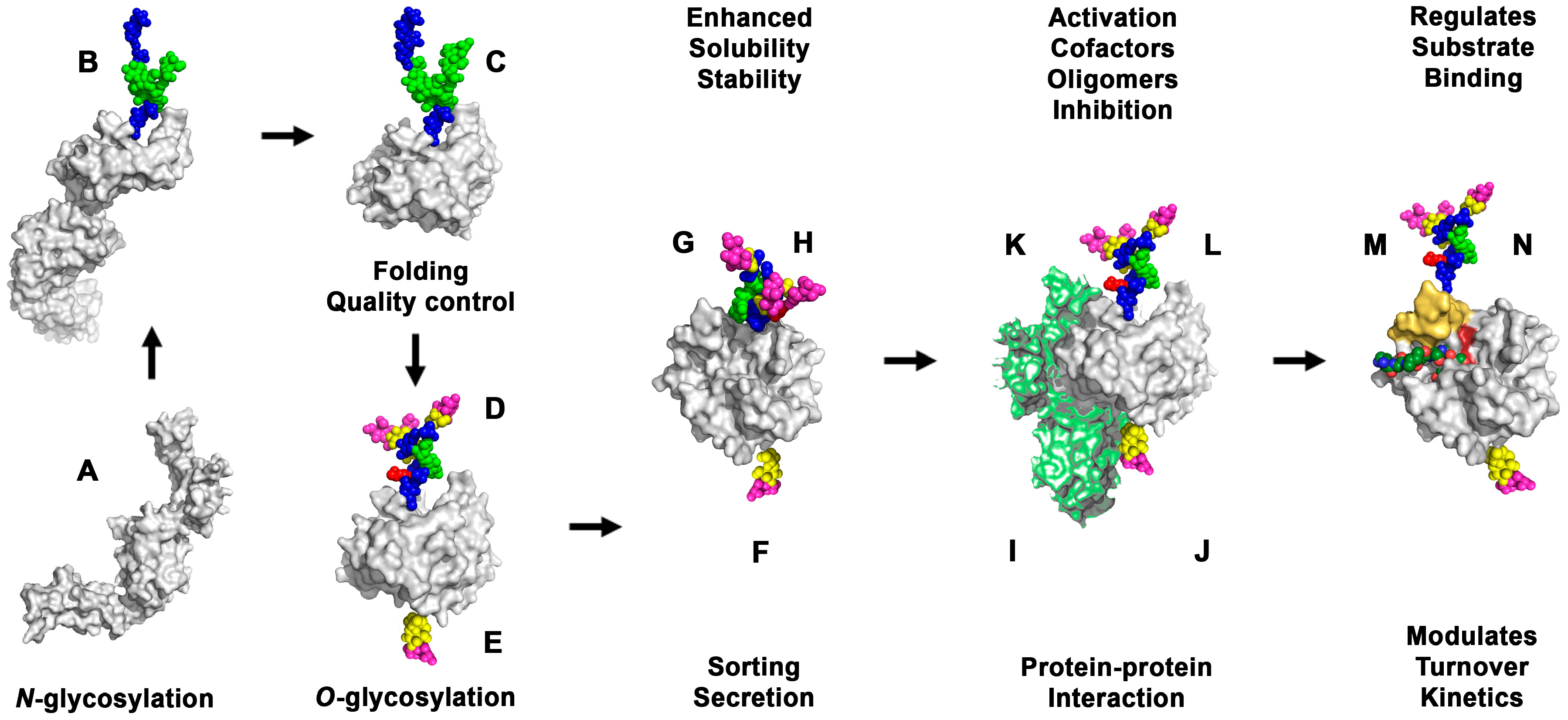
3.1. Effects of Glycosylation on Folding, Sub-Cellular Distribution and Secretion of Proteases

Usually, N-glycosylation is required for sufficient expression, efficient secretion, and protein trafficking, which holds true for numerous proteases. Glycans facilitate the folding of polypeptides, enhance the solubility of proteins and prevent aggregation [94]. Chymotrypsin C, a pancreatic regulator of trypsin, exhibits two N-glycans in the propeptide and the catalytic domain, which serve folding and secretion, but have no impact on activation and activity [95]. A comparison of a fully N-glycosylated Pseudomonas elastase with its triple mutant Asn43Gln/Asn212Gln/Asn280Gln in the catalytic domain showed virtually no differences in enzyme activity, in contrast to significantly reduced secretion levels [96]. Inspection of this elastase structure (1EZM) revealed that the glycans are remote from the active site, in line with the assumption that the glycans are responsible for proper folding alone as basis for secretion [96,97]. In addition, N-glycosylation can be crucial in recombinant expression, as demonstrated for the trypsin-like human KLK5, which could be only expressed in insect cells with a single core N-glycan, while expression in E. coli cells and subsequent in vitro refolding was not feasible [98]. Similarly, human legumain expression for structure–function studies was largely improved in Leishmania cells, which generate short core N-glycans [52]. In the case of ADAMTS-9, three N-linked glycosylation sites in the propeptide are critical for proper secretion and subcellular localization [99]. Similarly, a mutagenesis study of the human aspartic peptidase renin, which regulates blood pressure by cleavage of angiotensinogen, revealed that N-glycosylation at Asn5 and Asn75 plays only a role for secretion [100]. In addition, natural N-glycosylation regulates uptake and metabolic clearance, as shown for variants of rat renin [101].

3.2. Effects of Glycosylation on Activation and Stability of Proteases

N-Glycosylation influences the activation of proteases, both with glycans that are located in the propeptides and in the catalytic domain. Several human proteases possess N-glycosylated propeptides, such as KLKs 10 and 13 (Table 3). These glycans may regulate protease activation, similar as in the trypsin-like Der p 3, a dust mite allergen, whose N-glycan at position P3 with respect to the cleavage site slows down the activation process [102]. In addition, glycans in the catalytic domain of a proform can be crucial as demonstrated by tunicamycin induced loss of N-glycosylation at Asn968 and Asn1087 of rat corin, whose activation failed completely [103]. However, for a recombinant form of the human cysteine protease cathepsin B, no effect of N-glycosylation on activation, stability and enzymatic activity was found [104]. Many glycosylation sites protect against proteolysis in sensitive regions, which represents often autoproteolysis of the investigated proteases. For example, the aspartic protease cathepsin E lost rapidly activity and stability when its N-glycans were enzymatically cleaved [105]. Enzymatic removal of two N-glycans in the 75- and 99-loops from human mast cell chymase did not alter kinetic parameters, only the enzymatic activity decreased faster, suggesting that the glycans protect the exposed surface loops from autoproteolysis [106]. Even the branching of N-glycans can contribute to protein stability, as demonstrated for matriptase, which was more resistant to proteolysis by trypsin with fucose β1-6 linked to GlcNAc than with the unbranched N-glycan in the protease domain [107].

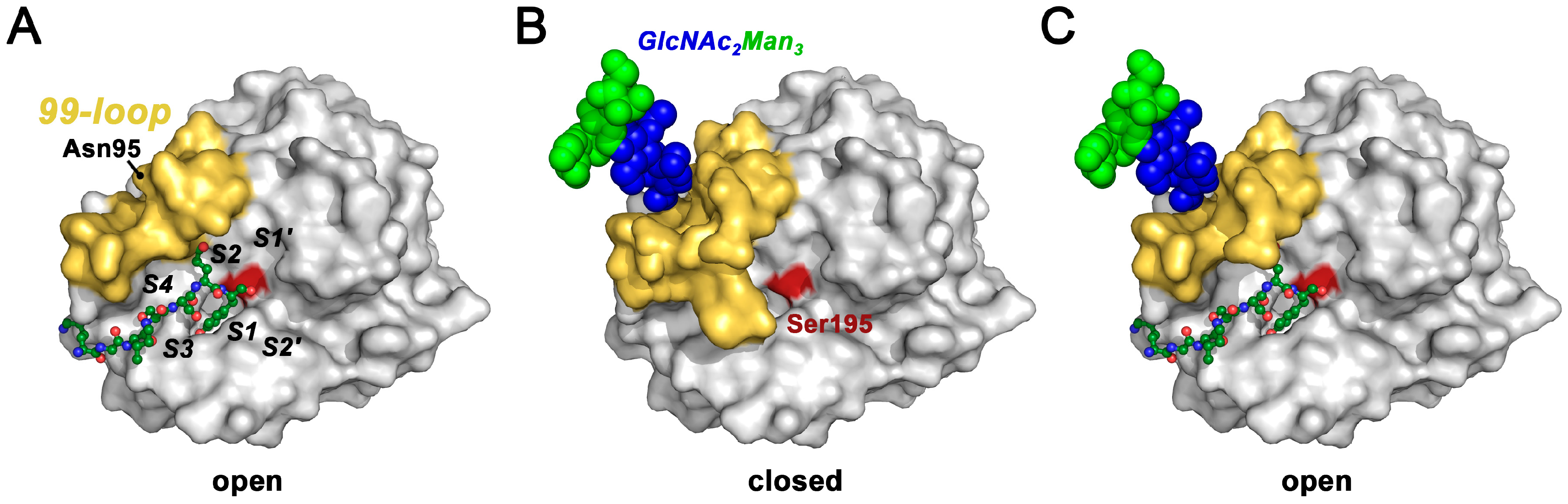
3.3. Effects of Glycosylation on Substrate Binding and Turnover

Regardless of the protease type, it is more likely that glycans in the vicinity of the active site influence substrate binding in the specificity subsites, which depend on numerous biophysical interactions (Figure 2A). Several examples of aspartic, metallo- and other proteases, which exhibit enzyme substrate (Michaelis) complex and tetrahedral intermediates during catalysis, confirm that the presence or absence of N-glycans can considerably alter substrate binding and turnover (Figure 2B–D). Nevertheless, the overall presence or absence of N-glycosylation plays no role for the function of some proteases, such as human complement factor I [108].
Numerous examples of N- and O-glycosylated and non-glycosylated variants of enzymes demonstrate that the presence of glycans can have very diverse effects. Single mutations of the glycosylated Asn632 and Asn651 of the metalloprotease endothelin-converting enzyme had no effect, while the double mutant was completely inactive [109]. A series of sequon mutants of mouse meprin-α showed that six out of ten N-glycans are critical for catalysis [110]. The completely deglycosylated snake venom RVV-X, a metalloprotease, which can activate coagulation factor X, exhibited no change in KM and a 130-fold reduced kcat [111]. Since all N-glycans of RVV-X are distant from the active site (PDB code 2E3X) and substrate binding was not hampered, one has to conclude that the overall conformation was significantly affected by their absence [112]. Complete deglycosylation of MT1-MMP (MMP-14) in the O-glycan rich linker resulted in a stable and active protease, which, however, failed to bind the tissue inhibitor of metalloproteinases-2 (TIMP-2) and could not form the MT1-MMP/TIMP-2/pro-MMP-2 activation complex [113].
By contrast, removal of glycans enhances the activity of several proteases, most likely because the accessibility of the active site increases. Insect cell expressed ADAM17 with short N-glycans has an up to 30-fold increased catalytic efficiency compared to a mammalian cell expressed variant with more complex N-glycans, which could interfere with substrate binding [121]. Nevertheless, O-glycosylation near the scissile bond of ADAM17 substrates enhances the turnover significantly [122]. N-glycosylated plasma kallikrein (KLKB1) has a lower catalytic efficiency than deglycosylated KLKB1, which exhibits an altered cleavage pattern for the substrate insulin [123]. The deletion of a single N-glycan in the kringle 2 domain of tPA form II enhanced fibrin binding and the resulting fibrinolytic activity by plasmin [124]. Fully deglycosylated tPA is nearly 4-times more active against small chromogenic substrates, whereby a higher mannose content or removal of sialic acids stimulates the activity as well [125,126]. Since the O-glycan in the EGF-like domain and the N-glycans in both kringle domains can hardly interfere with small substrates, the glycosylated Asn173 in the 176-loop of the protease domain is most likely critical for the observed effect.

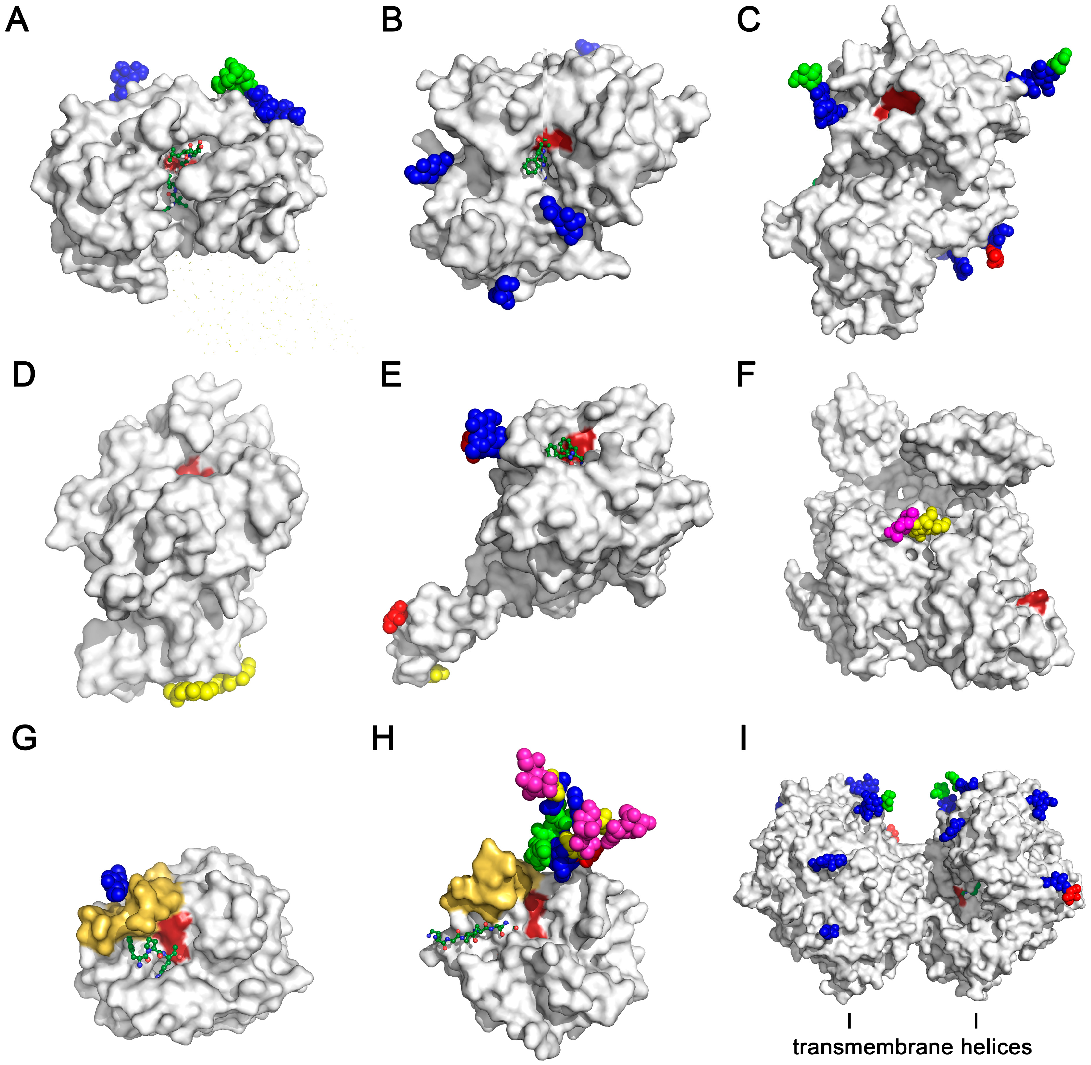
3.4. Effects of Glycosylation on Protease Structures

Structural studies explain to some extent how glycosylation influences the protein conformation. Small angle X-ray scattering data of an N-glycosylated fungal enzyme and its deglycosylated counterpart suggested that the glycans limit the torsion angle range of the polypeptide in general [127]. Currently, only a few studies provide sufficient structural and functional information on glycosylated enzymes and their non-glycosylated variants. Several studies of mammalian lipases, which are closely related to proteases, emphasize the stabilizing role of N-glycans, in particular of those close to the active site, for the conformation with the highest activity [128,129,130,131]. Thus, glycans that are located distant from the active site, as seen for the aspartic protease cathepsin D, cannot directly influence substrate recognition and turnover, while these N-glycans are important for protein interactions in lysosomal targeting (Figure 3A) [132].
A major consequence of the conformational stabilization conferred by N-glycosylation might be an efficient interaction with substrates and regulation of substrate access, which can enhance the specificity. For both cathepsin C and meprin-β a single N-glycan at the substrate binding cleft near the catalytic residues appears to be crucial for structure stabilization and substrate binding (Figure 3B,C) [110,133]. Although in some cases the exact role of individual glycans is unclear, it can be deduced that they support oligomerization or interaction with other proteins. The O-glycans of recombinant Carboxypeptidase N are compatible with substrate binding and tetramerization, however, their function has not been defined yet (Figure 3D) [134]. Double Ala mutants of the O-glycosylated Ser52 and Ser60 in the EGF-like domain of coagulation factor VIIa exhibited about 14% of the coagulant activity of wild-type FVIIa, while the amidolytic activity was unchanged, indicating that both O-glycans are relevant for the association with tissue factor (Figure 3E) [135]. A comparison of recombinant FVII and plasma FVII confirmed that N-glycans with terminal GalNAc instead of sialic acids enhance activity and association with tissue factor [136,137]. Subtle variations of the O-glycans at Thr346 (form 1) or Ser248 (form 2) and of the N-glycan at Asn288 in plasmin influence binding of the inhibitor α2-antiplasmin and the substrate fibrin (Figure 3E) [138,139]. As in several other kallikrein-related peptidases, KLK2 exhibits a single N-glycan in the 99-loop close to the substrate binding cleft, which regulates substrate turnover and could increase the specificity for larger protein substrates (Figure 3G) [140]. N-Glycosylation in the nearby 62-loop is known for KLK3, thrombin and human neutrophil cathepsin G, where it may modulate substrate binding in the prime side region (Figure 3H) [141]. Eventually, DPP4 is a dimeric transmembrane α/β-hydrolase with various receptor functions beyond its protease activity [142]. As for other integral membrane proteases its N-glycosylation may prevent unspecific protein interaction, such as aggregation, but individual glycans may have specific functions (Figure 3I).

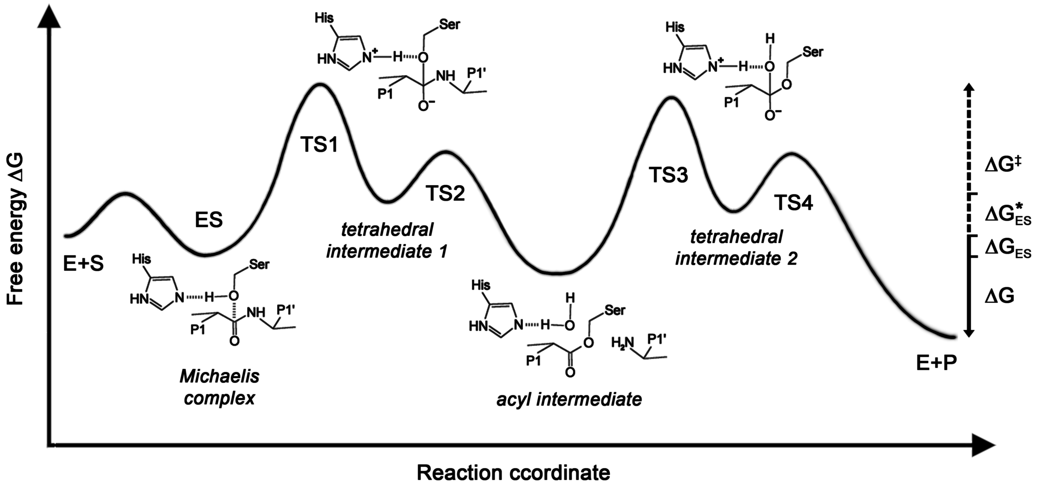
3.5. Effects of Glycosylation on Protease Mechanisms

Kinetic and structural data can provide valuable information on enzymatic mechanisms. The basic proteolytic mechanisms are well known and kinetic parameters are available for many substrates, e.g., in the BRENDA database [143]. However, both functional and structural data of glycosylated and non-glycosylated enzymes are scarce. Thus, a model of the possible effects of glycosylation on catalysis by a trypsin-like serine protease shall be described. In general, the Michaelis complex corresponds to the enzyme substrate complex (ES) and the first tetrahedral intermediate is close to the transition state (TS), while the acyl intermediate is the first intermediate product (Figure 2B). The distinct conformational states of these reaction steps were calculated for trypsin with a combined approach of quantum mechanics/molecular mechanics with molecular dynamics/free energy perturbation calculations, resulting in a free energy profile, which was extended by analyzing the role of Asp102 [118,144]. In addition, free energy profiles for the single steps of this mechanism have been determined, including enzyme product complexes, using rate constants from Michaelis−Menten kinetics, viscosity and isotope kinetic parameters (Figure 4) [145]. Although KM, kcat, and kcat/KM do not directly correspond to free energies in the reaction profile, they correlate, such as a low KM with a high ∆Gbind of the enzyme substrate complex formation, a high kcat with a low ∆G, and kcat/KM with the overall change of the free energy ∆G = ∆G − ∆GES [146,147].
Under certain conditions the free activation energy can be calculated from the catalytic efficiency [152]. Thus, free energy differences between a glycosylated protease and a glycan-free counterpart can be treated as the ones derived from the catalytic efficiency of enzyme mutants [151]. Based on the known catalytic mechanism, such experimental data could be employed to refine the corresponding mechanistic calculations in more detail as shown here.
N-glycosylated and glycan-free KLK2 were analyzed with respect to turnover of several small synthetic substrates, resulting always in a 5-fold increased KM compared with the KM of glycan-free KLK2, while the kcat was less affected [140]. This effect could be explained with an influence of the core glycan (GlcNAc2Man3) at Asn95 in the 99-loop close to the active site, favoring the formation of a type I β-turn over the Asx turn of glycan-free KLK2 [153,154]. Apparently, the flexible 99-loop is wide open in the glycan-free KLK2 crystal structure, whereas it may adopt a closed conformation due to the presence of the N-glycan, as in the related KLK1 structure (Figure 3G) [155,156]. The open loop allows for rapid binding of the substrate in the non-prime region at the specificity pockets S4 to S2, which can be explained by a higher kon rate, resulting in a lower KM = (koff + kcat)/kon. In addition, substrate binding requires a higher free activation energy ∆GES*, since the energy barrier of the N-glycosylated, closed loop has to be overcome. Interestingly, the lid-like 99-loop may also serve in fixing the substrate, as observed in a KLK3 acyl intermediate complex structure (Figure 5) [157].
Remarkably, N-glycosylated KLK2 was more efficient than the glycan-free variant in autoactivation and to some extent in degrading large protein substrates [140]. This finding can be partially explained by a stronger influence of the 99-loop on small synthetic substrate binding in the non-prime side, but the catalytic efficiency of N-glycosylated KLK2 might be enhanced by an optimally shaped active site for stabilizing the transition states. Thus, the decreased ∆G could depend on an increased kcat of the glycosylated enzyme as well. By contrast, N-glycans in close to the active site can have a different effect, as shown for porcine pepsin, in which artificial sequons were N-glycosylated, resulting in an overall stabilizing effect and a kcat decrease to about 30%, which was explained with a more rigid “flap” loop in favorable contact with the S1 and S2 specificity pockets [158]. However, the snake venom thrombin-like enzyme regulates its enzymatic activity, in particular the substrate access to the active site, by a distortion of the N-glycosylated 99-loop [159].
Similarly, the presence of the N-glycan in the 99-loop of KLK2 appears to regulate the substrate turnover by favoring the closed state (E) over the open E* state, as proposed by the conformational selection model, which is opposed to the induced fit model [155,160]. Thorough analyses of these two mechanistic principles conclude with the combined view of induced fit and conformational selection as extremes of one flux model [161,162]. This model may require further adaptations, as suggested by a molecular dynamics calculation of thrombin carrying a single N-glycan at Asn60G, resembling the one of KLK3 (Figure 3H). Seemingly, the N-glycan rigidifies distant surface regions that become more flexible upon heparin binding, such as the 62-, 148-, and 176-loops [163]. This phenomenon might be related to the long range interaction between protein sectors, i.e., residues in different segments of hydrolytic enzymes with remarkable functional consequences [164].

4. Conclusions and Outlook

Notably, not only the presence of a single glycan can influence kinetic parameters, but its composition as well. In 1988, a groundbreaking NMR study on the glycosylation of porcine, bovine and human plasmin, revealed the positions and composition of the O- and N-glycans [165]. The now well defined form 1 of human plasmin, in which the N-glycan at Asn288 with terminal sialic acids was altered to a high mannose glycan (GlcNAc2Man9), exhibited a kcat/KM of about 6%, due to interference with substrate binding in the kringle 3 domain (Figure 3F) [139,166]. Glycosylation variants of plasminogen and its activator tPA play a significant role in the activation and activity of the fibrinolytic system [167,168]. Glycans are important for the fine tuning of substrate recognition and binding, as demonstrated by two hybrid plasminogen activator variants, in which the epidermal growth factor-like domain of uPA preceded the kringle 2 and catalytic domain of tPA [169].
A summarizing overview of the most relevant effects of glycosylation on the physiology of proteases is depicted in Figure 6. Further evidence for the medical relevance of research on glycosylated proteases can be found in the kallikrein field. Natural, inhomogeneously glycosylated KLK3 and glycan-free KLK3 were equally active, recombinant glycosylated KLK3 was three times more active [170,171]. Intriguingly, glycosylation patterns of KLK3 and KLK6 seem to correlate with prostate and ovarian cancer stages [172,173,174]. Mass spectrometry determined forty glycan variants of KLK3 and eleven of KLK6 [174,175,176]. Recently, a glycan-specific immunoassay for cancer-related KLK3 variants has been established [177]. Already a decade ago, cancer-related glycans had been proposed as therapeutic targets [178]. Recently, it was demonstrated that variations of N-glycans reflect gene methylation and expression in cancer cells [179]. Altered glycosylation patterns were discovered in other diseases, such as diabetes, and may serve as markers or as targets for future therapies [180]. Biotechnological efforts aim at stabilizing proteases against autodegradation and “humanizing” glycosylation patterns of expression systems for pharmaceutical proteins, in order to abolish unfavorable immunogenic glycan-epitopes [5,181]. The study of effects of glycosylation on protease mechanisms could capitalize on the variation of Cys-linked glycans in the active site, as performed with neoglycoprotein variants of a bacterial subtilisin [182].
Despite the wealth of structural information from X-ray crystallography, this method struggles with glycosylated proteins, since they hamper crystallization and are often too flexible for building models of more than two sugar molecules. Novel methods or old techniques with new applications may facilitate the analysis of glycans on natural proteins, in particular NMR [165,184]. Thus, we are looking forward to a new era of combined efforts from different structural biological methods, which can elucidate the architecture and behavior of naturally glycosylated proteins. Eventually, this knowledge will be highly valuable for the development of better compounds with strongly reduced side-effects and biologically most compatible pharmaceuticals.

Acknowledgments

This study was supported by the Austrian Science Fund (FWF) with the grant P25003-B21. I would like to thank Hans Brandstetter for his encouragement in writing this review. I am also grateful to Brigitta Elsaesser for the preparation of Figure 2A.

Conflicts of Interest

The author declares no conflict of interest. The founding sponsors had no role in the design of the study; in the analyses and interpretation of the literature; in the writing of the manuscript, and in the decision to publish the results.

References

  1. Dell, A.; Galadari, A.; Sastre, F.; Hitchen, P. Similarities and Differences in the Glycosylation Mechanisms in Prokaryotes and Eukaryotes. Int. J. Microbiol. 2010, 2010, 148178. [Google Scholar] [CrossRef] [PubMed]
  2. Apweiler, R.; Hermjakob, H.; Sharon, N. On the frequency of protein glycosylation, as deduced from analysis of the SWISS-PROT database. Biochim. Biophys. Acta 1999, 1473, 4–8. [Google Scholar] [CrossRef]
  3. Varki, A.; Cummings, R.D.; Esko, J.D.; Freeze, H.H.; Stanley, P.; Bertozzi, C.R.; Hart, G.W.; Etzler, M.E. Essentials of Glycobiology, 2nd ed.; Varki, A., Cummings, R.D., Esko, J.D., Eds.; Cold Spring Harbor Laboratory Press: Cold Spring Harbor, NY, USA, 2009. [Google Scholar]
  4. Mitra, N.; Sinha, S.; Ramya, T.N.C.; Surolia, A. N-linked oligosaccharides as outfitters for glycoprotein folding, form and function. Trends Biochem. Sci. 2006, 31, 156–163. [Google Scholar] [CrossRef] [PubMed]
  5. Russell, D.; Oldham, N.J.; Davis, B.G. Site-selective chemical protein glycosylation protects from autolysis and proteolytic degradation. Carbohydr. Res. 2009, 344, 1508–1514. [Google Scholar] [CrossRef] [PubMed]
  6. Kozarsky, K.; Kingsley, D.; Krieger, M. Use of a mutant cell line to study the kinetics and function of O-linked glycosylation of low density lipoprotein receptors. Proc. Natl. Acad. Sci. USA 1988, 85, 4335–4339. [Google Scholar] [CrossRef] [PubMed]
  7. Reddy, P.; Krieger, M. Isolation and characterization of an extragenic suppressor of the low-density lipoprotein receptor-deficient phenotype of a Chinese hamster ovary cell mutant. Mol. Cell. Biol. 1989, 9, 4799–4806. [Google Scholar] [CrossRef] [PubMed]
  8. Schjoldager, K.T.B.G.; Clausen, H. Site-specific protein O-glycosylation modulates proprotein processing—Deciphering specific functions of the large polypeptide GalNAc-transferase gene family. Biochim. Biophys. Acta 2012, 1820, 2079–2094. [Google Scholar] [CrossRef] [PubMed]
  9. Moremen, K.W.; Tiemeyer, M.; Nairn, A.V. Vertebrate protein glycosylation: Diversity, synthesis and function. Nat. Rev. Mol. Cell Biol. 2012, 13, 448–462. [Google Scholar] [CrossRef] [PubMed]
  10. Rabinovich, G.A.; van Kooyk, Y.; Cobb, B.A. Glycobiology of immune responses. Ann. N. Y. Acad. Sci. 2012, 1253, 1–15. [Google Scholar] [CrossRef] [PubMed]
  11. Ohtsubo, K.; Marth, J.D. Glycosylation in Cellular Mechanisms of Health and Disease. Cell 2006, 126, 855–867. [Google Scholar] [CrossRef] [PubMed]
  12. Zhang, L.; Hagen, T.; Kelly, G. The cellular microenvironment and cell adhesion: A role for O-glycosylation. Biochem. Soc. Trans. 2011, 39, 378–382. [Google Scholar] [CrossRef] [PubMed]
  13. Martínek, V.; Sklenář, J.; Dračínský, M.; Šulc, M.; Hofbauerová, K.; Bezouška, K.; Frei, E.; Stiborová, M. Glycosylation Protects Proteins against Free Radicals Generated from Toxic Xenobiotics. Toxicol. Sci. 2010, 117, 359–374. [Google Scholar] [CrossRef] [PubMed]
  14. Zafar, S.; Nasir, A.; Bokhari, H. Computational analysis reveals abundance of potential glycoproteins in Archaea, Bacteria and Eukarya. Bioinformation 2011, 6, 352–355. [Google Scholar] [CrossRef] [PubMed]
  15. Gavel, Y.; Heijne, G. Sequence differences between glycosylated and non-glycosylated Asn-X-Thr/Ser acceptor sites: Implications for protein engineering. Protein Eng. 1990, 3, 433–442. [Google Scholar] [CrossRef] [PubMed]
  16. Zielinska, D.F.; Gnad, F.; Wiśniewski, J.R.; Mann, M. Precision Mapping of an In Vivo N-Glycoproteome Reveals Rigid Topological and Sequence Constraints. Cell 2010, 141, 897–907. [Google Scholar] [CrossRef] [PubMed]
  17. Stanley, P.; Schachter, H.; Taniguchi, N. Chapter 8: N-Glycans. In Essentials of Glycobiology, 2nd ed.; Varki, A., Cummings, R.D., Esko, J.D., Eds.; Cold Spring Harbor Laboratory Press: Cold Spring Harbor, NY, USA, 2009. [Google Scholar]
  18. Nyathi, Y.; Wilkinson, B.M.; Pool, M.R. Co-translational targeting and translocation of proteins to the endoplasmic reticulum. Biochim. Biophys. Acta 2013, 1833, 2392–2402. [Google Scholar] [CrossRef] [PubMed]
  19. Akopian, D.; Shen, K.; Zhang, X.; Shan, S.-O. Signal Recognition Particle: An Essential Protein-Targeting Machine. Annu. Rev. Biochem. 2013, 82, 693–721. [Google Scholar] [CrossRef] [PubMed]
  20. Aebi, M. N-linked protein glycosylation in the ER. Biochim. Biophys. Acta 2013, 1833, 2430–2437. [Google Scholar] [CrossRef] [PubMed]
  21. Oka, O.B.V.; Bulleid, N.J. Forming disulfides in the endoplasmic reticulum. Biochim. Biophys. Acta 2013, 1833, 2425–2429. [Google Scholar] [CrossRef] [PubMed]
  22. Gidalevitz, T.; Stevens, F.; Argon, Y. Orchestration of secretory protein folding by ER chaperones. Biochim. Biophys. Acta 2013, 1833, 2410–2424. [Google Scholar] [CrossRef] [PubMed]
  23. Appenzeller-Herzog, C.; Hauri, H.-P. The ER-Golgi intermediate compartment (ERGIC): In search of its identity and function. J. Cell Sci. 2006, 119, 2173–2183. [Google Scholar] [CrossRef] [PubMed]
  24. Anelli, T.; Sitia, R. Protein quality control in the early secretory pathway. EMBO J. 2008, 27, 315–327. [Google Scholar] [CrossRef] [PubMed]
  25. Kellokumpu, S.; Hassinen, A.; Glumoff, T. Glycosyltransferase complexes in eukaryotes: Long-known, prevalent but still unrecognized. Cell. Mol. Life Sci. 2016, 73, 305–325. [Google Scholar] [CrossRef] [PubMed]
  26. Kizuka, Y.; Taniguchi, N. Enzymes for N-Glycan Branching and Their Genetic and Nongenetic Regulation in Cancer. Biomolecules 2016, 6, 25. [Google Scholar] [CrossRef] [PubMed]
  27. Wopereis, S.; Lefeber, D.J.; Morava, É.; Wevers, R.A. Mechanisms in Protein O-Glycan Biosynthesis and Clinical and Molecular Aspects of Protein O-Glycan Biosynthesis Defects: A Review. Clin. Chem. 2006, 52, 574–600. [Google Scholar] [CrossRef] [PubMed]
  28. Lam, P.V.N.; Goldman, R.; Karagiannis, K.; Narsule, T.; Simonyan, V.; Soika, V.; Mazumder, R. Structure-based Comparative Analysis and Prediction of N-linked Glycosylation Sites in Evolutionarily Distant Eukaryotes. Genom. Proteom. Bioinform. 2013, 11, 96–104. [Google Scholar] [CrossRef] [PubMed]
  29. Gupta, R.; Brunak, S. Prediction of glycosylation across the human proteome and the correlation to protein function. In Pacific Symposium on Biocomputing; World Scientific Press: Lihue, HI, USA, 2002; pp. 310–322. [Google Scholar]
  30. Zielinska, D.F.; Gnad, F.; Schropp, K.; Wiśniewski Jacek, R.; Mann, M. Mapping N-Glycosylation Sites across Seven Evolutionarily Distant Species Reveals a Divergent Substrate Proteome Despite a Common Core Machinery. Mol. Cell 2012, 46, 542–548. [Google Scholar] [CrossRef] [PubMed]
  31. Christlet, T.H.T.; Veluraja, K. Database Analysis of O-Glycosylation Sites in Proteins. Biophys. J. 2001, 80, 952–960. [Google Scholar] [CrossRef]
  32. Hansen, J.E.; Lund, O.; Nielsen, J.O.; Hansen, J.-E.S.; Brunak, S. O-GLYCBASE: A Revised Database of O-Glycosylated Proteins. Nucleic Acids Res. 1996, 24, 248–252. [Google Scholar] [CrossRef] [PubMed]
  33. Steen, P.V.; Rudd, P.M.; Dwek, R.A.; Opdenakker, G. Concepts and Principles of O-Linked Glycosylation. Crit. Rev. Biochem. Mol. Biol. 1998, 33, 151–208. [Google Scholar] [CrossRef] [PubMed]
  34. Tian, E.; Ten Hagen, K.G. Recent insights into the biological roles of mucin-type O-glycosylation. Glycoconj. J. 2009, 26, 325–334. [Google Scholar] [CrossRef] [PubMed]
  35. Lommel, M.; Strahl, S. Protein O-mannosylation: Conserved from bacteria to humans. Glycobiology 2009, 19, 816–828. [Google Scholar] [CrossRef] [PubMed]
  36. Song, E.; Mechref, Y. LC–MS/MS Identification of the O-Glycosylation and Hydroxylation of Amino Acid Residues of Collagen α-1 (II) chain from Bovine Cartilage. J. Proteome Res. 2013, 12, 3599–3609. [Google Scholar] [CrossRef] [PubMed]
  37. Hijazi, M.; Velasquez, M.S.; Jamet, E.; Estevez, J.M.; Albenne, C. An update on post-translational modifications of hydroxyproline-rich glycoproteins: Towards a model highlighting their contribution to plant cell wall architecture. Front. Plant Sci. 2014, 5, 395. [Google Scholar] [CrossRef] [PubMed]
  38. Hofsteenge, J.; Blommers, M.; Hess, D.; Furmanek, A.; Miroshnichenko, O. The Four Terminal Components of the Complement System AreC-Mannosylated on Multiple Tryptophan Residues. J. Biol. Chem. 1999, 274, 32786–32794. [Google Scholar] [CrossRef] [PubMed]
  39. Ihara, Y.; Inai, Y.; Ikezaki, M.; Matsui, I.-S.L.; Manabe, S.; Ito, Y. C-Mannosylation: A Modification on Tryptophan in Cellular Proteins. In Glycoscience: Biology and Medicine; Endo, T., Seeberger, H.P., Hart, W.G., Wong, C.-H., Taniguchi, N., Eds.; Springer: Tokyo, Japan, 2015; pp. 1–8. [Google Scholar]
  40. Zurzolo, C.; Simons, K. Glycosylphosphatidylinositol-anchored proteins: Membrane organization and transport. Biochim. Biophys. Acta 2016, 1858, 632–639. [Google Scholar] [CrossRef] [PubMed]
  41. Nedić, O.; Rattan, S.I.S.; Grune, T.; Trougakos, I.P. Molecular effects of advanced glycation end products on cell signaling pathways, ageing and pathophysiology. Free Radic. Res. 2013, 47, 28–38. [Google Scholar] [CrossRef] [PubMed]
  42. Henning, C.; Glomb, M.A. Pathways of the Maillard reaction under physiological conditions. Glycoconj. J. 2016, 33, 499–512. [Google Scholar] [CrossRef] [PubMed]
  43. Consortium, T.U. UniProt: A hub for protein information. Nucleic Acids Res. 2015, 43, D204–D212. [Google Scholar] [CrossRef] [PubMed]
  44. Julenius, K.; Mølgaard, A.; Gupta, R.; Brunak, S. Prediction, conservation analysis, and structural characterization of mammalian mucin-type O-glycosylation sites. Glycobiology 2005, 15, 153–164. [Google Scholar] [CrossRef] [PubMed]
  45. Rawlings, N.D.; Barrett, A.J.; Bateman, A. MEROPS: The database of proteolytic enzymes, their substrates and inhibitors. Nucleic Acids Res. 2012, 40, D343–D350. [Google Scholar] [CrossRef] [PubMed]
  46. Schechter, I.; Berger, A. On the size of the active site in proteases. I. Papain. Biochem. Biophys. Res. Commun. 1967, 27, 157–162. [Google Scholar] [CrossRef]
  47. Ben Halima, S.; Mishra, S.; Raja, K.; Muruga, P.; Willem, M.; Baici, A.; Simons, K.; Brüstle, O.; Koch, P.; Haass, C.; et al. Specific Inhibition of β-Secretase Processing of the Alzheimer Disease Amyloid Precursor Protein. Cell Rep. 2016, 14, 2127–2141. [Google Scholar] [CrossRef] [PubMed]
  48. Wang, W.; Reichert, P.; Beyer, B.M.; Liu, J.; Lee, J.; Zhang, L.; Liu, Y.-H.; Taremi, S.S.; Le, H.V.; Strickland, C. Crystallization of glycosylated human BACE protease domain expressed in Trichoplusia ni. Biochim. Biophys. Acta 2004, 1698, 255–259. [Google Scholar] [CrossRef] [PubMed]
  49. Novinec, M.; Lenar, B.; Turk, B. Cysteine Cathepsin Activity Regulation by Glycosaminoglycans. BioMed Res. Int. 2014, 2014, 309718. [Google Scholar] [CrossRef] [PubMed]
  50. McLuskey, K.; Mottram, J.C. Comparative structural analysis of the caspase family with other clan CD cysteine peptidases. Biochem. J. 2015, 466, 219–232. [Google Scholar] [CrossRef] [PubMed]
  51. Campbell, R.L.; Davies, P.L. Structure–function relationships in calpains. Biochem. J. 2012, 447, 335–351. [Google Scholar] [CrossRef] [PubMed]
  52. Dall, E.; Brandstetter, H. Mechanistic and structural studies on legumain explain its zymogenicity, distinct activation pathways, and regulation. Proc. Natl. Acad. Sci. USA 2013, 110, 10940–10945. [Google Scholar] [CrossRef] [PubMed]
  53. Chen, J.-M.; Dando, P.M.; Rawlings, N.D.; Brown, M.A.; Young, N.E.; Stevens, R.A.; Hewitt, E.; Watts, C.; Barrett, A.J. Cloning, Isolation, and Characterization of Mammalian Legumain, an Asparaginyl Endopeptidase. J. Biol. Chem. 1997, 272, 8090–8098. [Google Scholar] [CrossRef] [PubMed]
  54. Klein, T.; Bischoff, R. Physiology and pathophysiology of matrix metalloproteases. Amino Acids 2011, 41, 271–290. [Google Scholar] [CrossRef] [PubMed]
  55. Bertini, I.; Calderone, V.; Fragai, M.; Jaiswal, R.; Luchinat, C.; Melikian, M.; Mylonas, E.; Svergun, D.I. Evidence of Reciprocal Reorientation of the Catalytic and Hemopexin-Like Domains of Full-Length MMP-12. J. Am. Chem. Soc. 2008, 130, 7011–7021. [Google Scholar] [CrossRef] [PubMed]
  56. Boon, L.; Ugarte-Berzal, E.; Vandooren, J.; Opdenakker, G. Glycosylation of matrix metalloproteases and tissue inhibitors: Present state, challenges and opportunities. Biochem. J. 2016, 473, 1471–1482. [Google Scholar] [CrossRef] [PubMed]
  57. Duellman, T.; Burnett, J.; Yang, J. Functional Roles of N-Linked Glycosylation of Human Matrix Metalloproteinase 9. Traffic 2015, 16, 1108–1126. [Google Scholar] [CrossRef] [PubMed]
  58. Mattu, T.S.; Royle, L.; Langridge, J.; Wormald, M.R.; van den Steen, P.E.; van Damme, J.; Opdenakker, G.; Harvey, D.J.; Dwek, R.A.; Rudd, P.M. O-Glycan Analysis of Natural Human Neutrophil Gelatinase B Using a Combination of Normal Phase-HPLC and Online Tandem Mass Spectrometry: Implications for the Domain Organization of the Enzyme. Biochemistry 2000, 39, 15695–15704. [Google Scholar] [CrossRef] [PubMed]
  59. Fry, S.A.; van den Steen, P.E.; Royle, L.; Wormald, M.R.; Leathem, A.J.; Opdenakker, G.; McDonnell, J.M.; Dwek, R.A.; Rudd, P.M. Cancer-Associated Glycoforms of Gelatinase B Exhibit a Decreased Level of Binding to Galectin-3. Biochemistry 2006, 45, 15249–15258. [Google Scholar] [CrossRef] [PubMed]
  60. Seals, D.F.; Courtneidge, S.A. The ADAMs family of metalloproteases: Multidomain proteins with multiple functions. Genes Dev. 2003, 17, 7–30. [Google Scholar] [CrossRef] [PubMed]
  61. Kelwick, R.; Desanlis, I.; Wheeler, G.N.; Edwards, D.R. The ADAMTS (A Disintegrin and Metalloproteinase with Thrombospondin motifs) family. Genome Biol. 2015, 16, 1–16. [Google Scholar] [CrossRef] [PubMed] [Green Version]
  62. Goth, C.K.; Halim, A.; Khetarpal, S.A.; Rader, D.J.; Clausen, H.; Schjoldager, K.T.-B.G. A systematic study of modulation of ADAM-mediated ectodomain shedding by site-specific O-glycosylation. Proc. Natl. Acad. Sci. USA 2015, 112, 14623–14628. [Google Scholar] [CrossRef] [PubMed]
  63. Arolas, J.L.; Broder, C.; Jefferson, T.; Guevara, T.; Sterchi, E.E.; Bode, W.; Stöcker, W.; Becker-Pauly, C.; Gomis-Rüth, F.X. Structural basis for the sheddase function of human meprin β metalloproteinase at the plasma membrane. Proc. Natl. Acad. Sci. USA 2012, 109, 16131–16136. [Google Scholar] [CrossRef] [PubMed]
  64. Broder, C.; Becker-Pauly, C. The metalloproteases meprin α and meprin β: Unique enzymes in inflammation, neurodegeneration, cancer and fibrosis. Biochem. J. 2013, 450, 253–264. [Google Scholar] [CrossRef] [PubMed]
  65. Bertenshaw, G.P.; Norcum, M.T.; Bond, J.S. Structure of Homo- and Hetero-oligomeric Meprin Metalloproteases: Dimers, tetramers, and high molecular mass multimers. J. Biol. Chem. 2003, 278, 2522–2532. [Google Scholar] [CrossRef] [PubMed]
  66. Fernández, D.; Pallarès, I.; Vendrell, J.; Avilés, F.X. Progress in metallocarboxypeptidases and their small molecular weight inhibitors. Biochimie 2010, 92, 1484–1500. [Google Scholar] [CrossRef] [PubMed]
  67. Wu, H.-Y.; Rong, Y.; Correia, K.; Min, J.; Morgan, J.I. Comparison of the Enzymatic and Functional Properties of Three Cytosolic Carboxypeptidase Family Members. J. Biol. Chem. 2015, 290, 1222–1232. [Google Scholar] [CrossRef] [PubMed]
  68. Zhang, X.; MacDonald, B.T.; Gao, H.; Shamashkin, M.; Coyle, A.J.; Martinez, R.V.; He, X. Characterization of Tiki, a New Family of Wnt-specific Metalloproteases. J. Biol. Chem. 2016, 291, 2435–2443. [Google Scholar] [CrossRef] [PubMed]
  69. Zheng, X.L.; Kitamoto, Y.; Sadler, J.E. Enteropeptidase, a type II transmembrane serine protease. Front. Biosci. 2009, 1, 242–249. [Google Scholar]
  70. Smith, S.A.; Travers, R.J.; Morrissey, J.H. How it all starts: Initiation of the clotting cascade. Crit. Rev. Biochem. Mol. Biol. 2015, 50, 326–336. [Google Scholar] [CrossRef] [PubMed]
  71. Johansson, L.; Karpf, D.M.; Hansen, L.; Pelzer, H.; Persson, E. Activation peptides prolong the murine plasma half-life of human factor VII. Blood 2011, 117, 3445–3452. [Google Scholar] [CrossRef] [PubMed]
  72. Begbie, M.E.; Mamdani, A.; Gataiance, S.; Eltringham-Smith, L.J.; Bhakta, V.; Hortelano, G.; Sheffield, W.P. An important role for the activation peptide domain in controlling factor IX levels in the blood of haemophilia B mice. Thromb. Haemost. 2005, 94, 1138–1147. [Google Scholar] [CrossRef] [PubMed]
  73. Aisina, R.; Mukhametova, L.; Gershkovich, K.; Varfolomeyev, S. The role of carbohydrate side chains of plasminogen in its activation by staphylokinase. Biochim. Biophys. Acta 2005, 1725, 370–376. [Google Scholar] [CrossRef] [PubMed]
  74. Law, R.H.P.; Caradoc-Davies, T.; Cowieson, N.; Horvath, A.J.; Quek, A.J.; Encarnacao, J.A.; Steer, D.; Cowan, A.; Zhang, Q.; Lu, B.G.C.; et al. The X-ray Crystal Structure of Full-Length Human Plasminogen. Cell Rep. 2012, 1, 185–190. [Google Scholar] [CrossRef] [PubMed]
  75. Buko, A.M.; Kentzer, E.J.; Petros, A.; Menon, G.; Zuiderweg, E.R.; Sarin, V.K. Characterization of a posttranslational fucosylation in the growth factor domain of urinary plasminogen activator. Proc. Natl. Acad. Sci. USA 1991, 88, 3992–3996. [Google Scholar] [CrossRef] [PubMed]
  76. Harris, R.J.; Leonard, C.K.; Guzzetta, A.W.; Spellman, M.W. Tissue plasminogen activator has an O-linked fucose attached to threonine-61 in the epidermal growth factor domain. Biochemistry 1991, 30, 2311–2314. [Google Scholar] [CrossRef] [PubMed]
  77. Lundwall, A.; Brattsand, M. Kallikrein-related peptidases. Cell. Mol. Life Sci. 2008, 65, 2019–2038. [Google Scholar] [CrossRef] [PubMed]
  78. Prassas, I.; Eissa, A.; Poda, G.; Diamandis, E.P. Unleashing the therapeutic potential of human kallikrein-related serine proteases. Nat. Rev. Drug Discov. 2015, 14, 183–202. [Google Scholar] [CrossRef] [PubMed]
  79. Sotiropoulou, G.; Pampalakis, G. Targeting the kallikrein-related peptidases for drug development. Trends Pharmacol. Sci. 2012, 33, 623–634. [Google Scholar] [CrossRef] [PubMed]
  80. Guo, S.; Skala, W.; Magdolen, V.; Brandstetter, H.; Goettig, P. Sweetened kallikrein-related peptidases (KLKs): Glycan trees as potential regulators of activation and activity. Biol. Chem. 2014, 395, 959–976. [Google Scholar] [CrossRef] [PubMed]
  81. Kalinska, M.; Meyer-Hoffert, U.; Kantyka, T.; Potempa, J. Kallikreins—The melting pot of activity and function. Biochimie 2016, 122, 270–282. [Google Scholar] [CrossRef] [PubMed]
  82. Seidah, N.G.; Sadr, M.S.; Chrétien, M.; Mbikay, M. The Multifaceted Proprotein Convertases: Their Unique, Redundant, Complementary, and Opposite Functions. J. Biol. Chem. 2013, 288, 21473–21481. [Google Scholar] [CrossRef] [PubMed]
  83. Gram Schjoldager, K.T.-B.; Vester-Christensen, M.B.; Goth, C.K.; Petersen, T.N.; Brunak, S.; Bennett, E.P.; Levery, S.B.; Clausen, H. A Systematic Study of Site-specific GalNAc-type O-Glycosylation Modulating Proprotein Convertase Processing. J. Biol. Chem. 2011, 286, 40122–40132. [Google Scholar] [CrossRef] [PubMed]
  84. Wujek, P.; Kida, E.; Walus, M.; Wisniewski, K.E.; Golabek, A.A. N-Glycosylation Is Crucial for Folding, Trafficking, and Stability of Human Tripeptidyl-peptidase 1. J. Biol. Chem. 2004, 279, 12827–12839. [Google Scholar] [CrossRef] [PubMed]
  85. Pal, A.; Kraetzner, R.; Gruene, T.; Grapp, M.; Schreiber, K.; Grønborg, M.; Urlaub, H.; Becker, S.; Asif, A.R.; Gärtner, J.; et al. Structure of Tripeptidyl-peptidase I Provides Insight into the Molecular Basis of Late Infantile Neuronal Ceroid Lipofuscinosis. J. Biol. Chem. 2009, 284, 3976–3984. [Google Scholar] [CrossRef] [PubMed]
  86. Engel, M.; Hoffmann, T.; Wagner, L.; Wermann, M.; Heiser, U.; Kiefersauer, R.; Huber, R.; Bode, W.; Demuth, H.-U.; Brandstetter, H. The crystal structure of dipeptidyl peptidase IV (CD26) reveals its functional regulation and enzymatic mechanism. Proc. Natl. Acad. Sci. USA 2003, 100, 5063–5068. [Google Scholar] [CrossRef] [PubMed]
  87. Aertgeerts, K.; Levin, I.; Shi, L.; Snell, G.P.; Jennings, A.; Prasad, G.S.; Zhang, Y.; Kraus, M.L.; Salakian, S.; Sridhar, V.; et al. Structural and Kinetic Analysis of the Substrate Specificity of Human Fibroblast Activation Protein α. J. Biol. Chem. 2005, 280, 19441–19444. [Google Scholar] [CrossRef] [PubMed]
  88. Aertgeerts, K.; Ye, S.; Shi, L.; Prasad, S.G.; Witmer, D.; Chi, E.; Sang, B.-C.; Wijnands, R.A.; Webb, D.R.; Swanson, R.V. N-linked glycosylation of dipeptidyl peptidase IV (CD26): Effects on enzyme activity, homodimer formation, and adenosine deaminase binding. Protein Sci. 2004, 13, 145–154. [Google Scholar] [CrossRef] [PubMed]
  89. Groll, M.; Ditzel, L.; Löwe, J.; Stock, D.; Bochtler, M.; Bartunik, H.D.; Huber, R. Structure of 20S proteasome from yeast at 2.4 Å resolution. Nature 1997, 386, 463–471. [Google Scholar] [CrossRef] [PubMed]
  90. Huber, E.M.; Basler, M.; Schwab, R.; Heinemeyer, W.; Kirk, C.J.; Groettrup, M.; Groll, M. Immuno- and Constitutive Proteasome Crystal Structures Reveal Differences in Substrate and Inhibitor Specificity. Cell 2012, 148, 727–738. [Google Scholar] [CrossRef] [PubMed]
  91. Van den Eynde, B.t.J.; Morel, S. Differential processing of class-I-restricted epitopes by the standard proteasome and the immunoproteasome. Curr. Opin. Immunol. 2001, 13, 147–153. [Google Scholar] [CrossRef]
  92. Kikuchi, J.; Iwafune, Y.; Akiyama, T.; Okayama, A.; Nakamura, H.; Arakawa, N.; Kimura, Y.; Hirano, H. Co- and post-translational modifications of the 26S proteasome in yeast. Proteomics 2010, 10, 2769–2779. [Google Scholar] [CrossRef] [PubMed]
  93. Overath, T.; Kuckelkorn, U.; Henklein, P.; Strehl, B.; Bonar, D.; Kloss, A.; Siele, D.; Kloetzel, P.-M.; Janek, K. Mapping of O-GlcNAc Sites of 20 S Proteasome Subunits and Hsp90 by a Novel Biotin-Cystamine Tag. Mol. Cell. Proteom. 2012, 11, 467–477. [Google Scholar] [CrossRef] [PubMed]
  94. Solá, R.J.; Griebenow, K. Effects of glycosylation on the stability of protein pharmaceuticals. J. Pharm. Sci. 2009, 98, 1223–1245. [Google Scholar] [CrossRef] [PubMed]
  95. Bence, M.; Sahin-Toth, M. Asparagine-linked glycosylation of human chymotrypsin C is required for folding and secretion but not for enzyme activity. FEBS J. 2011, 278, 4338–4350. [Google Scholar] [CrossRef] [PubMed]
  96. Han, M.; Wang, X.; Ding, H.; Jin, M.; Yu, L.; Wang, J.; Yu, X. The role of N-glycosylation sites in the activity, stability, and expression of the recombinant elastase expressed by Pichia pastoris. Enzym. Microb. Technol. 2014, 54, 32–37. [Google Scholar] [CrossRef] [PubMed]
  97. Thayer, M.M.; Flaherty, K.M.; McKay, D.B. Three-dimensional structure of the elastase of Pseudomonas aeruginosa at 1.5-A resolution. J. Biol. Chem. 1991, 266, 2864–2871. [Google Scholar] [PubMed]
  98. Debela, M.; Goettig, P.; Magdolen, V.; Huber, R.; Schechter, N.M.; Bode, W. Structural Basis of the Zinc Inhibition of Human Tissue Kallikrein 5. J. Mol. Biol. 2007, 373, 1017–1031. [Google Scholar] [CrossRef] [PubMed]
  99. Koo, B.-H.; Longpré, J.-M.; Somerville, R.P.T.; Alexander, J.P.; Leduc, R.; Apte, S.S. Regulation of ADAMTS9 Secretion and Enzymatic Activity by Its Propeptide. J. Biol. Chem. 2007, 282, 16146–16154. [Google Scholar] [CrossRef] [PubMed]
  100. Hori, H.; Yoshino, T.; Ishizuka, Y.; Yamauchi, T.; Shiratori, Y.; Nakagawa, S.; Umeyama, H.; Murakami, K. Characterization of N-linked oligosaccharides attached to human renin expressed in COS cells. Clin. Exp. Hypertens. A 1988, 10, 1147–1155. [Google Scholar] [PubMed]
  101. Kim, S.; Hiruma, M.; Ikemoto, F.; Yamamoto, K. Importance of glycosylation for hepatic clearance of renal renin. Am. J. Physiol. Endocrinol. Metab. 1988, 255, E642–E651. [Google Scholar]
  102. Dumez, M.-E.; Teller, N.; Mercier, F.; Tanaka, T.; Vandenberghe, I.; Vandenbranden, M.; Devreese, B.; Luxen, A.; Frère, J.-M.; Matagne, A.; et al. Activation Mechanism of Recombinant Der p 3 Allergen Zymogen: Contribution of Cysteine Protease Der p 1 and Effect of Propeptide Glycosylation. J. Biol. Chem. 2008, 283, 30606–30617. [Google Scholar] [CrossRef] [PubMed]
  103. Liao, X.; Wang, W.; Chen, S.; Wu, Q. Role of Glycosylation in Corin Zymogen Activation. J. Biol. Chem. 2007, 282, 27728–27735. [Google Scholar] [CrossRef] [PubMed]
  104. Mach, L.; Stüwe, K.; Hagen, A.; Ballaun, C.; Glössl, J. Proteolytic processing and glycosylation of cathepsin B. The role of the primary structure of the latent precursor and of the carbohydrate moiety for cell-type-specific molecular forms of the enzyme. Biochem. J. 1992, 282, 577–582. [Google Scholar] [CrossRef] [PubMed]
  105. Yasuda, Y.; Ikeda, S.; Sakai, H.; Tsukuba, T.; Okamoto, K.; Nishishita, K.; Akamine, A.; Kato, Y.; Yamamoto, K. Role of N-glycosylation in cathepsin E. Eur. J. Biochem. 1999, 266, 383–391. [Google Scholar] [CrossRef] [PubMed]
  106. Takao, K.; Takai, S.; Shiota, N.; Song, K.; Nishimura, K.; Ishihara, T.; Miyazaki, M. Lack of effect of carbohydrate depletion on some properties of human mast cell chymase. Biochim. Biophys. Acta 1999, 1427, 74–81. [Google Scholar] [CrossRef]
  107. Ihara, S.; Miyoshi, E.; Nakahara, S.; Sakiyama, H.; Ihara, H.; Akinaga, A.; Honke, K.; Dickson, R.B.; Lin, C.-Y.; Taniguchi, N. Addition of β1–6 GlcNAc branching to the oligosaccharide attached to Asn 772 in the serine protease domain of matriptase plays a pivotal role in its stability and resistance against trypsin. Glycobiology 2004, 14, 139–146. [Google Scholar] [CrossRef] [PubMed]
  108. Tsiftsoglou, S.A.; Arnold, J.N.; Roversi, P.; Crispin, M.D.; Radcliffe, C.; Lea, S.M.; Dwek, R.A.; Rudd, P.M.; Sim, R.B. Human complement factor I glycosylation: Structural and functional characterisation of the N-linked oligosaccharides. Biochim. Biophys. Acta 2006, 1764, 1757–1766. [Google Scholar] [CrossRef] [PubMed]
  109. Nelboeck, P.; Fuchs, M.; Bur, D.; Löffler, B.M. Glycosylation of Asn-632 and Asn-651 is important for functional expression of endothelin-converting enzyme-1. J. Cardiovasc. Pharmacol. 1998, 31, S4–S6. [Google Scholar] [CrossRef] [PubMed]
  110. Kadowaki, T.; Tsukuba, T.; Bertenshaw, G.P.; Bond, J.S. N-Linked Oligosaccharides on the Meprin A Metalloprotease Are Important for Secretion and Enzymatic Activity, but Not for Apical Targeting. J. Biol. Chem. 2000, 275, 25577–25584. [Google Scholar] [CrossRef] [PubMed]
  111. Gowda, D.C.; Jackson, C.M.; Kurzban, G.P.; McPhie, P.; Davidson, E.A. Core Sugar Residues of the N-Linked Oligosaccharides of Russell’s Viper Venom Factor X-Activator Maintain Functionally Active Polypeptide Structure. Biochemistry 1996, 35, 5833–5837. [Google Scholar] [CrossRef] [PubMed]
  112. Takeda, S.; Igarashi, T.; Mori, H. Crystal structure of RVV-X: An example of evolutionary gain of specificity by ADAM proteinases. FEBS Lett. 2007, 581, 5859–5864. [Google Scholar] [CrossRef] [PubMed]
  113. Wu, Y.I.; Munshi, H.G.; Sen, R.; Snipas, S.J.; Salvesen, G.S.; Fridman, R.; Stack, M.S. Glycosylation Broadens the Substrate Profile of Membrane Type 1 Matrix Metalloproteinase. J. Biol. Chem. 2004, 279, 8278–8289. [Google Scholar] [CrossRef] [PubMed]
  114. Chemical Computing Group. Molecular Operating Environment (MOE); 2015.10; Chemical Computing Group, Inc.: Montreal, QC, Canada, 2016. [Google Scholar]
  115. Barman, A.; Schürer, S.; Prabhakar, R. Computational Modeling of Substrate Specificity and Catalysis of the β-Secretase (BACE1) Enzyme. Biochemistry 2011, 50, 4337–4349. [Google Scholar] [CrossRef] [PubMed]
  116. Pelmenschikov, V.; Siegbahn, P.E.M. Catalytic Mechanism of Matrix Metalloproteinases: Two-Layered ONIOM Study. Inorg. Chem. 2002, 41, 5659–5666. [Google Scholar] [CrossRef] [PubMed]
  117. Hartley, B.S.; Neurath, H. Homologies in Serine Proteinases [and Discussion]. Philos. Trans. R. Soc. B 1970, 257, 77–87. [Google Scholar] [CrossRef]
  118. Ishida, T.; Kato, S. Theoretical Perspectives on the Reaction Mechanism of Serine Proteases: The Reaction Free Energy Profiles of the Acylation Process. J. Am. Chem. Soc. 2003, 125, 12035–12048. [Google Scholar] [CrossRef] [PubMed]
  119. Ma, S.; Devi-Kesavan, L.S.; Gao, J. Molecular Dynamics Simulations of the Catalytic Pathway of a Cysteine Protease: A Combined QM/MM Study of Human Cathepsin K. J. Am. Chem. Soc. 2007, 129, 13633–13645. [Google Scholar] [CrossRef] [PubMed]
  120. Huber, E.M.; Heinemeyer, W.; Li, X.; Arendt, C.S.; Hochstrasser, M.; Groll, M. A unified mechanism for proteolysis and autocatalytic activation in the 20S proteasome. Nat. Commun. 2016, 7, 10900. [Google Scholar] [CrossRef] [PubMed]
  121. Chavaroche, A.; Cudic, M.; Giulianotti, M.; Houghten, R.A.; Fields, G.B.; Minond, D. Glycosylation of a disintegrin and metalloprotease 17 affects its activity and inhibition. Anal. Biochem. 2014, 449, 68–75. [Google Scholar] [CrossRef] [PubMed]
  122. Minond, D.; Cudic, M.; Bionda, N.; Giulianotti, M.; Maida, L.; Houghten, R.A.; Fields, G.B. Discovery of Novel Inhibitors of a Disintegrin and Metalloprotease 17 (ADAM17) Using Glycosylated and Non-glycosylated Substrates. J. Biol. Chem. 2012, 287, 36473–36487. [Google Scholar] [CrossRef] [PubMed]
  123. Tang, J.; Yu, C.L.; Williams, S.R.; Springman, E.; Jeffery, D.; Sprengeler, P.A.; Estevez, A.; Sampang, J.; Shrader, W.; Spencer, J.; et al. Expression, Crystallization, and Three-dimensional Structure of the Catalytic Domain of Human Plasma Kallikrein. J. Biol. Chem. 2005, 280, 41077–41089. [Google Scholar] [CrossRef] [PubMed]
  124. Wilhelm, J.; Kalyan, N.K.; Lee, S.G.; Hum, W.T.; Rappaport, R.; Hung, P.P. Deglycosylation increases the fibrinolytic activity of a deletion mutant of tissue-type plasminogen activator. Thromb. Haemost. 1990, 63, 464–471. [Google Scholar] [PubMed]
  125. Howard, S.C.; Wittwer, A.J.; Welply, J.K. Oligosaccharides at each glycosylation site make structure-dependent contributions to biological properties of human tissue plasminogen activator. Glycobiology 1991, 1, 411–418. [Google Scholar] [CrossRef] [PubMed]
  126. Wittwer, A.J.; Howard, S.C.; Carr, L.S.; Harakas, N.K.; Feder, J.; Parekh, R.B.; Rudd, P.M.; Dwek, R.A.; Rademacher, T.W. Effects of N-glycosylation on in vitro activity of Bowes melanoma and human colon fibroblast-derived tissue plasminogen activator. Biochemistry 1989, 28, 7662–7669. [Google Scholar] [CrossRef] [PubMed]
  127. Høiberg-Nielsen, R.; Westh, P.; Arleth, L. The Effect of Glycosylation on Interparticle Interactions and Dimensions of Native and Denatured Phytase. Biophys. J. 2009, 96, 153–161. [Google Scholar] [CrossRef] [PubMed]
  128. Abouakil, N.; Mas, E.; Bruneau, N.; Benajiba, A.; Lombardo, D. Bile salt-dependent lipase biosynthesis in rat pancreatic AR 4–2 J cells. Essential requirement of N-linked oligosaccharide for secretion and expression of a fully active enzyme. J. Biol. Chem. 1993, 268, 25755–25763. [Google Scholar] [PubMed]
  129. Miller, G.C.; Long, C.J.; Bojilova, E.D.; Marchadier, D.; Badellino, K.O.; Blanchard, N.; Fuki, I.V.; Glick, J.M.; Rader, D.J. Role of N-linked glycosylation in the secretion and activity of endothelial lipase. J. Lipid Res. 2004, 45, 2080–2087. [Google Scholar] [CrossRef] [PubMed]
  130. Terzyan, S.; Wang, C.-S.; Downs, D.; Hunter, B.; Zhang, X.C. Crystal structure of the catalytic domain of human bile salt activated lipase. Protein Sci. 2000, 9, 1783–1790. [Google Scholar] [CrossRef] [PubMed]
  131. Wölle, J.; Jansen, H.; Smith, L.C.; Chan, L. Functional role of N-linked glycosylation in human hepatic lipase: Asparagine-56 is important for both enzyme activity and secretion. J. Lipid Res. 1993, 34, 2169–2176. [Google Scholar] [PubMed]
  132. Baldwin, E.T.; Bhat, T.N.; Gulnik, S.; Hosur, M.V.; Sowder, R.C.; Cachau, R.E.; Collins, J.; Silva, A.M.; Erickson, J.W. Crystal structures of native and inhibited forms of human cathepsin D: Implications for lysosomal targeting and drug design. Proc. Natl. Acad. Sci. USA 1993, 90, 6796–6800. [Google Scholar] [CrossRef] [PubMed]
  133. Turk, V.; Stoka, V.; Vasiljeva, O.; Renko, M.; Sun, T.; Turk, B.; Turk, D. Cysteine cathepsins: From structure, function and regulation to new frontiers. Biochim. Biophys. Acta 2012, 1824, 68–88. [Google Scholar] [CrossRef] [PubMed]
  134. Keil, C.; Maskos, K.; Than, M.; Hoopes, J.T.; Huber, R.; Tan, F.; Deddish, P.A.; Erdös, E.G.; Skidgel, R.A.; Bode, W. Crystal Structure of the Human Carboxypeptidase N (Kininase I) Catalytic Domain. J. Mol. Biol. 2007, 366, 504–516. [Google Scholar] [CrossRef] [PubMed]
  135. Iino, M.; Foster, D.C.; Kisiel, W. Functional Consequences of Mutations in Ser-52 and Ser-60 in Human Blood Coagulation Factor VII. Arch. Biochem. Biophys. 1998, 352, 182–192. [Google Scholar] [CrossRef] [PubMed]
  136. Sutkeviciute, I.; Mistiniene, E.; Sereikaite, J.; Bumelis, V.A. The influence of different glycosylation patterns on factor VII biological activity. Biochimie 2009, 91, 1123–1130. [Google Scholar] [CrossRef] [PubMed]
  137. Böhm, E.; Seyfried, B.K.; Dockal, M.; Graninger, M.; Hasslacher, M.; Neurath, M.; Konetschny, C.; Matthiessen, P.; Mitterer, A.; Scheiflinger, F. Differences in N-glycosylation of recombinant human coagulation factor VII derived from BHK, CHO, and HEK293 cells. BMC Biotechnol. 2015, 15, 1–15. [Google Scholar] [CrossRef] [PubMed]
  138. Ries, M.; Easton, R.L.; Longstaff, C.; Zenker, M.; Morris, H.R.; Dell, A.; Gaffney, P.J. Differences between neonates and adults in carbohydrate sequences and reaction kinetics of plasmin and α2-antiplasmin. Thromb. Res. 2002, 105, 247–256. [Google Scholar] [CrossRef]
  139. Xue, Y.; Bodin, C.; Olsson, K. Crystal structure of the native plasminogen reveals an activation-resistant compact conformation. J. Thromb. Haemost. 2012, 10, 1385–1396. [Google Scholar] [CrossRef] [PubMed]
  140. Guo, S.; Skala, W.; Magdolen, V.; Briza, P.; Biniossek, M.L.; Schilling, O.; Kellermann, J.; Brandstetter, H.; Goettig, P. A Single Glycan at the 99-Loop of Human Kallikrein-related Peptidase 2 Regulates Activation and Enzymatic Activity. J. Biol. Chem. 2016, 291, 593–604. [Google Scholar] [CrossRef] [PubMed]
  141. Loke, I.; Packer, N.H.; Thaysen-Andersen, M. Complementary LC-MS/MS-Based N-Glycan, N-Glycopeptide, and Intact N-Glycoprotein Profiling Reveals Unconventional Asn71-Glycosylation of Human Neutrophil Cathepsin G. Biomolecules 2015, 5, 1832–1854. [Google Scholar] [CrossRef] [PubMed]
  142. Rasmussen, H.B.; Branner, S.; Wiberg, F.C.; Wagtmann, N. Crystal structure of human dipeptidyl peptidase IV/CD26 in complex with a substrate analog. Nat. Struct. Mol. Biol. 2003, 10, 19–25. [Google Scholar] [CrossRef] [PubMed]
  143. Chang, A.; Schomburg, I.; Placzek, S.; Jeske, L.; Ulbrich, M.; Xiao, M.; Sensen, C.W.; Schomburg, D. BRENDA in 2015: Exciting developments in its 25th year of existence. Nucleic Acids Res. 2015, 43, D439–D446. [Google Scholar] [CrossRef] [PubMed]
  144. Ishida, T.; Kato, S. Role of Asp102 in the Catalytic Relay System of Serine Proteases: A Theoretical Study. J. Am. Chem. Soc. 2004, 126, 7111–7118. [Google Scholar] [CrossRef] [PubMed]
  145. Toney, M.D. Common Enzymological Experiments Allow Free Energy Profile Determination. Biochemistry 2013, 52, 5952–5965. [Google Scholar] [CrossRef] [PubMed]
  146. Chu, W.-T.; Wang, J. Energy Landscape Topography Reveals the Underlying Link Between Binding Specificity and Activity of Enzymes. Sci. Rep. 2016, 6, 27808. [Google Scholar] [CrossRef] [PubMed]
  147. Chandrasekhar, S. Thermodynamic analysis of enzyme catalysed reactions: New insights into the Michaelis–Menten equation. Res. Chem. Intermed. 2002, 28, 265–275. [Google Scholar] [CrossRef]
  148. De Castro, I.N.; Alonso, F.J. Energy diagrams for enzyme catalysed reactions: A confusing point in the textbooks. Biochem. Educ. 1997, 25, 87–89. [Google Scholar] [CrossRef]
  149. Hudáky, P.; Perczel, A. A self-stabilized model of the chymotrypsin catalytic pocket. The energy profile of the overall catalytic cycle. Proteins Struct. Funct. Bioinform. 2006, 62, 749–759. [Google Scholar] [CrossRef] [PubMed]
  150. Scheiner, S.; Lipscomb, W.N. Molecular orbital studies of enzyme activity: Catalytic mechanism of serine proteinases. Proc. Natl. Acad. Sci. USA 1976, 73, 432–436. [Google Scholar] [CrossRef] [PubMed]
  151. Fersht, A.R.; Shi, J.-P.; Knill-Jones, J.; Lowe, D.M.; Wilkinson, A.J.; Blow, D.M.; Brick, P.; Carter, P.; Waye, M.M.Y.; Winter, G. Hydrogen bonding and biological specificity analysed by protein engineering. Nature 1985, 314, 235–238. [Google Scholar] [CrossRef] [PubMed]
  152. Siddiqui, K.S.; Bokhari, S.A.; Afzal, A.J.; Singh, S. A Novel Thermodynamic Relationship Based on Kramers Theory for Studying Enzyme Kinetics under High Viscosity. IUBMB Life 2004, 56, 403–407. [Google Scholar] [CrossRef] [PubMed]
  153. O’Connor, S.E.; Imperiali, B. A molecular basis for glycosylation-induced conformational switching. Chem. Biol. 1998, 5, 427–437. [Google Scholar] [CrossRef]
  154. Bosques, C.J.; Tschampel, S.M.; Woods, R.J.; Imperiali, B. Effects of Glycosylation on Peptide Conformation: A Synergistic Experimental and Computational Study. J. Am. Chem. Soc. 2004, 126, 8421–8425. [Google Scholar] [CrossRef] [PubMed]
  155. Skala, W.; Utzschneider, D.T.; Magdolen, V.; Debela, M.; Guo, S.; Craik, C.S.; Brandstetter, H.; Goettig, P. Structure-Function Analyses of Human Kallikrein-Related Peptidase 2 Establish the 99-Loop as Master Regulator of Activity. J. Biol. Chem. 2014, 289, 34267–34283. [Google Scholar] [CrossRef] [PubMed]
  156. Laxmikanthan, G.; Blaber, S.I.; Bernett, M.J.; Scarisbrick, I.A.; Juliano, M.A.; Blaber, M. 1.70 angstrom X-ray structure of human apo kallikrein 1: Structural changes upon peptide inhibitor/substrate binding. Proteins Struct. Funct. Bioinform. 2005, 58, 802–814. [Google Scholar] [CrossRef] [PubMed]
  157. Menez, R.; Michel, S.; Muller, B.H.; Bossus, M.; Ducancel, F.; Jolivet-Reynaud, C.; Stura, E.A. Crystal Structure of a Ternary Complex between Human Prostate-specific Antigen, Its Substrate Acyl Intermediate and an Activating Antibody. J. Mol. Biol. 2008, 376, 1021–1033. [Google Scholar] [CrossRef] [PubMed]
  158. Yoshimasu, M.A.; Tanaka, T.; Ahn, J.-K.; Yada, R.Y. Effect of N-linked glycosylation on the aspartic proteinase porcine pepsin expressed from Pichia pastoris. Glycobiology 2004, 14, 417–429. [Google Scholar] [CrossRef] [PubMed]
  159. Zeng, F.; Shen, B.; Zhu, Z.; Zhang, P.; Ji, Y.; Niu, L.; Li, X.; Teng, M. Crystal structure and activating effect on RyRs of AhV_TL-I, a glycosylated thrombin-like enzyme from Agkistrodon halys snake venom. Arch. Toxicol. 2013, 87, 535–545. [Google Scholar] [CrossRef] [PubMed]
  160. Vogt, A.D.; Di Cera, E. Conformational Selection Is a Dominant Mechanism of Ligand Binding. Biochemistry 2013, 52, 5723–5729. [Google Scholar] [CrossRef] [PubMed]
  161. Hammes, G.G.; Chang, Y.-C.; Oas, T.G. Conformational selection or induced fit: A flux description of reaction mechanism. Proc. Natl. Acad. Sci. USA 2009, 106, 13737–13741. [Google Scholar] [CrossRef] [PubMed]
  162. Michel, D. Conformational selection or induced fit? New insights from old principles. Biochimie 2016, 128–129, 48–54. [Google Scholar] [CrossRef] [PubMed]
  163. Pol-Fachin, L.; Verli, H. Structural glycobiology of heparin dynamics on the exosite 2 of coagulation cascade proteases: Implications for glycosaminoglycans antithrombotic activity. Glycobiology 2014, 24, 97–105. [Google Scholar] [CrossRef] [PubMed]
  164. Halabi, N.; Rivoire, O.; Leibler, S.; Ranganathan, R. Protein Sectors: Evolutionary Units of Three-Dimensional Structure. Cell 2009, 138, 774–786. [Google Scholar] [CrossRef] [PubMed]
  165. Marti, T.; Schaller, J.; Rickli, E.E.; Schmid, K.; Kamerling, J.P.; Gerwig, G.J.; van Halbeek, H.; Vliegenthart, J.F.G. The N-and O-linked carbohydrate chains of human, bovine and porcine plasminogen. Eur. J. Biochem. 1988, 173, 57–63. [Google Scholar] [CrossRef] [PubMed]
  166. Davidson, D.J.; Castellino, F.J. The influence of the nature of the asparagine 289-linked oligosaccharide on the activation by urokinase and lysine binding properties of natural and recombinant human plasminogens. J. Clin. Investig. 1993, 92, 249–254. [Google Scholar] [CrossRef] [PubMed]
  167. Rudd, P.M.; Woods, R.J.; Wormald, M.R.; Opdenakker, G.; Downing, A.K.; Campbell, I.D.; Dwek, R.A. The effects of variable glycosylation on the functional activities of ribonuclease, plasminogen and tissue plasminogen activator. Biochim. Biophys. Acta 1995, 1248, 1–10. [Google Scholar] [CrossRef]
  168. Parekh, R.B.; Dwek, R.A.; Rudd, P.M.; Thomas, J.R.; Rademacher, T.W.; Warren, T.; Wun, T.C.; Hebert, B.; Reitz, B. N-Glycosylation and in vitro enzymic activity of human recombinant tissue plasminogen activator expressed in Chinese hamster ovary cells and a murine cell line. Biochemistry 1989, 28, 7670–7679. [Google Scholar] [CrossRef] [PubMed]
  169. Asselbergs, F.A.M.; Bürgi, R.; van Oostrum, J. Functional effects of kringle 2 glycosylation in a hybrid plasminogen activator. Blood Coagul. Fibrinolysis 1993, 4, 27–33. [Google Scholar] [CrossRef] [PubMed]
  170. Bowman, K.K.; Clark, J.; Yu, L.; Mortara, K.; Radika, K.; Wang, J.; Zhan, H. Expression, Purification, and Characterization of Deglycosylated Human Pro-Prostate-Specific Antigen. Protein Expr. Purif. 2000, 20, 405–413. [Google Scholar] [CrossRef] [PubMed]
  171. Zhang, W.-M.; Leinonen, J.; Kalkkinen, N.; Stenman, U.-H. Prostate-specific antigen forms a complex with and cleaves α1-protease inhibitor in vitro. Prostate 1997, 33, 87–96. [Google Scholar] [CrossRef]
  172. Gilgunn, S.; Conroy, P.J.; Saldova, R.; Rudd, P.M.; O’Kennedy, R.J. Aberrant PSA glycosylation—A sweet predictor of prostate cancer. Nat. Rev. Urol. 2013, 10, 99–107. [Google Scholar] [CrossRef] [PubMed]
  173. Kuzmanov, U.; Jiang, N.X.; Smith, C.R.; Soosaipillai, A.; Diamandis, E.P. Differential N-glycosylation of Kallikrein 6 Derived from Ovarian Cancer Cells or the Central Nervous System. Mol. Cell. Proteom. 2009, 8, 791–798. [Google Scholar] [CrossRef] [PubMed]
  174. Kuzmanov, U.; Smith, C.R.; Batruch, I.; Soosaipillai, A.; Diamandis, A.; Diamandis, E.P. Separation of kallikrein 6 glycoprotein subpopulations in biological fluids by anion-exchange chromatography coupled to ELISA and identification by mass spectrometry. Proteomics 2012, 12, 799–809. [Google Scholar] [CrossRef] [PubMed]
  175. Behnken, H.N.; Ruthenbeck, A.; Schulz, J.-M.; Meyer, B. Glycan Analysis of Prostate Specific Antigen (PSA) Directly from the Intact Glycoprotein by HR-ESI/TOF-MS. J. Proteome Res. 2014, 13, 997–1001. [Google Scholar] [CrossRef] [PubMed]
  176. White, K.Y.; Rodemich, L.; Nyalwidhe, J.O.; Comunale, M.A.; Clements, M.A.; Lance, R.S.; Schellhammer, P.F.; Mehta, A.S.; Semmes, O.J.; Drake, R.R. Glycomic Characterization of Prostate-Specific Antigen and Prostatic Acid Phosphatase in Prostate Cancer and Benign Disease Seminal Plasma Fluids. J. Proteome Res. 2009, 8, 620–630. [Google Scholar] [CrossRef] [PubMed]
  177. Bhanushali, P.B.; Badgujar, S.B.; Tripathi, M.M.; Gupta, S.; Murthy, V.; Krishnasastry, M.V.; Puri, C.P. Development of glycan specific lectin based immunoassay for detection of prostate specific antigen. Int. J. Biol. Macromol. 2016, 86, 468–480. [Google Scholar] [CrossRef] [PubMed]
  178. Fuster, M.M.; Esko, J.D. The sweet and sour of cancer: Glycans as novel therapeutic targets. Nat. Rev. Cancer 2005, 5, 526–542. [Google Scholar] [CrossRef] [PubMed]
  179. Anugraham, M.; Jacob, F.; Nixdorf, S.; Everest-Dass, A.V.; Heinzelmann-Schwarz, V.; Packer, N.H. Specific Glycosylation of Membrane Proteins in Epithelial Ovarian Cancer Cell Lines: Glycan Structures Reflect Gene Expression and DNA Methylation Status. Mol. Cell. Proteom. 2014, 13, 2213–2232. [Google Scholar] [CrossRef] [PubMed]
  180. Żurawska-Płaksej, E.; Kratz, E.M.; Ferens-Sieczkowska, M.; Knapik-Kordecka, M.; Piwowar, A. Changes in glycosylation of human blood plasma chitotriosidase in patients with type 2 diabetes. Glycoconj. J. 2016, 33, 29–39. [Google Scholar] [CrossRef] [PubMed]
  181. Jacobs, P.P.; Callewaert, N. N-glycosylation engineering of biopharmaceutical expression systems. Curr. Mol. Med. 2009, 9, 774–800. [Google Scholar] [CrossRef] [PubMed]
  182. Davis, B.G.; Lloyd, R.C.; Jones, J.B. Controlled site-selective protein glycosylation for precise glycan structure–catalytic activity relationships. Bioorg. Med. Chem. 2000, 8, 1527–1535. [Google Scholar] [CrossRef]
  183. Stura, E.A.; Muller, B.H.; Bossus, M.; Michel, S.; Jolivet-Reynaud, C.; Ducancel, F. Crystal structure of human prostate-specific antigen in a sandwich antibody complex. J. Mol. Biol. 2011, 414, 530–544. [Google Scholar] [CrossRef] [PubMed]
  184. Schubert, M.; Walczak, M.J.; Aebi, M.; Wider, G. Posttranslational Modifications of Intact Proteins Detected by NMR Spectroscopy: Application to Glycosylation. Angew. Chem. Int. Ed. 2015, 54, 7096–7100. [Google Scholar] [CrossRef] [PubMed]
Figure 1. Examples of the most relevant types of glycosylation according to the literature [3,27]. (A) N-glycosylation of asparagine in sequons with the consensus sequence Asn-Xaa-Ser/Thr. N-glycans are generated by trimming and extending the common precursor GlcNAc2Man9Glu3. Small core glycans are mostly intermediates in mammalian glycan synthesis, but often occur in more primitive eukaryotes and insects, as used for recombinant expression. Mammalian N-glycans exhibit an enormous diversity, due to many possible combinations of branching sugars; (B) O-glycosylation at Ser and Thr is found in all kingdoms of life. There is no distinct consensus sequence, but proline-rich regions are favored, e.g., a typical O-glycan site would be Pro-Ser/Thr-Xaa-Yaa-Pro. A very common mammalian O-glycan is the mucin-type that starts with GalNAc and is extended by galactose and sialic acids or GlcNAc, with eight different cores known. In addition, the O-xylose linked, non-branched glucosamine glycans (GAG) or proteoglycans are a large and diverse glycan family. The displayed chondroitin can be phosphorylated and heavily sulfated, comprising up to fifty disaccharide units. O-GlcNAc glycans occur inside cells, even in the nucleus, while O-galactosylation is found at hydroxylysine residues (Hyl) of collagens.
Ijms 17 01969 g001
Figure 2. Substrate recognition and catalytic steps in proteases. (A) Two-dimensional interaction map of the model peptide AWTRVR-SILMHY with the specificity subsites S6-S6′ of a KLK protease, calculated with the MOE software [114]. The tryptic specificity is based on the electrostatic interaction of P1-Arg and Asp189 in the S1 pocket. Chymotryptic proteases prefer hydrophobic P1 side chains, such as Tyr or Phe; (B) The aspartic protease mechanism requires a pair of Asp residues, with one being the general base that activates a water molecule as in BACE-1 [115]. After substrate binding, the nucleophilic water attacks the scissile bond between P1 and P1′ at the carbonyl C atom; (C) Three major metalloprotease mechanisms are known, such as the favored one for MMP-3 [116]; and (D) Catalysis of a serine protease with chymotrypsinogen numbering [117]. Other serine protease clans exhibit different arrangements of the triad with similar mechanisms. The catalytic triad activates the Ser Oγ as nucleophile via an acid (Asp102) and a general base (His57), which activates a water molecule for hydrolysis of the acyl intermediate [118]. In addition, cysteine protease mechanisms are related, but often require only Cys-His dyads, since the Sγ is more nucleophilic than the Ser Oγ [119]. Similarly, threonine proteases have a nucleophilic Thr Oγ, while the N-terminus and/or a Lys side chain serve as bases [120]. MEROPS lists variations of catalytic residues and rare protease types [45].
Ijms 17 01969 g002
Figure 3. Selected human proteases shown as molecular surface with glycans as spheres according to the standard sugar coloring scheme (Figure 1). Active site residues are colored dark red, while inhibitors or substrates are shown as ball-and-stick models. (A) The aspartic protease cathepsin D with two N-glycans distant from the active sit cleft (1LYA/1LYB); (B) The papain-like cysteine protease cathepsin C has four N-glycans, one is located near the substrate binding region (1K3B/2DJF); (C) The metalloprotease meprin β carries seven N-glycans, with the one at Asn254 located at the substrate binding cleft near the catalytic Zn2+ (4GWM); (D) Carboxypeptidase N is another Zn2+-dependent protease, with three O-glycans distant to the active site (2NSM); (E) The trypsin-like factor VII is an important activator in the blood coagulation cascade, exhibiting an N-glycan in the catalytic domain and two O-glycans in the EGF-like domain 1 (1QFK); (F) Plasmin, another trypsin-like protease degrades fibrin clots and is O-glycosylated between kringle domains 3 and 4 (4DUR); (G) The trypsin-like KLK1 exhibits one N-linked GlcNAc of a core glycan (1SPJ), which is sufficient to rigidify the flexible 99-loop (yellow) at the active sit cleft, filled with the PPACK inhibitor of the closely related KLK2 (4NFF), in order to explain the effect of glycosylation on substrate binding. In glycan-free KLK2, the 99-loop is more flexible and open; (H) The chymotrypsin-like KLK3/PSA carries a triantennary N-glycan, which may enhance binding of natural substrates and inhibitors (3QUM/2ZCK). The flexible 99-loop resembles the one of KLK1 (Figure 3G) and is depicted in yellow; (I) DPP4 is a membrane anchored, dimeric α/β-hydrolase, with the shielded active site located inside a cavity. The glycans may prevent aggregation and could play a role in the receptor function of DPP4 (1N1M).
Ijms 17 01969 g003
Figure 4. Free energy profile for substrate turnover by the serine protease trypsin [118,144,148]. Various approaches find shifted energy levels and additional energy minima of intermediates and the enzyme product complex [149,150]. The y-axis represents the Gibb´s free energy of the process (∆G), which is temperature dependent and related to the reaction enthalpy and entropy: ∆G = ∆HTS. The reaction coordinate represents the progress of the reaction, not a real time process. E, S, and P denote enzyme, substrate and products, while TS1 to TS4 are transition states. Direct conversion of kcat and KM into free energy values is not feasible, while they show an inverse correlation, e.g., a high kcat with a lower free activation energy ∆G. Currently, the influence of glycosylation on the single mechanistic steps can only be estimated, but differences in the binding energy of the transition states can be calculated as for mutant enzymes [151]. For the substrate Bz-Pro-Phe-Arg-pNA with the kcat/KM values of N-glycosylated KLK2 (76,930 M−1·s−1) and of glycan-free KLK2 (5780 M−1·s−1) with ∆G = −RT·ln([kcat/KM]glyc/[kcat/KM]) the result is−6.7 KJ·mol−1 [140].
Ijms 17 01969 g004
Figure 5. The effect of N-glycosylation at Asn95 on the active site conformation of KLK2 according to crystal structure derived models and kinetic data [140]. (A) Glycan-free KLK2 expressed in E. coli exhibits a wide open 99-loop and access of substrates, depicted as green ball-and-stick model bound to the specificity subsites (S4 to S2′ specificity subsites are labeled); (B) N-glycosylation at Asn95 favors a closed 99-loop, which covers the non-prime side region, left to Ser195 (dark red) in the standard orientation, which prevents substrate binding; (C) The N-glycosylated 99-loop of KLK2 opens to a lesser extent than in the glycan-free variant. Thus, substrate binding to glycosylated KLK2 requires more free energy, resulting in a lower kon rate and higher KM.
Ijms 17 01969 g005
Figure 6. Effects of glycosylation on human proteases. After protein synthesis (A) unfolded proteases enter the endoplasmic reticulum (ER), N-glycans are linked (B); which supports folding (C); The N-glycans are trimmed and extended, accompanied by quality control (D); N-glycans are further modified and O-glycans are attached in the Golgi (E); Sorting leads to membrane anchoring or storage in secretory vesicles (F); After secretion, glycosylation prevents aggregation and unspecific binding (G); as well as proteolysis, which increases the stability and lifetime of proteases (H); Glycosylation regulates binding of: activators (I); cofactors (J) oligomer partners (K); inhibitors (L); and substrates (M); Eventually, glycosylation fine tunes turnover and kinetic parameters in enzymatic reactions (N) [183].
Ijms 17 01969 g006
Table 1. Human aspartic and cysteine proteases with experimentally confirmed or predicted glycosylation sites (O-glycosylation indicated in parentheses as “o”). MEROPS families are A1, A28 (clan AA), A22 (clan AD), C1, C12 (clan CA), C13 (clan CD), and C26 (PC). UniProtKB entries refer to the first example, following ones continue numerical or alphabetical.Selected structures are shown with Protein Data Bank (PDB) code in bold and numbers of N- and O-glycans per monomer.
FamilyUniProtKBAspartic ProteasesPDB (N-/O-Glycans)
A1BACE1_HUMANβ-Secretase 1, 2 -
RENI_HUMANRenin1HRN (1N)
CATD_HUMANCathepsin D(o), ED: 1LYA (2N), 1LYB (2N)
NAPSA_HUMANNapsin-A -
A22SPP2A_HUMANSignal peptide peptidase-like 2A, 2B, 2C -
HM13_HUMANMinor histocompatibility antigen H13 -
A28APRV1_HUMANRetroviral-like aspartic protease 1 -
Cysteine Proteases
C1CATB_HUMANCathepsin B, C, F, H, K, L2, S, W, ZC: 1K3B (4N), 2DJF (4N)
C12UCHL1_HUMANUbiquitin C-terminal hydrolase isozyme L1(o) -
C13LGMN_HUMANLegumain4AW9 (3N)
C26GGH_HUMANγ-Glutamyl hydrolase-
Table 2. Human metalloproteases with glycosylation sites (o: O-glycans) according to UniProtKB. MEROPS families are M1, M2, M10, M12, M13, M43 (clan MA), M14 (MC), M16 (ME), M19 (MJ), M20, M28 (MH), M24 (MG) and M50 (MM). M87 and M96 have not been assigned to a clan. UniProtKB entries refer to the first example, following ones continue numerical or alphabetical. Selected structures are shown with PDB code in bold and numbers of N- and O-sites per monomer. MB: membrane-bound.
FamilyUniProtKBMetallo ProteasesPDB (N-/O-Glycans)
M1AMPE_HUMANAminopeptidase E, N(o), QE: 4KX7 (8N), N: 4FYT (8N)
ERAP1_HUMANEndoplasmic reticulum aminopeptidase 1, 21: 2YD0 (3N), 2: 5AB0 (8N)
LCAP_HUMANLeucyl-cystinyl aminopeptidase4P8Q (8N)
TRHDE_HUMANTRH-degrading ectoenzyme -
M2ACE_HUMANAngiotensin-converting enzyme 1, ACE 21O8A (6N), ACE2: 2AJF (3N)
M10MMP1_HUMANMMP 1–3, 8, 9(o), 12, 13, 14(o), 15–17, 19, 21, 23, 26–28 -
M12ADAM2_HUMANADAM 2, 7–10, 12, 15, 17–23, 28–30, 32, 3333: 1R55 (1N)
ADEC1_HUMANADAM DEC1 -
ATS1_HUMANADAM-TS 1–4, 5(o), 6-10, 12, 13(o), 14–205: 2RJQ (1N) 13: 3GHN (3N/1O)
BMP1_HUMANBone morphogenetic protein 1 -
TLL1_HUMANTolloid-like protein 1, 2 -
MEP1A_HUMANMeprin A subunit α, β(o)4GWM (7N)
M13ECE1_HUMANEndothelin-converting enzyme 1, 2, -like 1 -
NEP_HUMANNeprilysin5JMY (4N)
KELL_HUMANKell protein -
MMEL1_HUMANMembrane metallo-endopeptidase-like 1 -
PHEX_HUMANPhosphate-regulating neutral endopeptidase -
M14CBPA4_HUMANCarboxypeptidase A4, A6, B2, D, EA4: 2BOA (1N),B2: 3D68 (4N)
Carboxypeptidase M, N(o), O, X1, ZM: 1UWY (1N) , N: 2NSM (3O)
M16IDE_HUMANInsulin-degrading enzyme -
M19DPEP1_HUMANDipeptidase 1, 2, 31: 1ITQ (2N)
M20P20D1_HUMANCarboxypeptidase PM20D1 -
CNDP1_HUMANβ-Ala-His dipeptidase -
M24MAP2_HUMANMethionine aminopeptidase 2(o) -
XPP2_HUMANXaa-Pro aminopeptidase 2 -
M28CBPQ_HUMANCarboxypeptidase Q -
ERMP1_HUMANEndoplasmic reticulum metallopeptidase 1 -
FOLH1_HUMANGlutamate carboxypeptidase 22C6C (6N)
NALD2_HUMANNAALADase 2, NAALADaseL2: 3FED (4N), L: 4TWE (6N)
M43PAPP1_HUMANPappalysin 1, 2 -
M50MBTP1_HUMANMB transcription factor site protease 1, 2 -
M87CLCA1_HUMANCa-activated chloride channel regulator 1, 2, 4 -
M96TIKI1_HUMANMetalloprotease TIKI1, TIKI2 -
Table 3. Glycosylated human serine protease family S1 (clan PA) members (o: O-glycans). UniProtKB entries refer to the first example, following ones continue numerical or alphabetical. Selected structures are shown with PDB code in bold and numbers of N- or O-glycosylation sites per monomer. SP: serine protease.
FamilyUniProtKBSerine ProteasesPDB (N-/O-Glycans)
S1C1R_HUMANComplement C1r, C1s, C2 C1r subcomponent-like protein1r: 1GPZ (2N), 1s: 1ELV (1N) 2: 2I6S (5N)
CFAB_HUMANComplement factor B, IB: 2OK5 (4N), I: 2XRC (6N)
CELA1_HUMANChymotrypsin-like elastase family A1, 3A, 3B -
CMA1_HUMANChymase1NN6 (2N)
CORIN_HUMANAtrial natriuretic peptide-converting enzyme -
CTRC_HUMANChymotrypsin-C, CTR-like protease 1 -
ELNE_HUMANNeutrophil elastase1PPG (2N)
ENTK_HUMANEnteropeptidase (Enterokinase)
FA7_HUMANCoagulation factor V, VII(o), IX(o), X(o)7: 1QFK (1N/2O) 9: 3KCG (3N)
Coagulation factor XI, XII(o)11: 5EOK (4N) 12: 4XE4 (1N)
GRAA_HUMANGranzyme A, B, H, MB: 1IAU (2N)
HABP2_HUMANHyaluronan-binding protein 2 -
HEPS_HUMANSerine protease hepsin -
HGFA_HUMANHepatocyte growth factor activator2R0L (1N)
KLK1_HUMANKallikrein-related peptidase 1(o), 2, 3(o), 4, 51: 1SPJ (1N) 3: 3QUM (1N/1O)
6–13, 155: 2PSX (1N)
KLKB1_HUMANPlasma kallikrein -
MASP1_HUMANMannan-binding lectin serine protease 13DEM (1N)
NETR_HUMANNeurotrypsin -
OVCH1_HUMANOvochymase-1, 2 -
PLMN_HUMANPlasminogen(o)4DUR (1O)
POLS2_HUMANPolyserase-2 -
PROC_HUMANVitamin K-dependent protein C(o) -
PRS23_HUMANSP 23, 27, 29, 35, 38, 41, 42, 44, 45, 47, 48, 55–5857: 4Q7Y (2N)
PRSS8_HUMANProstasin -
PRTN3_HUMANMyeloblastin1FUJ (4N)
ST14_HUMANMatriptase -
TEST_HUMANTestisin -
THRB_HUMANProthrombin5E8E (1N)
TMPS2_HUMANTransmembrane (TM) protease serine 2–7, 9 -
TMPSC_HUMANTM protease serine 12, 13 -
TM11A_HUMANTM protease serine 11A, 11B, 11D, 11E -
TPA_HUMANTissue-type plasminogen activator(o) -
TRYB1_HUMANTryptase α/β-1, β-2, γ, δB1: 2F9N (2N)
UROK_HUMANUrokinase-type plasminogen activator(o)2FD6 (3N)
Table 4. Glycosylated human serine and threonine proteases (o: O-glycans). Included are families S8, S53 (clan SB), S9, S10, S28 (SC), S60 (SR), T1 and T3 (clan PB) according MEROPS. UniProtKB entries refer to the first example, following ones continue numerical or alphabetical. Selected PDBs with glycans are indicated with numbers of N- or O-glycosylation sites per monomer. PPC: Proprotein convertase.
FamilyUniProtKBSerine ProteasesPDB (N-/O-Glycans)
S8FURIN_HUMANFurin -
NEC1_HUMANNeuroendocrine convertase 1(o), 2 -
PCSK4_HUMANPPC subtilisin/kexin type 4–7, 99: 4NE9 (1N)
S9DPP4_HUMANDipeptidyl peptidase 41N1M (6)
SEPR_HUMANProlyl endopeptidase FAP1Z68 (5N)
S10PPGB_HUMANCathepsin A4MWS (3N)
RISC_HUMANRetinoid-inducible serine carboxypeptidase -
S28DPP2_HUMANDipeptidyl peptidase3JYH (4N)
PCP_HUMANLysosomal Pro-X carboxypeptidase3N2Z (5N)
TSSP_HUMANThymus-specific serine protease -
S53TPP1_HUMANTripeptidyl-peptidase 13EE6 (4N)
S60TRFL_HUMANLactotransferrin1LGB (1N)
Threonine Proteases
T1PSA1_HUMANProteasome subunit α type-1(o), 5–7(o) -
PSB1_HUMANProteasome subunit β type-1(o), 6(o) -
T3GGT1_HUMANγ-Glutamyltransferase 1, 3, 5–71: 4GDX (6N)

