Proceeding Paper

Droplet Reactors with Bioluminescent Enzymes for Real-Time Water Pollution Monitoring †

by
Anton S. Yakimov
1,*,‡,
Ivan A. Denisov
1,‡,
Anton S. Bukatin
2,6,
Kirill A. Lukyanenko
1,3,
Kirill I. Belousov
2,
Igor V. Kukhtevich
4,5,
Elena N. Esimbekova
1,3,
Anatoly A. Evstrapov
6 and
Peter I. Belobrov
1
1
Siberian Federal University, 660041 Krasnoyarsk, Russia
2
Alferov Saint Petersburg National Research Academic University of the Russian Academy of Sciences, 194021 Saint Petersburg, Russia
3
Federal Research Center “Krasnoyarsk Science Center SB RAS”, 660036 Krasnoyarsk, Russia
4
Institute of Silicate Chemistry of the Russian Academy of Sciences, 199034 Saint Petersburg, Russia
5
Institute of Functional Epigenetics, Helmholtz Zentrum München, D-85764 Neuherberg, Germany
6
Institute for Analytical Instrumentation of the Russian Academy of Sciences, 190103 Saint Petersburg, Russia
*
Author to whom correspondence should be addressed.
Presented at the 1st International Electronic Conference on Biosensors, 2–17 November 2020; Available online: https://iecb2020.sciforum.net/.
These authors contributed equally to this work.
Proceedings 2020, 60(1), 54; https://doi.org/10.3390/IECB2020-07046
Published: 25 November 2020
(This article belongs to the Proceedings of The 1st International Electronic Conference on Biosensors)

Abstract

:
Early detection of pollutants in wastewater, water coming out of treatment facilities, drinking water, and water for agricultural needs is a challenging problem. Effective water quality monitoring requires development of new methods for express detection of pollutants. Enzymes from bioluminescent bacteria can be used for the development of new express enzyme-based bioassay systems. This work demonstrates, for the first time, a microfluidic chip to generate emulsion droplets containing two enzymes of the bacterial bioluminescent system (luciferase and NAD(P)H:FMN-oxidoreductase) with reaction substrates. The developed chip generated “water-in-oil” emulsion droplets with a volume of 0.1 μ L and a frequency of up to 12 droplets per second. A portable photomultiplier tube (PMT) was used to measure the bioluminescent signal in each individual droplet; the signal-to-noise ratio was 3000/1. The intensity of luminescence in droplets depended on the concentration of copper ions. The limit of detection (LOD) for copper sulfate was 1 mg/L. We showed that bioluminescent enzymatic reactions can be carried out in droplet reactors that can be applied for online monitoring of water quality. Thus, the suggested method of biological measurements has a good perspective for biosensing in general.

1. Introduction

The importance of monitoring water pollution for the presence of heavy metal ions has been identified by a number of international organizations [1]. The main sources of the pollution of water bodies are discharged industrial wastes, household wastes, and contaminated soil washing with groundwaters [2]. It is known that heavy metals tend to accumulate in living organisms, leading to various diseases of the nervous, immune, reproductive, and other systems [3,4]. The ions of Pb, Cd, Hg, Cr, As, and Cu are highly toxic pollutants [5,6]. Even a small dose of these substances can have a serious impact on the ecosystem of the water reservoir and its inhabitants, as well as on human health [7].
Optical bioassays are good candidates for early water pollution alarm systems [8]. There are many bioassays based on luminescent bacteria [9,10,11,12,13,14]. Compared to other living organisms used for bioassays (fish, crustaceans, nematodes, etc.), bacteria provide more rapid response and are less demanding on the storage conditions, which reduces the final cost of the analysis. At the same time, this system is not devoid of certain disadvantages associated with the inconstancy of the characteristics of bacterial cultures [15].
An alternative to bacteria is the use of their enzymes for the development of bioassays. There is a well-studied reagent from enzymatic systems of luminous bacteria based on luciferase from the bacteria Photobacterium Leiognathi and NAD(P)H:FMN-oxidoreductase from the bacteria Vibrio Fischeri [16]. Luciferase emits light at 490 nm in the presence of flavin mononucleotide (FMN), nicotinamide adenine dinucleotide reduced (NADH), a long-chain aliphatic aldehyde, and molecular oxygen [17,18]. The principle of the analysis is to register the quenching of the luminescence intensity in the presence of pollutants in samples. This biochemical luminescent reaction provides better reproducibility for series of pollutants; however, it requires mixing several reagent preparations with high accuracy. For real-time water pollution monitoring, the automation is essential, and it can be achieved with a droplet microfluidics technique. Emulsion droplets in such cases are considered as separate microreactors, each with independent bioassay measurements inside [19].
Our aim was to develop and test a microfluidic generator of droplets with enzymes and substrates of luminescent bacteria in the presence of copper ions of various concentrations to enable robust and inexpensive online water quality monitoring.

2. Methods

2.1. Reagents

The following reagents were used: Abil EM180 (Evonik Ind., Essen, Germany), Mineral oil M5310 (Sigma Aldrich, St. Louis, MO, USA), poly(dimethyl siloxane) (PDMS), and curing agent (Dow Corning, Sylgard 184), as well as SU-8 photoresist (MicroChem, SU-8 2025 or 3025), FMN (CHEBI: 17621, Serva, Heidelberg, Germany), reduced nicotinamide adenine dinucleotide (NADH) (CHEBI: 16908, Gerbu, Heidelberg, Germany), ethanol (CHEBI:16236, Merk, Darmstadt, Germany), tetradecanal (CHEBI:84067, Merck, Germany), and potassium phosphate buffer with pH 6.8 (CHEBI:63036, Fluka, Sweden). Lyophilized preparations of purified enzymes were produced at the Laboratory of Nanobiotechnology and Bioluminescence of the Institute of Biophysics SB RAS (Krasnoyarsk, Russia). One vial of preparation contained 0.5 mg of luciferase EC 1.14.14.3 (Photobacterium leiognathi) from a recombinant strain of Escherichia coli and 0.18 activity units of NAD(P)H:FMN-oxidoreductase EC 1.5.1.29 (Vibrio fischeri).

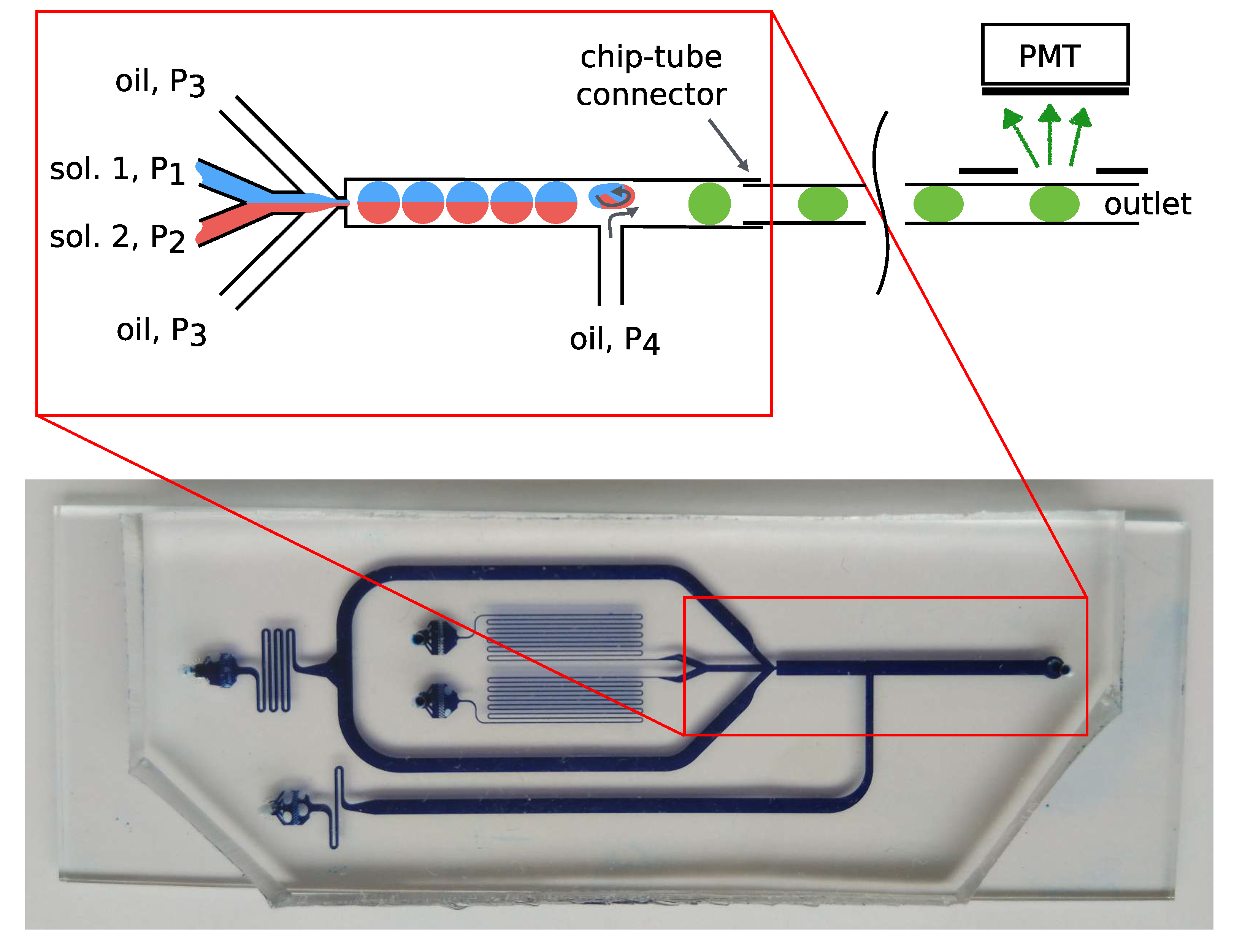
2.1.1. Microfluidic Chip

The final chip design was achieved through a series of experimental iterations and computer modeling. Numerical simulations were carried out in COMSOL Multiphysics (Comsol, Stockholm, Sweden) based on the standard model of multiphase fluids (flow and droplet formation) and diffusion of chemical compounds in two dimensions.
Microfluidic chips were made by the soft lithography method using PDMS and a Si/SU-8 mold. The channel depth was 250 μ m. To improve the quality of sealing, the chip and glass substrate were treated in oxygen plasma for 2 min. After that, they were kept at a temperature of 130 C for 10 min.
Due to the instability of the surface properties of PDMS after plasma treatment and glass hydrophilicity, the inner surfaces of microchannels were coated with a hydrophobic agent, AntiRain Repellent (Turtle Wax, Addison, IL, USA). It provided a surface wetting angle of approximately 100 . After that, the chips were also treated with mineral oil M5310 (Sigma Aldrich) and kept at a temperature of 130 C for 10 min to provide stable hydrophobic surface.

2.1.2. Fluid Management

All reagents were sampled into the microfluidic chip at a constant pressure in the range of 0–50 kPa using four ITV0001 electro-pneumatic regulators (SMC, Tokyo, Japan). These regulators were connected to the chip via 15 mL tubes with an air–liquid interface connector using flexible Tygon Tubing capillaries with an inner diameter of 0.5 mm. The tubes with liquids were thermally stabilized at 25 C.

2.1.3. Registration of Luminescence

The output channel of the microfluidic device was connected by a flexible capillary with an inner diameter of 0.5 mm to the H7828 PMT-based detector (Hamamatsu, Japan), which operated in the photon counting mode. The distance between the light-isolated camera with the PMT and the microfluidic chips was about 12 cm.
The peaks from the PMT were counted and visualized with an integration period of 0.1 s in a custom application developed by means of the BlackBox Component Builder framework (https://blackbox.oberon.org).

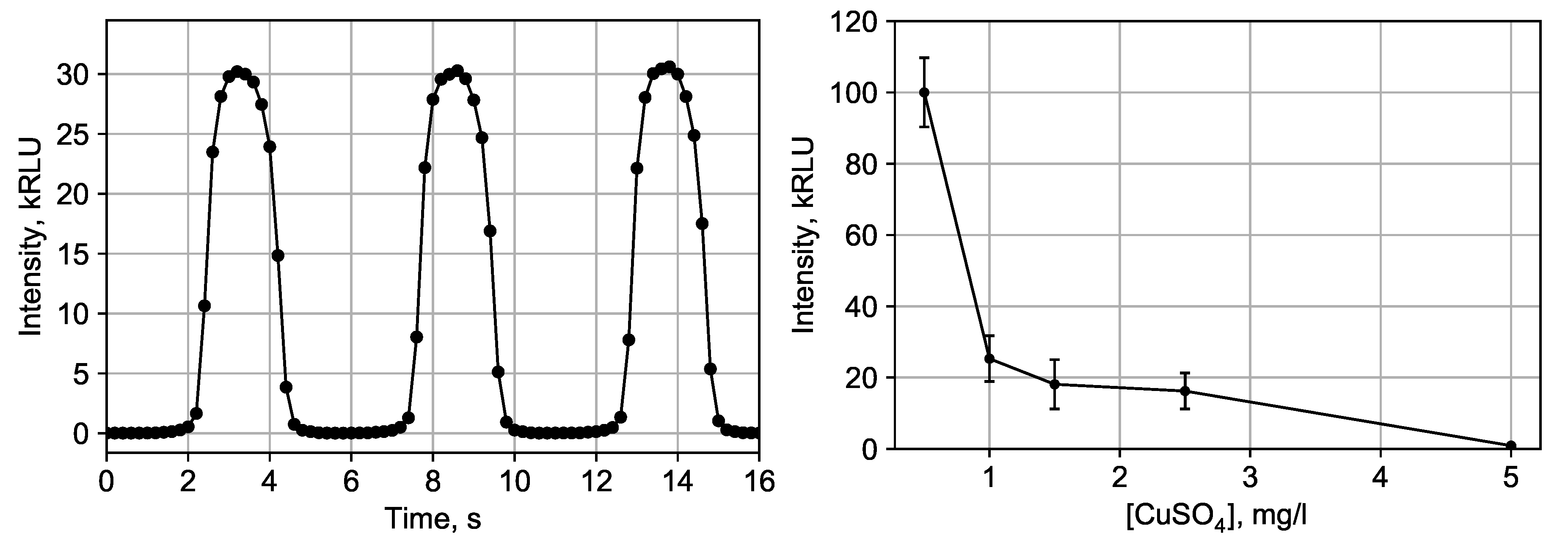
3. Results and Discussion

Preliminary estimation of possible light intensity from the bioluminescent reaction showed that the volume of the droplet reactor should be around 0.1 μ L. This value is bigger than the one that is commonly used in droplet microfluidics because of the low solubility of tetradecanal and necessity to have a significant volume of sample with pollutants to obtain the desired sensitivity.
Hereby, the microfluidic chip for the generation of stable emulsion with water droplets of around 0.1 μ L with a frequency of 0.1–0.5 Hz in oil flow with a regulated distance between droplets was developed (Figure 1). The design of the microfluidic droplet generator was built on the basis of the flow-focusing geometry, and contained two channels for packing the components of the bioluminescent reaction and water sample into droplets, as well as two channels for introducing oil, which acted as a continuous phase during the formation of an emulsion and allowed to adjust the distance between drops. Filters were formed near the inlet holes of the microchip, which consisted of arrays of microstructures with a characteristic distance between them of 5–20 μ m. Such a distance prevented the clogging of the working area of the droplet generator with dust microparticles. The depth of all channels in the microfluidic chip was 250 μ m.
The experimental setup for generation of emulsion droplets with bacterial luciferase was assembled. It consisted of the reduced pressure source, thermostable tanks with reagents, microfluidic chip, and PMT detector. The air under pressure displaced reagent solutions from tubes, and, thus, reagents were fed into the chip with a given pressure, where drops were formed. The pressures were selected so that droplets with a volume of about 0.12 μ L were formed, with a distance between them of 2 cm and a frequency of 0.2 Hz: P 1 = P 2 = P 3 = 16 kPa (taking into account the hydraulic resistance of 7 kPa), P 4 = 30 kPa. The distance of 2 cm between the drops was selected so that only one drop would be exposed to the detector through the aperture.
The droplets entered the detector, where their luminescence intensity was measured. For this, a light-isolated chamber was made in which a capillary with droplets flowing inside was placed. In this chamber, an aperture that was 5 mm wide was formed between the capillary and the PMT. The distance between the droplets of 2 cm made it possible to register each drop separately.
Due to the high solubility of tetradecanal in the mineral oil and low solubility in water, tetradecanal was diluted in the mineral oil in 10 mM concentration to prevent its transfer from the droplet microreactor. Abil EM180 surfactant was used in 0.75% concentration in mineral oil.
All reaction components were separated into two solutions to pack the bioluminescent system into an emulsion. Solution #1 contained: 0.05 mg of luciferase, 0.018 a.u. of NAD(P)H:FMN-oxidoreductase in 0.5 mL 0.143 M potassium phosphate buffer pH 6.8, 4 μ M NADH, and 388 μ L of water. Solution #2 contained: 331 μ L of water, 444 μ L of water sample, 63 μ L tetradecanal 0.25% in ethanol, and 50 μ L of 10 mM FMN. Droplets were formed by mixing solutions in a ratio close to 1:1. A detailed schematic of the microfluidic chip and the experimental circuit are shown in Figure 1.
The mixed flow was compressed by oil phase and formed droplets with equal volume parts of Sol. #1 and Sol. #2. After emulsion formation, in 20 s, the droplets reached the side distancing channel, which emitted extra oil. This oil provided separation and twisting of droplets for mixing of reagents inside droplets and, thus, activation of the luminescent reaction. Then, the droplets left the chip and flowed to the detector over t 2 ≈ 20 s. This time was enough for reaction kinetics to gain the highest luminescence intensity. The droplets passed through the detector aperture, where the luminescence intensity was recorded.
Figure 2 shows the signal recorded by the PMT for individual droplets passing by the detector aperture. The dependences of the relative intensity of luminescence on the concentration of copper ions in the sample were obtained. I C 50 for C u S O 4 was approximately 0.9 mg/L, and the limit of detection was approximately 5 mg/L.

4. Conclusions

It was shown that enzymatic testing could be perofmed in moving emulsion droplets generated in a microfluidic chip by measuring each individual droplet independently from the others.
The presented microfluidic droplet enzymatic assay can be used as a component of a multi-sensor bioassay [20] with rapid feedback for online water pollution monitoring. The studies carried out are applicable for the development of early warning systems at wastewater discharge sites of industrial enterprises or at water treatment plants in addition to the existing certified methods for the registration of water-soluble pollutants.

Author Contributions

Methodology, I.V.K., A.S.B., and E.N.E.; software, I.A.D.; investigation, A.S.Y., I.V.K., A.S.B. and I.A.D.; manuscript preparation, A.S.Y., I.A.D., and K.A.L.; computer simulation, K.I.B.; visualization, A.S.Y. and I.A.D.; supervision, P.I.B. and A.A.E. All authors have read and agreed to the published version of the manuscript.

Acknowledgments

The authors are grateful to Valentina A. Kratasyuk for the facility support.

Abbreviations

The following abbreviations are used in this manuscript:
NADHnicotinamide adenine dinucleotide reduced
FMNflavin mononucleotide
PDMSpoly(dimethyl siloxane)
PMTphotomultiplier tube

References

  1. Bansod, B.; Kumar, T.; Thakur, R.; Rana, S.; Singh, I. A review on various electrochemical techniques for heavy metal ions detection with different sensing platforms. Biosens. Bioelectron. 2017, 94, 443–455. [Google Scholar] [CrossRef] [PubMed]
  2. Roy, S.P. Overview of heavy metals and aquatic environment with notes on their recovery. Ecoscan 2010, 4, 235–240. [Google Scholar]
  3. Afkhami, A.; Soltani-Felehgari, F.; Madrakian, T.; Ghaedi, H.; Rezaeivala, M. Fabrication and application of a new modified electrochemical sensor using nano-silica and a newly synthesized Schiff base for simultaneous determination of Cd2+, Cu2+ and Hg2+ ions in water and some foodstuff samples. Anal. Chim. Acta 2013, 771, 21–30. [Google Scholar] [CrossRef] [PubMed]
  4. Tag, K.; Riedel, K.; Bauer, H.; Hanke, G.; Baronian, K.H.; Kunze, G. Amperometric detection of Cu2+ by yeast biosensors using flow injection analysis (FIA). Sens. Actuators B Chem. 2007, 122, 403–409. [Google Scholar] [CrossRef]
  5. Javed, M.; Ahmad, M.I.; Usmani, N.; Ahmad, M. Multiple biomarker responses (serum biochemistry, oxidative stress, genotoxicity and histopathology) in Channa punctatus exposed to heavy metal loaded waste water. Sci. Rep. 2017, 7, 1–7. [Google Scholar] [CrossRef] [PubMed]
  6. Triki, H.Z.; Laabir, M.; Lafabrie, C.; Malouche, D.; Bancon-Montigny, C.; Gonzalez, C.; Deidun, A.; Pringault, O.; Daly-Yahia, O.K. Do the levels of industrial pollutants influence the distribution and abundance of dinoflagellate cysts in the recently-deposited sediment of a Mediterranean coastal ecosystem? Sci. Total Environ. 2017, 595, 380–392. [Google Scholar] [CrossRef] [PubMed]
  7. Gumpu, M.B.; Sethuraman, S.; Krishnan, U.M.; Rayappan, J.B.B. A review on detection of heavy metal ions in water—An electrochemical approach. Sens. Actuators B Chem. 2015, 213, 515–533. [Google Scholar] [CrossRef]
  8. Long, F.; Zhu, A.; Shi, H. Recent Advances in Optical Biosensors for Environmental Monitoring and Early Warning. Sensors 2013, 13, 13928–13948. [Google Scholar] [CrossRef] [PubMed]
  9. Zurita, J.L.; Jos, A.; Cameán, A.M.; Salguero, M.; López-Artíguez, M.; Repetto, G. Ecotoxicological evaluation of sodium fluoroacetate on aquatic organisms and investigation of the effects on two fish cell lines. Chemosphere 2007, 67, 1–12. [Google Scholar] [CrossRef] [PubMed]
  10. Medvedeva, S.E.; Tyulkova, N.A.; Kuznetsov, A.M.; Rodicheva, E.K. Bioluminescent Bioassays Based on Luminous Bacteria. J. Sib. Fed. Univ. Biol. 2009, 4, 418–452. [Google Scholar]
  11. Woutersen, M.; Belkin, S.; Brouwer, B.; van Wezel, A.P.; Heringa, M.B. Are luminescent bacteria suitable for online detection and monitoring of toxic compounds in drinking water and its sources? Anal. Bioanal. Chem. 2011, 400, 915–929. [Google Scholar] [CrossRef] [PubMed]
  12. Elad, T.; Almog, R.; Yagur-Kroll, S.; Levkov, K.; Melamed, S.; Shacham-Diamand, Y.; Belkin, S. Online Monitoring of Water Toxicity by Use of Bioluminescent Reporter Bacterial Biochips. Environ. Sci. Technol. 2011, 45, 8536–8544. [Google Scholar] [CrossRef] [PubMed]
  13. Elad, T.; Belkin, S. Reporter Gene Assays in Ecotoxicology. In In Vitro Environmental Toxicology—Concepts, Application and Assessment; Reifferscheid, G., Buchinger, S., Eds.; Springer: Cham, Switzerland, 2016; Volume 157, pp. 135–157. [Google Scholar]
  14. Cevenini, L.; Calabretta, M.M.; Tarantino, G.; Michelini, E.; Roda, A. Smartphone-interfaced 3D printed toxicity biosensor integrating bioluminescent “sentinel cells”. Sens. Actuators B Chem. 2016, 225, 249–257. [Google Scholar] [CrossRef]
  15. Charrier, T.; Chapeau, C.; Bendria, L.; Picart, P.; Daniel, P.; Thouand, G. A multi-channel bioluminescent bacterial biosensor for the on-line detection of metals and toxicity. Part II: Technical development and proof of concept of the biosensor. Anal. Bioanal. Chem. 2011, 400, 1061–1070. [Google Scholar] [CrossRef] [PubMed]
  16. Kratasyuk, V.A.; Esimbekova, E.N.; Gladyshev, M.I.; Khromichek, E.B.; Kuznetsov, A.M.; Ivanova, E.A. The use of bioluminescent biotests for study of natural and laboratory aquatic ecosystems. Chemosphere 2001, 42, 909–915. [Google Scholar] [CrossRef]
  17. Hastings, J.; Nealson, K. Bacterial bioluminescence. Annu. Rev. Microbiol. 1977, 31, 549–595. [Google Scholar] [CrossRef] [PubMed]
  18. Petushkov, V.; Kratasyuk, G.; Rodionova, N.; Fish, A.; Belobrov, P. Two-enzyme NADH:FMN-oxidoreductase- luciferase system from luminescent bacteria. Biochem. Acad. Sci. USSR 1984, 49, 593–604. [Google Scholar]
  19. Mashaghi, S.; Abbaspourrad, A.; Weitz, D.A.; van Oijen, A.M. Droplet microfluidics: A tool for biology, chemistry and nanotechnology. TrAC Trends Anal. Chem. 2016, 82, 118–125. [Google Scholar] [CrossRef]
  20. Liu, C.; Guo, X.; Li, Z.; Wang, Y.; Wei, G. Multisensors Cooperative Detection Task Scheduling Algorithm Based on Hybrid Task Decomposition and MBPSO. Math. Probl. Eng. 2017, 2017, 1–11. [Google Scholar] [CrossRef]
Figure 1. The microfluidic chip with dye in channels and the scheme of experiment.
Proceedings 60 00054 g001
Figure 2. Signals from three droplets (left) and dependence of luminescence intensity on CuSO 4 concentration (right).
Proceedings 60 00054 g002
Publisher’s Note: MDPI stays neutral with regard to jurisdictional claims in published maps and institutional affiliations.

