Article

Novel Pathogenic Mutations Identified from Whole-Genome Sequencing in Unsolved Cases of Patients Affected with Inherited Retinal Diseases

1
Department of Molecular and Human Genetics, Baylor College of Medicine, Houston, TX 77030, USA
2
Human Genome Sequencing Center, Baylor College of Medicine, Houston, TX 77030, USA
3
Department of Ophthalmology, Casey Eye Institute, Oregon Health & Science University, Portland, OR 97239, USA
*
Author to whom correspondence should be addressed.
Genes 2023, 14(2), 447; https://doi.org/10.3390/genes14020447
Submission received: 16 January 2023 / Revised: 31 January 2023 / Accepted: 3 February 2023 / Published: 9 February 2023
(This article belongs to the Special Issue Feature Papers in Human Genomics and Genetic Diseases)

Abstract

:
Inherited retinal diseases (IRDs) are a diverse set of visual disorders that collectively represent a major cause of early-onset blindness. With the reduction in sequencing costs in recent years, whole-genome sequencing (WGS) is being used more frequently, particularly when targeted gene panels and whole-exome sequencing (WES) fail to detect pathogenic mutations in patients. In this study, we performed mutation screens using WGS for a cohort of 311 IRD patients whose mutations were undetermined. A total of nine putative pathogenic mutations in six IRD patients were identified, including six novel mutations. Among them, four were deep intronic mutations that affected mRNA splicing, while the other five affected protein-coding sequences. Our results suggested that the rate of resolution of unsolved cases via targeted gene panels and WES can be further enhanced with WGS; however, the overall improvement may be limited.

1. Introduction

Inherited retinal diseases (IRDs) are among the most severe and irreversible causes of blindness for millions of patients. It has been shown through the use of high-throughput sequencing technologies that IRDs exhibit a wide range of phenotypic and genetic heterogeneity with more than 280 genes and loci associated with autosomal-recessive, autosomal-dominant, X-linked, and mitochondrial inheritance (https://web.sph.uth.edu/RetNet/sum-dis.htm accessed on 10 January 2023). Due to the high heterogeneity in both genetic and clinical phenotypes, it is crucial to identify the pathogenic mutations for each IRD patient to provide improved diagnosis, prognosis, and genetic counseling. Consequently, the utilization of high-throughput sequencing techniques has become the standard of care for IRDs in recent years [1,2,3,4]. On the other hand, the retina is a favorable target for developing gene therapy due to its small volume, direct visibility, and immuno-privileged environment, as well as the various sensitive procedures that can be performed to assess its function [5]. As a result, with the rapid development of gene therapy that targets IRD diseases, accurate diagnoses at the molecular level are essential in matching patients with proper personalized treatment [5].
Genomic analysis is used as the diagnostic standard to distinguish among genes with diverse phenotypes that are attributed to variants. Previously, targeted gene panels and whole-exome sequencing (WES) were used for the detection of mutations in coding sequences and nearby splicing sites [6,7,8]. Whole-genome sequencing (WGS) affords significant advantages over traditional gene panels and WES because it provides more even and complete coverage for the entire genome by covering every base in both the coding and noncoding regions [9,10]. A recent study by Ellingford et al. indicated that the causal variant detection rate by WGS was significantly higher than that of targeted panels in IRD patients [7]. A study by Keren et al. demonstrated a 56% variant detection rate in IRD patients, which was considerably higher than the rate detected by WES [11]. Aziz et al. described that the proportion of false-positive variants for WES was greater (78%) than for WGS (17%) [12]. These studies not only emphasized the utility of WGS in the diagnosis of unresolved IRD cases following techniques such as targeted gene panels and WES, but several groups also identified novel mutations to establish underlying genetic causes for cryptic IRDs [8,11,13,14]. However, the increase in the diagnostic rate by WGS varied significantly among previous studies and ranged from a few percent to 24%. In this study, we examined the contribution of WGS in the diagnosis of a cohort of 311 IRD patients who were negative in targeted panel sequencing and WES.

2. Materials and Methods

2.1. Subjects

All probands in our cohort were clinically diagnosed with inherited retinal diseases by a qualified panel of ophthalmologists. Genetic counseling and DNA analysis via blood collection were conducted following the provision of written informed consent by the participants. DNA samples from probands were extracted using Qiagen blood genomic DNA extraction kits (Qiagen, Hilden, Germany). This study adhered to the tenets of the Declaration of Helsinki and was approved by the institutional review boards at every affiliated institution.

2.2. Whole-Genome Sequencing of IRD Patients

In order to identify pathogenic mutations, gene panel testing was performed for all patients in this cohort. Further in-depth analysis was conducted on patients who were not able to be assigned a certain molecular diagnosis. WGS results were processed at the Human Genome Sequencing Center at Baylor College of Medicine using a modified pipeline from our previous WES and WGS analyses [13,15]. WGS sequencing reads were aligned briefly with the Burrows–Wheeler Alignment (BWA) human genome assembly (hg19) [16], whereas single-nucleotide variants (SNVs) and insertion–deletion variants (INDELs) were identified using GATK4. To eliminate frequently occurring variants that were not likely to cause IRDs, a 0.5% population-frequency threshold was set. Coding region SNVs and INDELs were annotated with ANNOVAR and compared to the dbNSFP 3.5a database, while the remaining variants’ conservation was estimated in accordance with the UCSC Genome Browser’s phastCons.hg19.100way [17]. Prediction of the effect of coding variants was performed using CADD v1.3 [18,19].
WGS-consolidated SNVs from all patients were annotated and filtered according to the genomic alteration with a custom pipeline, and the prediction of intronic variant effects was performed with SpliceAI (spliceai-1.2.1) [20]. Each variant was given a score between 0 and 1 by the in silico variant-prediction engine SpliceAI; a higher score indicated greater confidence that the variant affected the splicing. Potential splicing variants were chosen with a prediction cutoff score of 0.5 after being restricted to previously reported IRD genes and analyzed in accordance with known inheritance patterns. One hit of the splicing variant was required for IRD genes associated with dominant or X-linked hemizygous diseases, while an additional coding or splice-affecting variant was needed for genes that were linked to recessive diseases (Supplementary Table S1). In order to assess only deep intronic splicing variants by removing those that were too close to canonical splice sites, these candidate variants were also filtered according to the distance from exon–intron junctions at >10 bp.

2.3. In Vitro Validation of Novel Intronic Variants

Following the prioritization of the variants, a minigene reporter assay (RHCglo minigene) was performed to analyze the splicing effects [21]. Site-directed mutagenesis was conducted with the vector using the WT amplicon for patients with greater than one mutation in the PCR-amplified region. These products were then cloned into the RHCglo vector, and the effects of splicing variants were assessed following the transfection of plasmids into HEK-293 cells and a subsequent RT-PCR assay [22]. Quantification of the DNA band intensity was performed using the ImageJ Gel Analysis program (https://imagej.nih.gov/ij/, accessed on 14 January 2022); the primer sequences for RT-PCR are listed in Supplementary Table S2.

3. Results

To explore the genetic variants in IRD patients, we analyzed the WGS data from a cohort of 311 IRD patients whose cases remained unsolved via panel sequencing or WES. Upon filtering as described in the Section 2, a total of nine candidate pathogenic variants (Table 1) in six IRD patients were identified (Table 2). Among these nine variants, three had been previously reported while the remaining six were novel. Moreover, five variants were exonic (one missense, three frameshift, and one indel), and four variants were deep intronic and found beyond 50 bp of the exon–intron boundary.

3.1. Validation of Novel Intronic Cryptic Splicing Variants

Among the identified variants in the IRD patients, two were novel intronic splicing variants that were predicted by SpliceAI to create both cryptic donor and acceptor sites (Table 1). To validate the SpliceAI prediction for the novel intronic splicing variants, we performed a minigene assay and revealed the functional impact of the identified candidate variants on mRNA splicing. An RHCglo minigene system was used to perform an in vitro functional splicing assay [20]. The results of the in vitro assay were consistent with the in silico prediction. Moreover, for the novel candidate splicing variants, the RT-PCR produced new bands as predicted by SpliceAI with different lengths as compared to the wild type. The details of each patient and the mutant alleles are described below.
A 34-year-old Caucasian female (MEP-123) was diagnosed with retinitis pigmentosa (RP) (Figure 1A) without a family history of the disease (Figure 1A and Table 2). The father (I:1) of the proband was affected by hearing loss. WGS identified two heterozygous variants in EYS, mutations in which led to autosomal recessive RP. One variant was a splicing variant (NM_001142800.2: c.2259+3291G>T) in intron 14 of EYS, and the second variant (NM_001142800.2:216delA) was a deletion of one nucleotide in exon 4 (Figure 1B and Table 1). Both variants were novel and are rare in the population (0.00003188 and absent) as estimated according to public databases such as gnomAD. The splicing variant c.2259+3291G>T was predicted to create a novel splicing donor site downstream of exon 14 causing an out-of-frame-insertion of a new cryptic exon of 59 bp between exons 14 and 15 (Figure 1C). Similarly, the frameshift introduced by the second variant led to premature stop that was likely to result in nonsense-mediated mRNA decay and therefore a null allele. EYS is located in the connecting cilium of the photoreceptor, and earlier studies proved that the disruption of EYS in zebrafish can cause an abnormality in the localization of the outer-segment proteins and degeneration of the photoreceptors, thus supporting its important role in the retinal architecture [26,27,28]. Taken together, these variants were likely pathogenic and caused RP in the patient.
Based on the in vitro minigene assay, the c.2259+3291G>T variant produced a major and intense band (Figure 1D). This major band was caused by the identified variant and was composed of the aberrant transcript of exon 14 and the addition of 58 bp downstream, which exactly matched the in silico prediction, while a minor and light band was identified as the wild type (Figure 1D). The mutant isoform carried the original exon and a cryptic exon of 58 bp, which perfectly matched the in silico prediction as confirmed by Sanger sequencing (Figure 1E). Thus, the c.2259+3291G>T variant is likely a pathogenic variant that causes retinitis pigmentosa.
A 50-year-old Caucasian male patient (MEP-395) was diagnosed with choroideremia (Figure 2A). He had no family history of the disease. His father (II:2) and paternal uncle (II:1) had partial hearing loss, and his mother (II:3) had slight night blindness (Figure 2A). WGS identified a hemizygous splicing variant (c.117-962G>C) in intron 2 of CHM as shown in the IGV plot (Figure 2B). The splicing variant c.117-962G>C was predicted to create a novel splicing donor site downstream of exon 2 causing an out-of-frame-insertion of a new cryptic exon of 115 bp between exons 2 and 3 (Figure 2C). The cryptic exon contained an early stop codon downstream of the cryptic acceptor splice site that resulted in the generation of a premature stop codon downstream of amino acid position 62 out of a total of 653 amino acids in the wild-type protein. CHM encodes Rab escort protein 1 (REP1), which is crucial for vesicle trafficking. In humans, any abnormality in CHM can cause the characteristic clinical phenotype choroideremia, which is a progressive centripetal retinal degenerative disease that appears only to affect the retinal pigment epithelium (RPE) layer [29,30,31]. All the functionally important domains of REP1 lie downstream of the mutant site, which may result in a very short REP1 protein with loss of all the functional domains. Consistent with the predicted results, the in vitro minigene assay for the c.117-962G>C variant produced two bands (Figure 2D). The relative band intensity of the mutant and wild type was 40% and 60%, respectively (Figure 2D). Of the two bands observed, the prominent band was caused by the identified variant and was composed of the atypical transcript of exon 2 and an addition of 114 bp downstream, which matched the in silico prediction, while the minor band was determined to be the wild type (Figure 2D). The minor isoform contained the original exon, while the major isoform consisted of the original exon plus a cryptic exon of 114 bp, which was confirmed by Sanger sequencing and exactly matched the in silico prediction (Figure 2E). Taken together, the c.2259+3291G>T variant was likely a pathogenic variant that caused choroideremia in patient MEP-395.

3.2. Patients Carrying Novel Coding Pathogenic Mutations

A 19-year-old Asian male (MEP-398) was diagnosed with RP with a paternal family history of minor vision problems (Figure 3A). Father (III:2) of the proband had partial hearing loss. WGS identified two heterozygous variants (NM_014714.4: c.1487C>T and c.1250_1271dup) in exon 13 and exon 11 of IFT140, respectively (Supplementary Figure S1A,B). The nonsynonymous sequence change caused by the coding variant c.1487C>T replaced a highly conserved threonine residue with methionine at codon 496 of the IFT140 protein (p.T496M), which was deemed to be novel and rare (0.000606) in the population database gnomAD. Moreover, multiple in silico algorithms predicted this variant to have a deleterious effect (GERP++ rank score = 0.89; REVEL score = 0.95). Another heterozygous variant was a duplication that was predicted to cause an early stop codon (p.S425Gfs*66) in IFT140. This variant was also novel and is extremely rare in the population: it was absent in the genome-sequencing database gnomAD. If translated, this variant is predicted to either produce a very short protein of only 490 instead of 1462 amino acids or cause nonsense-mediated decay (NMD) of mRNA. IFT140 is a subunit of IFT-A that plays a crucial role in the maintenance and development of the outer segments [32]. Mutations in IFT140 lead to autosomal recessive retinitis pigmentosa (ASRP) [33]. Taken together, both variants were likely to be pathogenic in the patient.
MEP-662 and MEP-663 were two affected Asian siblings that were diagnosed with cone–rod dystrophy (Figure 3A,C,D). WGS of the patient’s DNA identified compound heterozygous variants (NM_004928.3:c.634_635del and c.351_352insACCCTGCCGCGC) in both siblings in exon 6 and exon 4 of CFAP410/C21orf2, respectively (Supplementary Figure S2A–D). One of the variants (c.634_635del) was a novel frameshift variant that was predicted to cause an early stop codon at amino acid p.R212GfsTer, while the second variant (c.351_352insACCCTGCCGCGC) was a recurrent pathogenic mutation [23]. CFAP410/C21orf2 is a ubiquitous protein that has been associated with different cellular functions such as DNA damage repair, the regulation of cell morphology, and cytoskeletal organization [34,35]. Previous studies reported that variants in CFAP410/C21orf2 can cause retinitis pigmentosa and cone–rod dystrophy [36]. Moreover, based on the predictive effects of both variants, these were likely the disease-causing mutations in the patients.

3.3. Patient with Reported Pathogenic Mutations

MEP-082 was a 46-year-old Caucasian male who was diagnosed with ABCA4-related retinopathy (Figure 3A,E) and had no family history of retinal disease. However, his father (II:2) and a paternal uncle (II:1) were affected by amyotrophic lateral sclerosis (ALS) (Figure 3A). The genetic analysis via WGS identified two heterozygous known variants (NM_000350.3; c.5196+1137G>A and c.1555-2745A>G) in intron 36 and 11 of ABCA4, respectively. Both variants are rare in the population and had a very low frequency (0.000264 and 0.000397, respectively) in the genome sequencing database gnomAD (Supplementary Figure S1C,D). Both variants were previously reported as pathogenic mutations. The identified variant (c.5196+1137G>A) was present in trans with c.5196+1216C>A by haplotyping in a patient affected with Stargardt disease [24]. Interestingly, the second variant (c.1555:2745A>G) was detected along with the first variant (c.5196+1137G>A) and another variant (p.C54Y) in the same gene (ABCA4) in a patient affected with Stargardt disease [25]. In short, our identified variants (NM_000350.3; c.5196+1137G>A and c.1555-2745A>G) were reported previously and caused the disease in our patient.

4. Discussion

In this study, WGS was performed in 311 probands whose mutations remained unknown after targeted panel sequencing or WES. Following systematic screening criteria of causal variants in known genes, nine causative variants were identified. Out of nine variants, six were novel and three were previously described. Among the novel variants, one was missense, three were frameshift, and two were deep intronic splicing variants.
The two novel intronic splicing variants (the c.2259+3291G>T variant in EYS and the c.117-962G>C variant in CHM) activated cryptic donor and acceptor splice sites close to the mutation sites and thereby resulted in cryptic exon inclusion. Both novel splicing variants were deleterious for the following reasons: (1) they were rare in the population; (2) the predictions were further validated by a minigene assay; and (3) the clinical phenotypes were consistent with the genotypes. It is worth noting that two out of four of the intronic variants that we identified were novel and had not been reported previously, which suggested that a considerable portion (50%) of cryptic splicing mutations remain undiscovered by conventional sequencing techniques [13]. Most of the exonic variants (75%, 3/4) were frameshift, which was missed by targeted panel sequencing or WES due to various reasons such as out-of-date gene panels, the inadequacy of the annotation pipeline, or a lack of knowledge and evidence regarding the interpretation of IRD-associated genes at the time of the targeted panel sequencing or WES data analysis [37].
In previous studies, an improved mutation-detection rate via WGS was described compared to targeted panels and WES, although the improvement varied between a few percent and 24% [1,2,3,4,7,11,37,38,39,40]. It was found that the WGS was superior in the detection of SVs, variants in regulatory regions, and variants in GC-rich regions compared to WES [11]. In another recent study, Fadaie Z. et al. detected disease-causing variants in 24 out of 100 unsolved IRD cases, which was the highest percentage of variant detection by WGS to date [37]. However, it is worth noting that a significant portion of mutations missed by capture sequencing mapped to coding regions or near canonical splicing sites, which indicated that the improvement in WGS was not simply due to the improvement in the sequencing coverage. Factors that can influence the differences between the detection rates include the degree of prescreening performed, the panel design utilized, and the data analysis. In the cohort investigated in this study, a customized gene panel that included known deep intronic mutations was used in the initial screening. In addition, patients with SVs and large CNVs were not included in this report because they were described in our previous study [40]. As result, novel deep intronic mutations and small indels missed by our previous screen via panel sequencing and WES were only detected in 1.93% (6/311) of the unsolved patient cohort.
This study illustrated that while improvement is limited, WGS can improve the diagnostic rate for unsolved IRD cases. It is plausible that long-read WGS could provide a higher diagnostic yield by detecting causal variants that are missed by the short WGS technologies [37]. To further increase the diagnostic rate, the contribution of other types of mutations such as those that affect gene expression regulatory elements need to be systematically assessed. WGS coupled with a functional assay of candidate noncoding variants is essential to identify and confirm the pathogenicity of these noncoding mutations.
In conclusion, WGS offered some advantages over gene panel sequencing and WES in detecting deep intronic mutations, SVs and CNVs, and indels, therefore increasing the yield, although the improvement was limited. As a result, the majority of IRD cases remain unsolved despite the introduction of WGS. Several possibilities to increase the detection rate warrant further investigation; these include cryptic splicing sites missed by current prediction tools, SVs, gene regulatory elements and duplication regions that are hard to detect with short-read sequencing technology, and novel disease-associated genes.

Supplementary Materials

The following supporting information can be downloaded at: https://www.mdpi.com/article/10.3390/genes14020447/s1, Figure S1. IGV plots of probands affected with IRD: (A,B) IGV plots of MEP-398 in IFT140 (chr16:1630797G>A & chr16:1634305ins22) and (C-D) IGV plots of MEP-082 in gene ABCA4: (chr1:94484001C>T and chr1:94531618T>C); Figure S2. IGV plots of siblings affected with IRD: (A-B) IGV plots of MEP-662 in gene C21orf2/CFAP410 (chr21:45750712 CCT>C and chr21:45752937Gins12) and (C-D) IGV plots of MEP-663 in gene C21orf2/CFAP410 (chr21:45750712 CCT>C and chr21:45752937Gins12). Supplementary Table S1: SpliceAI prediction scores of novel splicing variants.; Supplementary Table S2: Primer sequences for RT-PCR.

Author Contributions

H.M.J.H. complied the data and designed and conceptualized the study; H.M.J.H. and M.W. conducted the WGS data analysis; Y.L. and X.Q. performed the experiments. H.M.J.H. and A.H. prepared the first draft of the manuscript; R.S., M.E.P., P.Y. and M.M. enrolled families and collected the data; R.C. conceptualized and supervised the study and revised the manuscript. All authors have read and agreed to the published version of the manuscript.

Funding

This study was funded by the National Eye Institute (EY022356, EY018571, EY002520, P30EY010572 and EY030499), the Retinal Research Foundation, an NIH shared instrument grant (S10OD023469), the Daljit S. and Elaine Sarkaria Charitable Foundation, an Unrestricted Grant from Research to Prevent Blindness (New York), Fighting Blindness Canada, and the Vision Health Research Network.

Institutional Review Board Statement

This study, which involved human participants, was reviewed and approved by the Baylor College of Medicine.

Informed Consent Statement

Informed consent was obtained from all participants.

Data Availability Statement

Due to the concerns of patients, all datasets for this manuscript are not publicly available. Requests to access the datasets should be directed to the corresponding author.

Acknowledgments

All the participants consented and we thank them for their cooperation.

Conflicts of Interest

The authors declare no conflict of interest.

References

  1. Huang, X.F.; Huang, F.; Wu, K.C.; Wu, J.; Chen, J.; Pang, C.P.; Lu, F.; Qu, J.; Jin, Z.B. Genotype-phenotype correlation and mutation spectrum in a large cohort of patients with inherited retinal dystrophy revealed by next-generation sequencing. Genet. Med. 2015, 17, 271–278. [Google Scholar] [CrossRef] [PubMed]
  2. Patel, N.; Aldahmesh, M.A.; Alkuraya, H.; Anazi, S.; Alsharif, H.; Khan, A.O.; Sunker, A.; Al-Mohsen, S.; Abboud, E.B.; Nowilaty, S.R.; et al. Expanding the clinical, allelic, and locus heterogeneity of retinal dystrophies. Genet. Med. 2016, 18, 554–562. [Google Scholar] [CrossRef] [PubMed]
  3. Eisenberger, T.; Neuhaus, C.; Khan, A.O.; Decker, C.; Preising, M.N.; Friedburg, C.; Bieg, A.; Gliem, M.; Charbel Issa, P.; Holz, F.G.; et al. Increasing the yield in targeted next-generation sequencing by implicating CNV analysis, non-coding exons and the overall variant load: The example of retinal dystrophies. PloS ONE 2013, 8, e78496. [Google Scholar] [CrossRef]
  4. Tiwari, A.; Bahr, A.; Bähr, L.; Fleischhauer, J.; Zinkernagel, M.S.; Winkler, N.; Barthelmes, D.; Berger, L.; Gerth-Kahlert, C.; Neidhardt, J.; et al. Next generation sequencing based identification of disease-associated mutations in Swiss patients with retinal dystrophies. Sci. Rep. 2016, 6, 28755. [Google Scholar] [CrossRef]
  5. Hu, M.L.; Edwards, T.L.; O’Hare, F.; Hickey, D.G.; Wang, J.H.; Liu, Z.; Ayton, L.N. Gene therapy for inherited retinal diseases: Progress and possibilities. Clin. Exp. Optom. 2021, 104, 444–454. [Google Scholar] [CrossRef]
  6. Consugar, M.B.; Navarro-Gomez, D.; Place, E.M.; Bujakowska, K.M.; Sousa, M.E.; Fonseca-Kelly, Z.D.; Taub, D.G.; Janessian, M.; Wang, D.Y.; Au, E.D.; et al. Panel-based genetic diagnostic testing for inherited eye diseases is highly accurate and reproducible, and more sensitive for variant detection, than exome sequencing. Genet. Med. 2015, 17, 253–261. [Google Scholar] [CrossRef]
  7. Ellingford, J.M.; Barton, S.; Bhaskar, S.; Williams, S.G.; Sergouniotis, P.I.; O’Sullivan, J.; Lamb, J.A.; Perveen, R.; Hall, G.; Newman, W.G.; et al. Whole Genome Sequencing Increases Molecular Diagnostic Yield Compared with Current Diagnostic Testing for Inherited Retinal Disease. Ophthalmology 2016, 123, 1143–1150. [Google Scholar] [CrossRef]
  8. Tehreem, R.; Chen, I.; Shah, M.R.; Li, Y.; Khan, M.A.; Afshan, K.; Chen, R.; Firasat, S. Exome Sequencing Identified Molecular Determinants of Retinal Dystrophies in Nine Consanguineous Pakistani Families. Genes 2022, 13, 1630. [Google Scholar] [CrossRef]
  9. Stavropoulos, D.J.; Merico, D.; Jobling, R.; Bowdin, S.; Monfared, N.; Thiruvahindrapuram, B.; Nalpathamkalam, T.; Pellecchia, G.; Yuen, R.K.C.; Szego, M.J.; et al. Whole Genome Sequencing Expands Diagnostic Utility and Improves Clinical Management in Pediatric Medicine. NPJ Genom. Med. 2016, 1, 15012. [Google Scholar] [CrossRef]
  10. Zou, G.; Zhang, T.; Cheng, X.; Igelman, A.D.; Wang, J.; Qian, X.; Fu, S.; Wang, K.; Koenekoop, R.K.; Fishman, G.A.; et al. Noncoding mutation in RPGRIP1 contributes to inherited retinal degenerations. Mol. Vis. 2021, 27, 95–106. [Google Scholar]
  11. Carss, K.J.; Arno, G.; Erwood, M.; Stephens, J.; Sanchis-Juan, A.; Hull, S.; Megy, K.; Grozeva, D.; Dewhurst, E.; Malka, S.; et al. Comprehensive Rare Variant Analysis via Whole-Genome Sequencing to Determine the Molecular Pathology of Inherited Retinal Disease. Am. J. Hum. Genet. 2017, 100, 75–90. [Google Scholar] [CrossRef] [PubMed] [Green Version]
  12. Belkadi, A.; Bolze, A.; Itan, Y.; Cobat, A.; Vincent, Q.B.; Antipenko, A.; Shang, L.; Boisson, B.; Casanova, J.L.; Abel, L. Whole-genome sequencing is more powerful than whole-exome sequencing for detecting exome variants. Proc. Natl. Acad. Sci. USA 2015, 112, 5473–5478. [Google Scholar] [CrossRef] [PubMed]
  13. Qian, X.; Wang, J.; Wang, M.; Igelman, A.D.; Jones, K.D.; Li, Y.; Wang, K.; Goetz, K.E.; Birch, D.G.; Yang, P.; et al. Identification of Deep-Intronic Splice Mutations in a Large Cohort of Patients with Inherited Retinal Diseases. Front. Genet. 2021, 12, 647400. [Google Scholar] [CrossRef] [PubMed]
  14. Branham, K.; Matsui, H.; Biswas, P.; Guru, A.A.; Hicks, M.; Suk, J.J.; Li, H.; Jakubosky, D.; Long, T.; Telenti, A.; et al. Establishing the involvement of the novel gene AGBL5 in retinitis pigmentosa by whole genome sequencing. Physiol. Genom. 2016, 48, 922–927. [Google Scholar] [CrossRef]
  15. Soens, Z.T.; Li, Y.; Zhao, L.; Eblimit, A.; Dharmat, R.; Li, Y.; Chen, Y.; Naqeeb, M.; Fajardo, N.; Lopez, I.; et al. Hypomorphic mutations identified in the candidate Leber congenital amaurosis gene CLUAP1. Genet. Med. 2016, 18, 1044–1051. [Google Scholar] [CrossRef]
  16. Li, H.; Durbin, R. Fast and accurate short read alignment with Burrows-Wheeler transform. Bioinformatics 2009, 25, 1754–1760. [Google Scholar] [CrossRef]
  17. Pollard, K.S.; Hubisz, M.J.; Rosenbloom, K.R.; Siepel, A. Detection of nonneutral substitution rates on mammalian phylogenies. Genome Res. 2010, 20, 110–121. [Google Scholar] [CrossRef]
  18. Rentzsch, P.; Witten, D.; Cooper, G.M.; Shendure, J.; Kircher, M. CADD: Predicting the deleteriousness of variants throughout the human genome. Nucleic Acids Res. 2019, 47, D886–D894. [Google Scholar] [CrossRef]
  19. Kircher, M.; Witten, D.M.; Jain, P.; O’Roak, B.J.; Cooper, G.M.; Shendure, J. A general framework for estimating the relative pathogenicity of human genetic variants. Nat. Genet. 2014, 46, 310–315. [Google Scholar] [CrossRef]
  20. Jaganathan, K.; Kyriazopoulou Panagiotopoulou, S.; McRae, J.F.; Darbandi, S.F.; Knowles, D.; Li, Y.I.; Kosmicki, J.A.; Arbelaez, J.; Cui, W.; Schwartz, G.B.; et al. Predicting Splicing from Primary Sequence with Deep Learning. Cell 2019, 176, 535–548.e524. [Google Scholar] [CrossRef]
  21. Singh, R.K.; Cooper, T.A. Pre-mRNA splicing in disease and therapeutics. Trends Mol. Med. 2012, 18, 472–482. [Google Scholar] [CrossRef] [PubMed] [Green Version]
  22. Soens, Z.T.; Branch, J.; Wu, S.; Yuan, Z.; Li, Y.; Li, H.; Wang, K.; Xu, M.; Rajan, L.; Motta, F.L.; et al. Leveraging splice-affecting variant predictors and a minigene validation system to identify Mendelian disease-causing variants among exon-captured variants of uncertain significance. Hum. Mutat. 2017, 38, 1521–1533. [Google Scholar] [CrossRef] [PubMed]
  23. Huang, L.; Xiao, X.; Li, S.; Jia, X.; Wang, P.; Sun, W.; Xu, Y.; Xin, W.; Guo, X.; Zhang, Q. Molecular genetics of cone-rod dystrophy in Chinese patients: New data from 61 probands and mutation overview of 163 probands. Exp. Eye Res. 2016, 146, 252–258. [Google Scholar] [CrossRef] [PubMed]
  24. Braun, T.A.; Mullins, R.F.; Wagner, A.H.; Andorf, J.L.; Johnston, R.M.; Bakall, B.B.; Deluca, A.P.; Fishman, G.A.; Lam, B.L.; Weleber, R.G.; et al. Non-exomic and synonymous variants in ABCA4 are an important cause of Stargardt disease. Hum. Mol. Genet. 2013, 22, 5136–5145. [Google Scholar] [CrossRef] [PubMed]
  25. Paavo, M.; Lee, W.; Allikmets, R.; Tsang, S.; Sparrow, J.R. Photoreceptor cells as a source of fundus autofluorescence in recessive Stargardt disease. J. Neurosci. Res. 2019, 97, 98–106. [Google Scholar] [CrossRef] [PubMed]
  26. Lu, Z.; Hu, X.; Liu, F.; Soares, D.C.; Liu, X.; Yu, S.; Gao, M.; Han, S.; Qin, Y.; Li, C.; et al. Ablation of EYS in zebrafish causes mislocalisation of outer segment proteins, F-actin disruption and cone-rod dystrophy. Sci. Rep. 2017, 7, 46098. [Google Scholar] [CrossRef] [PubMed]
  27. Messchaert, M.; Dona, M.; Broekman, S.; Peters, T.A.; Corral-Serrano, J.C.; Slijkerman, R.W.N.; van Wijk, E.; Collin, R.W.J. Eyes shut homolog is important for the maintenance of photoreceptor morphology and visual function in zebrafish. PloS ONE 2018, 13, e0200789. [Google Scholar] [CrossRef]
  28. Yu, M.; Liu, Y.; Li, J.; Natale, B.N.; Cao, S.; Wang, D.; Amack, J.D.; Hu, H. Eyes shut homolog is required for maintaining the ciliary pocket and survival of photoreceptors in zebrafish. Biol. Open 2016, 5, 1662–1673. [Google Scholar] [CrossRef]
  29. Cremers, F.P.; van de Pol, D.J.; van Kerkhoff, L.P.; Wieringa, B.; Ropers, H.H. Cloning of a gene that is rearranged in patients with choroideraemia. Nature 1990, 347, 674–677. [Google Scholar] [CrossRef]
  30. Seabra, M.C.; Brown, M.S.; Goldstein, J.L. Retinal degeneration in choroideremia: Deficiency of rab geranylgeranyl transferase. Science 1993, 259, 377–381. [Google Scholar] [CrossRef]
  31. Van den Hurk, J.A.; Schwartz, M.; van Bokhoven, H.; van de Pol, T.J.; Bogerd, L.; Pinckers, A.J.; Bleeker-Wagemakers, E.M.; Pawlowitzki, I.H.; Rüther, K.; Ropers, H.H.; et al. Molecular basis of choroideremia (CHM): Mutations involving the Rab escort protein-1 (REP-1) gene. Hum. Mutat. 1997, 9, 110–117. [Google Scholar] [CrossRef] [PubMed]
  32. Crouse, J.A.; Lopes, V.S.; Sanagustin, J.T.; Keady, B.T.; Williams, D.S.; Pazour, G.J. Distinct functions for IFT140 and IFT20 in opsin transport. Cytoskeleton 2014, 71, 302–310. [Google Scholar] [CrossRef] [PubMed]
  33. Hull, S.; Owen, N.; Islam, F.; Tracey-White, D.; Plagnol, V.; Holder, G.E.; Michaelides, M.; Carss, K.; Raymond, F.L.; Rozet, J.M.; et al. Nonsyndromic Retinal Dystrophy due to Bi-Allelic Mutations in the Ciliary Transport Gene IFT140. Investig. Ophthalmol. Vis. Sci. 2016, 57, 1053–1062. [Google Scholar] [CrossRef] [PubMed]
  34. Fang, X.; Lin, H.; Wang, X.; Zuo, Q.; Qin, J.; Zhang, P. The NEK1 interactor, C21ORF2, is required for efficient DNA damage repair. Acta Biochim. Biophys. Sin. 2015, 47, 834–841. [Google Scholar] [CrossRef]
  35. Bai, S.W.; Herrera-Abreu, M.T.; Rohn, J.L.; Racine, V.; Tajadura, V.; Suryavanshi, N.; Bechtel, S.; Wiemann, S.; Baum, B.; Ridley, A.J. Identification and characterization of a set of conserved and new regulators of cytoskeletal organization, cell morphology and migration. BMC Biol. 2011, 9, 54. [Google Scholar] [CrossRef]
  36. Suga, A.; Mizota, A.; Kato, M.; Kuniyoshi, K.; Yoshitake, K.; Sultan, W.; Yamazaki, M.; Shimomura, Y.; Ikeo, K.; Tsunoda, K.; et al. Identification of Novel Mutations in the LRR-Cap Domain of C21orf2 in Japanese Patients with Retinitis Pigmentosa and Cone-Rod Dystrophy. Investig. Ophthalmol. Vis. Sci. 2016, 57, 4255–4263. [Google Scholar] [CrossRef]
  37. Fadaie, Z.; Whelan, L.; Ben-Yosef, T.; Dockery, A.; Corradi, Z.; Gilissen, C.; Haer-Wigman, L.; Corominas, J.; Astuti, G.D.N.; de Rooij, L.; et al. Whole genome sequencing and in vitro splice assays reveal genetic causes for inherited retinal diseases. NPJ Genom. Med. 2021, 6, 97. [Google Scholar] [CrossRef]
  38. O’Sullivan, J.; Mullaney, B.G.; Bhaskar, S.S.; Dickerson, J.E.; Hall, G.; O’Grady, A.; Webster, A.; Ramsden, S.C.; Black, G.C. A paradigm shift in the delivery of services for diagnosis of inherited retinal disease. J. Med. Genet. 2012, 49, 322–326. [Google Scholar] [CrossRef]
  39. Khan, K.N.; Chana, R.; Ali, N.; Wright, G.; Webster, A.R.; Moore, A.T.; Michaelides, M. Advanced diagnostic genetic testing in inherited retinal disease: Experience from a single tertiary referral centre in the UK National Health Service. Clin. Genet. 2017, 91, 38–45. [Google Scholar] [CrossRef]
  40. Wen, S.; Wang, M.; Qian, X.; Li, Y.; Wang, K.; Choi, J.; Pennesi, M.E.; Yang, P.; Marra, M.; Koenekoop, R.K.; et al. Systematic assessment of the contribution of structural variants to inherited retinal diseases. bioRxiv 2023. [Google Scholar] [CrossRef]
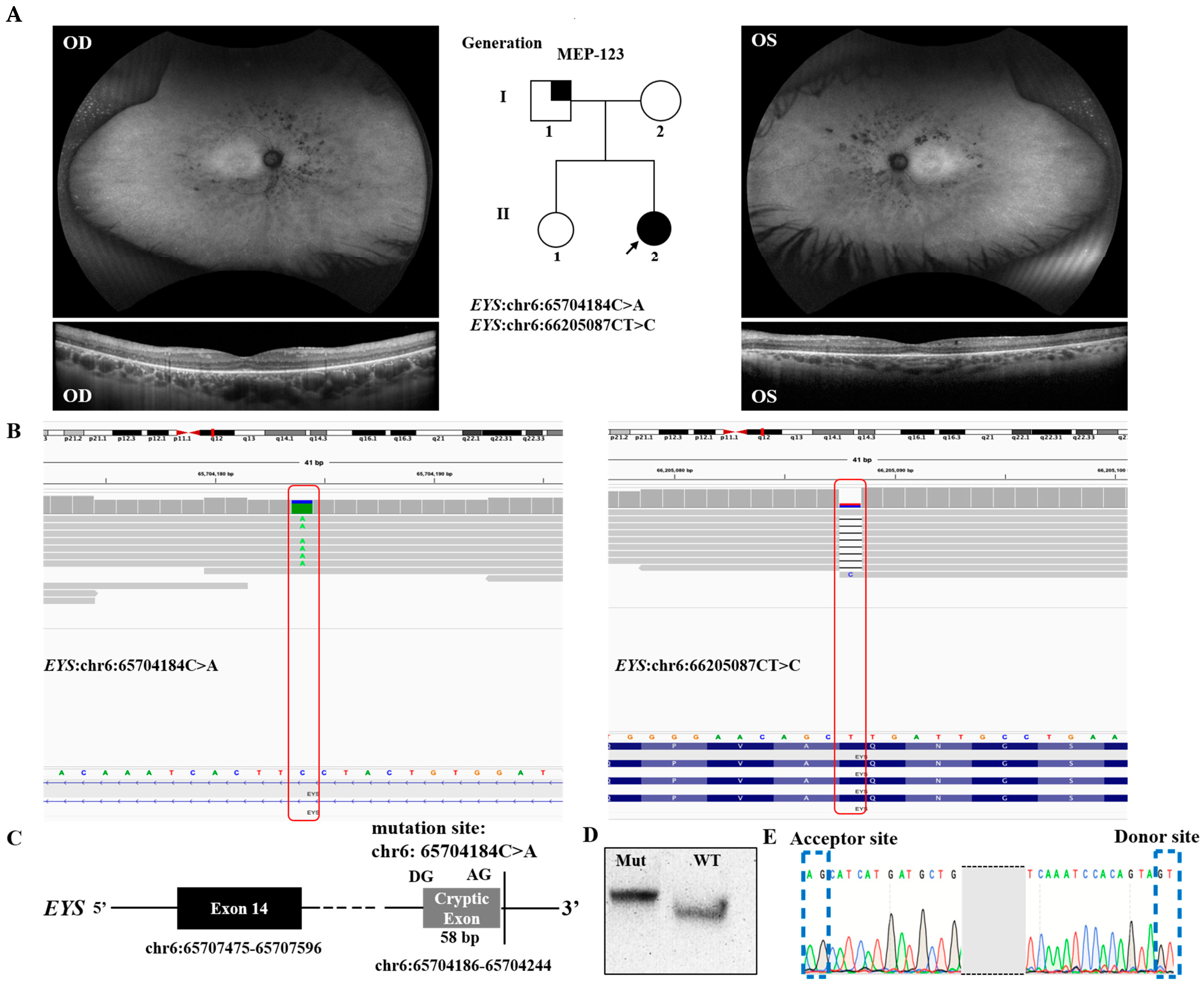
Figure 1. Clinical and functional validation of intronic splicing variant of EYS in MEP-123, who was affected with retinitis pigmentosa (RP). (A) Pedigree of proband, fundus autofluorescence (AF), and optical coherence tomography (OCT) for MEP-123. Symbols represent: females (circles), males (squares), affected individuals (filled symbols), unaffected individuals (open symbols), proband (arrowhead), and hearing loss (partially filled symbols). (B) IGV plots showing the mutant region. A proband (MEP-123) affected with RP had a pathogenic heterozygous deep intronic variant in EYS (chr6:65704184C>A). The individual also had a pathogenic frameshift variant NM_001142800.2: c.216delA (p.Q72fs). (C) Predictive results of novel splicing variant identified in proband. (D) Gel electrophoresis of reverse-transcription PCR (RT-PCR) of novel splicing variant and WT. (E) Confirmation of novel donor and acceptor sites of cryptic exon via Sanger sequencing. Blue dotted lines are showing donor and acceptor sites.
Genes 14 00447 g001
Figure 2. Clinical and functional validation of intronic splicing variant of CHM in MEP-395, who was affected with choroideremia. (A) Pedigree of proband, fundus autofluorescence (AF), and optical coherence tomography (OCT) for MEP-395. Symbols represent: females (circles), males (squares), affected individuals (filled symbols), unaffected individuals (open symbols), proband (arrowhead), and slight night blindness (star). (B) IGV plot showing the mutant region. A male proband (MEP-395) affected with choroideremia had a pathogenic hemizygous deep intronic variant in CHM (chrX:85237775C>G). (C) Predictive results of novel splicing variant identified in proband. (D) Gel electrophoresis of reverse-transcription PCR (RT-PCR) of novel splicing variant and WT. (E) Confirmation of novel donor and acceptor sites of cryptic exon via Sanger sequencing. Blue dotted lines are showing donor and acceptor sites.
Genes 14 00447 g002
Figure 3. Clinical data of probands affected with IRD. (A) Pedigrees of probands (MEP-398 and MEP-662/663) and MEP-082. Symbols represent: females (circles), males (squares), unknown sex (diamonds), affected individuals (filled symbols), unaffected individuals (open symbols), numbers of siblings (numbers in symbols), and deceased (oblique line through symbol) (B) Fundus autofluorescence (AF) and optical coherence tomography (OCT) of MEP-398. (C) AF and OCT of MEP-662. (D) AF and OCT of MEP-663. (E) AF and OCT of MEP-082.
Genes 14 00447 g003
Table 1. Identified variants in IRD patients.
Patient IDGeneGenomic
Variant
cDNA VariantProtein VariantZygosityVariant TypeReference
MEP-123EYSchr6:65704184C>ANM_001142800.2:c.2259+3291G>T-HeterozygousSplicingNovel
EYSchr6:
66205087CT>C
NM_001142800.2:c.216delANP_00113627.1:p.
A73Lfs*12
HeterozygousFrameshiftNovel
MEP-395CHMchrX:85237775C>GNM_000390.4:c.117-962G>C-HemizygousSplicingNovel
MEP-398
IFT140
chr16:1630797G>ANM_014714.4:c.1487C>TNP_055529.2:T496MHeterozygousMissenseNovel

IFT140
chr16:1634305ins22NM_014714.4:c.1250_1271dupNP_055529.2:p.
S425Gfs*66
HeterozygousFrameshiftNovel
MEP-662C21orf2/
CFAP410
chr21:45750712 CCT>CNM_004928.3:c.634_635delNP_004919.1:p.
R212Gfs
HeterozygousFrameshiftNovel
C21orf2/
CFAP410
chr21:45752937Gins12NM_004928.3:c.351_353dup12NP_004919.1:p.
L118delinsTLPRL
HeterozygousInsertion[23]
MEP-663C21orf2/
CFAP410
chr21:45750712 CCT>CNM_004928.3:c.634_635delNP_004919.1:p.
R212Gfs
HeterozygousFrameshiftNovel
C21orf2/
CFAP410
chr21:45752937Gins12NM_004928.3:c.352_352dup12NP_004919.1:p.
L118delinsTLPRL
HeterozygousInsertion[23]
MEP-082
ABCA4
chr1:94484001C>TNM_000350.3:c.5196+1137G>A-HeterozygousSplicing[24]

ABCA4
chr1:94531618T>CNM_000350.3:c.1555-2745A>G-HeterozygousSplicing[25]
Table 2. Clinical features of selected IRD patients.
Patient IDClinical DiagnosisGenderRaceAge (Years)Age of Onset (Years)BCVAProgressionOther SymptomsFamily History
RightLeft
MEP-123RPFCaucasian342020/50-20/50-Mild progression of cystoid macular edema, peripheral vision, and night blindnessNAIsolated case
MEP-395ChoroideremiaMCaucasian503820/20-220/80Mild progression of visual fieldNight blindness, legally blindMother with possible “slight night blindness”
MEP-398RPMAsian19920/25 + 220/20-2Patient had minimal progressionNyctalopia, midperipheral scotomas, midperipheral atrophy, reduced ERGs, visual fields, loss of outer retinal structures on OCT, high myopiaSome relatives with weak vision but not a similar phenotype
MEP-662CRDMAsian31220/70 + 120/70+1Gradual tilting of RNFL over timePrimary congenital glaucoma, subnormal visual acuity, severe cone and mild rod dysfunctionAffected sister with negative family history of similar eye problems
MEP-663CRDFAsian1920/150 20/150 + 1Low vision from cone–rod dystrophyHyperopia, nystagmus, astigmatism, intermittent exotropia OU, progressive cone–rod dystrophyAffected brother with negative family history of similar eye problems
MEP-082ABCA4 related retinopathyFCaucasian46NACF20/100Progression of retinopathy OU with foveal sparingDeceased in January 2022
Family history of ALS but not vision problems
RP, retinitis pigmentosa; CRD, cone–rod dystrophy; ALS, amyotrophic lateral sclerosis; CF, count fingers visual acuity; NA, not available.
Disclaimer/Publisher’s Note: The statements, opinions and data contained in all publications are solely those of the individual author(s) and contributor(s) and not of MDPI and/or the editor(s). MDPI and/or the editor(s) disclaim responsibility for any injury to people or property resulting from any ideas, methods, instructions or products referred to in the content.

