

Microbial Polysaccharide-Based Formulation with Silica Nanoparticles; A New Hydrogel Nanocomposite for 3D Printing
 ,
,  , , ,
, , ,  , and
, and
Abstract
:1. Introduction
2. Results and Discussion
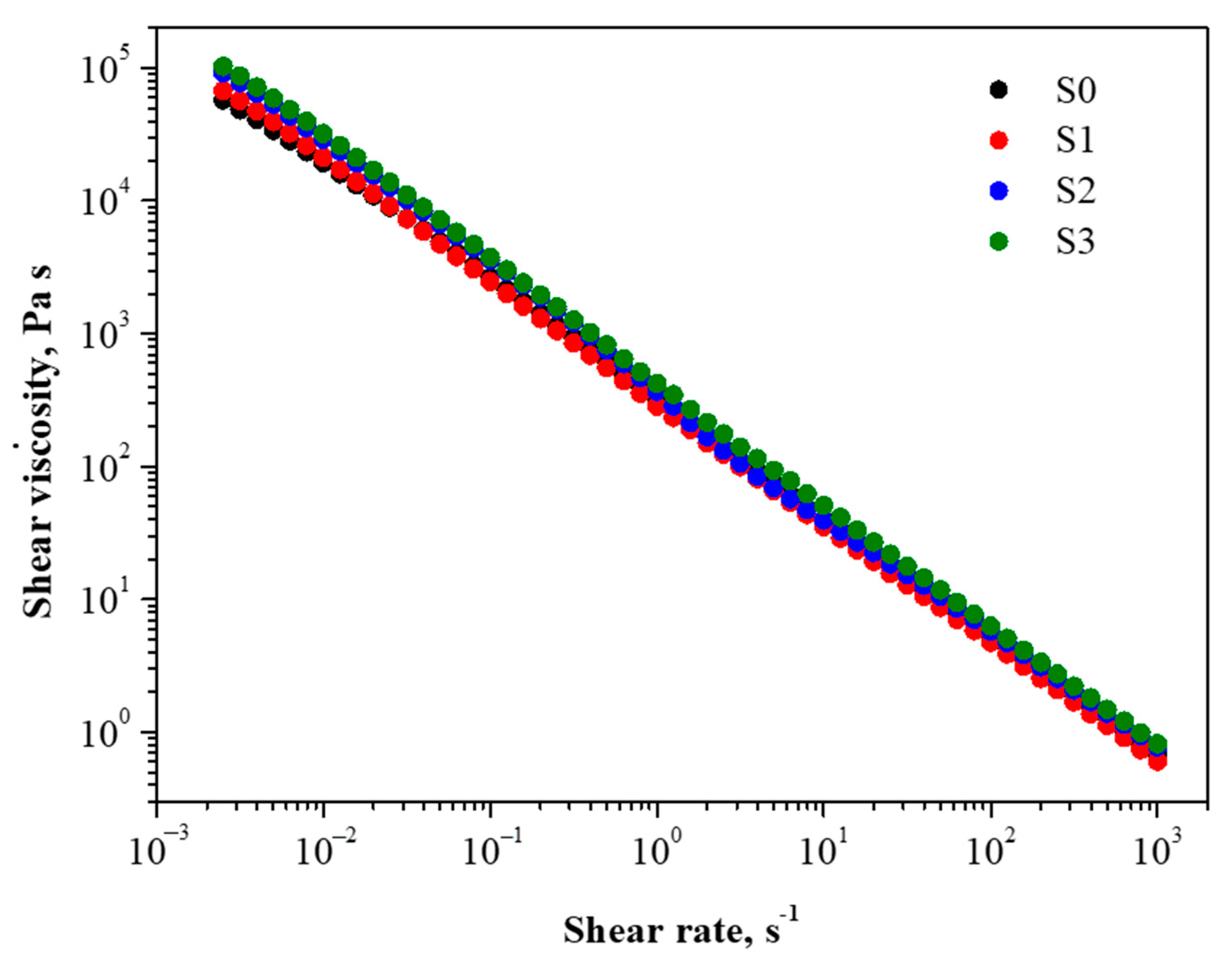
2.1. Rheological Behavior
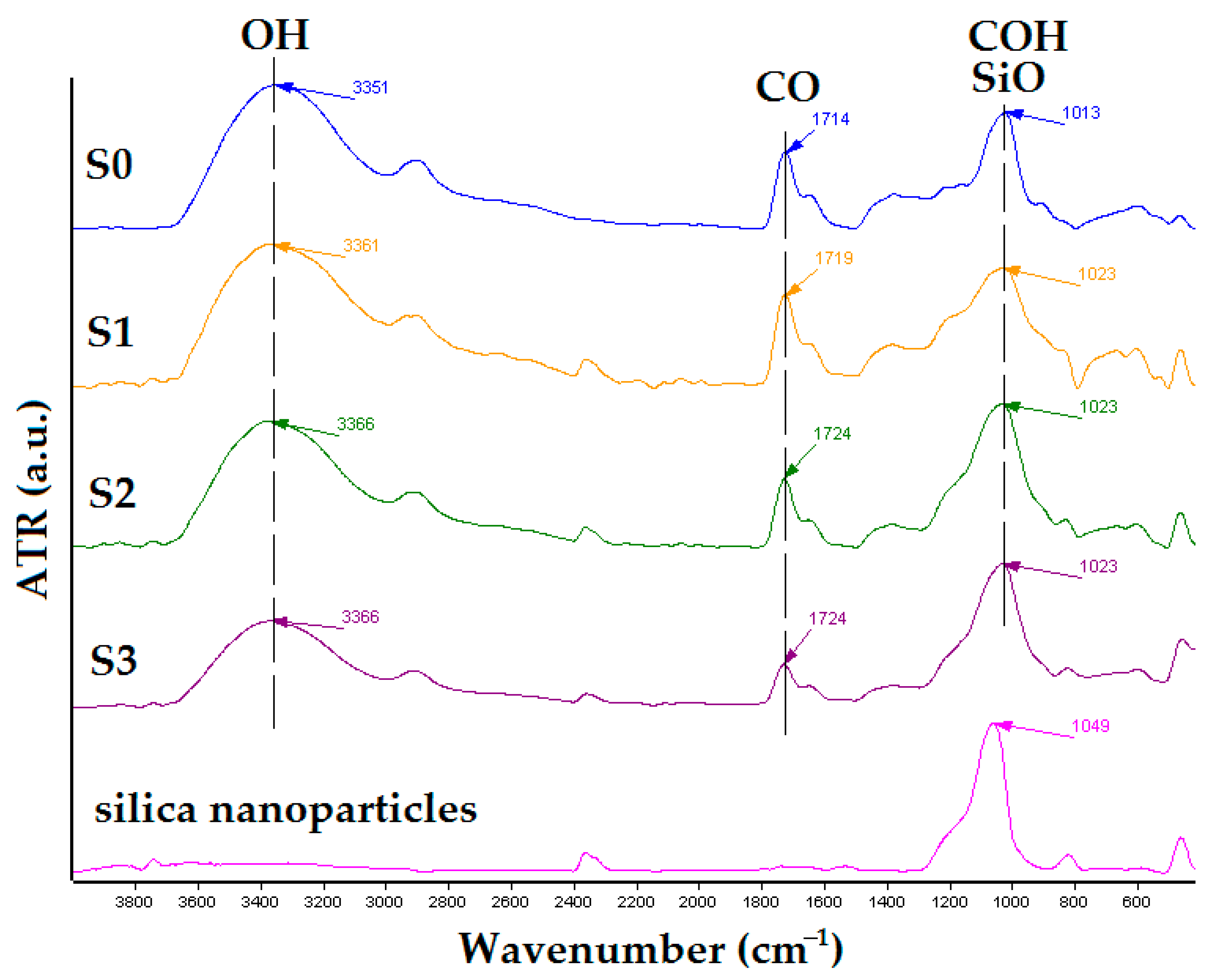
2.2. FT-IR Analyses
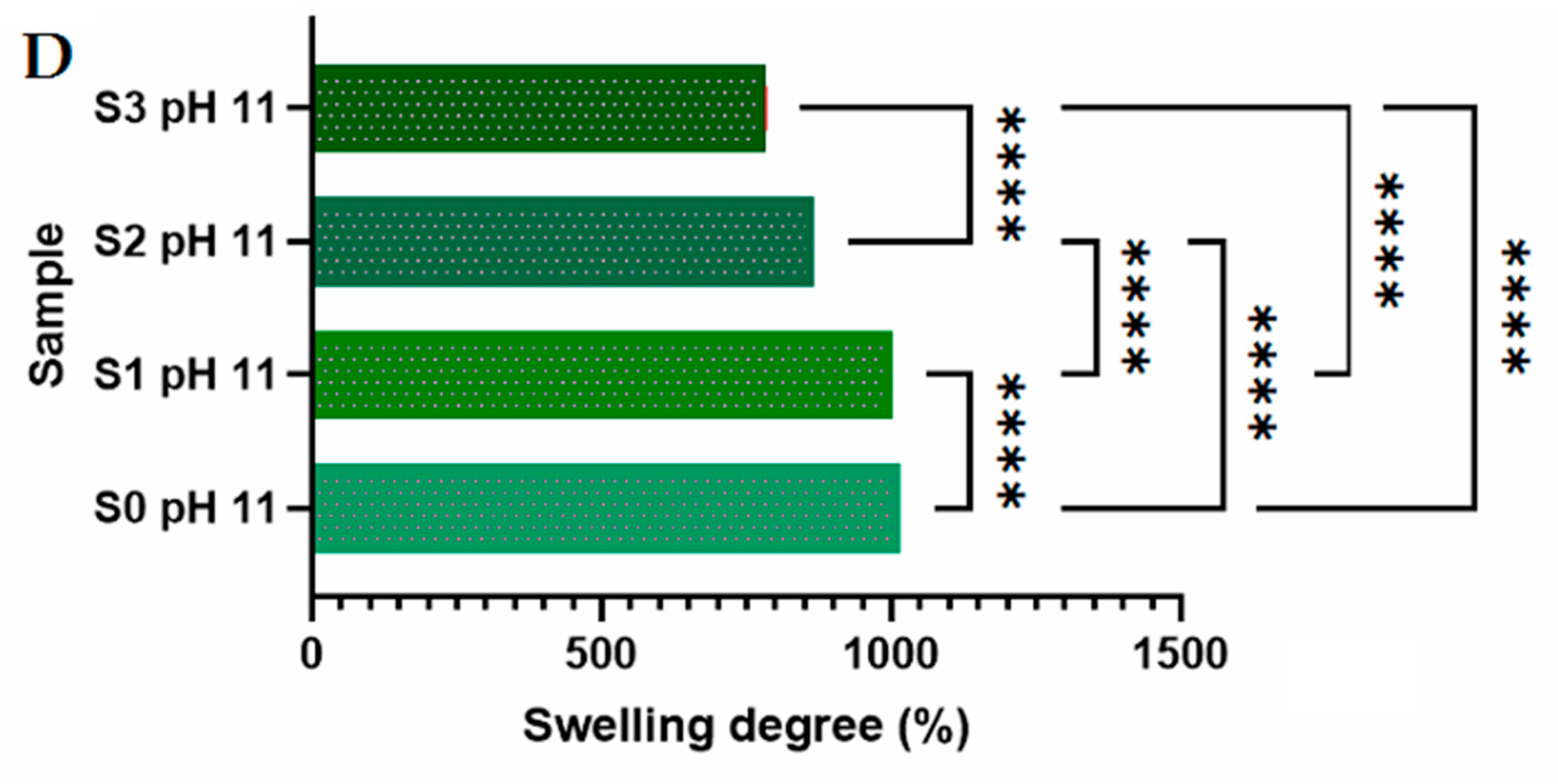
2.3. Swelling Behavior
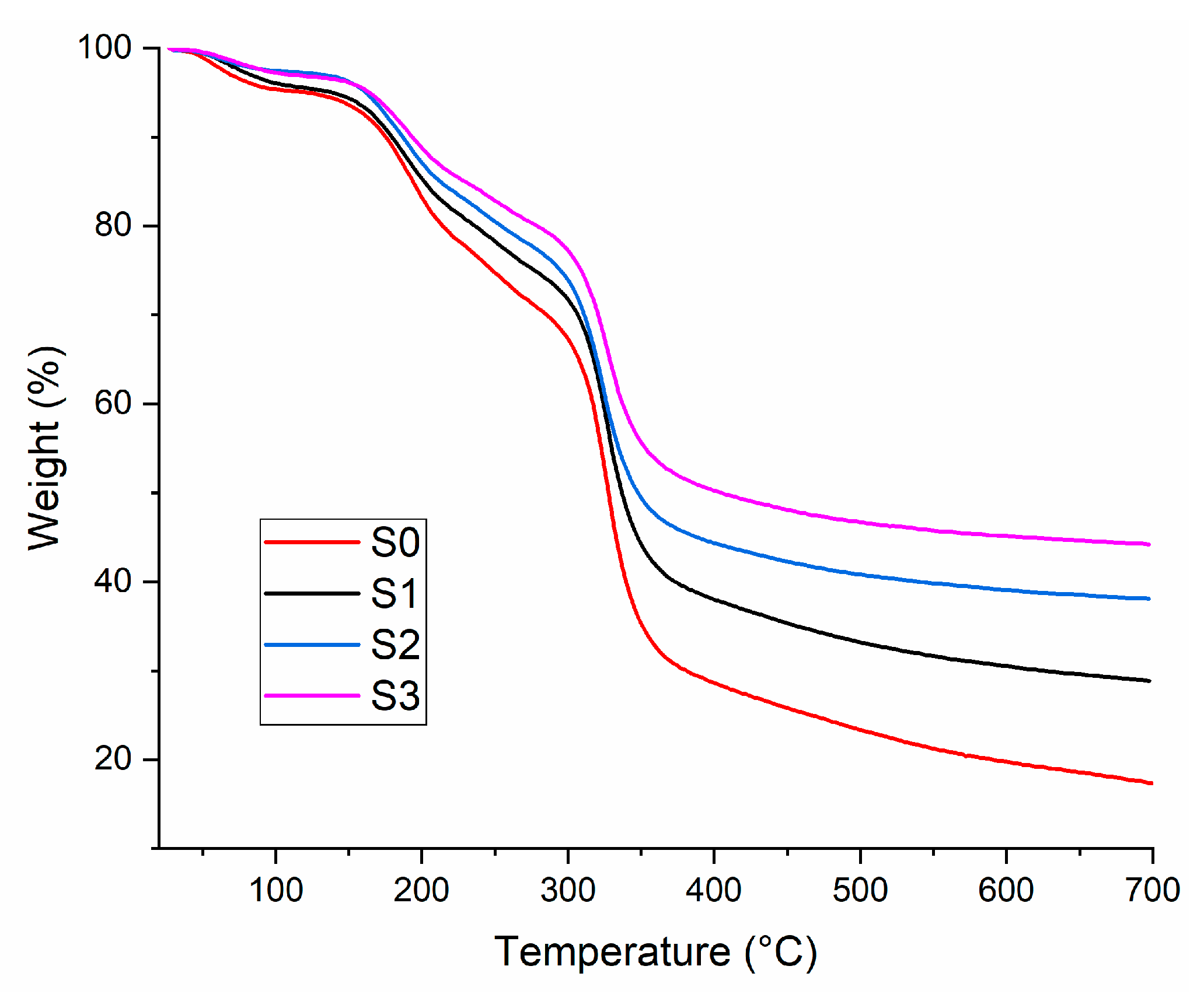
2.4. Thermal Properties
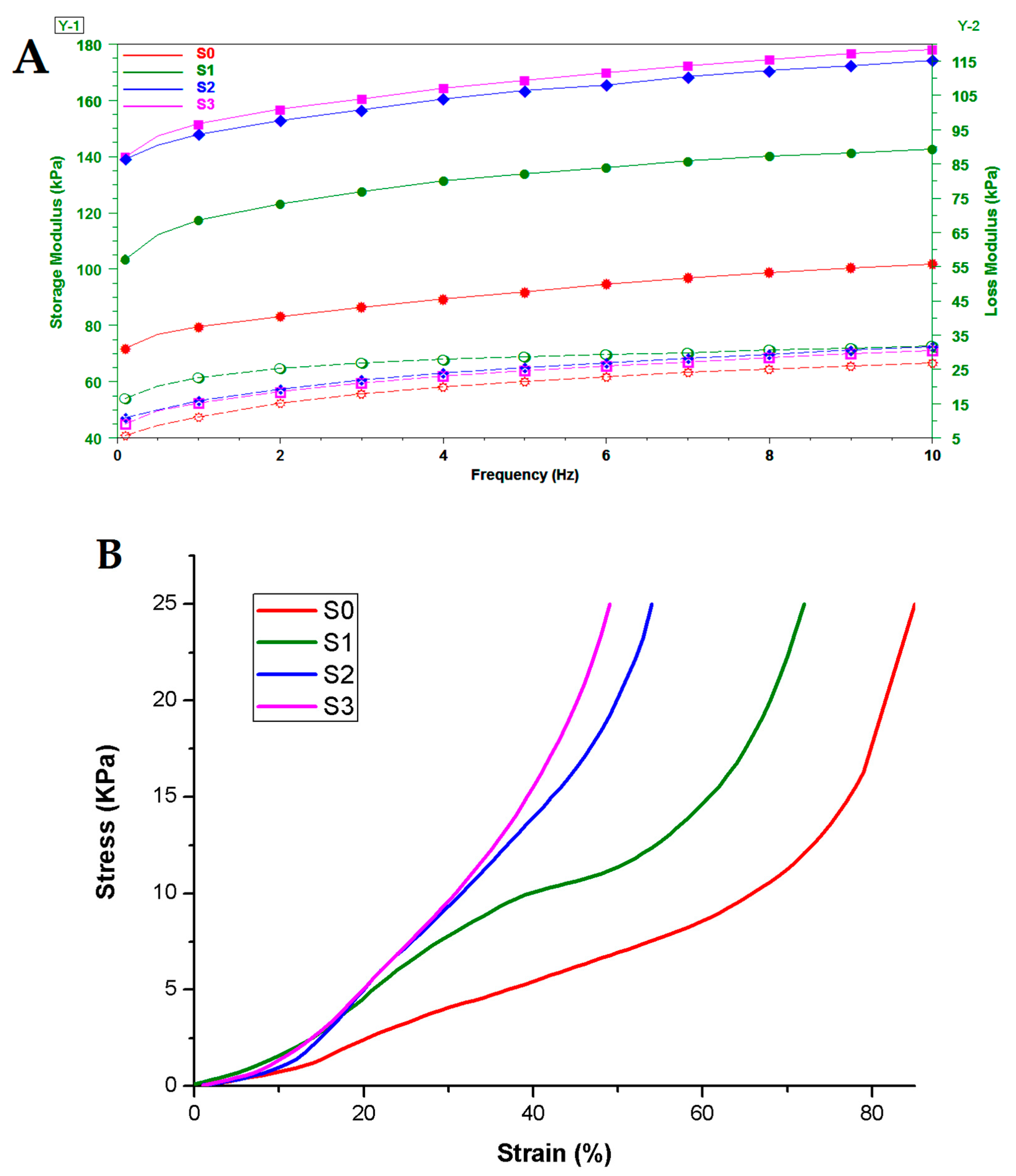
2.5. Mechanical Properties of the Crosslinked Materials in Wet Conditions
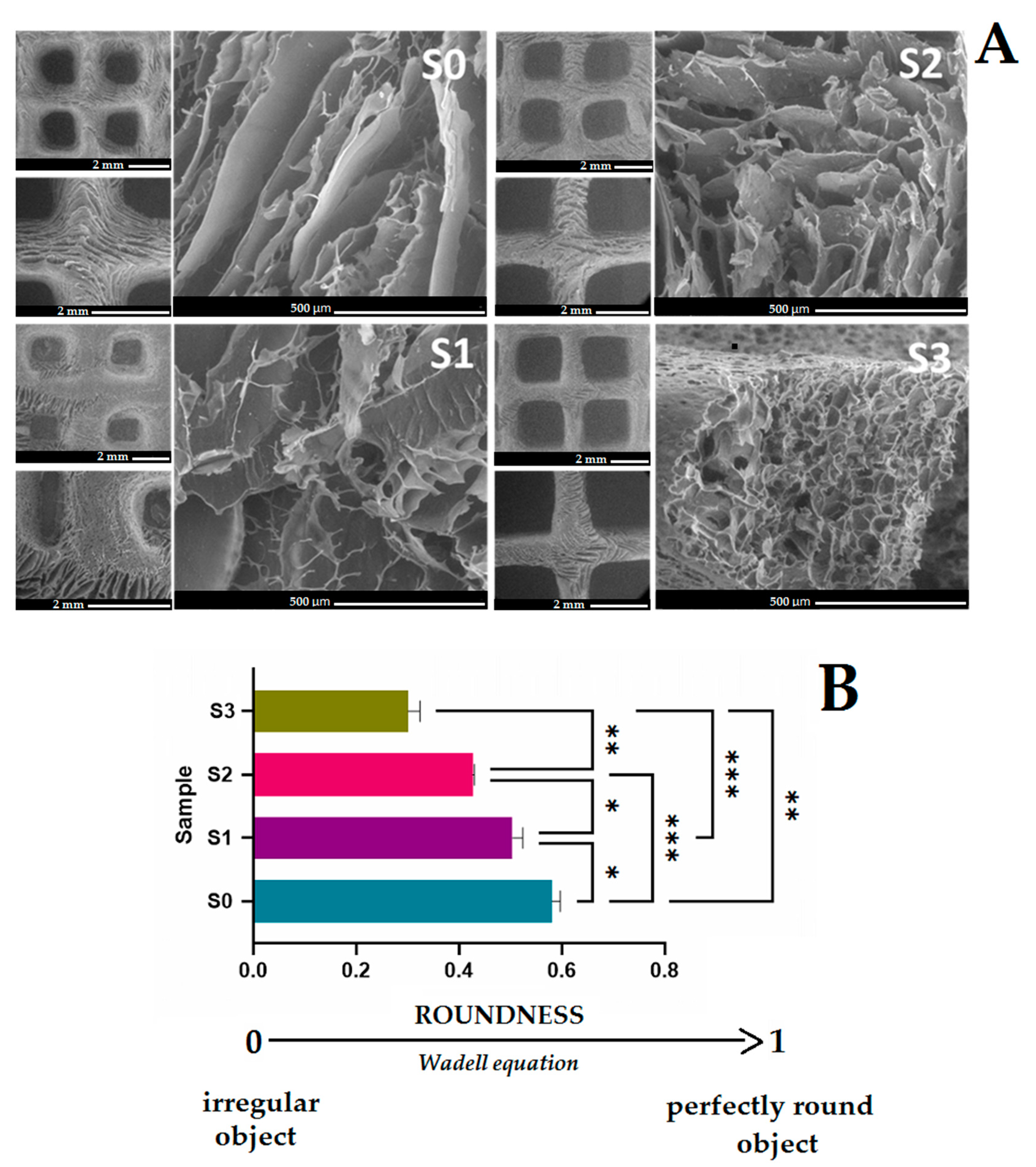
2.6. Morphological Evaluation
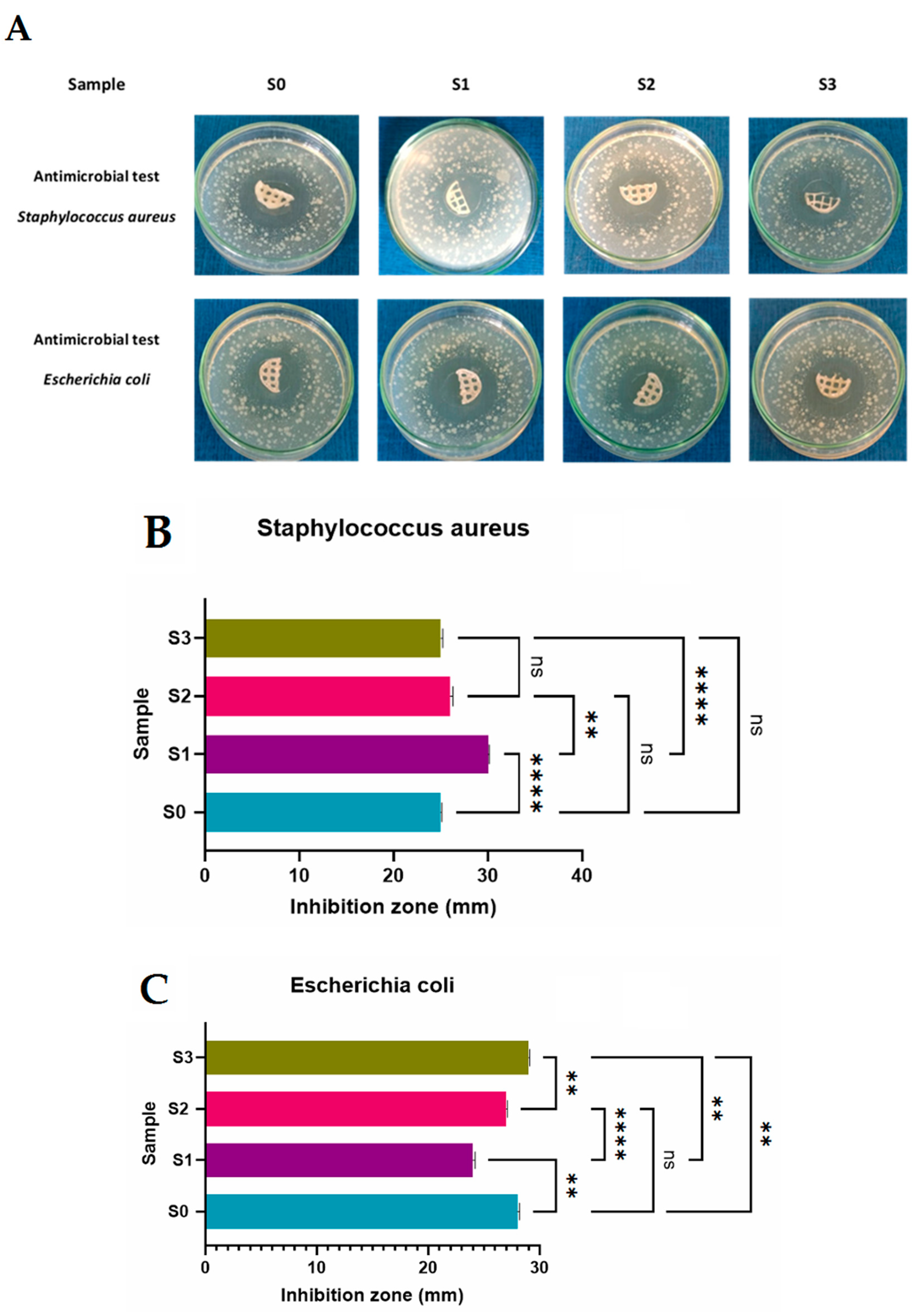
2.7. Antimicrobial Activity
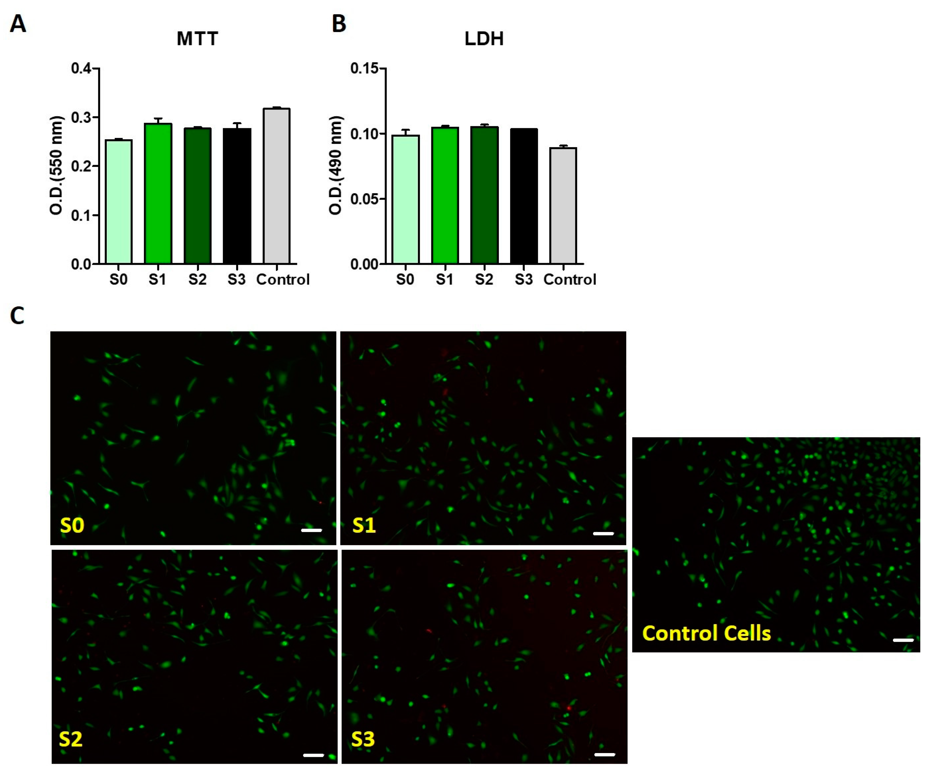
2.8. Biological Assessment of the Crosslinked 3D Printed Polysaccharide Constructs
3. Conclusions
4. Materials and Methods
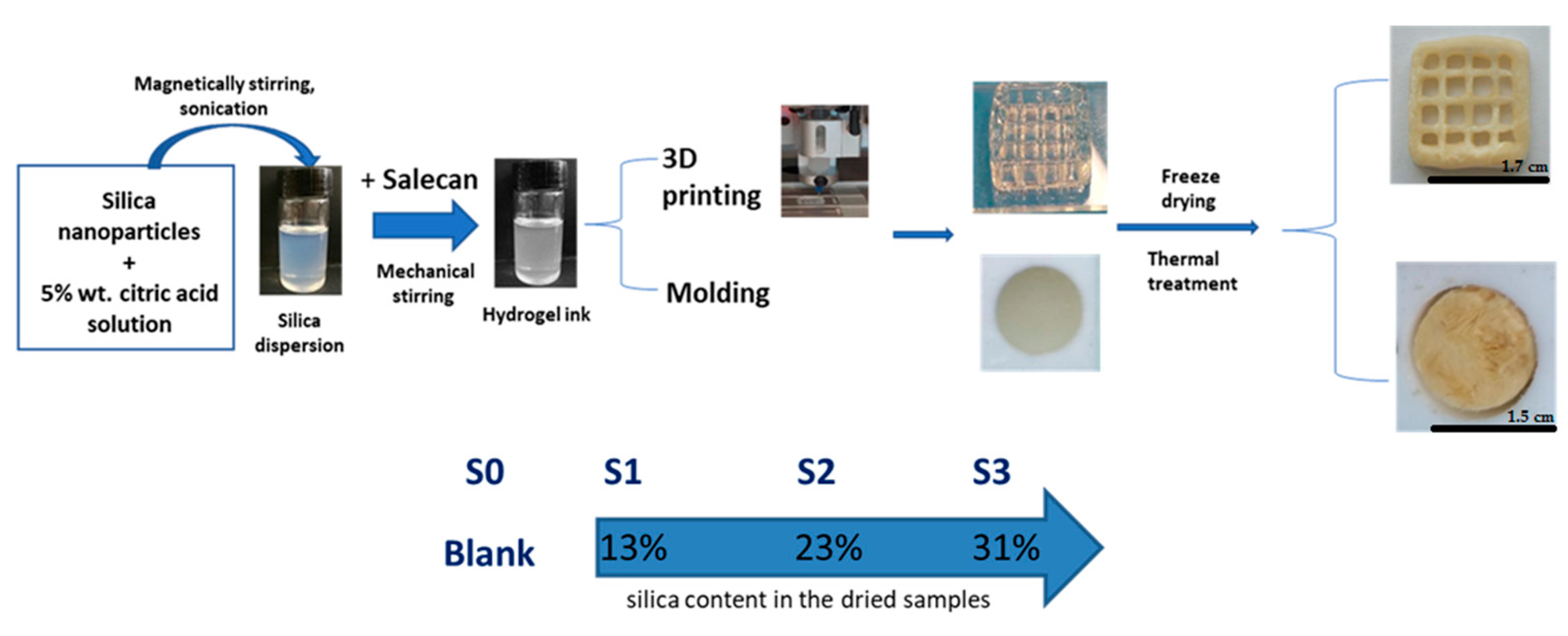
4.1. Materials and Synthesis
4.2. Methods
4.2.1. Rheological Behavior
4.2.2. Fourier Transform Infrared Spectrometry (FT-IR)
4.2.3. Swelling Behavior
4.2.4. Thermo Gravimetric Analysis (TGA)
4.2.5. Scanning Electron Microscopy (SEM)
4.2.6. Transmission Electron Microscopy (TEM)
4.2.7. Mechanical Tests
4.2.8. Antimicrobial Activity
4.2.9. Biological Assessment of the 3D Printed Salecan-Based Hydrogels
4.2.10. Statistical Analyses
Author Contributions
Funding
Institutional Review Board Statement
Informed Consent Statement
Data Availability Statement
Acknowledgments
Conflicts of Interest
References
- Bácskay, I.; Ujhelyi, Z.; Fehér, P.; Arany, P. The Evolution of the 3D-Printed Drug Delivery Systems: A Review. Pharmaceutics 2022, 14, 1312. [Google Scholar] [CrossRef] [PubMed]
- Norman, J.; Madurawe, R.D.; Moore, C.M.V.; Khan, M.A.; Khairuzzaman, A. A New Chapter in Pharmaceutical Manufacturing: 3D-Printed Drug Products. Adv. Drug Deliv. Rev. 2017, 108, 39–50. [Google Scholar] [CrossRef]
- Araújo, M.R.P.; Sa-Barreto, L.L.; Gratieri, T.; Gelfuso, G.M.; Cunha-Filho, M. The Digital Pharmacies Era: How 3D Printing Technology Using Fused Deposition Modeling Can Become a Reality. Pharmaceutics 2019, 11, 128. [Google Scholar] [CrossRef] [PubMed]
- Guarch-Pérez, C.; Shaqour, B.; Riool, M.; Verleije, B.; Beyers, K.; Vervaet, C.; Cos, P.; Zaat, S.A.J. 3D-Printed Gentamicin-Releasing Poly-ε-Caprolactone Composite Prevents Fracture-Related Staphylococcus Aureus Infection in Mice. Pharmaceutics 2022, 14, 1363. [Google Scholar] [CrossRef]
- Zhang, Q.; Zhou, J.; Zhi, P.; Liu, L.; Liu, C.; Fang, A.; Zhang, Q. 3D Printing Method for Bone Tissue Engineering Scaffold. Med. Nov. Technol. Devices 2023, 17, None. [Google Scholar] [CrossRef] [PubMed]
- Vikram Singh, A.; Hasan Dad Ansari, M.; Wang, S.; Laux, P.; Luch, A.; Kumar, A.; Patil, R.; Nussberger, S. The Adoption of Three-Dimensional Additive Manufacturing from Biomedical Material Design to 3D Organ Printing. Appl. Sci. 2019, 9, 811. [Google Scholar] [CrossRef]
- Miao, Y.; Chen, Y.; Luo, J.; Liu, X.; Yang, Q.; Shi, X.; Wang, Y. Black Phosphorus Nanosheets-Enabled DNA Hydrogel Integrating 3D-Printed Scaffold for Promoting Vascularized Bone Regeneration. Bioact. Mater. 2023, 21, 97–109. [Google Scholar] [CrossRef]
- Qi, X.; Hu, X.; Wei, W.; Yu, H.; Li, J.; Zhang, J.; Dong, W. Investigation of Salecan/Poly(Vinyl Alcohol) Hydrogels Prepared by Freeze/Thaw Method. Carbohydr. Polym. 2015, 118, 60–69. [Google Scholar] [CrossRef]
- Ianchis, R.; Ninciuleanu, C.; Gifu, I.C.; Alexandrescu, E.; Nistor, C.L.; Nitu, S.G.; Petcu, C. Hydrogel-Clay Nanocomposites as Carriers for Controlled Release. CMC 2018, 25. [Google Scholar] [CrossRef]
- El-Sherbiny, I.M. Enhanced PH-Responsive Carrier System Based on Alginate and Chemically Modified Carboxymethyl Chitosan for Oral Delivery of Protein Drugs: Preparation and in-Vitro Assessment. Carbohydr. Polym. 2010, 80, 1125–1136. [Google Scholar] [CrossRef]
- El-Sherbiny, I.M.; Yacoub, M.H. Hydrogel Scaffolds for Tissue Engineering: Progress and Challenges. Glob. Cardiol. Sci. Pract. 2013, 2013, 316–342. [Google Scholar] [CrossRef]
- Dragan, E.S.; Apopei, D.F. Multiresponsive Macroporous Semi-IPN Composite Hydrogels Based on Native or Anionically Modified Potato Starch. Carbohydr. Polym. 2013, 92, 23–32. [Google Scholar] [CrossRef]
- Almany, L.; Seliktar, D. Biosynthetic Hydrogel Scaffolds Made from Fibrinogen and Polyethylene Glycol for 3D Cell Cultures. Biomaterials 2005, 26, 2467–2477. [Google Scholar] [CrossRef]
- Dinu, M.V.; Přádný, M.; Drăgan, E.S.; Michálek, J. Morphogical and Swelling Properties of Porous Hydrogels Based on Poly(Hydroxyethyl Methacrylate) and Chitosan Modulated by Ice-Templating Process and Porogen Leaching. J. Polym. Res. 2013, 20, 285. [Google Scholar] [CrossRef]
- Aouada, F.A.; Moura, M.R.d.; Lopes da Silva, W.T.; Muniz, E.C.; Mattoso, L.H.C. Preparation and Characterization of Hydrophilic, Spectroscopic, and Kinetic Properties of Hydrogels Based on Polyacrylamide and Methylcellulose Polysaccharide. J. Appl. Polym. Sci. 2011, 120, 3004–3013. [Google Scholar] [CrossRef]
- Mandal, S.; Nagi, G.K.; Corcoran, A.A.; Agrawal, R.; Dubey, M.; Hunt, R.W. Algal Polysaccharides for 3D Printing: A Review. Carbohydr. Polym. 2023, 300, 120267. [Google Scholar] [CrossRef]
- Hu, X.; Wang, Y.; Zhang, L.; Xu, M.; Zhang, J.; Dong, W. Design of a PH-Sensitive Magnetic Composite Hydrogel Based on Salecan Graft Copolymer and Fe3O4@SiO2 Nanoparticles as Drug Carrier. Int. J. Biol. Macromol. 2018, 107, 1811–1820. [Google Scholar] [CrossRef] [PubMed]
- Li, F.; Wu, H.; Fan, L.; Zhang, H.; Zhang, H.; Gu, C. Study of Dual Responsive Poly[(Maleilated Dextran)-Graft-(N-Isopropylacrylamide)] Hydrogel Nanoparticles: Preparation, Characterization and Biological Evaluation. Polym. Int. 2009, 58, 1023–1033. [Google Scholar] [CrossRef]
- Thambi, T.; Phan, V.H.G.; Lee, D.S. Stimuli-Sensitive Injectable Hydrogels Based on Polysaccharides and Their Biomedical Applications-Thambi-2016-Macromolecular Rapid Communications-Wiley Online Library. Available online: https://onlinelibrary.wiley.com/doi/full/10.1002/marc.201600371 (accessed on 4 April 2023).
- Aderibigbe, B.; Buyana, B. Alginate in Wound Dressings. Pharmaceutics 2018, 10, 42. [Google Scholar] [CrossRef] [PubMed]
- Beltran-Vargas, N.E.; Peña-Mercado, E.; Sánchez-Gómez, C.; Garcia-Lorenzana, M.; Ruiz, J.-C.; Arroyo-Maya, I.; Huerta-Yepez, S.; Campos-Terán, J. Sodium Alginate/Chitosan Scaffolds for Cardiac Tissue Engineering: The Influence of Its Three-Dimensional Material Preparation and the Use of Gold Nanoparticles. Polymers 2022, 14, 3233. [Google Scholar] [CrossRef]
- Dai, L.; Cheng, T.; Duan, C.; Zhao, W.; Zhang, W.; Zou, X.; Aspler, J.; Ni, Y. 3D Printing Using Plant-Derived Cellulose and Its Derivatives: A Review. Carbohydr. Polym. 2019, 203, 71–86. [Google Scholar] [CrossRef] [PubMed]
- Tavakoli, S.; Kharaziha, M.; Kermanpur, A.; Mokhtari, H. Sprayable and Injectable Visible-Light Kappa-Carrageenan Hydrogel for In-Situ Soft Tissue Engineering. Int. J. Biol. Macromol. 2019, 138, 590–601. [Google Scholar] [CrossRef]
- Mohammed, A.S.A.; Naveed, M.; Jost, N. Polysaccharides; Classification, Chemical Properties, and Future Perspective Applications in Fields of Pharmacology and Biological Medicine (A Review of Current Applications and Upcoming Potentialities). J. Polym. Environ. 2021, 29, 2359–2371. [Google Scholar] [CrossRef]
- Ianchis, R.; Alexa, R.L.; Gifu, I.C.; Marin, M.M.; Alexandrescu, E.; Constantinescu, R.; Serafim, A.; Nistor, C.L.; Petcu, C. Novel Green Crosslinked Salecan Hydrogels and Preliminary Investigation of Their Use in 3D Printing. Pharmaceutics 2023, 15, 373. [Google Scholar] [CrossRef]
- Qi, X.; Wei, W.; Shen, J.; Dong, W. Salecan Polysaccharide-Based Hydrogels and Their Applications: A Review. J. Mater. Chem. B 2019, 7, 2577–2587. [Google Scholar] [CrossRef] [PubMed]
- Fan, Z.; Cheng, P.; Gao, Y.; Wang, D.; Jia, G.; Zhang, P.; Prakash, S.; Wang, Z.; Han, J. Understanding the Rheological Properties of a Novel Composite Salecan/Gellan Hydrogels. Food Hydrocoll. 2022, 123, 107162. [Google Scholar] [CrossRef]
- Fan, Z.; Cheng, P.; Prakash, S.; Zhang, P.; Mei, L.; Ji, S.; Wang, Z.; Han, J. Rheological Investigation of a Versatile Salecan/Curdlan Gel Matrix. Int. J. Biol. Macromol. 2021, 193, 2202–2209. [Google Scholar] [CrossRef] [PubMed]
- Fan, Z.; Cheng, P.; Chu, L.; Han, J. Exploring the Rheological and Structural Characteristics of Novel Pectin-Salecan Gels. Polymers 2022, 14, 4619. [Google Scholar] [CrossRef] [PubMed]
- Qi, X.; Su, T.; Tong, X.; Xiong, W.; Zeng, Q.; Qian, Y.; Zhou, Z.; Wu, X.; Li, Z.; Shen, L.; et al. Facile Formation of Salecan/Agarose Hydrogels with Tunable Structural Properties for Cell Culture. Carbohydr. Polym. 2019, 224, 115208. [Google Scholar] [CrossRef]
- Fan, Z.; Cheng, P.; Yin, G.; Wang, Z.; Han, J. In Situ Forming Oxidized Salecan/Gelatin Injectable Hydrogels for Vancomycin Delivery and 3D Cell Culture. J. Biomater. Sci. Polym. Ed. 2020, 31, 762–780. [Google Scholar] [CrossRef]
- Florian, P.E.; Icriverzi, M.; Ninciuleanu, C.M.; Alexandrescu, E.; Trica, B.; Preda, S.; Ianchis, R.; Roseanu, A. Salecan-Clay Based Polymer Nanocomposites for Chemotherapeutic Drug Delivery Systems; Characterization and In Vitro Biocompatibility Studies. Materials 2020, 13, 5389. [Google Scholar] [CrossRef] [PubMed]
- Ip, H.T.; Liu, L.; Hong, L.; Ngai, T. Synthesis of Polystyrene/Silica and Poly(Styrene-Co-Butyl Acrylate)/Silica Nanocomposite Particles by Pickering Emulsion Polymerization with Non-Functionalized Silica Nanoparticles. Colloids Surf. A Physicochem. Eng. Asp. 2022, 654, 130104. [Google Scholar] [CrossRef]
- Zhao, N.; Yan, L.; Zhao, X.; Chen, X.; Li, A.; Zheng, D.; Zhou, X.; Dai, X.; Xu, F.-J. Versatile Types of Organic/Inorganic Nanohybrids: From Strategic Design to Biomedical Applications. Chem. Rev. 2019, 119, 1666–1762. [Google Scholar] [CrossRef]
- Marin, M.M.; Ianchis, R.; Leu Alexa, R.; Gifu, I.C.; Kaya, M.G.A.; Savu, D.I.; Popescu, R.C.; Alexandrescu, E.; Ninciuleanu, C.M.; Preda, S.; et al. Development of New Collagen/Clay Composite Biomaterials. Int. J. Mol. Sci. 2022, 23, 401. [Google Scholar] [CrossRef] [PubMed]
- Huang, Y.; Li, P.; Zhao, R.; Zhao, L.; Liu, J.; Peng, S.; Fu, X.; Wang, X.; Luo, R.; Wang, R.; et al. Silica Nanoparticles: Biomedical Applications and Toxicity. Biomed. Pharmacother. 2022, 151, 113053. [Google Scholar] [CrossRef]
- Diab, R.; Canilho, N.; Pavel, I.A.; Haffner, F.B.; Girardon, M.; Pasc, A. Silica-Based Systems for Oral Delivery of Drugs, Macromolecules and Cells. Adv. Colloid Interface Sci. 2017, 249, 346–362. [Google Scholar] [CrossRef]
- Choi, Y.; Kim, J.; Yu, S.; Hong, S. PH- and Temperature-Responsive Radially Porous Silica Nanoparticles with High-Capacity Drug Loading for Controlled Drug Delivery. Nanotechnology 2020, 31, 335103. [Google Scholar] [CrossRef]
- Kiran Roopavath, U.; Soni, R.; Mahanta, U.; Suresh Deshpande, A.; Narayan Rath, S. 3D Printable SiO2 Nanoparticle Ink for Patient Specific Bone Regeneration. RSC Adv. 2019, 9, 23832–23842. [Google Scholar] [CrossRef]
- Florek, J.; Caillard, R.; Kleitz, F. Evaluation of Mesoporous Silica Nanoparticles for Oral Drug Delivery–Current Status and Perspective of MSNs Drug Carriers. Nanoscale 2017, 9, 15252–15277. [Google Scholar] [CrossRef]
- Hsu, S.-Y.; Morris, R.; Cheng, F. Signaling Pathways Regulated by Silica Nanoparticles. Molecules 2021, 26, 1398. [Google Scholar] [CrossRef]
- Shao, H.; Zhang, H.; Tian, Y.; Song, Z.; Lai, P.; Ai, L. Composition and Rheological Properties of Polysaccharide Extracted from Tamarind (Tamarindus indica L.) Seed. Molecules 2019, 24, 1218. [Google Scholar] [CrossRef] [PubMed]
- Unagolla, J.M.; Jayasuriya, A.C. Hydrogel-Based 3D Bioprinting: A Comprehensive Review on Cell-Laden Hydrogels, Bioink Formulations, and Future Perspectives. Appl. Mater. Today 2020, 18, 100479. [Google Scholar] [CrossRef] [PubMed]
- Gerezgiher, A.G.; Szabó, T. Crosslinking of Starch Using Citric Acid. J. Phys. Conf. Ser. 2022, 2315, 012036. [Google Scholar] [CrossRef]
- Caprarescu, S.; Radu, A.-L.; Purcar, V.; Sarbu, A.; Vaireanu, D.-I.; Ianchis, R.; Ghiurea, M. Removal of Copper Ions from Simulated Wastewaters Using Different Bicomponent Polymer Membranes. Water Air Soil Pollut 2014, 225, 2079. [Google Scholar] [CrossRef]
- Ninciuleanu, C.M.; Ianchiş, R.; Alexandrescu, E.; Mihăescu, C.I.; Scomoroşcenco, C.; Nistor, C.L.; Preda, S.; Petcu, C.; Teodorescu, M. The Effects of Monomer, Crosslinking Agent, and Filler Concentrations on the Viscoelastic and Swelling Properties of Poly(Methacrylic Acid) Hydrogels: A Comparison. Materials 2021, 14, 2305. [Google Scholar] [CrossRef]
- Ninciuleanu, C.M.; Ianchiș, R.; Alexandrescu, E.; Mihăescu, C.I.; Burlacu, S.; Trică, B.; Nistor, C.L.; Preda, S.; Scomoroscenco, C.; Gîfu, C.; et al. Adjusting Some Properties of Poly(Methacrylic Acid) (Nano)Composite Hydrogels by Means of Silicon-Containing Inorganic Fillers. Int. J. Mol. Sci. 2022, 23, 10320. [Google Scholar] [CrossRef]
- Asabuwa Ngwabebhoh, F.; Saha, N.; Nguyen, H.T.; Brodnjak, U.V.; Saha, T.; Lengalova, A.; Saha, P. Preparation and Characterization of Nonwoven Fibrous Biocomposites for Footwear Components. Polymers 2020, 12, 3016. [Google Scholar] [CrossRef]
- Boonmahitthisud, A.; Nakajima, L.; Nguyen, K.D.; Kobayashi, T. Composite Effect of Silica Nanoparticle on the Mechanical Properties of Cellulose-Based Hydrogels Derived from Cottonseed Hulls. J. Appl. Polym. Sci. 2017, 134. [Google Scholar] [CrossRef]
- Li, X.; Rombouts, W.; Van Der Gucht, J.; De Vries, R.; Dijksman, J.A. Mechanics of Composite Hydrogels Approaching Phase Separation. PLoS ONE 2019, 14, e0211059. [Google Scholar] [CrossRef] [PubMed]
- Xing, W.; Tang, Y. On Mechanical Properties of Nanocomposite Hydrogels: Searching for Superior Properties. Nano Mater. Sci. 2022, 4, 83–96. [Google Scholar] [CrossRef]
- Liu, J.; Zheng, H.; Poh, P.S.P.; Machens, H.-G.; Schilling, A.F. Hydrogels for Engineering of Perfusable Vascular Networks. Int. J. Mol. Sci. 2015, 16, 15997–16016. [Google Scholar] [CrossRef] [PubMed]
- Advanced BioMatrix-Stiffness Range of Living Tissue. Available online: https://advancedbiomatrix.com/stiffness-range-of-living-tissue.html (accessed on 4 April 2023).
- Low, Z.W.; Chee, P.L.; Kai, D.; Loh, X.J. The Role of Hydrogen Bonding in Alginate/Poly(Acrylamide-Co-Dimethylacrylamide) and Alginate/Poly(Ethylene Glycol) Methyl Ether Methacrylate-Based Tough Hybrid Hydrogels. RSC Adv. 2015, 5, 57678–57685. [Google Scholar] [CrossRef]
- Suflet, D.M.; Popescu, I.; Pelin, I.M.; Ichim, D.L.; Daraba, O.M.; Constantin, M.; Fundueanu, G. Dual Cross-Linked Chitosan/PVA Hydrogels Containing Silver Nanoparticles with Antimicrobial Properties. Pharmaceutics 2021, 13, 1461. [Google Scholar] [CrossRef]
- Hocken, A.; Beyer, F.L.; Lee, J.S.; Grim, B.J.; Mithaiwala, H.; Green, M.D. Covalently Integrated Silica Nanoparticles in Poly(Ethylene Glycol)-Based Acrylate Resins: Thermomechanical, Swelling, and Morphological Behavior. Soft Matter 2022, 18, 1019–1033. [Google Scholar] [CrossRef] [PubMed]
- Kato, K.; Matsui, D.; Mayumi, K.; Ito, K. Synthesis, Structure, and Mechanical Properties of Silica Nanocomposite Polyrotaxane Gels. Beilstein J. Org. Chem. 2015, 11, 2194–2201. [Google Scholar] [CrossRef]
- Ahn, S.; Kim, S.; Kim, B.C.; Shim, K.; Cho, B. Mechanical Properties of Silica Nanoparticle Reinforced Poly(Ethylene 2, 6-Naphthalate). Macromol. Res. 2004, 12, 293–302. [Google Scholar] [CrossRef]
- Loh, Q.L.; Choong, C. Three-Dimensional Scaffolds for Tissue Engineering Applications: Role of Porosity and Pore Size. Tissue Eng Part B Rev 2013, 19, 485–502. [Google Scholar] [CrossRef]
- Liu, W.; Li, Y.; Feng, S.; Ning, J.; Wang, J.; Gou, M.; Chen, H.; Xu, F.; Du, Y. Magnetically Controllable 3D Microtissues Based on Magnetic Microcryogels. Lab Chip 2014, 14, 2614–2625. [Google Scholar] [CrossRef]
- Wadell, H. Volume, Shape, and Roundness of Rock Particles. J. Geol. 1932, 40, 443–451. [Google Scholar] [CrossRef]
- Marin, M.M.; Albu Kaya, M.; Kaya, D.A.; Constantinescu, R.; Trica, B.; Gifu, I.C.; Alexandrescu, E.; Nistor, C.L.; Alexa, R.L.; Ianchis, R. Novel Nanocomposite Hydrogels Based on Crosslinked Microbial Polysaccharide as Potential Bioactive Wound Dressings. Materials 2023, 16, 982. [Google Scholar] [CrossRef]
- Botet-Carreras, A.; Marimon, M.B.; Millan-Solsona, R.; Aubets, E.; Ciudad, C.J.; Noé, V.; Montero, M.T.; Domènech, Ò.; Borrell, J.H. On the Uptake of Cationic Liposomes by Cells: From Changes in Elasticity to Internalization. Colloids Surf. B Biointerfaces 2023, 221, 112968. [Google Scholar] [CrossRef]
- Su, L.-C.; Xie, Z.; Zhang, Y.; Nguyen, K.T.; Yang, J. Study on the Antimicrobial Properties of Citrate-Based Biodegradable Polymers. Front. Bioeng. Biotechnol. 2014, 2, 23. [Google Scholar] [CrossRef] [PubMed]
- Wang, M.; Xu, P.; Lei, B. Engineering Multifunctional Bioactive Citrate-Based Biomaterials for Tissue Engineering. Bioact. Mater. 2023, 19, 511–537. [Google Scholar] [CrossRef]
- Xi, Y.; Guo, Y.; Wang, M.; Ge, J.; Liu, Y.; Niu, W.; Chen, M.; Xue, Y.; Winston, D.D.; Dai, W.; et al. Biomimetic Bioactive Multifunctional Poly(Citrate-Siloxane)-Based Nanofibrous Scaffolds Enable Efficient Multidrug-Resistant Bacterial Treatment/Non-Invasive Tracking In Vitro/In Vivo. Chem. Eng. J. 2020, 383, 123078. [Google Scholar] [CrossRef]
- Qi, X.; Su, T.; Zhang, M.; Tong, X.; Pan, W.; Zeng, Q.; Zhou, Z.; Shen, L.; He, X.; Shen, J. Macroporous Hydrogel Scaffolds with Tunable Physicochemical Properties for Tissue Engineering Constructed Using Renewable Polysaccharides. ACS Appl. Mater. Interfaces 2020, 12, 13256–13264. [Google Scholar] [CrossRef] [PubMed]
- Fan, Z.; Cheng, P.; Ling, L.; Han, J. Dynamic Bond Crosslinked Poly(γ-Glutamic Acid)/Salecan Derived Hydrogel as a Platform for 3D Cell Culture. Mater. Lett. 2020, 273, 127936. [Google Scholar] [CrossRef]
- Cruz-Matías, I.; Ayala, D.; Hiller, D.; Gutsch, S.; Zacharias, M.; Estradé, S.; Peiró, F. Sphericity and Roundness Computation for Particles Using the Extreme Vertices Model. J. Comput. Sci. 2019, 30, 28–40. [Google Scholar] [CrossRef]
- Marin, M.M.; Albu Kaya, M.G.; Iovu, H.; Stavarache, C.E.; Chelaru, C.; Constantinescu, R.R.; Dinu-Pîrvu, C.-E.; Ghica, M.V. Obtaining, Evaluation, and Optimization of Doxycycline-Loaded Microparticles Intended for the Local Treatment of Infectious Arthritis. Coatings 2020, 10, 990. [Google Scholar] [CrossRef]


| Samples | Step 1 | Step 2 | Step 3 | |||
|---|---|---|---|---|---|---|
| T, °C | Weight Loss, % | T, °C | Weight Loss, % | T, °C | Weight Loss, % | |
| S0 | 80–200 | 16.81 | 200–320 | 57.49 | 320–700 | 39.86 |
| S1 | 11.95 | 22.07 | 34.35 | |||
| S2 | 10.84 | 22.31 | 26.72 | |||
| S3 | 9.29 | 18.48 | 26.10 | |||
| Sample | Elastic Modulus (KPa) | ||||||||
|---|---|---|---|---|---|---|---|---|---|
| 1–6 KPa | STDV | R2 | 20–25 KPa | STDV | R2 | 25–35 KPa | STDV | R2 | |
| S0 | 6.57 | ±0.66 | 0.9906 | 12.74 | ±0.44 | 0.9990 | 13.46 | ±0.10 | 0.9971 |
| S1 | 15.35 | ±2.10 | 0.9979 | 24.37 | ±0.98 | 0.9988 | 25.90 | ±0.18 | 0.9977 |
| S2 | 7.32 | ±1.29 | 0.9913 | 27.38 | ±1.45 | 0.9998 | 31.09 | ±1.36 | 0.9993 |
| S3 | 9.42 | ±2.36 | 0.9988 | 27.47 | ±1.50 | 0.9977 | 32.14 | ±1.88 | 0.9984 |
| Sample | Salecan [g] | Citric Acid Solution 5% [mL] | Silica Nanoparticles [g] |
|---|---|---|---|
| S0 | 1.125 | 15 | - |
| S1 | 1.125 | 15 | 0.281 |
| S2 | 1.125 | 15 | 0.562 |
| S3 | 1.125 | 15 | 0.843 |
Disclaimer/Publisher’s Note: The statements, opinions and data contained in all publications are solely those of the individual author(s) and contributor(s) and not of MDPI and/or the editor(s). MDPI and/or the editor(s) disclaim responsibility for any injury to people or property resulting from any ideas, methods, instructions or products referred to in the content. |
© 2023 by the authors. Licensee MDPI, Basel, Switzerland. This article is an open access article distributed under the terms and conditions of the Creative Commons Attribution (CC BY) license (https://creativecommons.org/licenses/by/4.0/).
Share and Cite
Marin, M.M.; Gifu, I.C.; Pircalabioru, G.G.; Albu Kaya, M.; Constantinescu, R.R.; Alexa, R.L.; Trica, B.; Alexandrescu, E.; Nistor, C.L.; Petcu, C.; et al. Microbial Polysaccharide-Based Formulation with Silica Nanoparticles; A New Hydrogel Nanocomposite for 3D Printing. Gels 2023, 9, 425. https://doi.org/10.3390/gels9050425
Marin MM, Gifu IC, Pircalabioru GG, Albu Kaya M, Constantinescu RR, Alexa RL, Trica B, Alexandrescu E, Nistor CL, Petcu C, et al. Microbial Polysaccharide-Based Formulation with Silica Nanoparticles; A New Hydrogel Nanocomposite for 3D Printing. Gels. 2023; 9(5):425. https://doi.org/10.3390/gels9050425
Chicago/Turabian StyleMarin, Maria Minodora, Ioana Catalina Gifu, Gratiela Gradisteanu Pircalabioru, Madalina Albu Kaya, Rodica Roxana Constantinescu, Rebeca Leu Alexa, Bogdan Trica, Elvira Alexandrescu, Cristina Lavinia Nistor, Cristian Petcu, and et al. 2023. "Microbial Polysaccharide-Based Formulation with Silica Nanoparticles; A New Hydrogel Nanocomposite for 3D Printing" Gels 9, no. 5: 425. https://doi.org/10.3390/gels9050425