
Bioinspired Sandcastle Worm-Derived Peptide-Based Hybrid Hydrogel for Promoting the Formation of Liver Spheroids

 , and
, and 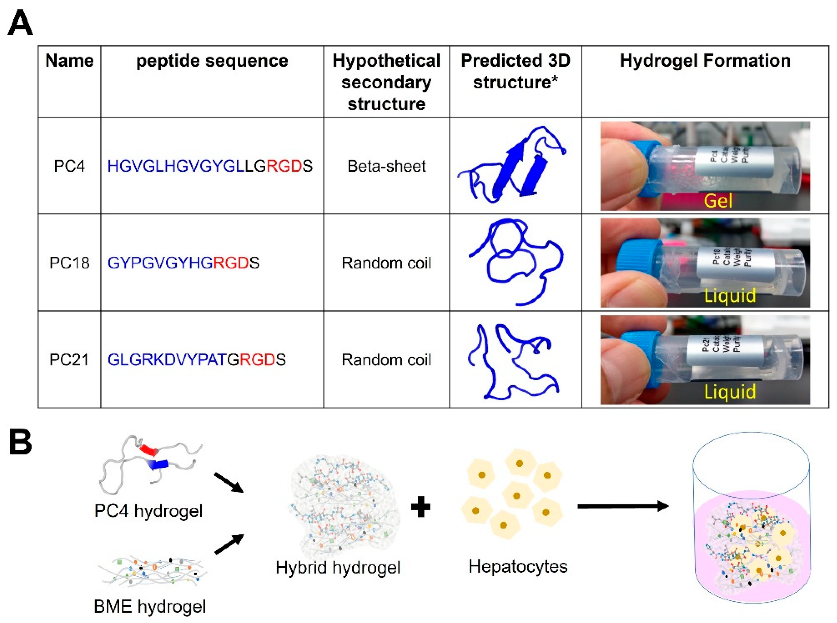
Abstract
:1. Introduction
2. Results and Discussion
2.1. Screening of Self-Assembling Peptides Inspired by Sandcastle Worm Adhesion Proteins
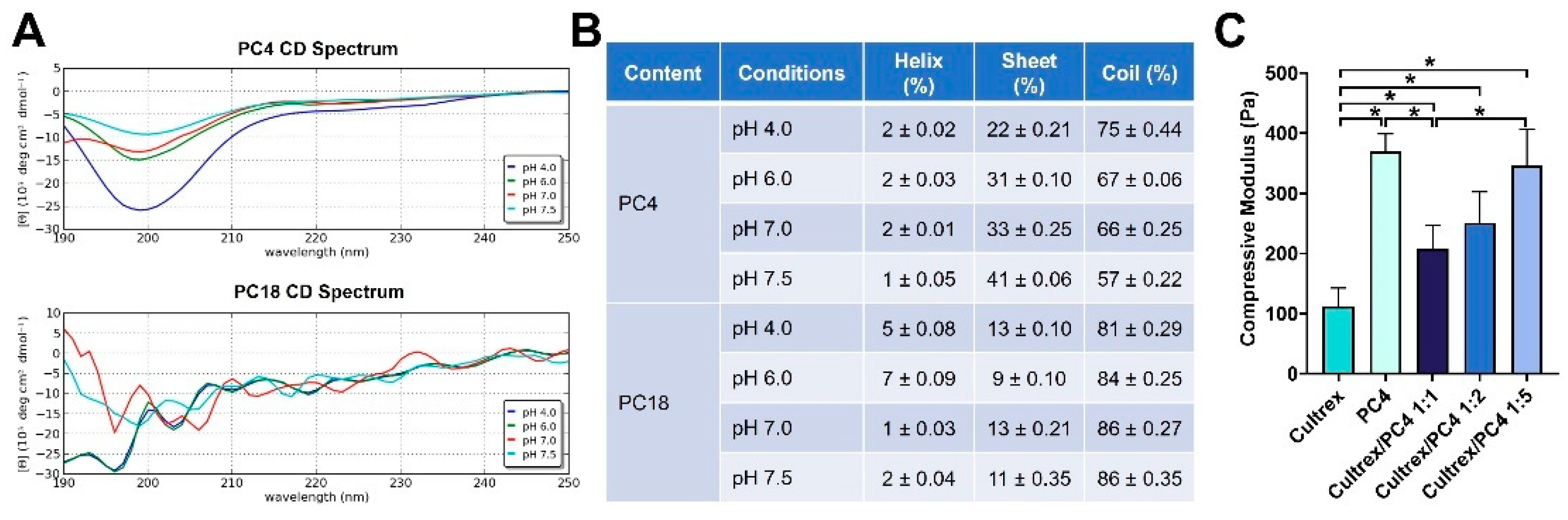
2.2. Mechanical Strength Evaluation of Self-Assembling Peptides
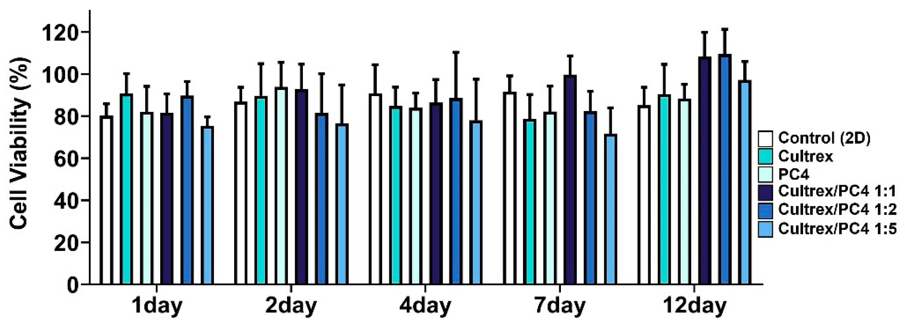
2.3. PC4/Cultrex Hybrid Hydrogels Are Biocompatible
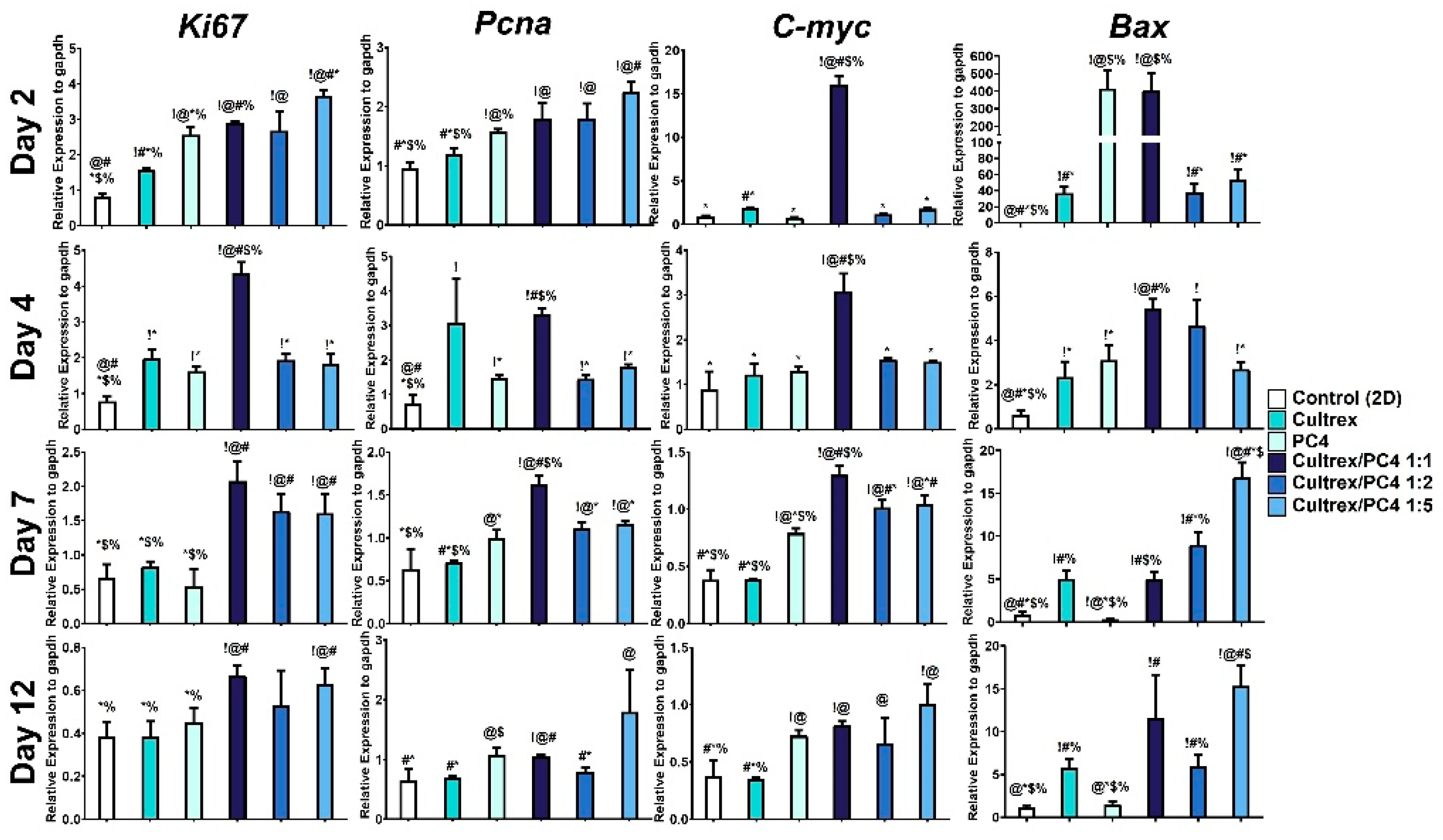
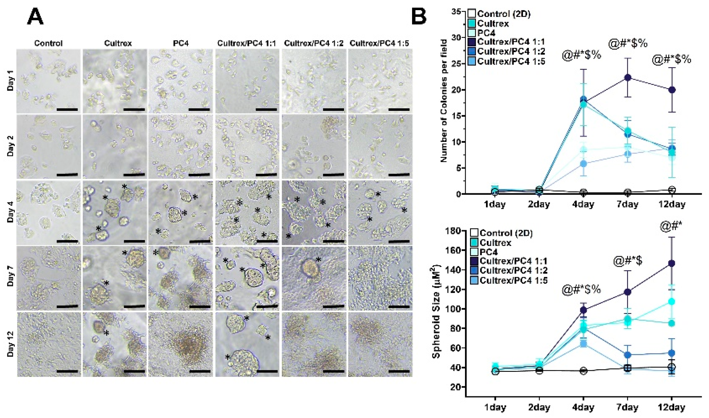
2.4. PC4/Cultrex Hybrid Hydrogels Significantly Promote Hepatic Spheroid Formation
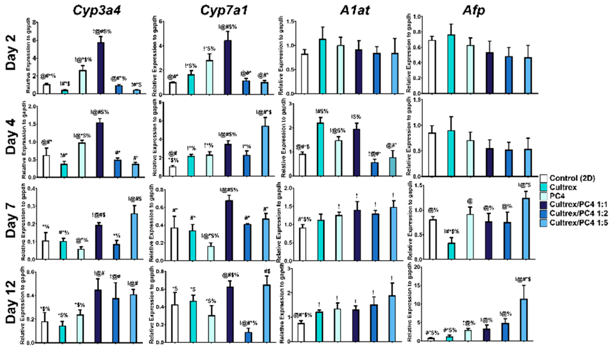
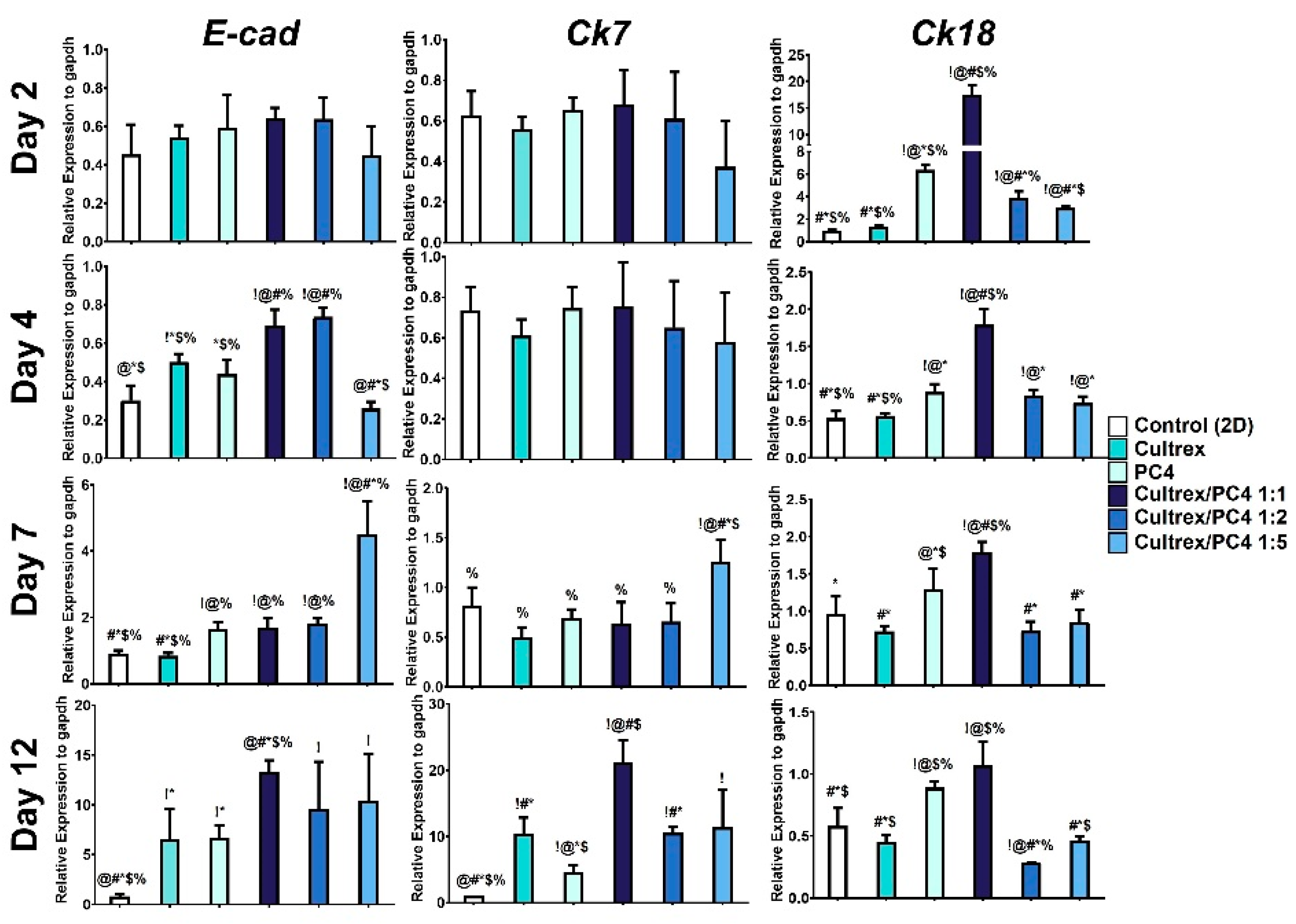
2.5. PC4/Cultrex Hybrid Hydrogels Significantly Enhanced the Liver-like Functionality of Hepatic Spheroids
3. Conclusions
4. Materials and Methods
4.1. Peptide Design
4.2. Hydrogel Preparation
4.3. Mechanical Characterisation of Hydrogels
4.4. Circular Dichroism Spectroscopy (CD)
4.5. In Vitro Cell Culture
4.6. MTT Assay
4.7. Spheroid Size Evaluation
4.8. Quantitative Real-Time Polymerase Chain Reaction
4.9. Statistics
Supplementary Materials
Author Contributions
Funding
Institutional Review Board Statement
Informed Consent Statement
Data Availability Statement
Conflicts of Interest
References
- Godoy, P.; Hewitt, N.J.; Albrecht, U.; Andersen, M.E.; Ansari, N.; Bhattacharya, S.; Bode, J.G.; Bolleyn, J.; Borner, C.; Boettger, J. Recent Advances in 2D and 3D in Vitro Systems Using Primary Hepatocytes, Alternative Hepatocyte Sources and Non-Parenchymal Liver Cells and Their Use in Investigating Mechanisms of Hepatotoxicity, Cell Signaling and ADME. Arch. Toxicol. 2013, 87, 1315–1530. [Google Scholar] [CrossRef] [Green Version]
- Calitz, C.; Hamman, J.H.; Fey, S.J.; Wrzesinski, K.; Gouws, C. Recent Advances in Three-Dimensional Cell Culturing to Assess Liver Function and Dysfunction: From a Drug Biotransformation and Toxicity Perspective. Toxicol. Mech. Methods 2018, 28, 369–385. [Google Scholar] [CrossRef]
- Griffith, L.G.; Swartz, M.A. Capturing Complex 3D Tissue Physiology in Vitro. Nat. Rev. Mol. Cell Biol. 2006, 7, 211–224. [Google Scholar] [CrossRef]
- Ijima, H.; Nakamura, S.; Bual, R.P.; Yoshida, K. Liver-Specific Extracellular Matrix Hydrogel Promotes Liver-Specific Functions of Hepatocytes in Vitro and Survival of Transplanted Hepatocytes in Vivo. J. Biosci. Bioeng. 2019, 128, 365–372. [Google Scholar] [CrossRef]
- Saheli, M.; Sepantafar, M.; Pournasr, B.; Farzaneh, Z.; Vosough, M.; Piryaei, A.; Baharvand, H. Three-dimensional Liver-derived Extracellular Matrix Hydrogel Promotes Liver Organoids Function. J. Cell. Biochem. 2018, 119, 4320–4333. [Google Scholar] [CrossRef]
- Hu, H.; Gehart, H.; Artegiani, B.; LÖpez-Iglesias, C.; Dekkers, F.; Basak, O.; van Es, J.; de Sousa Lopes, S.M.C.; Begthel, H.; Korving, J. Long-Term Expansion of Functional Mouse and Human Hepatocytes as 3D Organoids. Cell 2018, 175, 1591–1606. [Google Scholar] [CrossRef] [Green Version]
- Rose, S.; Ezan, F.; Cuvellier, M.; Bruyère, A.; Legagneux, V.; Langouët, S.; Baffet, G. Generation of Proliferating Human Adult Hepatocytes Using Optimized 3D Culture Conditions. Sci. Rep. 2021, 11, 515. [Google Scholar] [CrossRef]
- Garnier, D.; Li, R.; Delbos, F.; Fourrier, A.; Collet, C.; Guguen-Guillouzo, C.; Chesné, C.; Nguyen, T.H. Expansion of Human Primary Hepatocytes in Vitro through Their Amplification as Liver Progenitors in a 3D Organoid System. Sci. Rep. 2018, 8, 8222. [Google Scholar] [CrossRef]
- Kleinman, H.K.; Martin, G.R. Matrigel: Basement Membrane Matrix with Biological Activity; Elsevier: Amsterdam, The Netherlands, 2005; Volume 15, pp. 378–386. [Google Scholar]
- Kleinman, H.K.; McGarvey, M.L.; Liotta, L.A.; Robey, P.G.; Tryggvason, K.; Martin, G.R. Isolation and Characterization of Type IV Procollagen, Laminin, and Heparan Sulfate Proteoglycan from the EHS Sarcoma. Biochemistry 1982, 21, 6188–6193. [Google Scholar] [CrossRef]
- Czerwinski, M.; Spence, J.R. Hacking the Matrix. Cell Stem Cell 2017, 20, 9–10. [Google Scholar] [CrossRef] [Green Version]
- Hughes, C.S.; Postovit, L.M.; Lajoie, G.A. Matrigel: A Complex Protein Mixture Required for Optimal Growth of Cell Culture. Proteomics 2010, 10, 1886–1890. [Google Scholar] [CrossRef]
- Miner, J.H. Renal Basement Membrane Components. Kidney Int. 1999, 56, 2016–2024. [Google Scholar] [CrossRef] [Green Version]
- Wang, Z.; Lee, S.J.; Cheng, H.-J.; Yoo, J.J.; Atala, A. 3D Bioprinted Functional and Contractile Cardiac Tissue Constructs. Acta Biomater. 2018, 70, 48–56. [Google Scholar] [CrossRef]
- Sun, A.X.; Lin, H.; Fritch, M.R.; Shen, H.; Alexander, P.G.; DeHart, M.; Tuan, R.S. Chondrogenesis of Human Bone Marrow Mesenchymal Stem Cells in 3-Dimensional, Photocrosslinked Hydrogel Constructs: Effect of Cell Seeding Density and Material Stiffness. Acta Biomater. 2017, 58, 302–311. [Google Scholar] [CrossRef] [Green Version]
- Aisenbrey, E.A.; Murphy, W.L. Synthetic Alternatives to Matrigel. Nat. Rev. Mater. 2020, 5, 539–551. [Google Scholar] [CrossRef]
- Mazzocchi, A.; Devarasetty, M.; Huntwork, R.; Soker, S.; Skardal, A. Optimization of Collagen Type I-Hyaluronan Hybrid Bioink for 3D Bioprinted Liver Microenvironments. Biofabrication 2018, 11, 015003. [Google Scholar] [CrossRef]
- Ye, S.; Boeter, J.W.B.; Penning, L.C.; Spee, B.; Schneeberger, K. Hydrogels for Liver Tissue Engineering. Bioengineering 2019, 6, 59. [Google Scholar] [CrossRef] [Green Version]
- Wang, C.S.; Stewart, R.J. Localization of the Bioadhesive Precursors of the Sandcastle Worm, Phragmatopoma Californica (Fewkes). J. Exp. Biol. 2012, 215, 351–361. [Google Scholar] [CrossRef] [Green Version]
- Endrizzi, B.J.; Stewart, R.J. Glueomics: An Expression Survey of the Adhesive Gland of the Sandcastle Worm. J. Adhes. 2009, 85, 546–559. [Google Scholar] [CrossRef]
- Wang, C.S.; Stewart, R.J. Multipart Copolyelectrolyte Adhesive of the Sandcastle Worm, Phragmatopoma Californica (Fewkes): Catechol Oxidase Catalyzed Curing through Peptidyl-DOPA. Biomacromolecules 2013, 14, 1607–1617. [Google Scholar] [CrossRef]
- Yang, Y.J.; Jung, D.; Yang, B.; Hwang, B.H.; Cha, H.J. Aquatic Proteins with Repetitive Motifs Provide Insights to Bioengineering of Novel Biomaterials. Biotechnol. J. 2014, 9, 1493–1502. [Google Scholar] [CrossRef]
- Stewart, R.J.; Wang, C.S.; Song, I.T.; Jones, J.P. The Role of Coacervation and Phase Transitions in the Sandcastle Worm Adhesive System. Adv. Colloid Interface Sci. 2017, 239, 88–96. [Google Scholar] [CrossRef] [Green Version]
- Stewart, R.J.; Wang, C.S.; Shao, H. Complex Coacervates as a Foundation for Synthetic Underwater Adhesives. Adv. Colloid Interface Sci. 2011, 167, 85–93. [Google Scholar] [CrossRef] [Green Version]
- Shao, H.; Stewart, R.J. Biomimetic Underwater Adhesives with Environmentally Triggered Setting Mechanisms. Adv. Mater. 2010, 22, 729–733. [Google Scholar] [CrossRef] [Green Version]
- Timilsena, Y.P.; Akanbi, T.O.; Khalid, N.; Adhikari, B.; Barrow, C.J. Complex Coacervation: Principles, Mechanisms and Applications in Microencapsulation. Int. J. Biol. Macromol. 2019, 121, 1276–1286. [Google Scholar] [CrossRef]
- Sing, C.E.; Perry, S.L. Recent Progress in the Science of Complex Coacervation. Soft Matter 2020, 16, 2885–2914. [Google Scholar] [CrossRef] [Green Version]
- Yokoi, H.; Kinoshita, T.; Zhang, S. Dynamic Reassembly of Peptide RADA16 Nanofiber Scaffold. Proc. Natl. Acad. Sci. USA 2005, 102, 8414–8419. [Google Scholar] [CrossRef] [Green Version]
- Cormier, A.R.; Pang, X.; Zimmerman, M.I.; Zhou, H.-X.; Paravastu, A.K. Molecular Structure of RADA16-I Designer Self-Assembling Peptide Nanofibers. ACS Nano 2013, 7, 7562–7572. [Google Scholar] [CrossRef] [Green Version]
- Taraballi, F.; Campione, M.; Sassella, A.; Vescovi, A.; Paleari, A.; Hwang, W.; Gelain, F. Effect of Functionalization on the Self-Assembling Propensity of β-Sheet Forming Peptides. Soft Matter 2009, 5, 660–668. [Google Scholar] [CrossRef]
- Zhang, S.; Holmes, T.; Lockshin, C.; Rich, A. Spontaneous Assembly of a Self-Complementary Oligopeptide to Form a Stable Macroscopic Membrane. Proc. Natl. Acad. Sci. USA 1993, 90, 3334–3338. [Google Scholar] [CrossRef] [Green Version]
- Luo, Z.; Zhao, X.; Zhang, S. Self-organization of a Chiral D-EAK16 Designer Peptide into a 3D Nanofiber Scaffold. Macromol. Biosci. 2008, 8, 785–791. [Google Scholar] [CrossRef]
- Pereira, R.F.; Silva, M.M.; de Zea Bermudez, V. Bombyx Mori Silk Fibers: An Outstanding Family of Materials. Macromol. Mater. Eng. 2015, 300, 1171–1198. [Google Scholar] [CrossRef]
- Zhou, C.; Confalonieri, F.; Jacquet, M.; Perasso, R.; Li, Z.; Janin, J. Silk Fibroin: Structural Implications of a Remarkable Amino Acid Sequence. Proteins Struct. Funct. Bioinform. 2001, 44, 119–122. [Google Scholar] [CrossRef]
- Murphy, A.R.; Kaplan, D.L. Biomedical Applications of Chemically-Modified Silk Fibroin. J. Mater. Chem. 2009, 19, 6443–6450. [Google Scholar] [CrossRef] [Green Version]
- Lucas, F.; Shaw, J.; Smith, S. Comparative Studies of Fibroins: I. The Amino Acid Composition of Various Fibroins and Its Significance in Relation to Their Crystal Structure and Taxonomy. J. Mol. Biol. 1960, 2, 339–349. [Google Scholar] [CrossRef]
- Skoulas, D.; Stavroulaki, D.; Santorinaios, K.; Iatrou, H. Synthesis of Hybrid-Polypeptides m-PEO-b-Poly(His-Co-Gly) and m-PEO-b-Poly(His-Co-Ala) and Study of Their Structure and Aggregation. Influence of Hydrophobic Copolypeptides on the Properties of Poly(L-Histidine). Polymers 2017, 9, 564. [Google Scholar] [CrossRef] [Green Version]
- Kumar, A.; Mohanram, H.; Kong, K.W.; Goh, R.; Hoon, S.; Lescar, J.; Miserez, A. Supramolecular Propensity of Suckerin Proteins Is Driven by β-Sheets and Aromatic Interactions as Revealed by Solution NMR. Biomater. Sci. 2018, 6, 2440–2447. [Google Scholar] [CrossRef]
- Guerette, P.A.; Hoon, S.; Ding, D.; Amini, S.; Masic, A.; Ravi, V.; Venkatesh, B.; Weaver, J.C.; Miserez, A. Nanoconfined β-Sheets Mechanically Reinforce the Supra-Biomolecular Network of Robust Squid Sucker Ring Teeth. ACS Nano 2014, 8, 7170–7179. [Google Scholar] [CrossRef]
- Hiew, S.H.; Guerette, P.A.; Zvarec, O.J.; Phillips, M.; Zhou, F.; Su, H.; Pervushin, K.; Orner, B.P.; Miserez, A. Modular Peptides from the Thermoplastic Squid Sucker Ring Teeth Form Amyloid-like Cross-β Supramolecular Networks. Acta Biomater. 2016, 46, 41–54. [Google Scholar] [CrossRef] [Green Version]
- Pena-Francesch, A.; Florez, S.; Jung, H.; Sebastian, A.; Albert, I.; Curtis, W.; Demirel, M.C. Materials Fabrication from Native and Recombinant Thermoplastic Squid Proteins. Adv. Funct. Mater. 2014, 24, 7401–7409. [Google Scholar] [CrossRef]
- Deber, C.M.; Glibowicka, M.; Woolley, G.A. Conformations of Proline Residues in Membrane Environments. Biopolym. Orig. Res. Biomol. 1990, 29, 149–157. [Google Scholar] [CrossRef]
- Hurley, J.H.; Mason, D.A.; Matthews, B.W. Flexible-geometry Conformational Energy Maps for the Amino Acid Residue Preceding a Proline. Biopolym. Orig. Res. Biomol. 1992, 32, 1443–1446. [Google Scholar] [CrossRef]
- Smith, J.A.; Pease, L.G.; Kopple, K.D. Reverse Turns in Peptides and Protein. Crit. Rev. Biochem. 1980, 8, 315–399. [Google Scholar] [CrossRef]
- You, G.; Niu, G.; Long, H.; Zhang, C.; Liu, X. Elucidation of Interactions between Gelatin Aggregates and Hsian-Tsao Gum in Aqueous Solutions. Food Chem. 2020, 319, 126532. [Google Scholar] [CrossRef]
- Gelain, F.; Luo, Z.; Zhang, S. Self-Assembling Peptide EAK16 and RADA16 Nanofiber Scaffold Hydrogel. Chem. Rev. 2020, 120, 13434–13460. [Google Scholar] [CrossRef]
- Ding, X.; Zhao, H.; Li, Y.; Lee, A.L.; Li, Z.; Fu, M.; Li, C.; Yang, Y.Y.; Yuan, P. Synthetic Peptide Hydrogels as 3D Scaffolds for Tissue Engineering. Adv. Drug Deliv. Rev. 2020, 160, 78–104. [Google Scholar] [CrossRef]
- Guo, J.; Su, H.; Zeng, Y.; Liang, Y.-X.; Wong, W.M.; Ellis-Behnke, R.G.; So, K.-F.; Wu, W. Reknitting the Injured Spinal Cord by Self-Assembling Peptide Nanofiber Scaffold. Nanomed. Nanotechnol. Biol. Med. 2007, 3, 311–321. [Google Scholar] [CrossRef]
- Liu, X.; Wang, X.; Wang, X.; Ren, H.; He, J.; Qiao, L.; Cui, F.-Z. Functionalized Self-Assembling Peptide Nanofiber Hydrogels Mimic Stem Cell Niche to Control Human Adipose Stem Cell Behavior in Vitro. Acta Biomater. 2013, 9, 6798–6805. [Google Scholar] [CrossRef]
- Georges, P.C.; Janmey, P.A. Cell Type-Specific Response to Growth on Soft Materials. J. Appl. Physiol. 2005, 98, 1547–1553. [Google Scholar] [CrossRef] [Green Version]
- Lin, T.-Y.; Ki, C.S.; Lin, C.-C. Manipulating Hepatocellular Carcinoma Cell Fate in Orthogonally Cross-Linked Hydrogels. Biomaterials 2014, 35, 6898–6906. [Google Scholar] [CrossRef]
- Semler, E.J.; Lancin, P.A.; Dasgupta, A.; Moghe, P.V. Engineering Hepatocellular Morphogenesis and Function via Ligand-presenting Hydrogels with Graded Mechanical Compliance. Biotechnol. Bioeng. 2005, 89, 296–307. [Google Scholar] [CrossRef] [PubMed]
- Lee, B.H.; Kim, M.H.; Lee, J.H.; Seliktar, D.; Cho, N.-J.; Tan, L.P. Modulation of Huh7. 5 Spheroid Formation and Functionality Using Modified PEG-Based Hydrogels of Different Stiffness. PLoS ONE 2015, 10, e0118123. [Google Scholar]
- Bokhari, M.; Carnachan, R.J.; Cameron, N.R.; Przyborski, S.A. Culture of HepG2 Liver Cells on Three Dimensional Polystyrene Scaffolds Enhances Cell Structure and Function during Toxicological Challenge. J. Anat. 2007, 211, 567–576. [Google Scholar] [CrossRef]
- Zimoch, J.; Padial, J.S.; Klar, A.S.; Vallmajo-Martin, Q.; Meuli, M.; Biedermann, T.; Wilson, C.J.; Rowan, A.; Reichmann, E. Polyisocyanopeptide Hydrogels: A Novel Thermo-Responsive Hydrogel Supporting Pre-Vascularization and the Development of Organotypic Structures. Acta Biomater. 2018, 70, 129–139. [Google Scholar] [CrossRef] [PubMed]
- Kornev, V.A.; Grebenik, E.A.; Solovieva, A.B.; Dmitriev, R.I.; Timashev, P.S. Hydrogel-Assisted Neuroregeneration Approaches towards Brain Injury Therapy: A State-of-the-Art Review. Comput. Struct. Biotechnol. J. 2018, 16, 488–502. [Google Scholar] [CrossRef] [PubMed]
- Castillo Diaz, L.A.; Elsawy, M.; Saiani, A.; Gough, J.E.; Miller, A.F. Osteogenic Differentiation of Human Mesenchymal Stem Cells Promotes Mineralization within a Biodegradable Peptide Hydrogel. J. Tissue Eng. 2016, 7, 2041731416649789. [Google Scholar] [CrossRef] [PubMed]
- Anada, T.; Fukuda, J.; Sai, Y.; Suzuki, O. An Oxygen-Permeable Spheroid Culture System for the Prevention of Central Hypoxia and Necrosis of Spheroids. Biomaterials 2012, 33, 8430–8441. [Google Scholar] [CrossRef]
- Nath, S.; Devi, G.R. Three-Dimensional Culture Systems in Cancer Research: Focus on Tumor Spheroid Model. Pharmacol. Ther. 2016, 163, 94–108. [Google Scholar] [CrossRef] [Green Version]
- Kyffin, J.A.; Sharma, P.; Leedale, J.; Colley, H.E.; Murdoch, C.; Mistry, P.; Webb, S.D. Impact of Cell Types and Culture Methods on the Functionality of in Vitro Liver Systems–a Review of Cell Systems for Hepatotoxicity Assessment. Toxicol. Vitr. 2018, 48, 262–275. [Google Scholar] [CrossRef]
- Chang, T.T.; Hughes-Fulford, M. Monolayer and Spheroid Culture of Human Liver Hepatocellular Carcinoma Cell Line Cells Demonstrate Distinct Global Gene Expression Patterns and Functional Phenotypes. Tissue Eng. Part A 2009, 15, 559–567. [Google Scholar] [CrossRef]
- Knowles, B.B.; Howe, C.C.; Aden, D.P. Human Hepatocellular Carcinoma Cell Lines Secrete the Major Plasma Proteins and Hepatitis B Surface Antigen. Science 1980, 209, 497–499. [Google Scholar] [CrossRef] [PubMed]
- Shah, U.-K.; de Oliveira Mallia, J.; Singh, N.; Chapman, K.E.; Doak, S.H.; Jenkins, G.J. A Three-Dimensional in Vitro HepG2 Cells Liver Spheroid Model for Genotoxicity Studies. Mutat. Res./Genet. Toxicol. Environ. Mutagenesis 2018, 825, 51–58. [Google Scholar] [CrossRef] [PubMed] [Green Version]
- Terashima, J.; Goto, S.; Hattori, H.; Hoshi, S.; Ushirokawa, M.; Kudo, K.; Habano, W.; Ozawa, S. CYP1A1 and CYP1A2 Expression Levels Are Differentially Regulated in Three-Dimensional Spheroids of Liver Cancer Cells Compared to Two-Dimensional Monolayer Cultures. Drug Metab. Pharmacokinet. 2015, 30, 434–440. [Google Scholar] [CrossRef]
- Brown, B.N.; Badylak, S.F. Extracellular Matrix as an Inductive Scaffold for Functional Tissue Reconstruction. Transl. Res. 2014, 163, 268–285. [Google Scholar] [CrossRef] [PubMed] [Green Version]
- Rice, J.J.; Martino, M.M.; De Laporte, L.; Tortelli, F.; Briquez, P.S.; Hubbell, J.A. Engineering the Regenerative Microenvironment with Biomaterials. Adv. Healthc. Mater. 2013, 2, 57–71. [Google Scholar] [CrossRef]
- Underhill, G.H.; Chen, A.A.; Albrecht, D.R.; Bhatia, S.N. Assessment of Hepatocellular Function within PEG Hydrogels. Biomaterials 2007, 28, 256–270. [Google Scholar] [CrossRef]
- Zhou, M.; Smith, A.M.; Das, A.K.; Hodson, N.W.; Collins, R.F.; Ulijn, R.V.; Gough, J.E. Self-Assembled Peptide-Based Hydrogels as Scaffolds for Anchorage-Dependent Cells. Biomaterials 2009, 30, 2523–2530. [Google Scholar] [CrossRef]
- Loessner, D.; Stok, K.S.; Lutolf, M.P.; Hutmacher, D.W.; Clements, J.A.; Rizzi, S.C. Bioengineered 3D Platform to Explore Cell–ECM Interactions and Drug Resistance of Epithelial Ovarian Cancer Cells. Biomaterials 2010, 31, 8494–8506. [Google Scholar] [CrossRef] [Green Version]
- Wee, Y.S.; Weis, J.J.; Gahring, L.C.; Rogers, S.W.; Weis, J.H. Age-Related Onset of Obesity Corresponds with Metabolic Dysregulation and Altered Microglia Morphology in Mice Deficient for Ifitm Proteins. PLoS ONE 2015, 10, e0123218. [Google Scholar] [CrossRef]


Publisher’s Note: MDPI stays neutral with regard to jurisdictional claims in published maps and institutional affiliations. |
© 2022 by the authors. Licensee MDPI, Basel, Switzerland. This article is an open access article distributed under the terms and conditions of the Creative Commons Attribution (CC BY) license (https://creativecommons.org/licenses/by/4.0/).
Share and Cite
Chen, Y.-H.; Ku, Y.-H.; Wang, K.-C.; Chiang, H.-C.; Hsu, Y.-P.; Cheng, M.-T.; Wang, C.-S.; Wee, Y. Bioinspired Sandcastle Worm-Derived Peptide-Based Hybrid Hydrogel for Promoting the Formation of Liver Spheroids. Gels 2022, 8, 149. https://doi.org/10.3390/gels8030149
Chen Y-H, Ku Y-H, Wang K-C, Chiang H-C, Hsu Y-P, Cheng M-T, Wang C-S, Wee Y. Bioinspired Sandcastle Worm-Derived Peptide-Based Hybrid Hydrogel for Promoting the Formation of Liver Spheroids. Gels. 2022; 8(3):149. https://doi.org/10.3390/gels8030149
Chicago/Turabian StyleChen, Yu-Hsu, Yuan-Hao Ku, Kuo-Cheng Wang, Hung-Chi Chiang, Yu-Pao Hsu, Ming-Te Cheng, Ching-Shuen Wang, and Yinshen Wee. 2022. "Bioinspired Sandcastle Worm-Derived Peptide-Based Hybrid Hydrogel for Promoting the Formation of Liver Spheroids" Gels 8, no. 3: 149. https://doi.org/10.3390/gels8030149