
Diagnosis and Management of Button Battery Ingestion Complicated by Tracheo-Esophageal and Aorto-Esophageal Fistulas
 , , , , , ,
Abstract
:1. Introduction
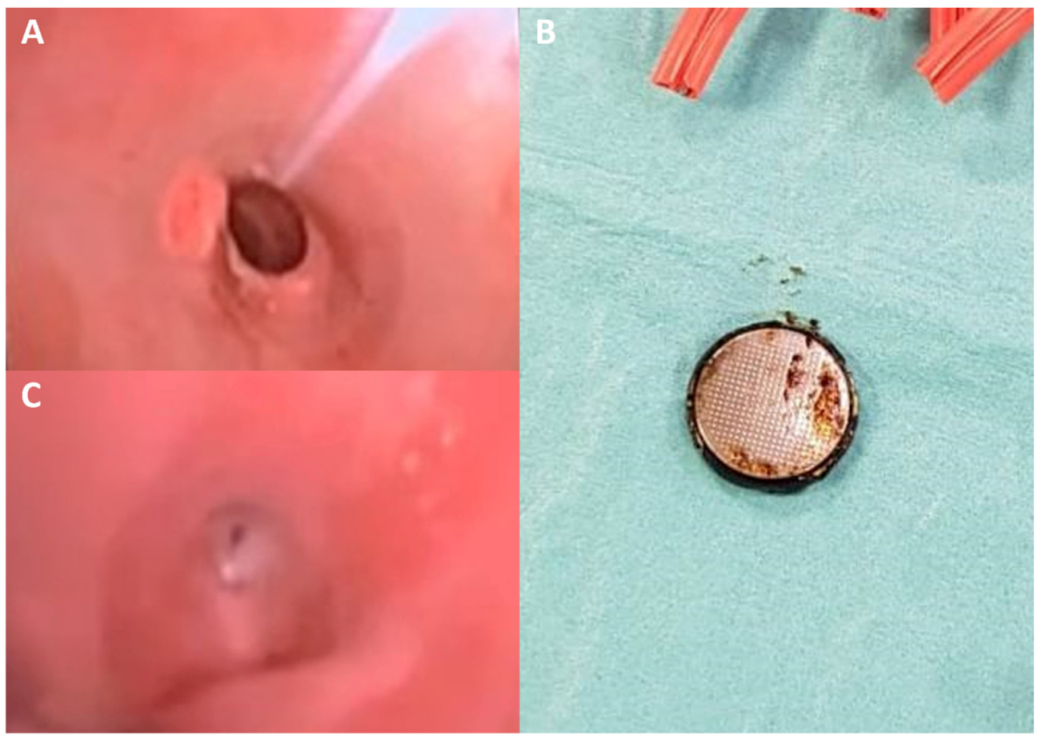
2. Case 1
3. Case 2
4. Discussion
Author Contributions
Funding
Institutional Review Board Statement
Informed Consent Statement
Conflicts of Interest
References
- National Capital Poison Center. Button Battery Ingestion Triage and Treatment Guideline. Available online: https://www.poison.org/battery/guideline (accessed on 20 August 2022).
- Litovitz, T.; Whitaker, N.; Clark, L. Preventing Battery Ingestions: An Analysis of 8648 Cases. Pediatrics 2010, 125, 1178–1183. [Google Scholar] [CrossRef] [PubMed]
- Yardeni, D.; Coran, A.G.; Golladay, E.S.; Yardeni, H. Severe esophageal damage due to button battery ingestion: Can it be prevented? Pediatr. Surg. Int. 2004, 20, 496–501. [Google Scholar] [CrossRef] [PubMed]
- Mubarak, A.; Benninga, M.A.; Broekaert, I.; Dolinsek, J.; Homan, M.; Mas, E.; Miele, E.; Pienar, C.; Thapar, N.; Thomson, M.; et al. Diagnosis, Management, and Prevention of Button Battery Ingestion in Childhood: A European Society for Paediatric Gastroenterology Hepatology and Nutrition Position Paper. J. Pediatr. Gastroenterol. Nutr. 2021, 73, 129–136. [Google Scholar] [CrossRef]
- Eliason, M.J.; Melzer, J.M.; Winters, J.R.; Gallagher, T.Q. Identifying predictive factors for long-term complications following button battery impactions: A case series and literature review. Int. J. Pediatr. Otorhinolaryngol. 2016, 87, 198–202. [Google Scholar] [CrossRef] [PubMed]
- Litovitz, T.; Whitaker, N.; Clark, L.; White, N.C.; Marsolek, M. Emerging Battery-Ingestion Hazard: Clinical Implications. Pediatrics 2010, 125, 1168–1177. [Google Scholar] [CrossRef]
- Alreheili, K.M.; Almutairi, M.; Alsaadi, A.; Ahmed, G.; Alhejili, A.; AlKhatrawi, T. A 2-Year-Old Boy Who Developed an Aortoesophageal Fistula After Swallowing a Button Battery, Managed Using a Novel Procedure with Vascular Plug Device as a Bridge to Definitive Surgical Repair. Am. J. Case Rep. 2021, 22, e931013. [Google Scholar] [CrossRef]
- Buttazzoni, E.; Gregori, D.; Paoli, B.; Soriani, N.; Baldas, S.; Rodriguez, H.; Lorenzoni, G. Symptoms associated with button batteries injuries in children: An epidemiological review. Int. J. Pediatr. Otorhinolaryngol. 2015, 79, 2200–2207. [Google Scholar] [CrossRef]
- Kramer, R.E.; Lerner, D.G.; Lin, T.; Manfredi, M.; Shah, M.; Stephen, T.C.; Gibbons, T.E.; Pall, H.; Sahn, B.; McOmber, M.; et al. Management of Ingested Foreign Bodies in Children: A clinical report of the NASPGHAN Endoscopy Committee. J. Pediatr. Gastroenterol. Nutr. 2015, 60, 562–574. [Google Scholar] [CrossRef]
- Poupore, N.S.; Shih, M.C.; Nguyen, S.A.; Brennan, E.A.; Clemmens, C.S.; Pecha, P.P.; McDuffie, L.A.; Carroll, W.W. Evaluating the management timeline of tracheoesophageal fistulas secondary to button batteries: A systematic review. Int. J. Pediatr. Otorhinolaryngol. 2022, 157, 111100. [Google Scholar] [CrossRef]
- Skrzypczak, P.; Nogal, P.; Sosnowska, P.; Szydłowski, J.; Mańkowski, P. Multi-stage treatment of esophago-tracheal injury after button battery ingestion. J. Pediatr. Surg. Case Rep. 2020, 61, 101584. [Google Scholar] [CrossRef]
- Bibas, B.J.; Cardoso, P.F.G.; Minamoto, H.; Pêgo-Fernandes, P.M. Surgery for intrathoracic tracheoesophageal and bronchoesophageal fistula. Ann. Transl. Med. 2018, 6, 210. [Google Scholar] [CrossRef]
- Guelfguat, M.; Kaplinskiy, V.; Reddy, S.H.; DiPoce, J. Clinical Guidelines for Imaging and Reporting Ingested Foreign Bodies. Am. J. Roentgenol. 2014, 203, 37–53. [Google Scholar] [CrossRef]
- Park, S.-J.; Burns, H. Button battery injury: An update. Aust. J. Gen. Pract. 2022, 51, 471–475. [Google Scholar] [CrossRef]
- Martin, S.S.; Wichmann, J.L.; Scholtz, J.-E.; Leithner, D.; D’Angelo, T.; Weyer, H.; Booz, C.; Lenga, L.; Vogl, T.J.; Albrecht, M.H. Noise-Optimized Virtual Monoenergetic Dual-Energy CT Improves Diagnostic Accuracy for the Detection of Active Arterial Bleeding of the Abdomen. J. Vasc. Interv. Radiol. 2017, 28, 1257–1266. [Google Scholar] [CrossRef]
- Spiers, A.; Jamil, S.; Whan, E.; Forbes, D.; Gollow, I.; Andrews, D. Survival of patient after aorto-oesophageal fistula following button battery ingestion. ANZ J. Surg. 2012, 82, 186–187. [Google Scholar] [CrossRef]
- Granata, A.; Gandolfo, C.; Acierno, C.; Piazza, M.; Burgio, G.; Traina, M. Button battery removed from the stomach resulting in a missed aortoesophageal fistula—A multidisciplinary approach to rescuing a very young patient: A case report. J. Med. Case Rep. 2018, 12, 318. [Google Scholar] [CrossRef]
- Mahajan, S.; Jaswal, V.; Thingnam, S.K.S.; Dogra, N. Successful surgical management of an aorto-oesophageal fistula caused by button battery ingestion. Eur. J. Cardio-Thoracic Surg. 2018, 55, 790–791. [Google Scholar] [CrossRef]
- Bartkevics, M.; Stankovic, Z.; Schibli, S.; Fluri, S.; Berger, S.; Schmidli, J.; Kadner, A. A Near Miss and Salvage Management of Aortoesophageal Fistula Secondary to Cell Battery Ingestion. World J. Pediatr. Congenit. Heart Surg. 2019, 11, 120–122. [Google Scholar] [CrossRef]
- Sinclair, E.M.; Stevens, J.P.; McElhanon, B.; Meisel, J.A.; Santore, M.T.; Chahine, A.A.; Riedesel, E.L. Development and repair of aorto-esophageal fistula following esophageal button battery impaction: A case report. J. Pediatr. Surg. Case Rep. 2021, 66, 101782. [Google Scholar] [CrossRef]
- Tobias, J.D.; Wakimoto, M.; Willer, B.L.; Mckee, C.; Nafiu, O.O. Successful management of an aorto-esophageal fistula following button battery ingestion: A case report and review of the literature. Saudi J. Anaesth. 2021, 15, 193–198. [Google Scholar] [CrossRef]
- Gibbs, H.; Sethia, R.; McConnell, P.I.; Aldrink, J.H.; Shinoka, T.; Williams, K.; Jatana, K.R. Survival of Toddler with Aortoesophageal Fistula after Button Battery Ingestion. Case Rep. Otolaryngol. 2021, 2021, 5557054. [Google Scholar] [CrossRef] [PubMed]
- Muhieldin, M.A.; Larson, C.; DeCaen, A.; Alrajhi, Y.; El-Andari, R.; Perry, T.; Ben Sivarajan, V.; Cave, D.; Al-Aklabi, M. Surgical repair of massive hemorrhage secondary to button battery ingestion causing aortoesophageal fistula. J. Card. Surg. 2022, 37, 2112–2114. [Google Scholar] [CrossRef]


| Sex | Age (Years) | Battery Size | Length of BB Exposure | Clinical Presentation | Diagnosis | Management | |
|---|---|---|---|---|---|---|---|
| Spiers (2012) [16] | M | 1 | 20 mm | 14 h | Hematemesis | CTA | Surgical repair |
| Granata (2018) [17] | F | 2 | Unknown | Unknown | Hematemesis and hemodynamic shock | Angiogram | Endovascular stent |
| Mahajan (2019) [18] | F | 3 | Unknown | Unknown | Hematemesis | CTA | Surgical repair |
| Bartkevics (2020) [19] | F | 1 | 20 mm | Unknown | Hematemesis and melena | CTA | Surgical repair |
| Sinclair (2021) [20] | F | 6 | 21 mm | 6 h | Hematemesis | Angiogram | Endovascular stent |
| Wakimoto (2021) [21] | F | 1.5 | 23.5 mm | Unknown | Hematemesis | CTA | Surgical repair |
| Alreheili (2021) [7] | M | 2.5 | 20 mm | 16 h | Hematemesis, melena and nasal bleeding | CTA | Vascular plug device |
| Gibbs (2021) [22] | F | 1.5 | 20 mm | Unknown | Hematemesis | CTA | Surgical repair |
| Muhieldin (2022) [23] | F | 1.5 | 21.6 mm | Unknown | Hematemesis | During operation | Surgical repair |
| Current Case | M | 1 | 20 mm | Unknown | Hematemesis and cardio-circulatory arrest | CTA | Surgical repair |
Publisher’s Note: MDPI stays neutral with regard to jurisdictional claims in published maps and institutional affiliations. |
© 2022 by the authors. Licensee MDPI, Basel, Switzerland. This article is an open access article distributed under the terms and conditions of the Creative Commons Attribution (CC BY) license (https://creativecommons.org/licenses/by/4.0/).
Share and Cite
Lanzafame, L.R.M.; Blandino, A.; Cicero, G.; Romeo, P.; Agati, S.; Zanai, R.; Celona, A.; Booz, C.; Koch, V.; Mazziotti, S.; et al. Diagnosis and Management of Button Battery Ingestion Complicated by Tracheo-Esophageal and Aorto-Esophageal Fistulas. Diagnostics 2022, 12, 2369. https://doi.org/10.3390/diagnostics12102369
Lanzafame LRM, Blandino A, Cicero G, Romeo P, Agati S, Zanai R, Celona A, Booz C, Koch V, Mazziotti S, et al. Diagnosis and Management of Button Battery Ingestion Complicated by Tracheo-Esophageal and Aorto-Esophageal Fistulas. Diagnostics. 2022; 12(10):2369. https://doi.org/10.3390/diagnostics12102369
Chicago/Turabian StyleLanzafame, Ludovica R. M., Alfredo Blandino, Giuseppe Cicero, Placido Romeo, Salvatore Agati, Rosanna Zanai, Antonio Celona, Christian Booz, Vitali Koch, Silvio Mazziotti, and et al. 2022. "Diagnosis and Management of Button Battery Ingestion Complicated by Tracheo-Esophageal and Aorto-Esophageal Fistulas" Diagnostics 12, no. 10: 2369. https://doi.org/10.3390/diagnostics12102369