Article

Algal Diversity in Paramecium bursaria: Species Identification, Detection of Choricystis parasitica, and Assessment of the Interaction Specificity

by
Felicitas E. Flemming
1,
Alexey Potekhin
2,3,
Thomas Pröschold
4 and
Martina Schrallhammer
1,*
1
Microbiology, Institute of Biology II, Albert Ludwig University of Freiburg, 79104 Freiburg, Germany
2
Department of Microbiology, Faculty of Biology, Saint Petersburg State University, 199034 Saint Petersburg, Russia
3
Laboratory of Cellular and Molecular Protistology, Zoological Institute RAS, 199034 Saint Petersburg, Russia
4
Research Department for Limnology, University of Innsbruck, 5310 Mondsee, Austria
*
Author to whom correspondence should be addressed.
Diversity 2020, 12(8), 287; https://doi.org/10.3390/d12080287
Submission received: 6 May 2020 / Revised: 17 July 2020 / Accepted: 19 July 2020 / Published: 23 July 2020
(This article belongs to the Special Issue Biodiversity of Ciliates and their Symbionts)

Abstract

:
The ‘green’ ciliate Paramecium bursaria lives in mutualistic symbiosis with green algae belonging to the species Chlorella variabilis or Micractinium conductrix. We analysed the diversity of algal endosymbionts and their P. bursaria hosts in nine strains from geographically diverse origins. Therefore, their phylogenies using different molecular markers were inferred. The green paramecia belong to different syngens of P. bursaria. The intracellular algae were assigned to Chl. variabilis, M. conductrix or, surprisingly, Choricystis parasitica. This usually free-living alga co-occurs with M. conductrix in the host’s cytoplasm. Addressing the potential status of Chor. parasitica as second additional endosymbiont, we determined if it is capable of symbiosis establishment and replication within a host cell. Symbiont-free P. bursaria were generated by cycloheximid treatment. Those aposymbiotic P. bursaria were used for experimental infections to investigate the symbiosis specificity not only between P. bursaria and Chor. parasitica but including also Chl. variabilis and M. conductrix. For each algae we observed the uptake and incorporation in individual perialgal vacuoles. These host-symbiont associations are stable since more than five months. Thus, Chor. parasitica and P. bursaria can form an intimate and long-term interaction. This study provides new insights into the diversity of P. bursaria algal symbionts.

Graphical Abstract

1. Introduction

The genus Paramecium Müller [1] (Peniculida, Oligohymenophorea, Ciliophora) includes two species that are known to maintain intracellular algae. Paramecium chlorelligerum Kahl [2,3] lives in symbiotic association with green algae belonging to the genus Meyerella Fawley & K. Fawley [4] (Chlorellaceae, Trebouxiophyceae, Chlorophyta) and is considered as an endemic, extremely rare species [3,5]. The other Paramecium species with algal symbionts is Paramecium bursaria Focke [6], the ’green’ Paramecium, frequently found in freshwater habitats around the world [7]. Evolutionary, this species has been assumed to be the first that diverged within the genus [8,9]. This green ciliate harbours hundreds of symbiotic green algae within its cytoplasm [7,10,11,12,13,14] either belonging to Chlorella variabilis Shihira & R. W. Krauss [15] or to Micractinium conductrix (K. Brandt) Pröschold & Darienko [16,17] (Chlorellaceae, Trebouxiophyceae, Chlorophyta).
The algae escape lysosomal fusion by incorporation in perialgal vacuoles [18,19,20]. In this facultative, mutualistic symbiosis, the algae provide photosynthesis products for their host [21,22,23] and also form a protective layer against UV-radiation damage [24]. Moreover, they profit from protection against the lytic Paramecium bursaria chlorovirus [25,26]. In addition, the host increases its symbiont’s motility by providing transport to brightly illuminated areas optimal for photosynthesis [27]. The facultative nature of this symbiosis allows the separate cultivation of both organisms, making the association an easy-to-access model system to study symbiotic interactions [28,29,30]. Albeit under laboratory conditions the symbiosis is not obligate, it is the natural condition in the environment. Both algae have not been retrieved as free-living organisms from nature so far and, therefore, are considered as highly adapted to their endosymbiotic lifestyle [31]. Similarly, aposymbiotic P. bursaria cells have been isolated from natural freshwater sources only on rare occasions [32]. As Chl. variabilis and M. conductrix apparently are mainly restricted to their endosymbiotic lifestyle within P. bursaria, these associations have been considered as highly specific. However, recent studies report these microalgae as intracellular symbionts from different host organisms such as Tetrahymena [33], Hydra [34], Frontonia vernalis [35], and sponges [10]. Furthermore, P. bursaria can also harbour different organisms either co-occurring with Chlorella-like algae, i.e., bacteria [36,37], or in their place, i.e., yeasts [38,39]. Therefore, the symbiotic relationship between P. bursaria and its intracellular green algae might not be as exclusive as previously assumed.
An interesting case of double algal infection in P. bursaria by Chlorella and another green alga was reported by two Japanese groups [31,40]. The intracellularly uncommon, additional alga was characterized as Choricystis parasitica (K. Brandt) Pröschold & Darienko [16,17] (Trebouxiophyceae, Chlorophyta). Cells of this alga were observed between the trichocysts near the host’s cortex [31,40]. This localization corresponds to that of the natural algal endosymbionts’. The cortex area is devoid of lysosomes, thus an intracellular symbiont is likely protected here against lysosomal fusion [41] and hence against host defence mechanisms, which was interpreted as an advanced symbiotic status [31,40]. Due to its small cell size (1.5–3.0 μm in length, 1.0–1.5 μ m in width; [10]), Chor. parasitica will be referred to as picoalga.
Characterization of symbiotic systems often requires interdisciplinary expertise to fulfill the standards of the respective scientific disciplines. Molecular characterization of a single or more phylogenetic markers is typically part of current descriptions of new taxa. Sequence analysis allows evolutionary interpretation in absence of fossil records and furthermore facilitates the assignment of new isolates to described taxa. The choice of the molecular markers depends on multiple factors and varies between organism groups and the desired level of phylogenetic resolution. In case of P. bursaria, the SSU (small subunit or 18S ribosomal RNA) gene sequence is commonly applied for phylogenetic analyses of members of the genus Paramecium [5,42,43,44]. Some recognized Paramecium species are actually sibling or cryptic species complexes, i.e., their members are morphologically indistinguishable but reproductively isolated and thus are called syngens. The most prominent example are the syngens of the Paramecium aurelia complex, which was first studied in detail by Sonneborn [45]. Those syngens are nowadays recognized as separate species. Similarly, the existence of multiple syngens was described for P. bursaria [46]. Bomford’s description of P. bursaria has been the most extensive one, unfortunately his strain collection was lost. Only few strains remain and are scattered across different laboratories. A representative collection of P. bursaria strains assigned to five syngens, R1 to R5 [47], is maintained at the RC CCM collection (World Data Centre for Microorganisms, RN 1171), Saint Petersburg State University, Saint Petersburg, Russia. Molecular phylogenetic analyses confirmed P. bursaria to be a complex of at least five cryptic species [47,48,49,50] using different molecular markers (e.g., ITS1-5.8S-ITS2-5’LSU fragments, mitochondrial cytochrome c oxidase subunit I gene, histone H4 gene; [5,48,51]).
Based on molecular phylogenetic analyses of SSU and ITS (internal transcribed spacer) regions of nuclear-encoded rRNA genes, the genus and species concepts within the Chlorellaceae remain provisional due to the lack of bootstrap support in molecular analyses [10,52,53,54,55]. The family is divided into two major clades: (i) the well-supported Parachlorella clade and (ii) the moderately supported Chlorella clade [56,57]. Furthermore, the ITS2 region has been used for species delineation of members of the Chlorellaceae [10,50,55,58]. The two common endosymbionts of P. bursaria, Chl. variabilis and M. conductrix, both vary in the ITS2 secondary structure and can be differentiated using their ITS2 barcodes [50].
This study addresses the diversity of intracellular algae in P. bursaria. Therefore, ciliate hosts and algal endosymbionts were identified using different molecular markers. In order to analyse the interaction between the three different algal species detected and P. bursaria, experimental re- and cross-infections were performed and the fate of the intracellular algae was monitored. Differences in the establishment of stable associations will allow to draw conclusions regarding the compatibility of the different algae as endosymbionts of P. bursaria and the specificity of such interactions.

2. Materials and Methods

2.1. Strains and Cultivation

All P. bursaria strains (Table 1) were cultivated in Dryl’s solution (0.1 M Na2HPO4, 0.1 M NaH2PO4, 0.1 M CaCl2, 0.1 M sodium citrate, pH 6.8) at 20 °C in a light chamber (EHRET Type KLT/S 4, Ehret, Emmendingen, Germany) with a 12 h/12 h light-dark cycle. The cultures were fed weekly with the bacterium Raoultella planticola DMSZ 3069 as food organism (bacterized CM). Therefore, the bacteria were inoculated in 0.25% Cerophyll medium (CM; [59] prepared from wheat grass powder (GSE-Vertrieb), Saarbrücken, Germany) and a stigmasterol (Sigma-Aldrich, Munich, Germany) concentration of 500 μ g/L. After two days of incubation at 20 °C, the bacterized CM was ready for use.

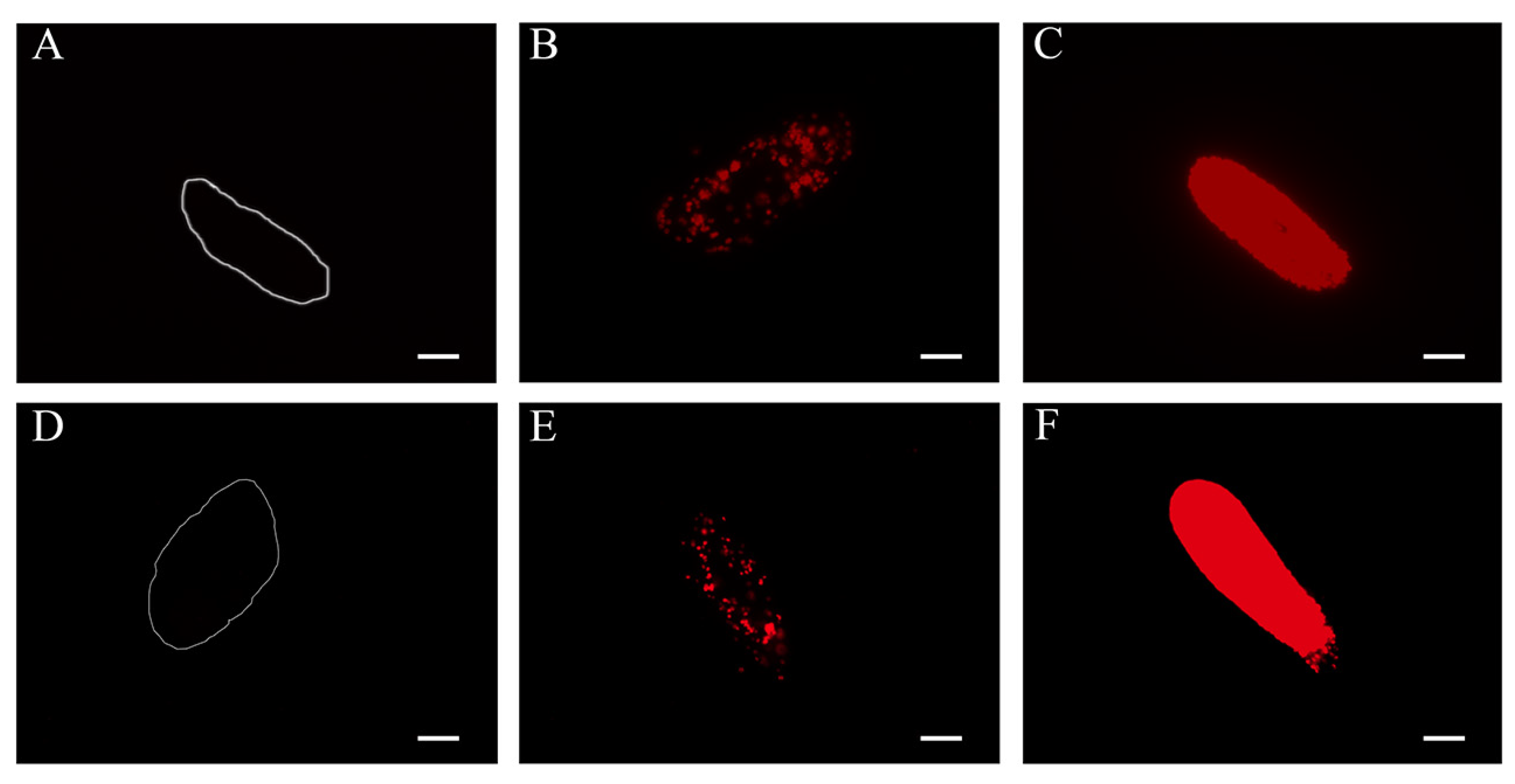
2.2. Observation and Size Determination of Intracellular Algae

The intracellular algae were investigated by detecting the autofluorescence of the chlorophyll by fluorescence microscopy (Axio Imager.M2, Zeiss, Jena, Germany). Therefore, Paramecium cells were fixed with Bouin’s fixative solution (Sigma-Aldrich) and transferred to a microscope slide. Cells were examined using the Cy5 channel (EX BP 640/30, BS FT 660, EM BP 690/50) and an exposure time ranging from 3000 to 7000 ms in order to discriminate between the individual intracellular algae. To determine the size of the intracellular micro- and picoalgae, the diameter of 15 cells per strain was measured.

2.3. DNA Extraction and Amplification of Molecular Markers

All polymerase chain reactions (PCR) were performed with a PeqStar 2× thermocycler (Peqlab, VWR International, Darmstadt, Germany) using TaKaRa reagents and TaKaRa ExTaq polymerase (TaKaRa Bio, Otsu, Japan). Primer sequences (Table 2) and amplification protocols (Supplementary Tables S1 and S2) are provided. The SSU-ITS1-5.8S-ITS2-5’LSU rRNA gene region of P. bursaria was amplified using the primer combinations Penic_F82 and Penic_R1280 and Penic_F661 and 28S_R457. Primers Penic_F82, Penic_F661 and 28S_R457 were used for sequencing.
For the intracellular algae, the nuclear encoded SSU gene and the ITS region were amplified using a semi-nested PCR approach to obtain sufficient DNA amounts for direct sequencing. For the initial amplification (primer combination: AF and ITS055R) as well as for the two semi-nested amplification reactions (primer combinations: AF and Chlo_R841 and Chlo_G800F and ITS055R) the protocol described by Pröschold and colleagues [10] was used. AF, Chlo_F238, Chlo_G800F, Chlo_R841 and ITS055R were used as sequencing primers. For strains Scot and Old-Pf amplification was carried out with primer combination Chlo_G800F and ITS055R (Supplementary Tables S1 and S2) and both primers for sequencing. Additionally, a diagnostic PCR allowing to discriminate between Chl. variabilis and M. conductrix was carried out as described elsewhere [50].
To amplify the SSU gene of Choricystis from strain Frieds the primer combination Chori_F238 and Chori_R841 was used. Those primers were designed to specifically match Choricystis-like algae based on preliminary sequence data. Chori_F238 was used for sequencing.
Purified PCR products (QIAquick PCR Purification Kit, Qiagen, Hilden, Germany) were sequenced at GATC Biotech (Konstanz, Germany). Accession numbers of obtained sequences are provided in Table 1.

2.4. Molecular Characterization of Paramecium bursaria

In order to confirm our morphological identification of the ciliates as members of P. bursaria, we sequenced the SSU rRNA gene sequence and the ITS region of our newly obtained strains. Both markers are commonly applied for characterization at generic and species level [5,42,43,44,47,50]. The obtained sequences were imported into two datasets: (i) The SSU sequences were included into a dataset of representatives of most Paramecium species received from the SILVA SSU Ref NR 99 release 138 database [64]. In addition, nine other members of Peniculidae were included as outgroup resulting in a dataset of 59 sequences with a length of 1493 bp. (ii) The ITS sequences were included into an alignment of 32 P. bursaria strains (506 bp) as described in previous studies [47,50]. For syngen assignment, we added sequences of additional strains (Ard10, Bob2, Ek), which were tested for their mating behaviour by RC CCM to increase the number of sequenced strains per syngen. The ITS sequences were aligned according to their secondary structures.
The best fitting evolutionary models for these datasets were determined using the automated model selection tool implemented in PAUP (version 4.0a, built 167) [65]. The best model (SSU: GTR+I+G; ITS: K81uf+I) was chosen following the Akaike Information Criterion (AIC). For the Bayesian analyses, Bayesian Information Criteria (BIC) were chosen (SSU: GTR+I+G; ITS: GTR+I) for calculation in MrBayes (version 3.2.6 x64; [66]). Non-paramectric Maximum Likelihood (ML) analysis was estimated on 1000 pseudoreplicates (PHYML 2.4.5 from the ARB software package; [67]). Bayesian Interference (BI) analysis was carried out running three runs with one cold and three heated Markov Chain Monte Carlo chains for 1,000,000 generations and sampling the first 25% of the generations as burn-in.

2.5. Molecular Characterization of Green Algal Endosymbionts in Paramecium bursaria

For the molecular characterization of the algal endosymbionts we used sequences covering the SSU-ITS1-5.8S-ITS2 region for discrimination at species level in accordance with previous studies [10,55,56,62,68,69]. Especially the ITS2 region of this nuclear encoded ribosomal operon is often used for species discrimination between members of the Chlorellaceae [53,55,56,70]. The here performed amplification of the ITS2 region covered helices I-III but only partial helix IV. The analysed dataset comprised 50 sequences of representative members of the Chlorellaceae, which were aligned according to the secondary structures predicted by the software mfold [71], following the approach of Pröschold and colleagues [10,55]. This software used the thermodynamic model (minimal energy) for RNA folding. The best fitting evolutionary model for this dataset was predicted and ML and BI analyses were conducted as described above except that BI analysis was run for 5,000,000 generations. Additionally, the Neighbour Joining method (implemented in PAUP) was used. The analysis was run with 1000 bootstrap replicates and a 50% majority rule consensus tree was calculated.
Furthermore, we compared the algal endosymbionts’ secondary structures of ITS2 helices I-III to that of the type strains of Chl. variabilis and M. conductrix. In case of the symbionts of strains Bob2, Ek, and RanNy these algae were identified by means of diagnostic PCR (Supplementary Figure S1).
To identify the intracellular picoalgae of P. bursaria observed in strain Frieds, we used the obtained partial SSU gene sequence and compared it with 41 representative sequences for the Trebouxia lineage (Trebouxiophyceae; following Darienko and colleagues [62]) with the Prasiola clade (four species) and the genus Neocystis (two species) as outgroup. The alignment was obtained as described for Paramecium SSU and comprised 948 positions. Model selection (TIM+I+G) for this dataset was performed as detailed above. Since this model is not implemented in MrBayes, it was substituted with GTR+I+G. ML and BI analyses were performed as described above for P. bursaria. The settings of all used evolutionary models are provided in Supplementary Table S3.

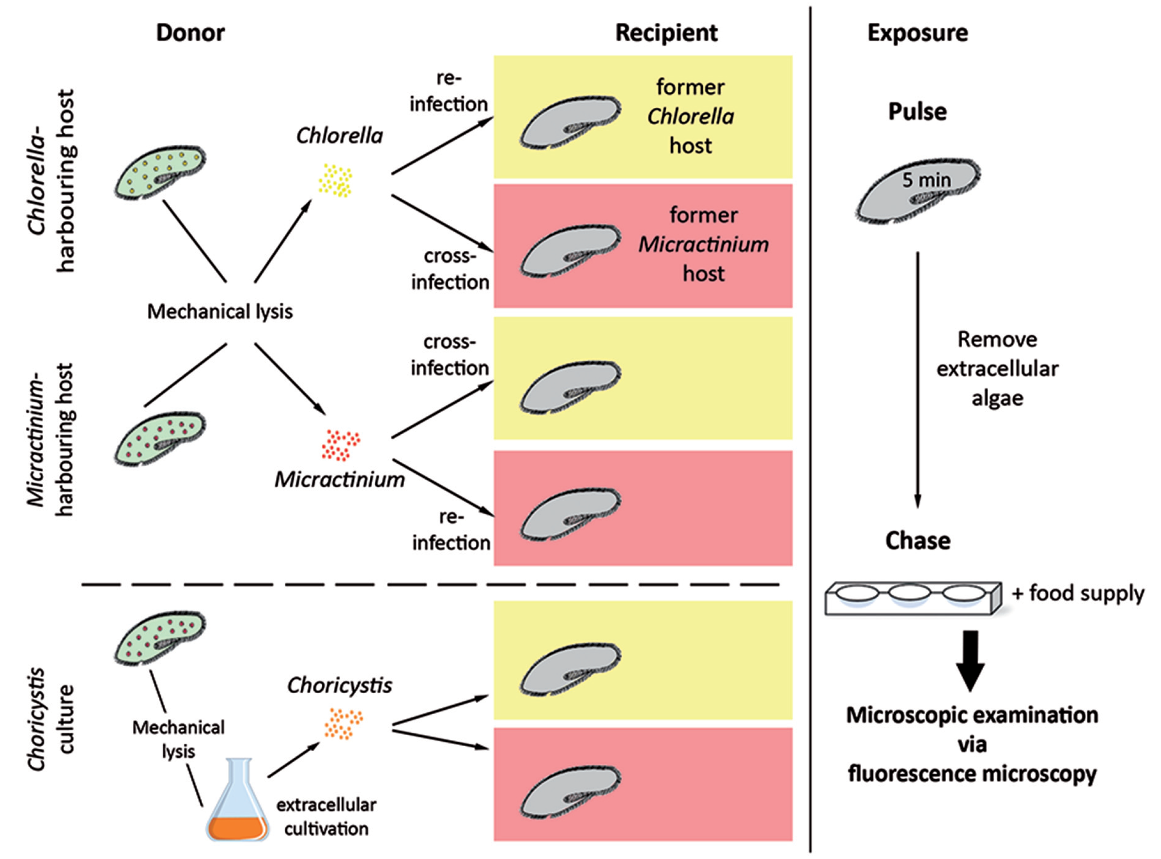
2.6. Establishment of Aposymbiotic Paramecium bursaria

Algal-free P. bursaria were generated by treatment with cycloheximid (Roth, Karlsruhe, Germany). Therefore, symbiotic P. bursaria cultures not fed for at least seven days were incubated in 10 μ g/mL cycloheximid solution and kept under constant light conditions for approximately three days, then the cells were washed with Dryl’s solution by filtering over a 10 μ m membrane and fed with bacterized CM. This filtering step was repeated three to five times over the course of two weeks to assure complete elimination of the algae. If necessary, a second consecutive cycloheximid treatment was performed seven to ten days after the initial exposure. Microscopic inspection via fluorescence microscopy confirmed the elimination of intracellular algae. Aposymbiotic status was confirmed if after 10 seconds of exposure time no autofluorescence signal of the algae’s chloroplasts was detected.

2.7. Pulse-Chase Infection Experiments

To investigate the symbiosis specificity between P. bursaria and three different symbiotic algal species, we exposed aposymbiotic cells (strains JPN, RanNy, Scot; affiliated to different syngens, see below) to isolated Chl. variabilis, M. conductrix, or Chor. parasitica (Figure 1). Aposymbiotic P. bursaria generated by treatment with cycloheximid were exposed (recipient) to isolated algae obtained from symbiotic P. bursaria cultures (donor; Supplementary Table S4). In re-infection assays, the supplied algae belonged to the same algal species as the recipient strain originally harboured, while in cross-infection experiments a different species was provided. We followed the fate of the algae after their uptake and differentiated between digestion, expulsion, and endosymbiotic maintenance by fluorescence microscopy using the above listed parameters. Each experiment comprised three replicates. In total, 20 aposymbiotic Paramecium cells were exposed to a suspension of the respective alga for 5 min with a ratio of 3 × 10³ algae per ciliate cell. Then the paramecia were washed and transferred to 200 μ L Dryl’s solution inoculated with 20 μ L of bacterized CM. 20 μ L bacterized CM were added every second day for seven to ten days. Afterwards, the amount of supplied food was adjusted with respect to cell densities. A regular weekly feeding schedule was implemented after two to three weeks post infection (p.i.). The establishment of the symbiosis was monitored using the fluorescence signal of the algae’s chlorophyll as described above. Instead of Bouin’s solution, the Paramecium cells were fixed with 2% paraformaldehyde (PFA), which resulted in lower background signal and thus an improved signal to noise ratio. To monitor the process of re-establishment of symbiosis, P. bursaria cells were microscopically screened after two, four, seven, ten, and 14 days. Successful infection was confirmed when individually enclosed algae were localized beneath the host’s cell cortex.

3. Results

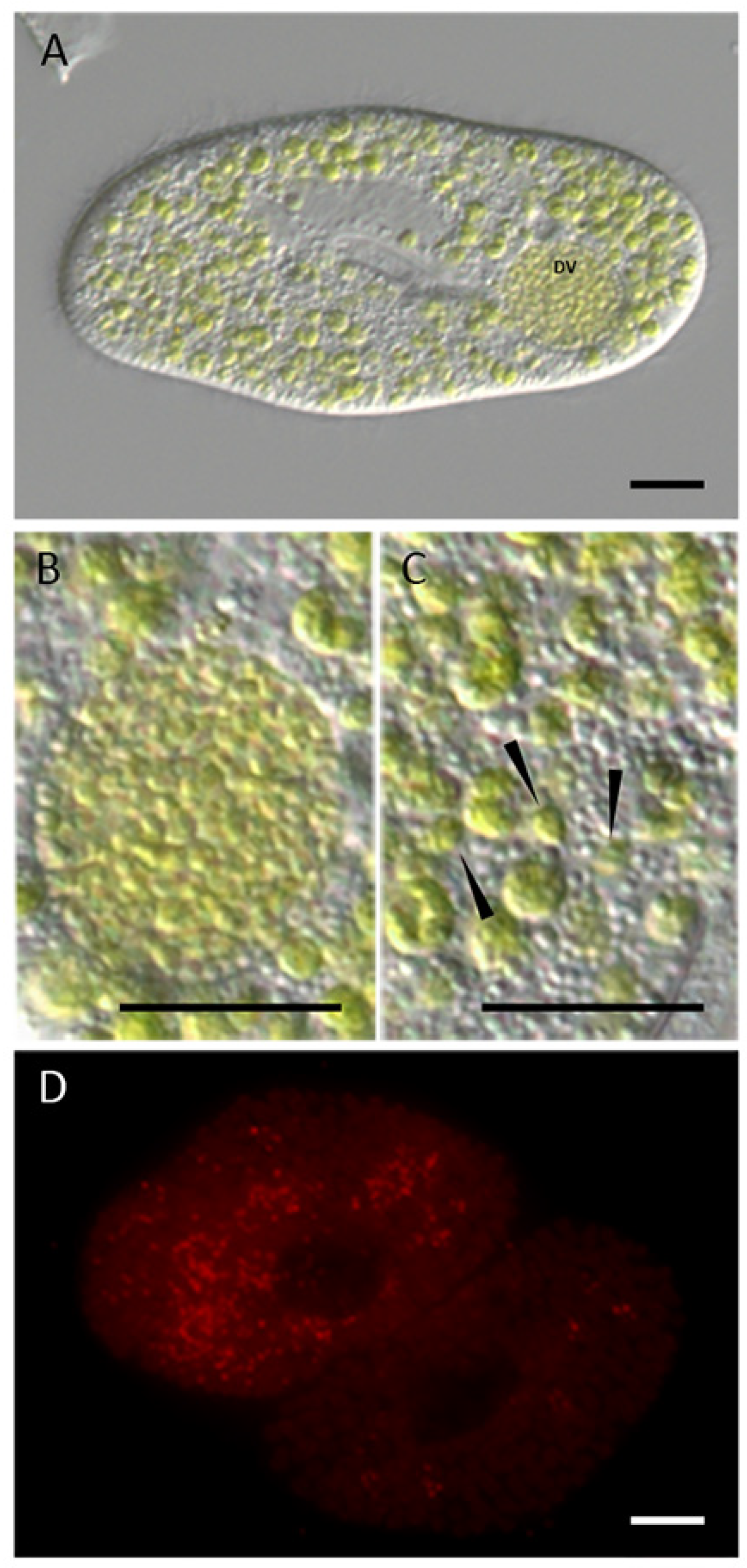
3.1. Microscopic Investigation of the Intracellular Algae

Microscopic observations of the nine P. bursaria strains (Table 1) confirmed the presence of intracellular green algae (data not shown). In case of strain Frieds, algal cells belonging to two size categories were detected (Figure 2). Algae belonging to the larger category showed cell diameters of 4.08 ± 0.02 μ m (referred to as microalgae) while the smaller algae had a diameter of 1.40 ± 0.20 μ m (picoalgae). The picoalgae were localized individually throughout the cytoplasm, especially near the host’s cell cortex. In addition, several of these picoalgae were observed aggregated in digestive vacuoles (Figure 2B). In a single occasion we even followed the fate of a digestive vacuole comprising more than 20 algal cells being emptied into the surrounding medium and thus Chor. parasitica were expelled.

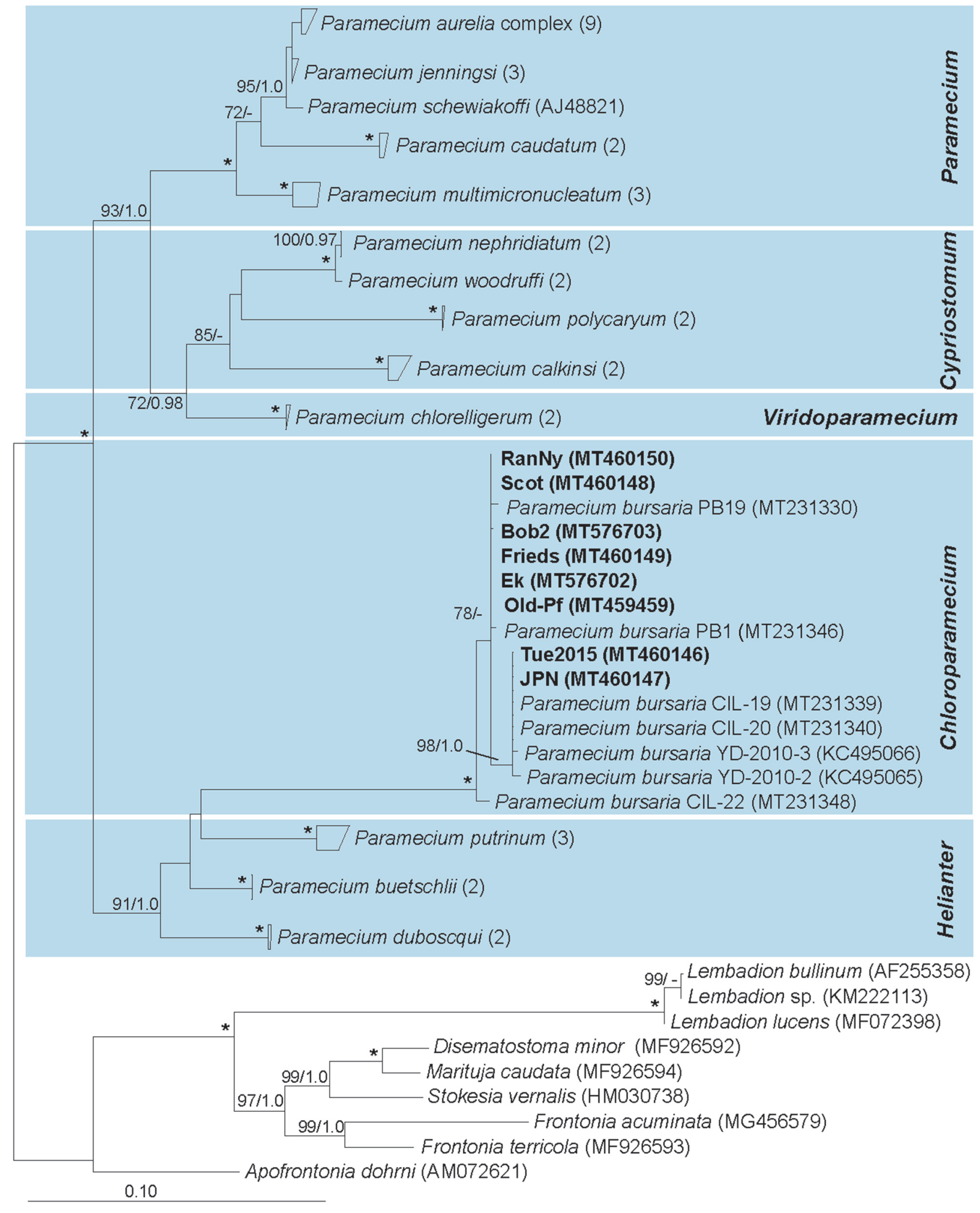
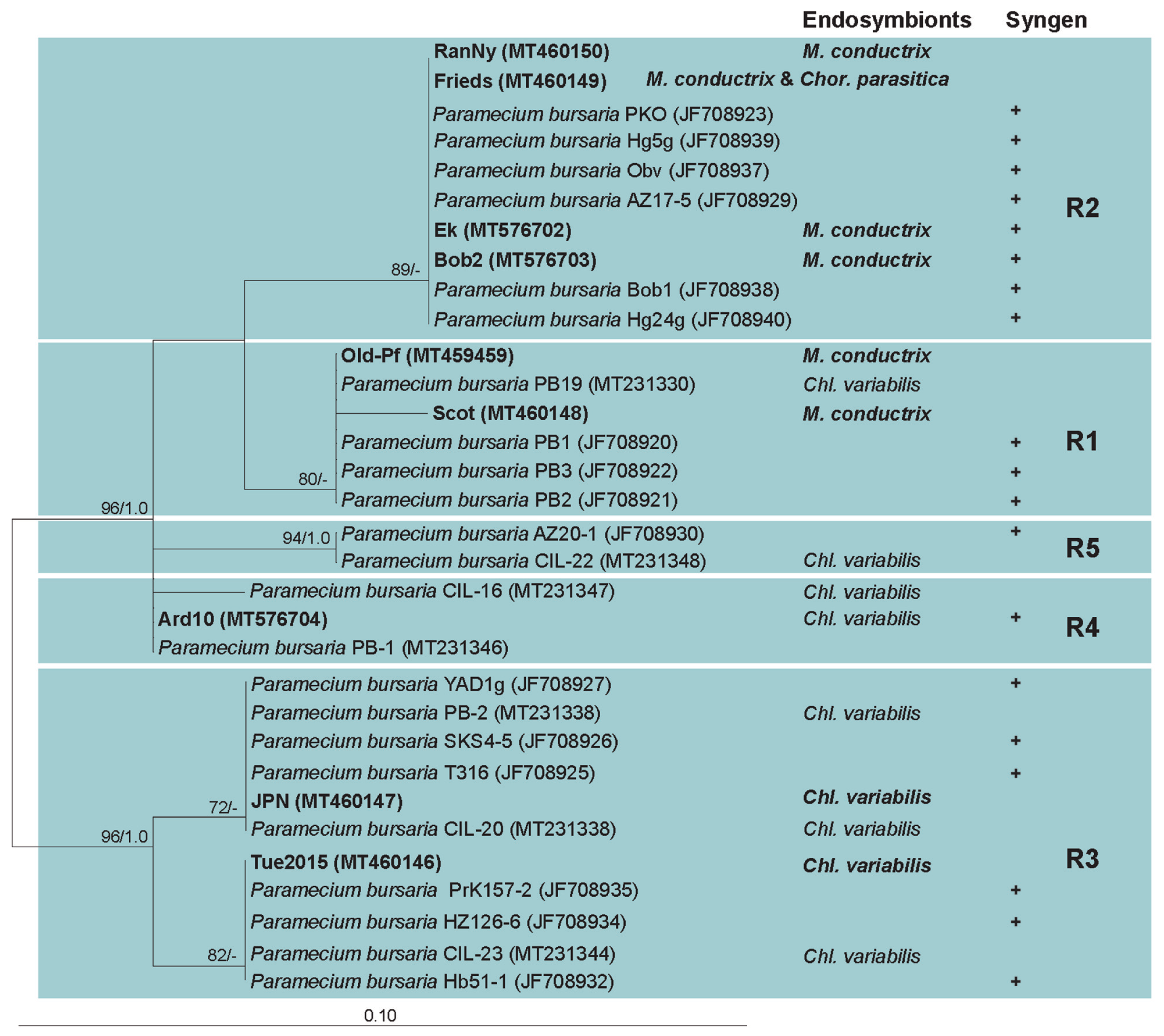
3.2. Paramecium bursaria and the Five Syngens

The phylogenetic inference of Paramecium species based on SSU rRNA gene sequences (Figure 3) recovers the expected topology and composition of the five subgenera of this genus. All here obtained sequences cluster within the monophyletic and maximal supported subgenus Chloroparamecium. Subgroups within the P. bursaria clade can be observed (Figure 3), but the achieved resolution does not allow unambiguous identification of syngen affiliations. In order to discriminate between the five syngens of P. bursaria, we analysed the ITS1-5.8S-ITS2-5’LSU region alone (Figure 4). While the here characterized sequences span from SSU till the beginning of the LSU (except for strain Ard10), the majority of publicly available sequences of P. bursaria strains with known syngen affiliation are limited to the ITS region. Sequences obtained here cluster either with (Figure 4) P. bursaria syngen R1 (Old-Pf and Scot), R2 (Bob2, Ek, Frieds, and RanNy), R3 (JPN and Tue2015), or R4 (Ard10). A potential correlation between the host’s syngen affiliation and the present algal symbiont can be observed. Strains affiliated to syngens R3, R4, and R5 harbour Chl. variabilis while those assigned to syngen R2 contain M. conductrix. Only in case of syngen R1 both algae have been described as symbionts.

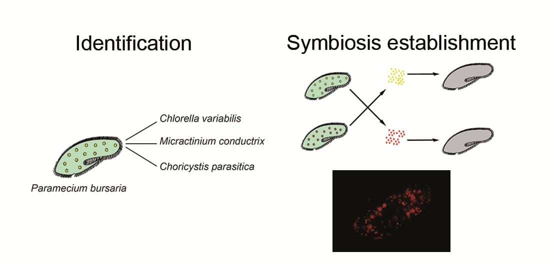
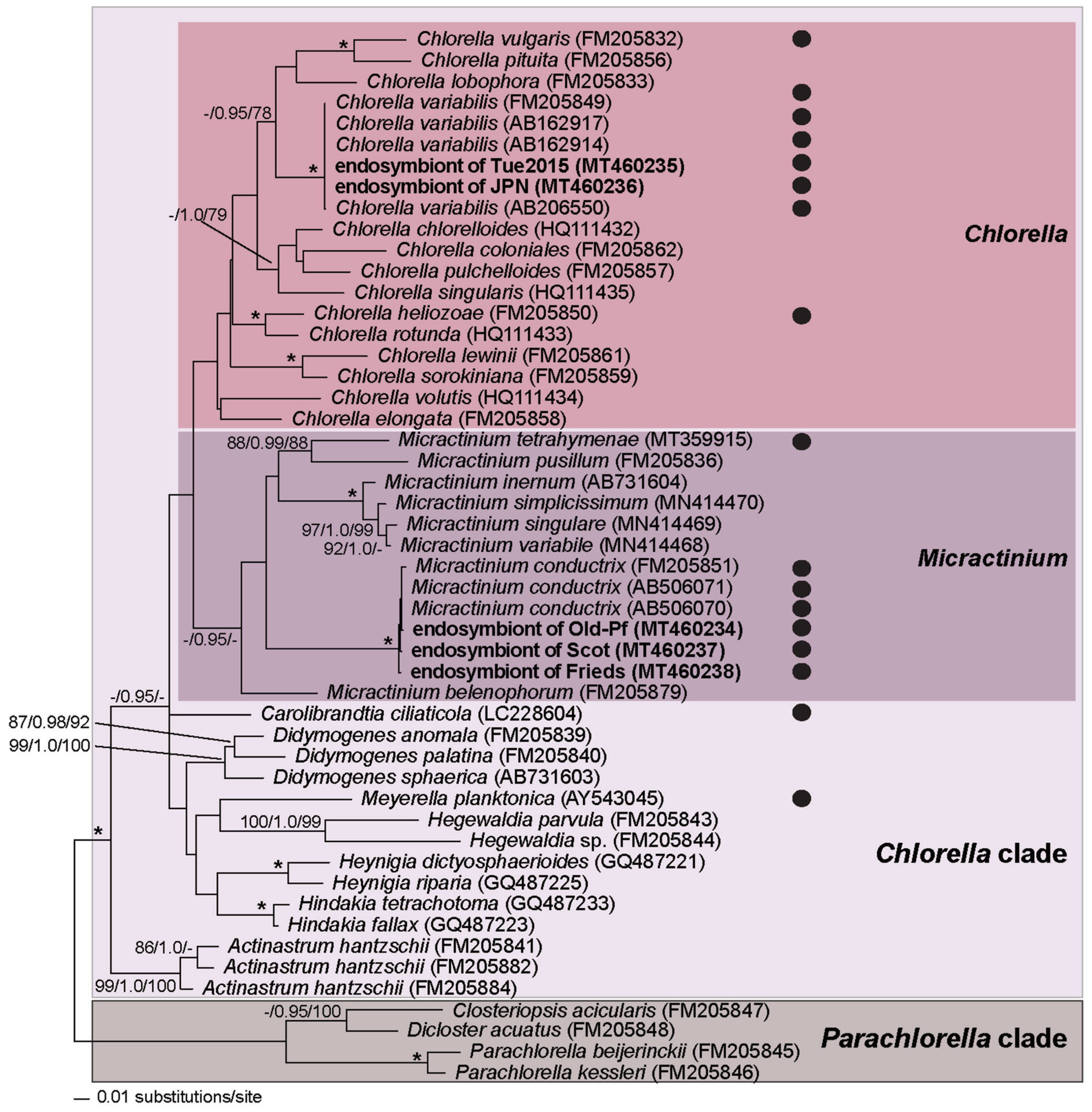
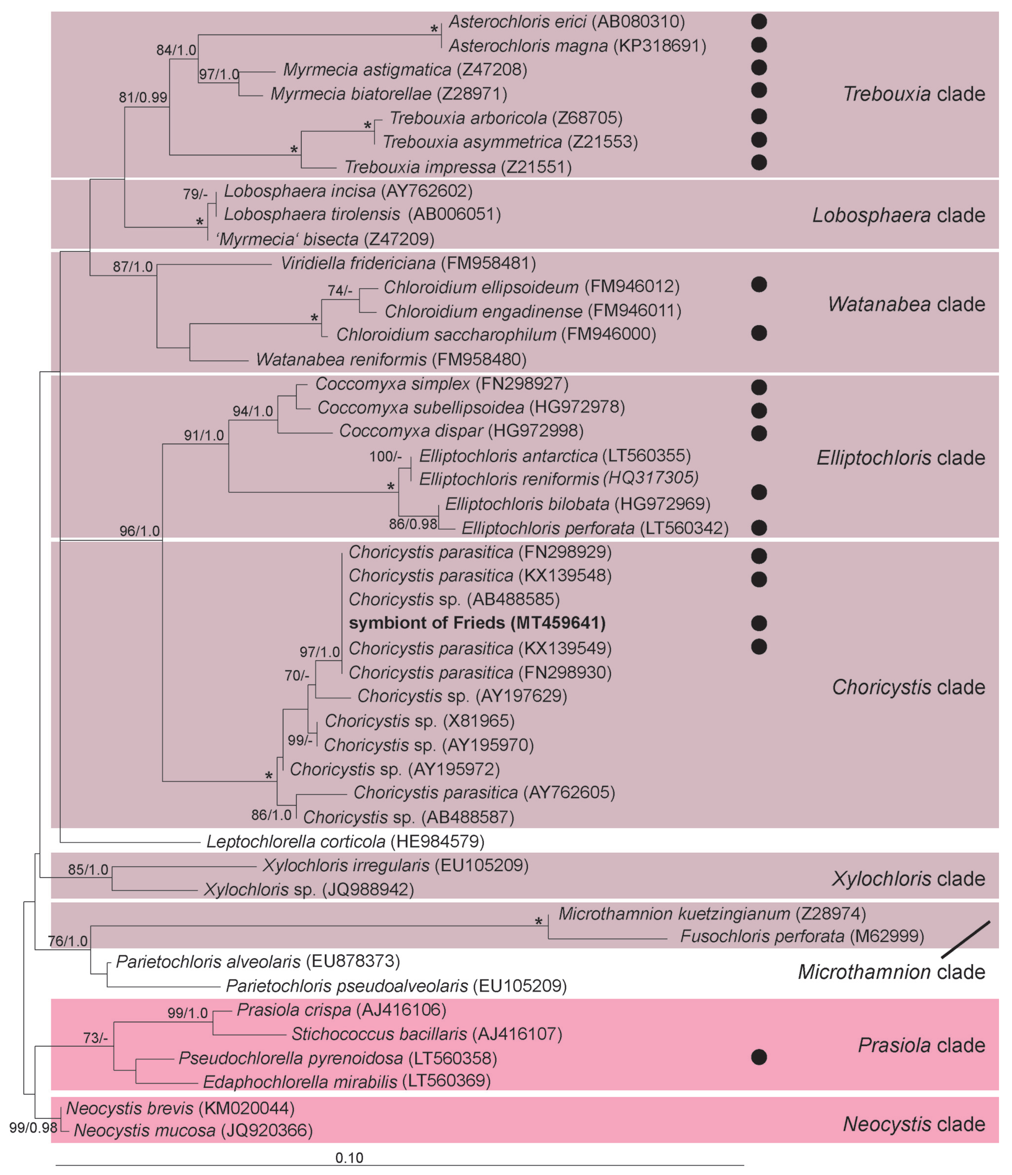
3.3. Chlorella variabilis, Micractinium conductrix, and Choricystis parasitica as Paramecium’s Endosymbionts

Phylogenetic inference of the intracellular microalgae of P. bursaria based on sequences spanning the SSU-ITS1-5.8S-ITS2 region (Figure 5) reveals the genera Chlorella and Micractinium as two distinct monophyletic clades. The intracellular microalgal strains JPN and Tue2015 cluster with other Chl. variabilis strains with maximum support in the phylogenetic analyses. Frieds, Old-Pf, and Scot affiliate with sequences belonging to M. conductrix with maximum support.
Comparing the conserved regions of ITS2 helices I-III of Chl. variabilis (barcode CVAR, [50]) to that of strains JPN and Tue2015, respectively M. conductrix (barcode MCON, [50]) to strains Frieds, Old-Pf, and Scot, no differences were observed (data not shown).
The sequence of the second intracellular alga detected in strain Frieds clusters with other sequences of the genus Choricystis as monophyletic sister group to the Elliptochloris clade with high support in both analyses (Figure 6). The obtained sequence is identical to that of several other Chor. parasitica strains.

3.4. Establishment of Symbioses

We followed the fate of isolated algae after their uptake and differentiated between digestion, expulsion, and endosymbiotic maintenance by aposymbiotic P. bursaria cells via fluorescence microscopy. Within five days after exposure of aposymbiotic P. bursaria cells (Figure 7A,D), digestive vacuoles were observed in all paramecia predominantly occurring in the central part of the Paramecium cell with more than one enclosed alga regardless of the supplied species. Multiple perialgal vacuoles enclosing single algal cells were observed throughout the entire host cytoplasm. After 12 to 15 days p.i., numerous perialgal vacuoles were localized in the cytoplasm near the host’s cell cortex in each examined Paramecium cell (Figure 7B,E). Successful re-establishment of symbiosis was achieved for all tested combinations (Supplementary Table S4) regardless of host background (former Chlorella or Micractinium host), syngen affiliation, and algal identity (Chl. variabilis, M. conductrix, or Chor. parasitica). Thus, we observed no differences between re- versus cross-infection and no preference of certain P. bursaria syngens for specific algae. After four months of cultivation, infected P. bursaria cells harbour numerous (200–500) intracellular algal symbionts. The observed symbiotic conditions are comparable to the natural state (Figure 7C,F). These cultures are stable since over five months and are maintained to date in the laboratory.

4. Discussion

4.1. Symbiotic Relationships and Specificity of Paramecium bursaria and Its Green Algal Endosymbionts

Previous studies showed that aposymbiotic P. bursaria are able to re-establish symbiosis with their intracellular microalgae [19,72]. After uptake via phagocytosis, most of the algae are incorporated in digestive vacuoles. Some manage to escape the host’s lysosomal fusion by ’budding off’ into the cytoplasm being enclosed in perialgal vacuoles. Subsequently, those horizontally transmitted algae are distributed throughout the host cell as well as beneath the cell cortex [73]. Based on the characterization of natural P. bursaria-algae symbioses, it was speculated that the occurrence of certain intracellular algae species might depend on the geographic origin of P. bursaria. The observed endosymbionts separated into two groups [10,12,13,74,75,76], i.e., the “American/Southern” (later on described as Chl. variabilis) and the “European/Northern” (M. conductrix) group. Thus, a correlation between endosymbiont and host population was assumed. Another recent study presents contradicting results reporting the first Chl. variabilis from European P. bursaria [50].
Actual testing the specificity of symbiotic partners accepted by P. bursaria ([77], this study) revealed that all tested intracellular algae derived from this ciliate were accepted as endosymbionts. On the other hand, free-living algae and those obtained from Hydra were digested [77]. A competition experiment revealed a clear preference of the aposymbiotic P. bursaria strain for its native Chlorella strain [77]. In the here conducted re- and cross-infection experiments without of a competitive setting, algae-free P. bursaria did not show a preference for their original symbiont or symbiont species. Aposymbiotic strains formerly harbouring Chl. variabilis maintained M. conductrix as endosymbiont, and vice versa. The picoalga Chor. parasitica, naturally sharing the cytoplasm of its host with M. conductrix, was able to form a stable symbiosis independently of co-occurring microalgae and with cells naturally harbouring Chl. variabilis. Even though P. bursaria establishes an intimate and long-term stable symbiotic association with Chor. parasitica, the role of the picoalga for P. bursaria remains unclear. An evaluation of the symbiotic relationship between P. bursaria and the intracellular picoalga Chor. parasitica awaits further detailed analyses as current findings do not allow a placement of this interaction on the mutualism-parasitism continuum.

4.2. Molecular Characterization of Paramecium bursaria and Its Green Algal Endosymbionts

Unsurprisingly, the novel isolates of ‘green’ ciliates were unambiguously confirmed as P. bursaria. The phylogenetic tree inferred from SSU sequences is in overall good agreement with those previously published [5,42,44]. It already indicates intraspecific diversity, which becomes obvious in the analysis of the ITS region whose results reflect previous findings [47,50]. The analysis of the currently available ITS data might be interpreted that certain P. bursaria syngens harbour preferentially certain algal symbiont species. On the other hand, the number of symbiotic systems with both partners fully characterized is small, therefore this result should be considered very cautiously. It is likely that the picture will change with an increased amount of characterized strains. A revision of the P. bursaria species complex and conclusions about the suitability of certain syngens as hosts for specific algae await future studies.
The here characterized algal symbionts Chl. variabilis and M. conductrix are morphologically nearly identical [50]. However, they clearly differ in size from the additional algal symbiont detected in strain Frieds. The observed sizes in case of the microalgae were at the minimal range described for M. conductrix (5.0–12.0 μ m, [10]) and in the typical size range for intracellular Chor. parasitica (1.5–3.0 μ m) in case of the picoalgae. In the phylogenetic inference based on SSU-ITS sequences, the genera Chlorella and Micractinium were recovered as monophyletic and maximum support was obtained for the associations of the here studied microalgae with either Chl. variabilis or M. conductrix. The tree topology is nearly identical to that of previous studies [62,68] except for small differences lacking support in each analysis. The recovery of both algal species as endosymbiont of P. bursaria was not surprising, the detection of picoalgae in strain Frieds and their identification as Chor. parasitica more so. This is the first report of these picoalgae co-occurring with M. conductrix in the cytoplasm of P. bursaria and only the third report of Chor. parasitica as potential endosymbiont of P. bursaria. In previous observations the co-occuring primary symbiont was identified as Chlorella [31,40].

5. Conclusions

Paramecium bursaria is well adapted to harbour green algae as symbionts. Almost exclusively, either Chl. variabilis or M. conductrix are found in high abundances in the cytoplasm of this ciliate. One route to address the specificity of these associations is the molecular characterizations of host and symbionts. This approach provides information which symbioses are ecologically and evolutionary successful in the studied habitat. It is important to stress that therefore host and symbiont should be characterized in parallel, which is not always the case. Molecular approaches need to be combined with microscopical observations to ensure that indeed all partners are accounted for and none is overlooked or dismissed as environmental contamination. This might easily happen with minor symbionts in multiple infections or with much smaller and intracellularly less abundant picoalgae. Experiments to test the ability for symbiosis formation provide an additional perspective to the observed occurrence of associations in the environment. Infection assays can examine which interactions can be formed at e.g., species or genotype level. Thus, they provide insights into the genetic broadness of potentially realizable symbioses under constant laboratory conditions and allow to entangle the impact of additional biotic or abiotic factors shaping the formation and occurrence of symbioses. The present work can serve as a roadmap how such analyses can be conducted in regard to molecular as well as physiological characterization.

Supplementary Materials

The following are available online at https://www.mdpi.com/1424-2818/12/8/287/s1, Table S1: Primer combinations and their PCR program specifications. Table S2: General PCR program to amplify the SSU rRNA gene. Table S3: Model parameters as defined by PAUP. Table S4: Combinations of aposymbiotic P. bursaria strains and algae used in the re- and cross-infection experiments in this study. Figure S1: Diagnostic PCR for identification of Micractinium conductrix.

Author Contributions

Conceptualization and supervision, M.S.; methodology, F.E.F., T.P., and M.S.; formal analysis, F.E.F.; resources, A.P., T.P., and M.S.; data curation, F.E.F. and M.S.; visualization, F.E.F.; funding acquisition, F.E.F., T.P., and M.S.; project administration, T.P. and M.S.; writing—original draft preparation, F.E.F. and M.S.; writing—review and editing, F.E.F. and M.S. All authors read and approved the final version of the manuscript.

Funding

This research received external funding from the Albert Ludwig University of Freiburg (Innovationsfond, project number 2100297401; MS), the Austrian Science Fund (FWF, project P28333-B25; TP), the Wissenschaftliche Gesellschaft Freiburg i. Breisgau (MS), the Grünewald-Zuberbier-Stiftung (FEF), and from the European Community’s H2020 Programme H2020-MSCA-RISE 2019 under grant agreement number 872767 (MS).

Acknowledgments

We thank Bettina Sonntag for the opportunity to work at the Limnological Research Station Mondsee, Austria. We also thank Tatyana Darienko for her kind assistance with the morphology of green algae and in addition Birgit Süssenbach for preliminary molecular work and Chantal Bleile for her contributions to graphical design.

Conflicts of Interest

The authors declare no conflict of interest.

References

  1. Müller, O.F. Vermium Terrestrium et Fluviatilium, seu Animalium Infusoriorum, Helminthicorum et Testaceorum non Marinorum Succincta Historia; Heineck et Faber: Havniae et Lipsiae, Germany, 1773; Volume 1, p. 74. [Google Scholar]
  2. Kahl, A. Urtiere oder Protozoa. I. Wimpertiere oder Ciliata (Infusoria). 4. Peritricha und Chonotricha. Die Tierwelt der angrenzenden Meeresteile 1935, 30, 651–886. [Google Scholar]
  3. Kreutz, M.; Stoeck, T.; Foissner, W. Morphological and molecular characterization of Paramecium (Viridoparamecium nov. subgen.) chlorelligerum Kahl (Ciliophora). J. Eukaryot. Microbiol. 2012, 59, 548–563. [Google Scholar] [CrossRef] [PubMed] [Green Version]
  4. Fawley, M.W.; Fawley, K.P.; Owen, H.A. Diversity and ecology of small coccoid green algae from Lake Itasca, Minnesota, USA, including Meyerella planktonica, gen. et sp. nov. Phycologia 2005, 44, 35–48. [Google Scholar] [CrossRef]
  5. Lanzoni, O.; Fokin, S.I.; Lebedeva, N.; Migunova, A.; Petroni, G.; Potekhin, A. Rare freshwater ciliate Paramecium chlorelligerum Kahl, 1935 and its macronuclear symbiotic bacterium “Candidatus Holospora parva”. PLoS ONE 2016, 11, e0167928. [Google Scholar] [CrossRef]
  6. Focke, G. Ueber einige Organisationsverhältnisse bei polygastrischen Infusorien und Räderthieren. Oken. Isis 1836, 10, 785–787. [Google Scholar]
  7. Fujishima, M. Endosymbionts in Paramecium; Springer Science and Business Media: Berlin, Germany, 2009; Volume 12. [Google Scholar]
  8. Strüder-Kypke, M.C.; Wright, A.D.G.; Fokin, S.I.; Lynn, D.H. Phylogenetic relationships of the subclass Peniculia (Oligohymenophorea, Ciliophora) inferred from small subunit rRNA gene sequences. J. Eukaryot. Microbiol. 2000, 47, 419–429. [Google Scholar] [CrossRef]
  9. Fokin, S.I.; Przyboś, E.; Chivilev, S.M.; Beier, C.L.; Horn, M.; Skotarczak, B.; Wodecka, B.; Fujishima, M. Morphological and molecular investigations of Paramecium schewiakoffi sp. nov. (Ciliophora, Oligohymenophorea) and current status of distribution and taxonomy of Paramecium spp. Eur. J. Protistol. 2004, 40, 225–243. [Google Scholar] [CrossRef]
  10. Pröschold, T.; Darienko, T.; Silva, P.C.; Reisser, W.; Krienitz, L. The systematics of Zoochlorella revisited employing an integrative approach. Environ. Microbiol. 2011, 13, 350–364. [Google Scholar] [CrossRef]
  11. Vorobyev, K.; Andronov, E.; Rautian, M.; Skoblo, I.; Migunova, A.; Kvitko, K. An atypical Chlorella symbiont Paramecium bursaria. Protistology 2009, 6, 39–44. [Google Scholar]
  12. Hoshina, R.; Imamura, N. Phylogenetic position of endosymbiotic green algae in Paramecium bursaria Ehrenberg Japan. Plant Biol. 2004, 6, 447–453. [Google Scholar] [CrossRef]
  13. Gaponova, I.N.; Andronov, E.E.; Migunova, A.V.; Vorobyev, K.P.; Chizhevskaja, E.P.; Kvitko, K.V. Genomic dactyloscopy of Chlorella sp., symbionts of Paramecium bursaria. Protistology 2007, 4, 311–317. [Google Scholar]
  14. Migunova, A.; Kvitko, K.; Prokosheva, M.; Litvinov, D. Influence of temperature on duplication Paramecium bursaria, Chlorella, PBCV-1 viruses in system of threefold symbiosis. Vestnic St. Peterburg Univ. 2000, 3, 65–75. [Google Scholar]
  15. Shihira, I.; Krauss, R.W. Chlorella: Physiology and Taxonomy of Forty-One Isolates; University of Maryland: College Park, MD, USA, 1965; pp. 1–97. [Google Scholar]
  16. Brandt, K. Ueber das Zusammenleben von Thieren und Algen. Arch. Anat. Physiol. 1881, 1881, 570–574. [Google Scholar]
  17. Pröschold, T.; Bock, C.; Luo, W.; Krienitz, L. Polyphyletic distribution of bristle formation in Chlorellaceae: Micractinium, Diacanthos, Didymogenes and Hegewaldia gen. nov. (Trebouxiophyceae, Chlorophyta). Phycol. Res. 2010, 58, 1–8. [Google Scholar] [CrossRef]
  18. Karakashian, S.J.; Rudzinska, M.A. Inhibition of lysosomal fusion with symbiont-containing vacuoles in Paramecium bursaria. Exp. Cell Res. 1981, 131, 387–393. [Google Scholar] [CrossRef]
  19. Kodama, Y.; Fujishima, M. Infection of Paramecium bursaria by symbiotic Chlorella species. In Endosymbionts in Paramecium; Springer: Berlin, Germany, 2009; pp. 31–55. [Google Scholar]
  20. Kodama, Y.; Fujishima, M. Secondary symbiosis between Paramecium and Chlorella cells. In International Review of Cell and Molecular Biology; Elsevier: Amsterdam, The Netherlands, 2010; Volume 279, pp. 33–77. [Google Scholar]
  21. He, M.; Wang, J.; Fan, X.; Liu, X.; Shi, W.; Huang, N.; Zhao, F.; Miao, M. Genetic basis for the establishment of endosymbiosis in Paramecium. ISME J. 2019, 13, 1360–1369. [Google Scholar] [CrossRef]
  22. Reisser, W. Die stoffwechselphysiologischen Beziehungen zwischen Paramecium bursaria Ehrberg und Chlorella spec. der Paramecium bursaria-Symbiose. Arch. Microbiol. 1976, 107, 357–360. [Google Scholar] [CrossRef]
  23. Brown, J.A.; Nielsen, P.J. Transfer of photosynthetically produced carbohydrate from endosymbiotic Chlorellae Paramecium bursaria. J. Protozool. 1974, 21, 569–570. [Google Scholar] [CrossRef]
  24. Summerer, M.; Sonntag, B.; Hörtnagl, P.; Sommaruga, R. Symbiotic ciliates receive protection against UV damage from their algae: A test with Paramecium bursaria and Chlorella. Protist 2009, 160, 233–243. [Google Scholar] [CrossRef]
  25. Dunigan, D.D.; Al-Sammak, M.; Al-Ameeli, Z.; Agarkova, I.V.; DeLong, J.P.; Van Etten, J.L. Chloroviruses lure hosts through long-distance chemical signaling. J. Virol. 2019, 93, e01688-18. [Google Scholar] [CrossRef] [Green Version]
  26. Coy, S.R.; Alsante, A.N.; Van Etten, J.L.; Wilhelm, S.W. Cryopreservation of Paramecium bursaria Chlorella Virus-1 during an active infection cycle of its host. PLoS ONE 2019, 14, e0211755. [Google Scholar] [CrossRef] [PubMed] [Green Version]
  27. Hoshina, R.; Imamura, N. Origins of algal symbionts of Paramecium bursaria. In Endosymbionts in Paramecium; Springer: Berlin, Germany, 2009; pp. 1–29. [Google Scholar]
  28. Sommaruga, R.; Sonntag, B. Photobiological aspects of the mutualistic association between Paramecium bursaria and Chlorella. In Endosymbionts in Paramecium; Springer: Berlin, Germany, 2009; pp. 111–130. [Google Scholar]
  29. Kodama, Y.; Suzuki, H.; Dohra, H.; Sugii, M.; Kitazume, T.; Yamaguchi, K.; Shigenobu, S.; Fujishima, M. Comparison of gene expression of Paramecium bursaria with and without Chlorella variabilis symbionts. BMC Genom. 2014, 15, 183. [Google Scholar] [CrossRef] [PubMed] [Green Version]
  30. Lowe, C.D.; Minter, E.J.; Cameron, D.D.; Brockhurst, M.A. Shining a light on exploitative host control in a photosynthetic endosymbiosis. Curr. Biol. 2016, 26, 207–211. [Google Scholar] [CrossRef] [PubMed] [Green Version]
  31. Hoshina, R.; Fujiwara, Y. Photobiont flexibility in Paramecium bursaria: Double and triple photobiont co-habitation. Adv. Microbiol. 2012, 2, 227. [Google Scholar] [CrossRef] [Green Version]
  32. Tonooka, Y.; Watanabe, T. A natural strain of Paramecium bursaria lacking symbiotic algae. Eur. J. Protistol. 2002, 38, 55–58. [Google Scholar] [CrossRef]
  33. Nakajima, T.; Matsubara, T.; Ohta, Y.; Miyake, D. Exploitation or cooperation? Evolution of a host (ciliate)-benefiting alga in a long-term experimental microcosm culture. Biosystems 2013, 113, 127–139. [Google Scholar] [CrossRef]
  34. Ishikawa, M.; Yuyama, I.; Shimizu, H.; Nozawa, M.; Ikeo, K.; Gojobori, T. Different endosymbiotic interactions in two hydra species reflect the evolutionary history of endosymbiosis. Genome Biol. Evol. 2016, 8, 2155–2163. [Google Scholar] [CrossRef] [Green Version]
  35. Esteban, G.F.; Fenchel, T.; Finlay, B.J. Mixotrophy in ciliates. Protist 2010, 161, 621–641. [Google Scholar] [CrossRef]
  36. Rautian, M.S.; Wackerow-Kouzova, N.D. Phylogenetic placement of two previously described intranuclear bacteria from the ciliate Paramecium bursaria (Protozoa, Ciliophora): ‘Holospora acuminata’ and ‘Holospora curviuscula’. Int. J. Syst. Evol. Microbiol. 2013, 63, 1930–1933. [Google Scholar] [CrossRef] [Green Version]
  37. Gong, J.; Qing, Y.; Guo, X.; Warren, A. “Candidatus Sonnebornia yantaiensis”, a member of candidate division OD1, as intracellular bacteria of the ciliated protist Paramecium bursaria (Ciliophora, Oligohymenophorea). Syst. Appl. Microbiol. 2014, 37, 35–41. [Google Scholar] [CrossRef]
  38. Görtz, H. Infections of Paramecium bursaria with bacteria and yeasts. J. Cell. Sci. 1982, 58, 445–453. [Google Scholar] [PubMed]
  39. Suzaki, T.; Omura, G.; Görtz, H. Infection of symbiont-free Paramecium bursaria with yeasts. Jpn. J. Protozool. 2003, 36, 17–18. [Google Scholar]
  40. Nakahara, M.; Handa, S.; Watanabe, S.; Deguchil, H. Choricystis minor as a new symbiont of simultaneous two-species association with Paramecium bursaria and implications for its phylogeny. Symbiosis 2004, 36, 127–151. [Google Scholar]
  41. Szokoli, F.; Sabaneyeva, E.; Castelli, M.; Krenek, S.; Schrallhammer, M.; Soares, C.A.; da Silva-Neto, I.D.; Berendonk, T.U.; Petroni, G. “Candidatus Fokinia solitaria”, a novel “stand-alone” symbiotic lineage of Midichloriaceae (Rickettsiales). PLoS ONE 2016, 11, e0145743. [Google Scholar] [CrossRef]
  42. Krenek, S.; Berendonk, T.U.; Fokin, S.I. New Paramecium (Ciliophora, Oligohymenophorea) congeners shape our view its biodiversity. Org. Divers. Ecol. 2015, 15, 215–233. [Google Scholar] [CrossRef]
  43. Da Silva Paiva, T.; do Nascimento Borges, B.; Harada, M.L.; da Silva-Neto, I.D. Description and molecular phylogeny of Paramecium grohmannae sp. nov. (Ciliophora, Peniculida) from a wastewater treatment plant in Brazil. Rev. Bras. De Zoociências 2016, 17, 7–19. [Google Scholar]
  44. Castelli, M.; Serra, V.; Senra, M.V.; Basuri, C.K.; Soares, C.A.; Fokin, S.I.; Modeo, L.; Petroni, G. The hidden world of Rickettsiales symbionts: “Candidatus Spectririckettsia obscura,” a novel bacterium found in Brazilian and Indian Paramecium caudatum. Microb. Ecol. 2019, 77, 748–758. [Google Scholar] [CrossRef]
  45. Sonneborn, T.M. Mating types in Paramecium aurelia: Diverse conditions for mating in different stocks; occurrence, number and interrelations of the types. Proc. Am. Philosoph. Soc. 1938, 79, 411–434. [Google Scholar]
  46. Bomford, R. The syngens of Paramecium bursaria: New mating types and intersyngenic mating reactions. J. Protozool. 1966, 13, 497–501. [Google Scholar] [CrossRef]
  47. Greczek-Stachura, M.; Potekhin, A.; Przyboś, E.; Rautian, M.; Skoblo, I.; Tarcz, S. Identification of Paramecium Bursaria syngens through molecular markers–comparative analysis of three loci in the nuclear and mitochondrial DNA. Protist 2012, 163, 671–685. [Google Scholar] [CrossRef]
  48. Strüder-Kypke, M.C.; Lynn, D.H. Comparative analysis of the mitochondrial cytochrome c oxidase subunit I (COI) gene in ciliates (Alveolata, Ciliophora) and evaluation of its suitability as a biodiversity marker. Syst. Biodivers. 2010, 8, 131–148. [Google Scholar] [CrossRef]
  49. Tasneem, F.; Shakoori, F.R.; Ilyas, M.; Shahzad, N.; Potekhin, A.; Shakoori, A.R. Genetic diversity of Paramecium species on the basis of multiple loci analysis and ITS secondary structure models. J. Cell. Biochem. 2019. [Google Scholar] [CrossRef] [PubMed]
  50. Spanner, C.; Darienko, T.; Biehler, T.; Sonntag, B.; Pröschold, T. Endosymbiotic green algae in Paramecium Bursaria: A new isolation method and a simple diagnostic PCR approach for the identification. Diversity 2020, 12, 240. [Google Scholar] [CrossRef]
  51. Potekhin, A.; Mayén-Estrada, R. Paramecium diversity and a new member of the Paramecium Aurelia species complex described from Mexico. Diversity 2020, 12, 197. [Google Scholar] [CrossRef]
  52. Hoshina, R.; Fujiwara, Y. Molecular characterization of Chlorella cultures of the National Institute for Environmental Studies Culture Collection with description of Micractinium inermum sp. nov., Didymogenes sphaerica sp. nov., and Didymogenes soliella sp. nov. (Chlorellaceae, Trebouxiophyceae). Phycol. Res. 2013, 61, 124–132. [Google Scholar]
  53. Krienitz, L.; Bock, C. Present state of the systematics of planktonic coccoid green algae of inland waters. Hydrobiologia 2012, 698, 295–326. [Google Scholar] [CrossRef]
  54. Luo, W.; Pröschold, T.; Bock, C.; Krienitz, L. Generic concept in Chlorella-related coccoid green algae (Chlorophyta, Trebouxiophyceae). Plant Biol. 2010, 12, 545–553. [Google Scholar] [CrossRef]
  55. Pröschold, T.; Pitsch, G.; Darienko, T. Micractinium tetrahymenae (Trebouxiophyceae, Chlorophyta), a new endosymbiont isolated from ciliates. Diversity 2020, 12, 200. [Google Scholar] [CrossRef]
  56. Heeg, J.S.; Wolf, M. ITS2 and 18S rDNA sequence-structure phylogeny of Chlorella and allies (Chlorophyta, Trebouxiophyceae, Chlorellaceae). Plant Gene 2015, 4, 20–28. [Google Scholar] [CrossRef] [Green Version]
  57. Krienitz, L.; Hegewald, E.H.; Hepperle, D.; Huss, V.A.; Rohr, T.; Wolf, M. Phylogenetic relationship of Chlorella and Parachlorella gen. nov. (Chlorophyta, Trebouxiophyceae). Phycology 2004, 43, 529–542. [Google Scholar] [CrossRef]
  58. Krienitz, L.; Bock, C.; Kotut, K.; Proschold, T. Genotypic diversity of Dictyosphaerium-morphospecies (Chlorellaceae, Trebouxiophyceae) in African inland waters, including the description of four new genera. Fottea 2012, 12, 231–253. [Google Scholar] [CrossRef] [Green Version]
  59. Bella, C.; Koehler, L.; Grosser, K.; Berendonk, T.U.; Petroni, G.; Schrallhammer, M. Fitness impact of obligate intranuclear bacterial symbionts depends on host growth phase. Front. Microbiol. 2016, 7, 2084. [Google Scholar] [CrossRef] [PubMed] [Green Version]
  60. Zagata, P.; Greczek-Stachura, M.; Tarcz, S.; Rautian, M. The evolutionary relationships between endosymbiotic green algae of Paramecium bursaria syngens originating from different geographical locations. Folia Biol. 2016, 64, 47–54. [Google Scholar] [CrossRef] [PubMed] [Green Version]
  61. Fokin, S.; Andreoli, I.; Verni, F.; Petroni, G. Apofrontonia dohrni sp. n. and the phylogenetic relationships within Peniculia (Protista, Ciliophora, Oligohymenophorea). Zool. Scr. 2006, 35, 289–300. [Google Scholar] [CrossRef]
  62. Darienko, T.; Rad-Menéndez, C.; Campbell, C.; Pröschold, T. Are there any true marine Chlorella species? Molecular phylogenetic assessment and ecology of marine Chlorella-like organisms, including a description of Droopiella gen. nov. Syst. Biodivers. 2019, 17, 811–829. [Google Scholar] [CrossRef] [PubMed]
  63. Marin, M.; Castro, B.; Gaitan, A.; Preisig, O.; Wingfield, B.; Wingfield, M. Relationships of Ceratocystis fimbriata isolates from Colombian coffee-growing regions based on molecular data and pathogenicity. J. Phytopathol. 2003, 151, 395–405. [Google Scholar] [CrossRef]
  64. Quast, C.; Pruesse, E.; Yilmaz, P.; Gerken, J.; Schweer, T.; Yarza, P.; Peplies, J.; Glöckner, F.O. The SILVA ribosomal RNA gene database project: Improved data processing and web-based tools. Nucleic Acids Res. 2012, 41, D590–D596. [Google Scholar] [CrossRef]
  65. Swofford, D. PAUP*. Phylogenetic analysis using parsimony (* and other, odels). Version 4.0 b10 for Macintosh; Sinauer Associates Inc.: Sunderland, MA, USA, 2002. [Google Scholar]
  66. Huelsenbeck, J.P.; Ronquist, F. MRBAYES: Bayesian inference of phylogenetic trees. Bioinformatics 2001, 17, 754–755. [Google Scholar] [CrossRef] [Green Version]
  67. Guindon, S.; Gascuel, O. A simple, fast, and accurate algorithm to estimate large phylogenies by maximum likelihood. Syst. Biol. 2003, 52, 696–704. [Google Scholar] [CrossRef] [Green Version]
  68. Darienko, T.; Gustavs, L.; Pröschold, T. Species concept and nomenclatural changes within the genera Elliptochloris and Pseudochlorella (Trebouxiophyceae) based on an integrative approach. J. Phycol. 2016, 52, 1125–1145. [Google Scholar] [CrossRef]
  69. Chae, H.; Lim, S.; Kim, H.S.; Choi, H.G.; Kim, J.H.; Chae, H.; Lim, S.; Kim, H.S.; Choi, H.G.; Kim, J.H. Morphology and phylogenetic relationships of Micractinium (Chlorellaceae, Trebouxiophyceae) taxa, including three new species from Antarctica. Algae 2019, 34, 267–275. [Google Scholar] [CrossRef] [Green Version]
  70. Hodač, L.; Hallmann, C.; Spitzer, K.; Elster, J.; Faßhauer, F.; Brinkmann, N.; Lepka, D.; Diwan, V.; Friedl, T. Widespread green algae Chlorella and Stichococcus exhibit polar-temperate and tropical-temperate biogeography. FEMS Microbiol. Ecol. 2016, 92, 1–16. [Google Scholar] [CrossRef] [PubMed] [Green Version]
  71. Mathews, D.H.; Burkard, M.E.; Freier, S.M.; Wyatt, J.R.; Turner, D.H. Predicting oligonucleotide affinity to nucleic acid targets. RNA 1999, 5, 1458–1469. [Google Scholar] [CrossRef] [PubMed] [Green Version]
  72. Meier, R.; Wiessner, W. Infection of algae-free Paramecium bursaria symbiotic Chlorella sp. isolated from green paramecia: II. A timed study. J. Cell Sci. 1989, 93, 571–579. [Google Scholar] [CrossRef]
  73. Kodama, Y.; Fujishima, M. Characteristics of the digestive vacuole membrane of the alga-bearing ciliate Paramecium bursaria. Protist 2012, 163, 658–670. [Google Scholar] [CrossRef]
  74. Hoshina, R.; Kato, Y.; Kamako, S.; Imamura, N. Genetic evidence of “American” and “European” type symbiotic algae of Paramecium bursaria Ehrenberg. Plant Biol. 2005, 7, 526–532. [Google Scholar] [CrossRef]
  75. Hoshina, R.; Imamura, N. Multiple origins of the symbioses in Paramecium bursaria. Protist 2008, 159, 53–63. [Google Scholar] [CrossRef]
  76. Hoshina, R.; Iwataki, M.; Imamura, N. Chlorella variabilis and Micractinium reisseri sp. nov. (Chlorellaceae, Trebouxiophyceae): Redescription of the endosymbiotic green algae of Paramecium bursaria (Peniculia, Oligohymenophorea) in the 120th year. Phycol. Res. 2010, 58, 188–201. [Google Scholar] [CrossRef]
  77. Summerer, M.; Sonntag, B.; Sommaruga, R. An experimental test of the symbiosis specificity between the ciliate Paramecium bursaria and strains of the unicellular green alga Chlorella. Environ. Microbiol. 2007, 9, 2117–2122. [Google Scholar] [CrossRef]
Figure 1. Pulse-chase infection experiments to assess the symbiosis specificity between Paramecium bursaria and its symbiotic algae. Symbiotic cultures of P. bursaria (donor) were mechanically lysed to obtain algal suspensions of Chlorella variabilis and Micractinium conductrix, which were used in re- and cross-infection experiments, respectively. Detailed information about strains used in the experiments (algae as well as host) is provided in Supplementary Table S4. Algal culture of Choricystis parasitica was obtained via mechanical lysis of P. bursaria strain Frieds for subsequent extracellular cultivation. This algal culture was then used in infection experiments with aposymbiotic P. bursaria cells (recipient). After 5 min of exposure (pulse), the paramecia were washed to remove extracellular algae and placed into fresh medium inoculated with food bacteria (chase). Status of symbiosis was monitored regularly via autofluorescence microscopy.
Diversity 12 00287 g001
Figure 2. Paramecium bursaria and its intracellular algae. (A) P. bursaria cell of strain Frieds with micro- and picoalgae distributed throughout the cytoplasm. (B) Digestive vacuole of the same cell incorporating multiple picoalgae. (C) Micro- and picoalgae simultaneously situated within the host’s cytoplasm. Arrows indicate intracellular picoalgae. (D) Autofluorescence signal of the chloroplast from intracellular picoalgae of P. bursaria strain Frieds. Scale bars: 20 μ m.
Diversity 12 00287 g002
Figure 3. Molecular phylogeny of the genus Paramecium based on SSU rRNA gene sequences. Maximum likelihood tree based on 1493 aligned positions. GTR+I+G was used as evolutionary model. The respective subgenera are highlighted. Other members of Peniculida were used as outgroup. Bootstrap values above 70% and Bayesian Interference values above 0.95 are indicated. Asterisks indicate maximum support in both analyses. Numbers in brackets represent the number of sequences included in collapsed groups. Sequences marked in bold were obtained in this study.
Diversity 12 00287 g003
Figure 4. Syngen affiliation of Paramecium bursaria strains based on the internal transcribed spacer (ITS) region spanning ITS1-5.8S-ITS2. Unrooted Maximum likelihood tree based on 506 aligned positions is shown. GTR+I was used as evolutionary model. Bootstrap values above 70 % and Bayesian Inference values above 0.95 are indicated. The five syngens are highlighted as R1 to R5, a plus marks a strain whose mating behaviour was previously experimentally determined. The identity of the algal symbiont if known is provided. Sequences marked in bold were obtained in this study.
Diversity 12 00287 g004
Figure 5. Molecular identification of microalgal endosymbionts. Phylogeny of members of the Chlorellaceae based on sequences covering the SSU rRNA gene and the internal transcribed spacer region spanning ITS1-5.8S-ITS2. The shown tree was inferred by the Neighbour Joining method based on 2224 aligned positions. GTR+I+G was used as evolutionary model. Bootstrap values above 70 % and Bayesian Interference values above 0.95 are indicated (ML/BI/NJ). Asterisks indicate maximum support in all analyses. Members of the Parachlorella clade were selected as outgroup. Black circles indicate species capable of living in symbiosis. The sequences marked in bold were obtained in this study.
Diversity 12 00287 g005
Figure 6. Molecular phylogeny of Choricystis parasitica and closely related members of the Chlorellaceae based on partial SSU gene sequences. Maximum likelihood tree based on 948 aligned positions. GTR+I+G was used as evolutionary model. Bootstrap values above 70 % and Bayesian Inference values above 0.95 are indicated. Asterisks indicate maximum support in both analyses. Dark circles indicate species capable of living in symbiosis, in case of Choricystis the symbiotic strains are highlighted. Species belonging to the Prasiola and Neocystis clades were chosen as outgroup. The obtained sequence is marked in bold.
Diversity 12 00287 g006
Figure 7. Establishment of symbiosis between aposymbiotic Paramecium bursaria and intracellular algae. Re-establishment of symbiosis between aposymbiotic P. bursaria RanNy with Micractinium conductrix obtained from strain Scot; (AC) and aposymbiotic P. bursaria JPN with Choricystis parasitica (DF). Aposymbiotic Paramecium cells (A,D) obtained after treatment with cycloheximide. The white outline corresponds to the ciliate’s cell shape based on bright field microscopy (not shown). Symbiotic status of P. bursaria after four to seven days (B,E). Successfully established symbiosis after two (C) to four (F) weeks. Representative examples are shown. Arrows indicate digestive vacuoles. Scale bars: 20 μ m.
Diversity 12 00287 g007
Table 1. Paramecium bursaria and algal strains used in this study. Obtained sequences and their accession numbers are indicated.
StrainHostSyngenAcc. Number
(Region)
Algal
Symbiont
Acc. Number
(Region)
Collection
Date
Collected
by
JPN 1P. bursariaR3MT460147
(SSU-ITS-LSU part.)
Chlorella
variabilis
MT460236
(SSU-ITS)
September 2014L. Koehler
Tue2015 2P. bursariaR3MT460146
(SSU-ITS-LSU part.)
Chlorella
variabilis
MT460235
(SSU-ITS)
-K. Eisler
Frieds 3P. bursariaR2MT460149
(SSU-ITS-LSU part.)
1st Micractinium
conductrix
MT460238
(SSU-ITS)
August 2014M. Witt
2nd Choricystis
parasitica
MT459641
(partial SSU)
Old-Pf 4P. bursariaR1MT459459
(SSU-ITS-LSU part.)
Micractinium
conductrix
MT460234
(SSU-ITS)
August 2014K. Grosser
RanNy 5P. bursariaR2MT460150
(SSU-ITS-LSU part.)
Micractinium
conductrix
-
(diagn. PCR)
Jul. 2016K. Grosser
Scot 6P. bursariaR1MT460148
(SSU-ITS-LSU part.)
Micractinium
conductrix
MT460237
(SSU-ITS)
July 2010M. Schrall-
hammer
Ek 7P. bursariaR2MT576702
(SSU-ITS-LSU part.)
Micractinium
conductrix
-
(diagn. PCR)
September 2009A. Potekhin
Bob2 8P. bursariaR2MT576703
(SSU-ITS-LSU part.)
Micractinium
conductrix
-
(diagn. PCR)
August 2006A. Potekhin
Ard10 9P. bursariaR4MT576704
(ITS-LSU part.)
Chlorella *
variabilis
KM203667
(LSU partial)
April 2006V.
Yashchenko
1: Pond at Rurikojy temple, Yamaguchi, Japan; 2: Culture collection of Dr. K. Eisler, University of Tübingen, Germany; 3: Friedrichsee, Saxony-Anhalt, Germany; 4: Pond ’Pferdetränke’, Oldenburg, Germany; 5: Nymphensee, Rangsdorf, Germany; 6: Loch Katrine, Scotland, United Kingdom; 7: Saint Petersburg, pond in the park, Russia; 8: Forest pond, Vyborg, Saint Petersburg region, Russia; 9: Pond, Ardmoore, Oklahoma, USA; * Zagata et al., 2016 [60]; part.: partial.
Table 2. Oligonucleotides used for molecular characterization of both P. bursaria and symbiotic algae.
PrimerSequence [5′-3′]Literature
Penic_F82GAAACTGCGAATGGCTCStrüder-Kypke et al., 2000 [8]
Penic_F661ATAGATGGGGGCATTAGTmod. from Fokin et al., 2006 [61]
Penic_R1280CGACACGTCCTAACAAGAFokin et al., 2006 [61]
28S_R457CTTTCCTTCGYAGTACTW. Ludwig, pers. commun.
AFTCGACAATCTGGTGGATCCTGCCAGTPröschold et al., 2001 [10]
Chlo_F238GCCCTATCAACTTTCGATGthis study
Chlo_G500FGAATGAGTACAATCTAAACCCCTTAACDarienko et al., 2019 [62]
Chlo_G800FCCTGTTGGTCTGTAGGAGTGGAGTAATGDarienko et al., 2019 [62]
Chlo_F1074GGGTTGCCTTGTCAGGthis study
ITS055RCTCCTTGGTCCGTGTTTCAAGACGGGMarin et al., 2003 [63]
Chlo_G800RCATTACTCCGCTCCTACAGACCAACAGGDarienko et al., 2019 [62]
Chlo_R841CGGAGTCATCGAAGAAACthis study
Chori_F238GCCCTATCAACTTTCAACCthis study
Chori_R841TGGGGGGGTCATCAAAGGthis study

