Article

Impacts of Egg White Assisted Combustion and Ceramic Methods on Structural, Morphological and Magnetic Properties of Nickel Manganite System

by
Nasrallah Deraz
1,*,
Omar Hamed Abd-Elkader
2,3,* and
Mohamed Yassin
4
1
Physical Chemistry Department, National Research Centre, El Behooth St., Giza 12622, Egypt
2
Physics and Astronomy Department, Science College, King Saud University, P.O. Box 2455, Riyadh 11451, Saudi Arabia
3
Physics Division, Electron Microscope and Thin Films Department, National Research Centre, El Behooth St., Giza 12622, Egypt
4
Botany and Microbiology Department, Science College, King Saud University, P.O. Box 2455, Riyadh 11451, Saudi Arabia
*
Authors to whom correspondence should be addressed.
Crystals 2020, 10(6), 489; https://doi.org/10.3390/cryst10060489
Submission received: 22 April 2020 / Revised: 31 May 2020 / Accepted: 5 June 2020 / Published: 7 June 2020

Abstract

:
This study focuses on two different techniques for preparation of nickel manganite (NiMn2O4) system. One approach is conventional ceramic method, and the second is based on replacing the ceramic route with green synthesis mediated by egg white. The goal of this strategy is produce a single nanomagnetic phase of NiMn2O4 using a mostly simple one-step method with specific characteristics as seen in the second route compared to the ceramic method. The as synthesized system was characterized by using various techniques such as X-ray diffraction (XRD), scanning electron micrographs (SEM), and Energy dispersive X-ray spectrometry (EDS). XRD, EDS and SEM analyses confirm a successful synthesis of NiMn2O4 single phase with cubic spinel and sponge crystal structures. The particles are polycrystalline in their nature and average crystallite size ranged between 76 and 90 nm. Egg white assisted combustion method imparted amelioration in the system crystallization, size of grain, distribution of cation and magnetic properties of the as prepared materials. The magnetic mensuration suggested that the obtained nickel manganite shows ferromagnetism at room temperature with an optimum value (3.56 emu/g) of saturation magnetization.

1. Introduction

Nowadays, the temperature measurement application based on nanosized materials attracts the scientists’ attention due to their unique the physical and chemical properties. In other words, temperature-sensitive nanomaterials present promising applications in various fields such as electronics, biology and medicine because of their specific electrical and magnetic properties [1,2,3,4]. On the other side, ceramic thermistor materials with a negative temperature coefficient (NTC) have been widely used in various applications such as temperature sensing, system safety, automotive, control systems, voltage management, speech volume limitation, ultra-high frequency monitoring equipment and very low radiant power detection [5]. Nickel manganite (NiMn2O4), which exhibits a partly inverse cubic spinel structure, is among these products.
NiMn2O4 spinel’s remarkable physical electrical and magnetic properties benefit in its ability to distribute the cations among the available tetrahedral (A) and octahedral (B) sublattices. Several cation configurations have been proposed in the literature for NiMn2O4 which appear to depend on the conditions of synthesis [6,7,8,9]. Particular cations, depending on their oxidation state, may occupy either the A-sites or the B-sites or both, ionic radii and energetic preference, culminating in two distinct sublattic A and B attached by oxygen ions. The presence of these sublattices includes metals in different oxidation states may lead to long range magnetic and conducting interactions. Indeed, NiMn2O4 has been synthesized in various forms such as powders, films, thick films and single crystals [10,11,12,13]. However, NiMn2O4 ceramic thermistor material can be synthesized in the miniaturized form using different fabrication techniques. Some of these techniques are solid-state reaction, ball milling and sol-gel methods [14,15,16,17,18]. Unfortunately, almost all of these techniques need long time, higher temperature [19,20,21,22].
Nickel manganite could also be produced by ceramic route via a solid state reaction between oxides Ni and Mn [18]. The rate determining step in the diffusion of solid-solid interaction between species Ni and Mn is their thermal migration through the thin manganite film which covers the grain surfaces of reacting oxides Ni and Mn. This film serves as an energy barrier to prevent large amount of manganite from developing. However, the promotion of this diffusion can be achieved by various techniques such as doping, heat treatment, protracted heat treatment period and preparation route [23,24,25].
However, the author found no reports in literature about egg white assisted green synthesis of NiMn2O4 system. In addition, we have also employed this strategy recently in preparation of Mn3O4 NPs [26]. The present analysis was devoted for synthesis of NiMn2O4 by using both egg white assisted combustion method and ceramic method. Characterization of the produced NiMn2O4 and magnetic properties were also ascertained. The egg white assisted combustion method can be more controlled in terms of structure, morphology, magnetization, and size distribution compared to the ceramic method as will be seen.

2. Experimental

2.1. Preparation of Nickel Manganite System

Two samples have been prepared of combined Ni/Mn oxides. A first sample (S1) was prepared through ceramic method by stirring equimolar amount of hexahydrate nickel nitrate and tetrahydrate manganese nitrate. The solution obtained was first stirred at 60 °C to evaporate the water and maximize the viscosity. The mixture was then heated to 120 °C, to establish a gel. The corresponding precursor gel was calcined for 15 min in a furnace at 350 °C generating a black powder as a final product. The reference point for the second sample synthesis procedure (S2) was the construction of an egg white-based precursor from hexahydrate of nickel nitrate and tetrahydrate of manganese nitrate. The reagents have been dissolved in water, given the stoichiometric ratio Mn/Ni = 2. Egg white (5 mL) was applied to the mixture until it received a clear solution. The mixture obtained was first stirred at 60 °C to evaporate the water and increase the viscosity. The solution was then heated to 120 °C, to make a gel. The resulting precursor gel was calcined for 15 min in a furnace at 350 °C. When a crucible temperature was achieved, a huge amount of foams were produced and spark appeared at one corner spreading through the mass, producing a voluminous and fluffy substance in the container containing more black powder. The heat treatment of the final black powders for the S1 and S2 samples at 950 °C for 2 h resulted in preparation of the S3 and S4 samples, respectively. The chemicals used in this study have been of analytical grade provided by Prolabo Company [27].

2.2. Characterization Techniques

A computerized Shimadzu thermal analyzer (TGA 60 Japan) (Shimadzu, Osaka, Osaka, Japan) was used to perform thermogravimetery (TG) and differential derivative thermogravimetry analysis (DTG) [28]. The specimens were examined under air at a flow rate of 30 mL min−1. Persistent weights of the as-prepared samples (almost 48 mg) were used to avoid the peak shape and temperature effect of sample weight. For this technique alumina (α-Al2O3) has been used as the reference source. Heating rate was set at 10 °C min−1.
A BRUKER D8 (Bruker, Karlsruhe, Karlsruhe, Germany) advance diffractometer (Germany) was used to perform an X-ray data collection of the various mixed solids [29]. The patterns were recorded at 40 kV with Cu Kα radiation (1.54056 Å) and 40 mA with a scanning speed of 2° min−1 in 2θ in the range from 10° to 80°. The crystallite size of NiMn2O4 present in the solids investigated was based on an extension of the X-ray diffraction line and measured using Scherrer equation [30].
Where d is the average crystallite size of the material under examination, B is the Scherrer constant (0.89), λ is the wavelength of the X-ray beam used, β is the full width half maximum of diffraction (FWHM) and θ is the Bragg angle.
d = B λ β   cos   θ
Perkin-Elmer Spectrophotometer (type 1430) was used to evaluate Fourier- transform infrared spectrum (FTIR) of different solids [31]. From 4000 to 400 cm−1 the spectra for IR were recorded. Two mg of each solid sample is combined with 200 mg of KBr grade vacuum-dried IR. The mixture was distributed in a vibratory ball mill by grinding for 3 min and placed 13 mm in diameter in a steel die, subjected to a pressure of 12 tonnes. The sample disks were mounted in the holder of the IR spectrometer with double grating.
The JEOL JAX-840A electron micro-analyzer has captured scanning electron micrographs (SEM) (JEOL, Akishima, Tokyo, Japan) [32]. The samples were dispersed in ethanol and then ultrasonically treated to disperse single particles over a small carbon coated double stick tape piece over mount holder.
Energy dispersive X-ray spectrometry (EDS) was provided on the JEOL (JED- 2200 Series) electron (JEOL, Akishima, Tokyo, Japan) microscope with a Delta kevex device attached [33]. The parameters were as follows: 15 kV voltage acceleration, 100 s time accumulation, 8 mm width of window. The composition of surface molars was calculated by the Asa technique, Zaf-correction, Gaussian reconstruction. Vibrating sample magnetometer (VSM; 9600-1 LDJ, USA) (Weistron, West Hollywood, Hollywood, USA) was used to investigate the magnetic properties of the examined solids in a max. field of 20 kOe [34].

3. Results and Discussion

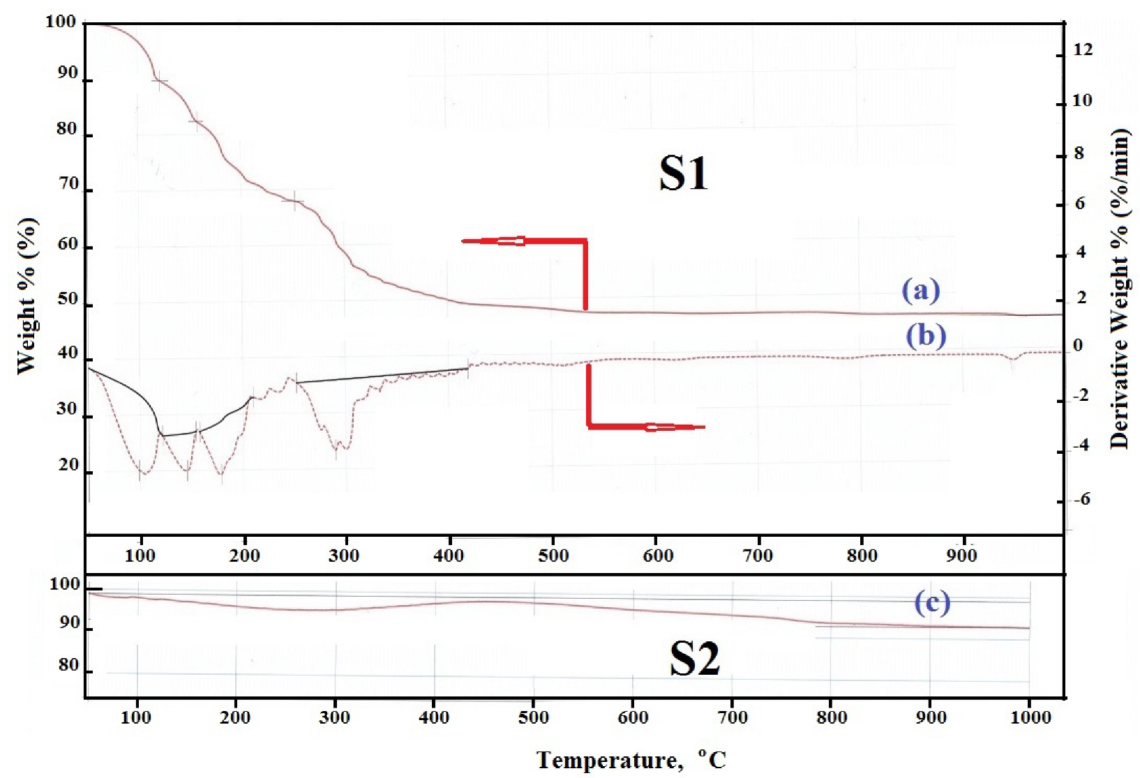
3.1. Thermal Analysis

TG and DTG curves of mixture of nickel and manganese nitrates, (S1), and TG of this mixture treated with 5 mL egg weight, (S2), are shown in Figure 1. Investigation of TG and DTG curves, Figure 1a,b, for the S1 sample revealed that: (i) the thermal treatment of the S1 sample resulted in dehydration, decomposition and solid state reaction processes which finish at about 1000 °C. (ii) Nickel nitrate hydrate dehydration as well as manganese nitrate hydrate occurs at room temperature but the path of dehydration is different in both cases. In case of the S1 sample, the dehydration of both nickel and manganese nitrates goes through three well-separated stages with subsequent release of both moisture and hydration water [35]. These stages confirmed by DTG peaks in the ranges of 50–120 °C, 121–152 °C and 153–251 °C with weight loss of 10.27%, 7.34% and 14.42%, respectively. (iii) The decomposition of anhydrous Ni and Mn nitrates obtained from hydrated nitrates is one-stage process occurring between 252 °C and 332 °C with a weight loss of 15.69% [35]. This observation confirms by a sharp endothermic peak maximized at 290 °C as shown in Figure 1b. This weight loss may be attributed to the decomposition of anhydrous mixed nitrates in order to manufacture the resulting oxides with release of nitrogen oxides indicating the completion of nitrate group’s decomposition. The final product for decomposition mixed hydrated nitrates is mixture of MnO2 and NiO. (iv) Slightly decrease of the sample mass was observed in the range from 333 °C to 1000 °C with a weight loss of 6.43%. This may be due to solid –solid interaction between the produced NiO and MnO2 yielding NiMn2O4 solid along the following reaction:
NiO + 2MnO2 ↔ NiMn2O4 + 0.5O2
TG curve, Figure 1c, for the S2 sample displays two stages containing different processes. This curve can be discussed as following: (i) the first step in this curve was observed in the range from 50 °C to 399 °C with losing weight 3%. This weight loss could be attributed to complete removal of moisture. Bearing in mind that the S2 sample was prepared via egg white assisted combustion method following by heating at 350 °C for 15 min, this sample has been exposed to both dehydration and decomposition processes with subsequent release of the crystallization water and nitrogen oxides due to presence of egg white as fuel. Based on the results of thermal analysis for the S1 sample, the S2 sample may contain mixture of NiO and MnO2 crystals. (2) The second step in TG curve of S2 sample starts from 450 °C to 1000 °C with a weight loss of 6.18% which agrees, approximately, with the theoretical value (6.43%) and confirms the complete conversion of NiO and MnO2 to NiMn2O4.

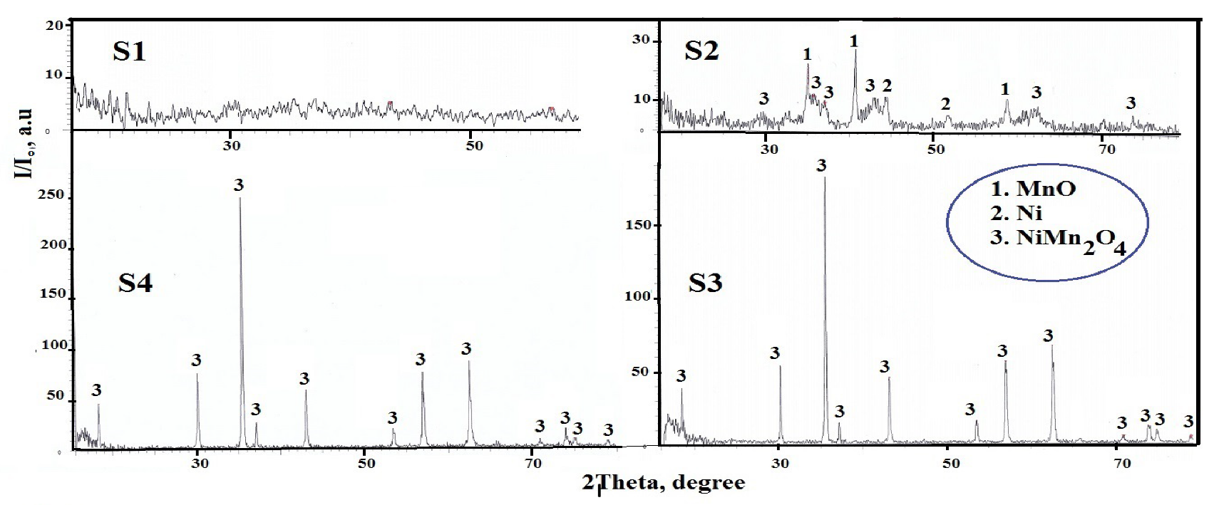
3.2. Phase Formation and Structural Analysis
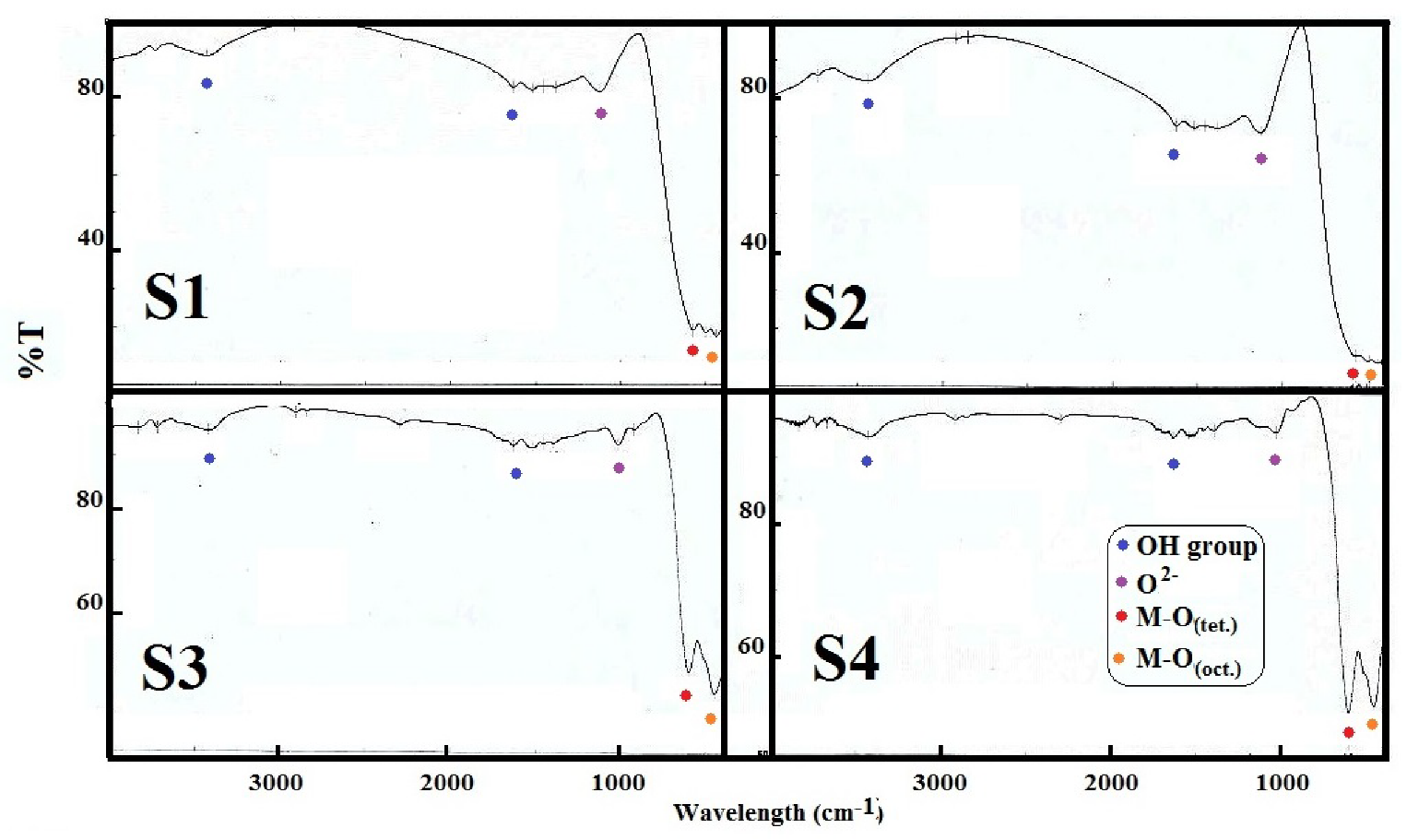
Determination of phase formation and different structural parameters of the as prepared materials carried out by using XRD technique. XRD patterns for the S1, S2, S3 and S4 samples are shown graphically in Figure 2. Enquiry of this figure revealed that: (i) The S1 sample is amorphous depending on the absence of any specified peak in its XRD pattern. (ii) Complementing the S2, S3 and S4 of XRD patterns with the data recorded in the reports (PDF No. 07-0230), (PDF No. 87-0712) and (PDF No. 71-0850) reveals that the prepared sample is crystalline and that the prevailing diffraction lines correspond to the lattices MnO, Ni and NiMn2O4, respectively. (iii) In case of the S2 sample, the addition of egg white as fuel resulted in crystallization the constituents of the mixed solids leading to formation of MnO, Ni and NiMn2O4 phases. In other words, the presence in egg white led to reduction of some nickel and manganese species with subsequent formation of Ni and MnO, respectively. Consequently, the S2 sample consisted entirely of MnO (major phase) and NiMn2O4 (minor phase) with presence small amount of Ni phase. This indicates that the presence of egg white stimulates the solid–solid interaction between the most species of nickel and manganese yielding NiMn2O4 phase. (iv) The augmentation in the preparation temperature to 950 °C for 2 h as shown in the S3 and S4 samples led to complete conversion of Ni and Mn species via solid state reaction between them yielding NiMn2O4 with cubic spinel type structure. In addition, the peaks height of NiMn2O4 for the S4 sample is greater than that of the S3 sample depending upon the promotion effect of egg white during the preparation process. In other words, the egg white assisted combustion method resulted in enhancement of the crystallinity of NiMn2O4 solid compared to ceramic method. This will in turn be reflected in an improvement in magnetic properties of NiMn2O4 solid.
Depending on Rietveld analysis of X-ray powder diffraction data, the actions of calcination temperature and fuel on the NiMn2O4 lattice structural parameters have been determined and tabled in Table 1 [36]. This table shows the values of, crystallite size (d), lattice constant (a), X-ray density (Dx), distance between reacting ions (LA and LB), ionic radius (rA, rB) and bond lengths (A–O and B–O) at tetrahedral (A) and octahedral (B) NiMn2O4 spinel sites. However, the rise in calcination temperature to 950 °C led to an increase in d values, a, LA, LB, rA, rB, A–O and B–O of NiMn2O4 lattice as shown in the S2 and S4 samples. Opposite result was observed in the value of Dx. Specific behaviors were found while comparing the samples from S3 and S4. In other words, the treatment with egg white at 950 °C led to an improvement in the values of previous structural parameters, except for the value of Dx. This confirms that preparing the NiMn2O4 solid with the help of egg white is much better than preparing it in a ceramic route.

3.3. FTIR Analysis

FTIR analysis of the S1, S2, S3 and S4 samples was shown in Figure 3. Study of the figure revealed: (i) Spectra of these samples display different bands located at 3438–3436, 1642–1636, 1034–1022, 602–596 and 451–444 cm−1. These bands enabled us to identify of functional group and structure elucidation for the investigated samples. (ii) The observed broad bands assigned to the mixed oxides at 3438–3436 cm−1 and 1642–1636 cm−1 may be attributed to the stretching and bending vibration of OH group for H2O [37]. However, the bands which appeared in the 1034–1022 cm−1 range may be correlated with the chemisorption’s of O2− on the sample surface during the cooling process: [38,39,40]. (iii) The spectra show sharp bands at 602–596 cm−1 and 451–444 cm−1. These bands are corresponded to the spinel structure of NiMn2O4 [41]. It is known that the characteristic bands of single and/or mixed oxides in the region of 4000 and 400 cm−1 are usually assigned to the vibration of metallic ions in the crystal lattice [41]. The spinel materials, however, are known to have two fundamental IR active modes in the vibration spectrum, which are high frequency bands around 600 cm−1 at the tetrahedral (A) site and low frequency bands around 400 cm−1 at octahedral (B) sites [42]. (iv)The divalent octahedral metal ion and oxygen ion complexes are suggested by the presence of the shoulder band at 513 cm−1 [43]. This shoulder band may be due to the presence of Ni2+ ions at B-site in this study [44,45]. (v) Slightly change in the position and intensity peaks of IR bands especially the characteristic bands for the spinel structure as shown in Table 2. This change is due to the difference in the preparation methods. Indeed, the intensity bands of the S4 sample prepared by combustion method are greater than that of the S3 sample prepared by ceramic method.
These results enabled us to investigate the cation distribution of the as synthesized spinel nickel manganite. Indeed, NiMn2O4 lattice have partially inverse spinel structure depending upon presence of Ni2+ ions at B-site and Mn3+ ions at A-site and B-site [44,45]. It can be suggested that this structure for the NiMn2O4 lattice can be obeyed the following formula:
(Mn3+)tet[Ni2+Mn3+]octO4
But, various factors such as preparation method brought about different changes in distribution of these cations between the mentioned sites with subsequent appearance of various valances of previous ions. Consequently, various authors found that NiMn2O4 lattice may be existence in random spinel structure with presence of both nickel and manganese ions in A- site and B-site. In this study, the calcination temperature and preparation method resulted in a portion of the Ni2+ cations migrate from octahedral to tetragonal interstices of cubic spinel structure. This migration will lead to presence of synergetic effect with subsequent appearance of both Mn2+ and Mn4+ ions as following:
Ni2+ + Mn3+ ↔ Ni3+ + Mn2+
Ni3+ + Mn3+ ↔ Ni2+ + Mn4+
The Mn2+ cations therefore switch to the tetrahedral sites to compensate for the Ni2+ vacancies.
Furthermore, the Mn2+ ions have a strong tendency to occupy A -sites while Ni2+ and Mn3+ occupy the B site preferentially [44,45]. Taking part account that some Ni2+ ions already present in A-site, we can be speculated the cation distribution in NiMn2O4 lattice as following:
(Nix2+Mny 2+)tet[Ni1−x2+Mn1−y3+Mny4+]octO4
In addition, the samples (S3 and S4) XRD patterns calcined at 950 °C showed distortion in the cubic spinel structure that is known from the peaks observed at (2 2 0) and (4 4 0) reflective planes. In addition, these planes’ intensities are more responsive to the cations on tetrahedral and octahedral sites, respectively [46]. The maximum increase in the intensity of (2 2 0) and (4 4 0) planes for the S4 sample attained 37.8% and 28%, respectively. This having found confirms that the egg white presence increased the migration of the Mn2+ cations to the tetrahedral sites to compensate for the Ni2+ vacancies.

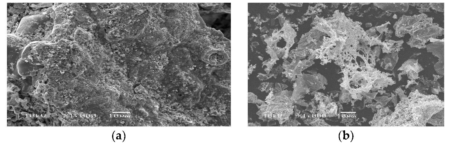
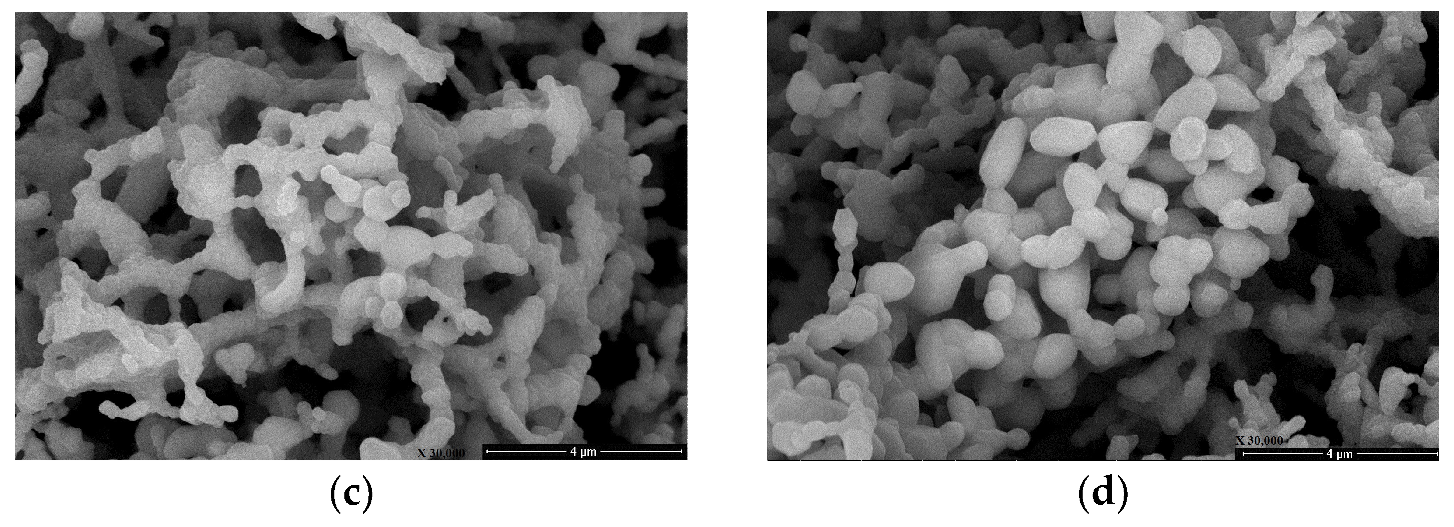
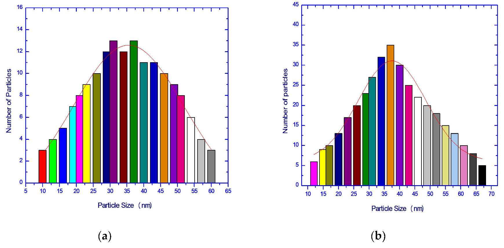
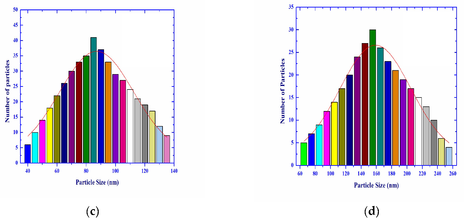
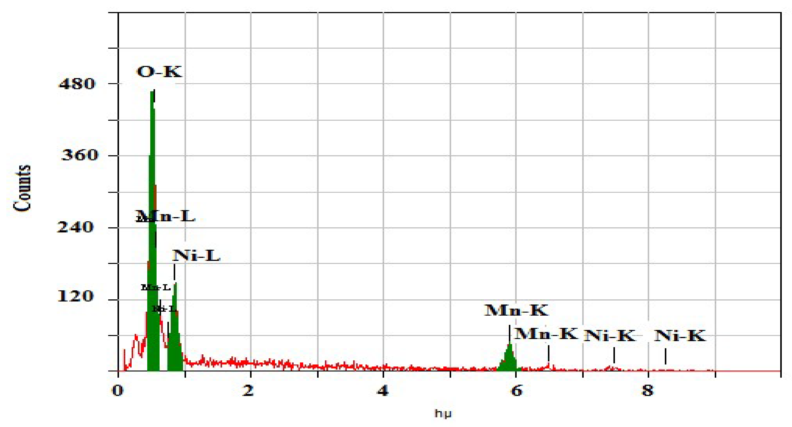
3.4. Surface Morphology and Compositional Analysis

Effects of the calcination temperature and egg white as fuel on the morphological characteristics of Ni-Mn mixed oxides were determined and shown in Figure 4a–d. Figure 4a represents the S1 sample heated at 350 °C for 15 min. As shown in this figure, eutectic crystal Ni-Mn nitrates was gray milky substance that looked like smooth block. Based on the photos, we could make the assumption that Ni and Mn oxides were well dispersed in eutectic crystal with some wrinkles and folds containing spherical particles. The SEM photograph (Figure 4b) of the S2 sample shows spongy fragile like structure containing pores and voids. Figure 4a,b shows well dispersed and homogeneous distributed particles in both eutectic crystals and spongy structure with no chemical reaction.
In addition, the calcination of the synthesized samples at 950 °C for 2 h brought about clearly modifications in their morphological features namely, size, shape, crystallization, structure and arrangement of grains as shown Figure 4c,d. Coexistence of nickel and manganese oxides together at high temperature stimulates the solid –solid interaction between them yielding cubic spinel nickel manganite particles with polyhedron structure. In Figure 5, there were histograms for particle size distribution of the as S1, S2, S3 and S4 samples based on SEM micrographs [47]. This figure shows that the average size ranged between 35 and 160 nm for different prepared materials. This indicates that the preparation of the sample by egg white assisted combustion method was enhanced the grain growth process. This result coincides with XRD measurement which indicates that the crystallite size of S4 crystals is greater than that of S3 crystals. The chemical composition of the samples as prepared could be further established by an EDS analysis that was attached to the SEM. For the S4 analysis, the representative EDS spectrum indicates that the mixed oxides consisted entirely of elements Ni, Mn and O as shown in Figure 6.

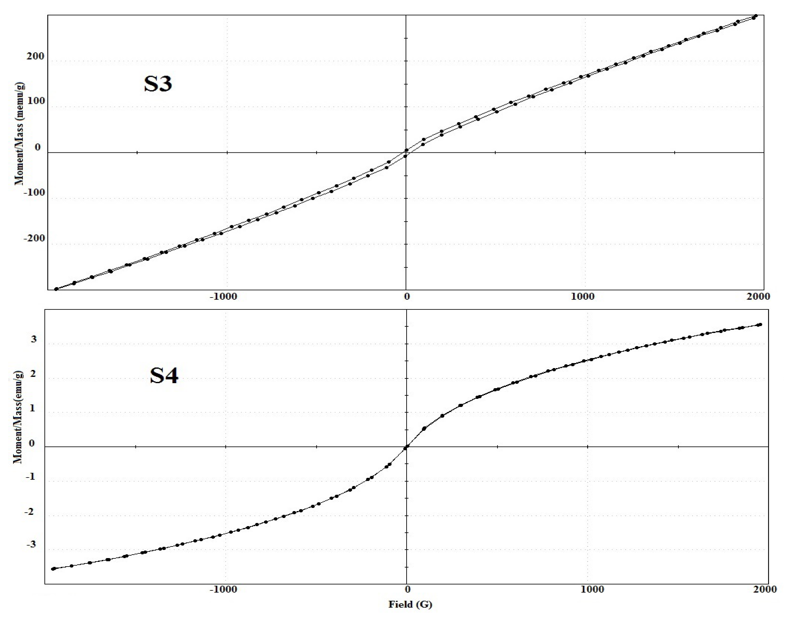
3.5. Magnetic Studies

VSM magnetometer has indeed been analyses the magnetic properties of the S3 and S4 samples. The observed curves of the hysteresis loop (M–H) of these samples are shown in Figure 7. Table 3 shows the relative magnetic properties for these samples, such as saturation magnetization (Ms), coercive field (Hc), retentivity (Mr/Ms), squareness ratio (Mr/Ms), anisotropic constant (Ka), and magnetic moment (μm). Inspection of this table revealed that: (i) the investigated materials display room temperature ferromagnetism (RTFM) behavior. The oxygen vacancies and size effect play an important role in introducing ferromagnetic of NiMn2O4 solid [48,49]. The egg white treatment followed by heating at 950 °C improves the sample crystallinity, increases the grain size, and decreases the number of spin suspension bonds on the surface, which lead to an increase in the net spin moment and Ms. (ii) For the S4 sample the values of Ms, Mr, μm and Ka were higher than for the S3 sample. Opposite behavior was noted in Mr and Mr/Ms values. This indicates that the method of combustion assisted by egg white enhances the magnetic properties of solid NiMn2O4 compared to that prepared by ceramic method.
However, the Zener double exchange effect (ZDE) cannot be ignored in improving the magnetic properties of NiMn2O4 solid through super interaction between ions of the same element in different valence states and distributed randomly over crystallographically equivalent lattice sites [48,49]. In addition, the existence of the Mnn+1/Mnn+ redox couple located in the semi-conductor spinel B-sites was considered the responsible factor of the controlled-valence conduction mechanism [50]. In particular, the contribution of super-exchange interaction between Mn3+–O2−–Mn4+ (B–B interaction) is suggested to be more pronounced in the case of the S4 sample. Opposite behavior was observed for super exchange interaction between Mn3+–O2−–Ni2+ (A–B interaction) because Mn2+ was increased through the partial reduction of Mn3+ by increasing the calcination temperature via egg white assisted combustion method. Thus, as the ratio of Mn4+/Mn3+ increases, as the value of Ms, Mr, μm and Ka increases as shown in the S4 sample.

4. Conclusions

For the first time: egg white assisted combustion route resulted in formation of NiMn2O4 solid with a good room temperature ferromagnetism behavior. In this study, the comparison between the preparation methods of NiMn2O4 solid was in favor of the combustion method at the expense of the ceramic method. From XRD results, the as synthesized material at 950 °C consisted entirely of NiMn2O4 NPs as a single phase with cubic spinel structure. The aspects and morphology of the prepared samples were characterized using techniques such as FTIR, EDS and SEM. In the future, this green process for synthesizing from of NiMn2O4 nanoparticles may also be applied to the manufacture of other metal oxides of industrial significance.

Author Contributions

Conceptualization, N.D. and O.H.A.-E.; methodology, N.D. and O.H.A.-E.; software, O.H.A.-E.; validation, N.D., O.H.A.-E. and M.Y.; formal analysis, N.D. and O.H.A.-E.; investigation, N.D. and O.H.A.-E.; resources, N.D. and O.H.A.-E.; data curation, N.D. and O.H.A.-E.; writing—original draft preparation, N.D. and O.H.A.-E.; writing—review and editing, N.D., O.H.A.-E. and M.Y.; visualization, N.D. and O.H.A.-E.; supervision, N.D. and O.H.A.-E.; project administration, O.H.A.-E.; funding acquisition, O.H.A.-E. All authors have read and agreed to the published version of the manuscript.

Funding

This research was funded by Deanship of Scientific Research, research group (RGP-306), King Saud University, Kingdom of Saudi Arabia.

Acknowledgments

The authors would like to extend their sincere appreciation to the Deanship of Scientific Research at King Saud University, Kingdom of Saudi Arabia, for funding the research group (RGP-306).

Conflicts of Interest

The authors declare no conflict of interest.

References

  1. Rozenberg, G.; Bäck, T.; Eiben, A.E.; Kok, J.N.; Spaink, H.P. Nanotechnology: Science and Computation; Springer: Berlin/Heidelberg, Germany, 2006; pp. 31–35. [Google Scholar]
  2. Miller, J.C.; Serrato, G.A.; Kundahl, J.K. The Handbook of Nanotechnology; John Wiley & Sons, Inc.: Hoboken, NJ, USA, 2004; pp. 22–34. [Google Scholar]
  3. Bergeson, L.L.; Clough, S.R.; Mackay, C.E.; Stephen, R.C.; Hoyt, M.; Chen, J.; Henery, K.; Hamblen, J. Nanotechnology and the Environment; Taylor and Fracis Group: New York, NY, USA, 2009; pp. 50–60. [Google Scholar]
  4. Kalantar, K.Z.; Fry, B. Nanotechnology-Enabled Sensors; Springer Science + Business Media, LLC: Sydeny, Australia, 2008; pp. 1–12. [Google Scholar]
  5. Feteira, A. Negative Temperature Coefficient Resistance (NTCR) Ceramic Thermistors: An Industrial Perspective. J. Am. Ceram. Soc. 2009, 92, 967–983. [Google Scholar] [CrossRef]
  6. Gillot, B.; Baudour, J.L.; Bouree, K.F.; Metz, R.; Legros, R.; Rousset, A. Ionic configuration and cation distribution in cubic nickel manganite spinels NixMn3−xO4 (0.57 < x < 1) in relation with thermal histories. Solid State Ion 1992, 58, 155–161. [Google Scholar]
  7. Hashemi, T.; Brinkman, W. Transport and x-ray photoelectron spectroscopy properties of (Ni1−xCux)Mn2O4 and Ni(Mn2−yCuy)O4. J. Mat. Res. 1992, 7, 1278–1282. [Google Scholar] [CrossRef]
  8. Elbadraoui, E.; Baudour, J.L.; Leroux, C.; Fritsch, S.; Bouree, F.; Guillot, B.; Rousset, A. Cation distribution, short-range order and small polaronhopping conduction in nickel manganites, from a neutrondiffraction study. Phys. Stat. Solidi 1999, 212, 129–139. [Google Scholar] [CrossRef]
  9. Almeida, J.M.A.; Meneses, C.T.; Menezes, A.S.; Jardim, R.F.; Sasaki, J.M. Synthesis and characterization of NiMn2O4 nanoparticles using gelatin as organic precursor. J. Magn. Magn. Mater. 2008, 320, e304–e307. [Google Scholar] [CrossRef]
  10. Fang, D.; Wang, Z.; Yang, P.; Liu, W.; Chen, C.; Winnubst, A.J.A. Preparation of Ultra-Fine Nickel Manganite Powders and Ceramics by a Solid-State Coordination Reaction. J. Am. Ceram. Soc. 2006, 89, 230–235. [Google Scholar] [CrossRef]
  11. Iagtap, S.; Rane, S.; Gosavi, G.; Amalnekar, D. Preparation, characterization and electrical properties of spinel-type environment friendly thick film NTC thermistors. J. Eur. Ceram. Soc. 2008, 28, 2501–2507. [Google Scholar]
  12. Groen, W.A.; Metzmacher, C.; Zaspalisa, V.; Huppertz, P.; Schuurman, S. Aging of NTC ceramics in the system Mn–Ni–Fe–O. J. Eur. Ceram. 2001, 21, 1793–1796. [Google Scholar] [CrossRef]
  13. Asbrink, S.; Waskowska, A.; Drozd, M.; Talik, E. Physical properties and X-ray diffraction of a NiMn2O4 single crystal below and above the ferromagnetic transition at Tc =145K. Phys. Chem. Solids 1997, 58, 725–729. [Google Scholar] [CrossRef]
  14. Díez, A.; Schmidt, R.; Sagua, A.E.; Frechero, M.A.; Matesanz, E.; Leon, C.; Morán, E. Structure and physical properties of nickel manganite NiMn2O4 obtained from nickel permanganate precursor. J. Eur. Ceram. Soc. 2010, 30, 2617–2624. [Google Scholar] [CrossRef]
  15. Aguilar-Garib, J.A.; Sánchez-de-Jesús, F.; Bolarín-Miró, A.M.; Ham-Hernández, S. Synthesis of NiMn2O4 assisted by high-energy ball milling of NiO-MnO powders. J. Ceram. Proc. Res. 2011, 12, 721–726. [Google Scholar]
  16. Durán, P.; Tartaj, J.; Rubio, F.; Peña, O.; Moure, C. Preparation and sintering behaviour of spinel-type CoxNiMn2−xO4 (0.2 ≤ x ≤ 1.2) by the ethylene glycol–metal nitrate polymerized complex process. J. Eur. Ceram. Soc. 2005, 15, 3021–3025. [Google Scholar] [CrossRef]
  17. Schmidt, R.; Brinkman, A.W. Preparation and characterisation of NiMn2O4 films. Int. J. Inorg. Mater. 2001, 3, 1215–1217. [Google Scholar] [CrossRef]
  18. Mehandjiev, D.; Naydenov, A.; Ivanov, G. Catalytic oxidation of CO and C6H6 on alumina-supported Cu-Cr and Co-Cr oxide catalysts in the presence of ozone. Appl. Catal. A Gen. 2001, 206, 13–18. [Google Scholar] [CrossRef]
  19. Shena, Y.I.; Nakayamaa, T.; Araia, M.; Yanagisawa, O.; Izumia, M. Magnetic phase transition and physical properties of spinel-type nickel manganese oxide. J. Phys. Chem. Solids 2002, 63, 947–950. [Google Scholar] [CrossRef]
  20. Ashcroft, G.; Terry, I.; Gover, R. Study of the preparation conditions for NiMn2O4 grown from hydroxide precursors. J. Eur. Ceram. Soc. 2006, 26, 901–908. [Google Scholar] [CrossRef]
  21. Zhao, C.; Wang, B.; Yang, P.; Winnubst, L.; Chen, C. Effects of Cu and Zn co-doping on the electrical properties of Ni0.5Mn2.5O4. J. Eur. Ceram. Soc. 2008, 28, 35–40. [Google Scholar] [CrossRef]
  22. Jung, H.R.; Lee, S.G.; Lee, D.G.; Kim, K.M. Characterization and site distributions of cation in nickel manganite thermistors. J. Ceram. Process Res. 2016, 17, 758–762. [Google Scholar]
  23. Deraz, N.M.; Abdeltawab, A.A.; Selim, M.M.; El-Shafey, O.; El-Asmy, A.A.; Al-Deyab, S.S. Precipitation-deposition assisted fabrication and characterization of nano-sized zinc manganite. J. Ind. Eng. Chem. 2014, 20, 2901–2904. [Google Scholar] [CrossRef]
  24. Deraz, N.M.; Abd-Elkader, O.H. Preparation of nanocrystalline ZnMn2O4 system by Sol gel route. Asian J. Chem. 2014, 26, 2125–2128. [Google Scholar] [CrossRef]
  25. Deraz, N.M.; Abd-Elkader, O.H. Effects of Precursor on Preparation and Properties of Nano-Crystalline Hopcalite Particles. Asian J. Chem. 2014, 26, 2133–2137. [Google Scholar] [CrossRef]
  26. Deraz, N.M. Green synthesis, Characterization and magnetic properties of hausmannite nano particles. Acta Phys. Pol. A 2019, 136, 147–150. [Google Scholar] [CrossRef]
  27. Karoui, S. Inflammatory Bowel Disease: Advances in Pathogenesis and Management; InTech: Rijeka, Croatia, 2011; p. 166. [Google Scholar]
  28. Huang, X.H.; Huang, X.Y.; Mao, H.K.; Yin, Z.X. Applied Mechanics and Mechatronics Automation; Trans Tech Publications Ltd.: Stafa-Zurich, Switzerland, 2012; p. 223. [Google Scholar]
  29. Sugimoto, K.; Dinnebier, R.E.; Schlechtb, T. Structure determination of Mg3(OH)5Cl·4H2O (F5 phase) from laboratory powder diffraction data and its impact on the analysis of problematic magnesia floors. Acta Cryst. 2007, 63, 805–811. [Google Scholar] [CrossRef] [PubMed]
  30. Cullity, B.D. Elements of X-ray Diffraction; Addison-Wesly Publishing Co. Inc.: Singapore, 1976; Chapter 14. [Google Scholar]
  31. El Nadi, M.L. Modern Trends in Physics. In Proceedings of the 4th International Conference on Modern Trends in Physics Research, Cairo, Egypt, 12–16 October 2010; World Scientific Publishing Co. Pte Ltd.: Singapore, 2013; p. 111. [Google Scholar]
  32. Kostecki, P.T.; Calabrese, E.J.; Bonzouuntas, M. Hydrocarbon Contaminated Soils; Lewis Publishers: Boca Raton, FL, USA, 1992; Volume 2, p. 346. [Google Scholar]
  33. Joseph, I.; Goldstein, D.E.; Newbury, J.R.; Michael, N.W.M.; Ritchie, J.H.J.; Scott, D.C.J. Scanning Electron Microscopy and X-ray Microanalysis, 4th ed.; Springer: New York, NY, USA, 2018; pp. 39–63. [Google Scholar]
  34. Azimi, G.; Kim, H.; Alam, S.; Ouchi, T.; Neale, R.; Alafara, N.; BabaRare, A. Metal Technology; Springer: Cham, Switzerland, 2019; p. 43. [Google Scholar]
  35. Małecka, B.; Lacz, A.; Drozdz, E.; Malecki, A. Thermal decomposition of d-metal nitrates supported on alumina. J. Therm. Anal. Calorim. 2015, 119, 1053–1061. [Google Scholar] [CrossRef] [Green Version]
  36. Boultif, A.; Louer, D. Powder pattern indexing with the dichotomy method. J. Appl. Cryst. 2004, 37, 724–731. [Google Scholar] [CrossRef]
  37. Wang, Z.; Liu, Q.; Yu, J.; Wu, T.; Wang, G. Surface structure and catalytic behavior of silica-supported copper catalysts prepared by impregnation and sol–gel methods. Appl. Catal. A General 2003, 239, 87–94. [Google Scholar] [CrossRef]
  38. Wei, Z.; Qiao, H.; Yang, H.; Zhang, C.; Yan, X. Characterization of NiO nanoparticles by anodic arc plasma method. J. Alloys Compd. 2009, 479, 855–858. [Google Scholar] [CrossRef]
  39. Deraz, N.M. Characterization and catalytic performance of pure and Li2O-doped CuO/CeO2 catalysts. Appl. Surf. Sci. 2009, 255, 3884–3890. [Google Scholar] [CrossRef]
  40. Salavati-Niasari, M.; Mir, N.; Davar, F. Synthesis and characterization of NiO nanoclusters via thermal decomposition. Polyhedron 2009, 28, 1111–1114. [Google Scholar] [CrossRef]
  41. Waldron, R.D. Infrared Spectra of Ferrites. Phys. Rev. 1955, 99, 1725–1727. [Google Scholar] [CrossRef]
  42. Ouaguenouni, N.H.; Benadda, A.; Kiennemann, A.; Barama, A. Preparation and catalytic activity of nickel–manganese oxide catalysts in the reaction of partial oxidation of methane. Comptes Rendus Chim. 2009, 12, 740–747. [Google Scholar] [CrossRef]
  43. Kavitha, N.; Lakshmi, P.V.A. Synthesis, characterization and thermogravimetric analysis of Co(II), Ni(II), Cu(II) and Zn(II) complexes supported by ONNO tetradentate Schiff base ligand derived from hydrazino benzoxazine. J. Saudi Chem. Soc. 2017, 21, S457–S466. [Google Scholar] [CrossRef] [Green Version]
  44. Berchmansa, L.J.; Selvana, R.K.; Kumara, P.N.S.; Augustina, C.O. Structural and electrical properties of Ni1−xMgxFe2O4 synthesized by citrate gel process. J. Magn. Magn. Mater. 2004, 279, 103–110. [Google Scholar] [CrossRef]
  45. Deraz, N.M.; Shaban, S. Optimization of catalytic, surface and magnetic properties of nanocrystalline manganese ferrite. J. Anal. Appl. Pyrol. 2009, 86, 173–179. [Google Scholar] [CrossRef]
  46. Alarifi, A.; Deraz, N.M.; Shaban, S. Structural, morphological and magnetic properties of NiFe2O4 nanoparticles. J. Alloys Compd. 2009, 486, 501–506. [Google Scholar] [CrossRef]
  47. Rasband, W.S. ImageJ; U.S. National Institutes of Health: Bethesda, MD, USA, 1997.
  48. Xu, Q.; Gao, D.; Zhang, J.; Yang, Z.; Zhang, Z.; Rao, J.; Xue, D. Observation of room temperature ferromagnetism in pure La2O3 nanoparticles. Appl. Phys. A 2014, 116, 1293–1298. [Google Scholar] [CrossRef]
  49. Kisan, B.; Alagarsamy, P. Room temperature ferromagnetism in finite sized ZnO nanoparticles. Physica B 2014, 448, 115–119. [Google Scholar] [CrossRef]
  50. Verwey, E.J.W. Oxidic Semiconductors in Semi-Conducting Materials; Henisch, H.K., Ed.; Butterworths Sc Pub Ltd.: London, UK, 1951; p. 151. [Google Scholar]
Figure 1. Thermal analysis of the prepared samples; (a) TG of S1, (b) DTG of S1 and (c) TG of S2.
Crystals 10 00489 g001
Figure 2. XRD patterns of S1, S2, S3 and S4 samples.
Crystals 10 00489 g002
Figure 3. FTIR patterns of S1, S2, S3 and S4 samples.
Crystals 10 00489 g003
Figure 4. SEM images of the prepared samples; (a) S1, (b) S2, (c) S3 and (d) S4 samples.
Crystals 10 00489 g004aCrystals 10 00489 g004b
Figure 5. Histograms of particle size distribution based on SEM micrographs images for the prepared samples; (a) S1, (b) S2, (c) S3 and (d) S4 samples.
Crystals 10 00489 g005aCrystals 10 00489 g005b
Figure 6. EDS pattern of S4 sample.
Crystals 10 00489 g006
Figure 7. M-H curves for the S3 and S4 samples.
Crystals 10 00489 g007
Table 1. Lattice parameters of NiMn2O4 Phase.
SampleS2S3S4
Parameters
d, (nm)507690
a, (nm)0.83510.83570.8381
V, (nm3)0.58240.58740.5887
Dx, g/cm35.3035.2585.246
LA (nm)0.36160.36260.3629
LB (nm)0.29510.29600.2962
A-O (nm)0.19090.19150.1916
B-O (nm)0.21550.21610.2162
rA (nm)0.05980.05950.0596
rB (nm)0.08350.08410.0842
I222/I4400.6250.8190.875
Table 2. Characteristic infrared frequencies of the as prepared materials.
Functional GroupsWavelengths, cm−1Intensity, %
OHstret.3438–343694.76–84.47
OHbend.1642–163694.10–73.19
Chemisorbed O21034–102294.10–72.31
M-Otet.602–59651.90–13.13
M-Ooct.451–44452.9–11.70
Table 3. The magnetic properties (Ms, Mr, Mr/Ms, Ka and Hc) of the S3 and S4 samples.
SamplesMs
(emu/g)
Mr
(emu/g)
Mr/Ms
(emu/g)
Hc
(Oe)
μmAnisotropy Constant (Ka)
J/m3
S30.29865.57 × 10−318.66 × 10−3215.160.012465.56
S43.558812.57 × 10−33.53 × 10−323.040.148283.69

