SHH Signaling Pathway Drives Pediatric Bone Sarcoma Progression


INSERM UMR1238, PHY-OS, “Bone sarcomas and remodeling of calcified tissues”, Nantes University, 44000 Nantes, France
*
Author to whom correspondence should be addressed.
Cells 2020, 9(3), 536; https://doi.org/10.3390/cells9030536
Submission received: 30 January 2020
/
Revised: 19 February 2020
/
Accepted: 23 February 2020
/
Published: 26 February 2020
(This article belongs to the Special Issue Molecular and Cellular Mechanisms of Cancers: Bone Sarcomas)
Abstract
:Primary bone tumors can be divided into two classes, benign and malignant. Among the latter group, osteosarcoma and Ewing sarcoma are the most prevalent malignant primary bone tumors in children and adolescents. Despite intensive efforts to improve treatments, almost 40% of patients succumb to the disease. Specifically, the clinical outcome for metastatic osteosarcoma or Ewing sarcoma remains poor; less than 30% of patients who present metastases will survive 5 years after initial diagnosis. One common and specific point of these bone tumors is their ability to deregulate bone homeostasis and remodeling and divert them to their benefit. Over the past years, considerable interest in the Sonic Hedgehog (SHH) pathway has taken place within the cancer research community. The activation of this SHH cascade can be done through different ways and, schematically, two pathways can be described, the canonical and the non-canonical. This review discusses the current knowledge about the involvement of the SHH signaling pathway in skeletal development, pediatric bone sarcoma progression and the related therapeutic options that may be possible for these tumors.
1. Introduction: Osteosarcoma and Ewing Sarcoma
Osteosarcoma (OS) and Ewing sarcoma (ES) are the two most common malignant primary bone tumors observed in children, adolescents and young adults, occurring primarily in the second decade of life (median age occurrence of 18 and 15 years, respectively, for OS and ES). They are qualified as rare tumors as they represent about 6% of all childhood malignancies and less than 0.2% of all cancers, whatever the age.
Osteosarcoma is the most frequent type of malignant primary tumor of the bone, with an incidence of three in one million. Boys are slightly more affected than girls. About 80% of osteosarcoma occur at the metaphysis of long bones, with the most common sites being the distal femur, the proximal tibia and the proximal humerus. Conventional OS, defined by cells that produce varying amounts of osteoid matrix (mineralized or not), is the most common histologic subtype, accounting for approximately 75% of all cases [1,2,3]. It is then divided in three major histologic subtypes: osteoblastic (50%), chondroblastic (25%) and fibroblastic (25%) OS. This rare cancer (around 600 cases per year in Europe) presents radiographically-mixed lesions with lytic and sclerotic territories, in variable proportion depending on the tumors [4]. Whole-genome sequencing analyses have shown that OS displays high rates of genetic alterations, containing many somatic mutations and copy number alterations. TP53 and RB1 show recurrent somatic alterations in concordant studies, suggesting that they could be key players in bone oncogenesis [5].
Ewing sarcoma is the second most common malignant primary bone tumor in children, adolescents and young adults. The annual incidence is estimated at 1–3 in one million. Staging procedures identify about 25% of patients that have metastases at diagnosis. Typically, ES arises in bone sites, with the most common sites including the long bones (47%), pelvis (26%), chest wall and spine. In long bones, ES predominantly occurs at the diaphysis. The osseous mass may coexist with a soft tissue component, resulting in a Coleman triangle or a multilayered periosteal reaction. On the contrary to OS, ES can occasionally develop in soft tissues [6]. The Ewing sarcoma family is characterized as tumors consisting of small round malignant cells that may exhibit different levels of neural differentiation. Ewing sarcoma, malignant peripheral neuroectodermal tumor, Askin tumor and atypical Ewing sarcoma are now summarized under the term Ewing sarcoma. Tumor-specific, non-random gene rearrangements of chromosome 22 are found in more than 95% cases. The most common rearrangement is the t(11 :22)(q24 :q12) translocation, found in 85% of these tumors [7].
2. Current Main Treatments and Perspectives
Significant improvement in overall survival has been achieved with the use of multi-agent chemotherapy in conjunction with local therapy approaches, namely surgery and radiotherapy in the case of Ewing sarcoma.
However, despite multi-agent chemotherapy and aggressive surgical resection sparing the limb, 30% of OS patients with localized disease and 80% with metastatic disease at diagnosis will relapse [8]. Standard therapy consists of the surgical removal of any resectable primary tumor and metastases, combined with neoadjuvant and adjuvant regimens of multi-agent chemotherapy, including four agents: doxorubicin (adriamycin), cisplatin, high-dose methotrexate and ifosfamide for patients with high-risk or metastatic disease. Surgery is the only adequate available treatment option to achieve local control in osteosarcoma. Indeed, due to the low radiosensitivity of osteosarcoma, radiotherapy does not play a major role in the multimodal treatment. However, there may be some situations where its use may be beneficial, for example, when primary lesions are not accessible for surgery. This modern multimodal therapy yields 75% survival at 5 years for osteosarcoma patients without overt metastasis at diagnosis, but the outcome for metastatic, relapsed disease (locally or distal) or for patients not responding to chemotherapy remains poor, at less than 30% survival at 5 years.
Before the era of combination chemotherapy, the prognosis of ES patients was poor, with more than 90% of patients dying from secondary metastases [9]. The current standard of care for ES consists mainly of chemotherapy with 5 drugs (vincristine/doxorubicin/cyclophosphamide alternating with ifosfamide/etoposide) and associated with local surgery or radiotherapy when surgery is difficult [10]. While this intensive multi-agent chemotherapy has improved survival compared with the pre-chemotherapy era, there have been few recent improvements in the outcome for ES patients with either localized disease or those who present with metastatic disease at diagnosis. In addition, it has been difficult even for therapies that prove to be beneficial, such as mifamurtide in osteosarcoma, to obtain regulatory approval [11].
However, great advances have been made recently in understanding the molecular basis of pathogenesis and the progression of OS and ES. This new understanding has been achieved in parallel with an explosion of novel therapies developed to specifically inhibit cancer-associated genes and pathways. Identification of key regulatory pathways and molecular biomarkers has yielded dramatic changes in the outcome for several adult cancers, but childhood cancers, in general, and bone sarcoma, in particular, have been largely sidelined in this revolution.
To help make these important discoveries relevant for pediatric bone sarcoma, it is important to have an understanding of the role of each signaling pathway in the biology of the disease as well as of the available agents that target these processes. Priority was given to the pathways for which there is good information available about the relevance to OS and ES. The main novel therapies that can be applied in children’s bone sarcomas involve (i) surface markers for OS (receptor tyrosine kinases, such as insulin-like growth factor receptor type I, human epidermal growth factor receptor 2 and the ERBB family, platelet-derived growth factor receptor), (ii) intracellular signaling pathways (Ezrin, mammalian target of rapamycin, steroid receptor co-activator, Notch, Hedgehog, histone deacetylase inhibitors, Ras, MDM2), (iii) targeting bone metabolism (bisphosphonates, conjugated radioisotopes, denosumab), and (iv) environmental and immune interactions of bone sarcoma (immunotherapy: mifamurtide, sargramostim, other immunomodulators, such as interferon-γ, other immunotherapies including tumor vaccine, targeted therapy using antibodies for GD2 tumor antigens). Immunotherapy approaches are promising for pediatric sarcoma treatment. Environmental interactions involving the matrix and vasculature are also attracting. Part of the pathogenesis of bone sarcomas includes the ability to invade through extracellular matrix tissues and to recruit a new blood supply as tumors grow and disseminate [12]. These activities proceed by hijacking normal biological processes that are then exploited by tumor cells to facilitate their growth and spread [13]. Inhibition of MMP2 and MMP9 affects OS tumor growth and metastasis formation [14,15]. Preclinical efficacy of VEGF-based therapeutics, including anti-VEGF antibodies and small molecule inhibitors against VEGFR, have been confirmed in pediatric bone sarcomas [16]. Very recently, a strategy using regorafenib, a multi-kinase inhibitor of angiogenic (VEGF-R1-3, TIE-2), stromal (PDGFR-β, FGFR) and oncogenic kinases (KIT, RET, and RAF), was tested. Two French and American clinical trials were conducted in patients with recurrent, progressive, metastatic OS after failure of conventional chemotherapy and they demonstrated a significant extension of progression-free survival in both trials [17,18].
3. SHH Signaling Pathway
The Hedgehog (HH) signaling pathway is a major evolutionarily conserved pathway that regulates key events during the development process and tissue homeostasis, such as bone [19]. The HH gene was discovered in 1980 by Nusslein-Volhard and Wieschaus through the analysis of Drosophila melanogaster [20]. In the early of 1990s, three HH ligands were described in vertebrates: Sonic, Indian and Desert Hedgehog, differentially expressed depending on the animal species. In humans, Sonic Hedgehog (SHH), the main ligand, is produced and transported into the endoplasmic reticulum (ER) and Golgi apparatus in which it undergoes autoprocessing [21]. SHH, initially synthetized as a 45kDa precursor, is thus cleaved into a 19 kDa (SHH-N) and a 26 kDa (SHH-C) secreted peptide. SHH-N is modified by the addition of lipid (palmitoyl and cholesterol) and mediates signaling in vertebrates and invertebrates [22]. SHH-C is believed to possess any biological activity under physiological condition [23]. Of note, SHH at the cell surface can be released by lipid bilayer membrane vesicles, called exosomes [24,25]. The activation of the SHH cascade can be done through different pathways: schematically, two pathways can be described, the canonical (Figure 1) and the non-canonical.
The canonical cascade is based on the binding of SHH on the 12-transmembrane receptor Patch (Ptch) [26], which, in the absence of ligand, continuously inhibits the activity of the G-coupled receptor Smoothened (SMO). In the presence of ligand, Ptch undergoes endocytosis, which leads to its degradation in the lysosome [27], resulting in the activation of SMO. Suppressor of fused homolog (Sufu) is a crucial inhibitor of the SHH cascade [28]. Indeed, Sufu sequesters glioma-associated oncogene homolog (Gli) in the cytosol [29]. The mammalian Gli gene family comprises three isoforms: Gli1, Gli2 and Gli3 (Figure 1). Of note, the human Gli1 gene was first identified by Volgestein and colleagues as a putative oncogene amplified in glioblastoma [30]. SMO activation generates an intracellular signal that induces the dissociation of the Gli-Sufu complex and promotes the translocation of Gli proteins in the nucleus where they act as transcriptional factors by binding to a consensus sequence of 5’-GACCACCCA-3’ [31,32] through the DNA-binding domain, which consists of five C2H2-Kruppel-type zinc-finger motifs [33,34]. The transcriptional activity of Gli promotes the expression of various genes among them: SHH cascade compounds, such as Gli1 and Ptch1, pro-proliferation genes, such as Cyclin D1 and Myc, or cell cycle regulators, such as CCND2 and CCNE1 [35].
The non-canonical cascade exists to elicit various cellular responses, especially in cancers [36]. One of the mechanisms of non-canonical SHH signaling involves the activation of Gli transcriptional factors independently of SHH, Ptch and SMO. For example, epidermal growth factor receptor (EGFR) can induce the activation of Gli through the extracellular signal-regulated kinase (ERK) pathway during oncogenic transformation [37]. Ras signaling induces Gli1-transcription in gastric cancer [38]. The TGF-β cascade can also enhance Gli expression, especially Gli2, via Smad3-dependant mechanisms in melanoma [39,40,41]. Regarding bone sarcoma, it has been established that Gli1 is a direct transcriptional target of EWS-Fli1 in Ewing Sarcoma [42].
4. SHH and Skeletal Development
The skeleton is composed of five different mineralized tissues. The bone and cartilage tissues constitute the main part of the skeleton and the enamel, dentin and cementum are dental-specific tissues. Embryologically, the cells forming these tissues derive from the neural crest for the craniofacial skeleton (most skull bones and teeth) and from the paraxial (somatic, intermediary and lateral) mesoderm for the axial and appendicular skeletons (Figure 2). SHH was shown to be a crucial factor for the migration and the predetermination of progenitor cells for skeletal tissues that originated from either the neural crests or mesoderm [43,44,45,46,47,48,49,50,51]. Indeed, any perturbation of SHH expression/function during these early steps of embryonic development leads to severe alterations of the skeleton development with reduction in the size or absence of certain elements of the craniofacial, axial or appendicular skeletons [49,52,53,54,55,56,57,58,59,60,61,62,63,64]. Such alterations of SHH, whatever their causes, are often lethal before birth as they are also associated with the defective formation of important none-skeletal organs.
During the following embryonic steps of skeleton formation, corresponding to the morphogenesis of the different skeletal elements, which is governed by numerous sequential interactions between tissues (epithelial-mesenchymal, neuro-mesenchymal, etc.), SHH was shown to be an important morphogen [49,65,66,67,68,69]. Indeed, SHH expression was associated with different organization centers participating with other factors of the FGF, BMP and WNT families to determine morphogenic fields.
In the craniofacial skeleton, SHH is expressed in the first branchial arch [49,70], participating successively to determine Meckel’s cartilage formation [45,71] (Figure 3A) and the dental regions (Figure 3B) [72,73,74,75,76,77] in the frontonasal prominences [78] that govern the formation of the different upper facial structures (Figure 3C) and in the cranium sutures that control the harmonious growth of the different bones of the skull with the underlying forebrain [79,80,81,82].
In the appendicular skeleton, SHH is implicated in the morphogenesis of the limb [83,84], which can be subdivided into three stages called the bud, the pallet and the rotations. The limb buds appear as expansions of the lateral mesoderm covered by the ectoderm (Figure 3D). A thickening of the ectoderm at the distal end of each bud forms the apical ectodermal ridge (AER). This ridge plays a fundamental role in elongating the bud (Figure 3E). The underlying mesenchyme is called the progression zone (Figure 3E). Another important mesenchymal area in the posterior part of the bud is called the zone of polarizing activity (ZPA) that determines the developing bud along the anterior/posterior axis (Figure 3E). SHH is expressed in the ZPA [85], whose function is the early determination of the proximal-distal identity of the mesenchymal cells from the limb bud [86,87,88,89,90] with an important consequence for the autopod and zeugopod polarization (Figure 3E). The pallet stage is characterized by the rapid elongation of the bud and the formation in its distal part of a flattened structure: the pallet. As the pallet grows, inter-digital furrows appear, resulting from the apoptosis of mesoderm cells that separate pre-cartilage blanks from fingers and toes. The digits that form in the posterior region are known to be under the influence of SHH [89,91,92,93]. Any perturbations in the SHH expression level or pattern during the bud and pallet stages of limb morphogenesis, whatever their genetic or environmental origins, lead to a large spectrum of limb dysmorphic features ranging from the absence of most of the autopod to polydactylism [94,95,96,97]. During the last stage of the limb morphogenesis, the rotation stage, the arm and leg blanks rotate so that the elbow is laterally oriented and the knee is ventral. To date, SHH has not been implicated in the rotation stage.
In the axial skeleton, which derives from the somitic mesoderm (Figure 2), SHH also plays a major morphogenetic role. Embryonic precursors of cartilage in the ribs and vertebrae are localized within the somite. Somites are paired segmented structures that form epithelial spheres from the ventral region, which cells leave to form the sclerotome. The ventral portion of the sclerotome surrounds the notochord and forms the rudiment of the vertebral body while the dorsal portion surrounds the neural tube and forms the rudiment of the vertebral arches. SHH, produced by the notochord, was shown as the primary signal for sclerotome induction. In cases of SHH expression disruptions in the notochord, the axial skeleton formation is always severely affected with presence of caudal dysgenesis, cyclopia and rib absence [98,99,100,101,102].
After morphogenesis, the organogenesis of the skeletal elements is initiated. SHH was implicated in both tooth and bone organogenesis. Concerning the tooth, SHH was expressed by the main organization centers, called enamel knocks, that control the proliferation and functional differentiation of the cells secreting the enamel and the dentine, respectively called the ameloblast and odontoblast. Defects in SHH expression in the enamel knocks lead to severe alterations of the size and the shape of the teeth and to disruptions of the polarity and organization of the ameloblast and odontoblast layers [75]. Regarding bone organogenesis, three bone formation processes exist: the endochondral, the intramembranous and the periosteal apposition. Endochondral bone formation is the process by which growing cartilage in the extremities of long bone is systematically replaced by bone. This process is implicated in the growth in the length of long bone. The periosteal apposition enables the growth in the width of the long bone and is secondary to the endochondral bone formation. The membranous bone formation, which is implicated in the formation of flat bones (mainly in the skull), is initiated by a condensation of mesenchymal stem cells that gradually differentiate into osteoprogenitor then osteoblast cells and directly secrete a bone matrix without requiring a cartilaginous phase.
Regarding endochondral bone formation, SHH has not been directly implicated, in contrast to IHH (Indian Hedgehog), which plays a major role in the differentiation of chondroblast cells of the growth plate [103,104,105]. In intramembranous bone formation and the periosteal apposition, several studies have established that SHH can modulate the differentiation and activation of osteoblastic cells via its expression in the primary cilia [106,107,108]. Interestingly, studies focusing on the healing and/or regeneration of bone and cartilage tissues have established that SHH was able to improve the neo-differentiation of osteoblasts and chondroblasts with an interesting therapeutic potential [109,110,111,112,113].
5. SHH Signaling in OS et ES
Beside its critical role in multiple developmental processes of organs and tissues, such as bone (see above), the Hedgehog-Gli signaling pathway is aberrantly activated in several human cancers [35,36,114,115,116,117,118].
Mutations in some of the genes involved in the SHH cascade, such as SMO, Ptch or Sufu, have been associated with the development of cancers [119,120,121]. For example, mutations of Ptch were originally described in Gorlin syndrome, a rare hereditary disease with an autosomal dominant transmission, characterized by the association of multiple basal cell carcinoma, medulloblastoma and rhabdomyosarcoma [122,123]. Sporadic mutations of SHH have been described in basal cell carcinoma, medulloblastoma and in breast carcinoma cell lines [117,124].
The SHH pathway can be also activated without mutations of the genes involved in the SHH cascade, mainly through a paracrine effect of SHH and, thus, on the canonical cascade [35,36,115,116,117,118]. Schematically, Gli transcriptional factor may act at different steps of tumorigenesis [118]. The main properties of Gli family proteins are to stimulate cell proliferation and tumor growth in many cancers [35,36,115,116,117,118]. The SHH cascade was also implicated in the ability of cells to (i) acquire a migratory phenotype and (ii) migrate, invade adjacent tissue and, thus, to metastasis [35,36,115,116,117,118].
In the context of bone tumors, the relevance of the HH pathway emerged progressively in the last decade in osteosarcoma (OS) and Ewing sarcoma bone tumors. Several preclinical studies highlighted the overexpression of HH components in OS cell lines. Ligands SHH, DHH and IHH, Receptors Ptch1 and SMO, and transcription factors Gli1 and Gli2 are significantly overexpressed in human osteosarcoma cell lines, both at the mRNA and protein level [125]. Importantly, these findings were also validated in OS tumor patients; several studies investigated the HH pathway on human samples mainly by RT-PCR analysis of HH component genes. In a small cohort of 12 patients at diagnosis, expression of SMO, PTCH1 and Gli2 transcripts were variously increased [126]. Recently, an exploratory study of surgical biopsies of 43 high-grade OS also revealed high gene expression of IHH, PTCH1 and Gli1, and moderated expression of SMO, normalized to normal osteoblast samples [127]. The levels of IHH, SMO, PTCH1 and Gli1 were correlated with each other in small tumors, indicative of a ligand-independent activation whereas ligand-dependent activation of the HH pathway was suggested in large tumors, as high expression of IHH and PTCH1 were correlated. Importantly, this study aimed at evaluating the prognostic value of the expression of the HH pathway and, despite no statistically differences due to the limited sample size, suggested that the expression of IHH could be predictive of outcome in combination with tumor size, as patients with lower IHH expression and small tumors had better survival compared to patients with high IHH expression and large tumors. A positive correlation was also identified between Gli1 expression and chemotherapy-induced necrosis, indicative of increased responsiveness to treatment in patients stratified into the high Gli1 expression group. Furthermore, Yang et al. [128] showed, in an immunohistochemistry study on a tissue-microarray of 58 OS samples, that overall survival was shortened in patients with high Gli2 expression and that, during the 5 years of follow-up, living patients significantly expressed lower Gli2 compared to patients who died. Altogether, these results suggested that expression of HH signaling components might be useful as a prognostic but also as a theranostic factor.
The functional significance of altered HH signaling in OS has been deciphered in several models of cell lines and in preclinical animal models. SMO-dependent HH signaling regulates the cell cycle in OS, favoring progression into G1 through regulation of cyclin D1 and E1. Moreover, inhibition of SMO by cyclopamine, a specific inhibitor of SMO, or by genetic invalidation of SMO, prevented cell proliferation in vitro and tumor growth in vivo [126]. Additional studies focused on the role of the transcription factor Gli2 in OS development and progression. In SaOS2 and 143B OS cell lines, inhibition of Gli2 prevented OS proliferation in vitro, through inhibition of cell cycle proteins cyclin D1, pRb and SKP2 [129]. Importantly, overexpression of potent activated Gli2 in immortalized human mesenchymal stem cells stimulated their proliferation, reinforcing a role for Gli2 in osteosarcoma genesis as osteosarcoma cells are of mesenchymal origin. In addition, invalidation of Gli2 in KHOS and U2OS cell lines has been shown to induce a decrease in OS proliferation both in 2D and 3D cultures and to promote OS cell sensitivity to chemotherapies doxorubicin and methotrexate [128]. Use of GANT61, a Gli1/2 antagonist, also displayed an inhibitory effect on OS cell lines [130]. Several strategies targeting the HH signaling pathway in preclinical animal models have reinforced the relevance of this pathway in OS. In a 143B-induced xenograft model, targeting of Gli2 by genetic invalidation in tumor cells inhibited tumor growth and provided a significant survival benefit [129]. In patient-derived xenograft (PDX) models, Lo et al. treated mice orally with IPI-926, a semi-synthetic analog of cyclopamin and small-molecule inhibitor of SMO, and showed a specific inhibition of ligand-dependent HH pathway and a significant decrease in tumor weight and volume [130]. Targeting Gli proteins with arsenic trioxide (ATO) in a nude mouse model of subcutaneously-grafted 143B OS cells also prevented OS growth [131]. Arsenic trioxide was initially described as a potent antagonist of the HH pathway by preventing the accumulation of Gli2 in primary cilia by reducing its stability [132]. However, this chemical was also identified as an inhibitor of the transcriptional activity of Gli1/-2 [133] in Ewing sarcoma. In OS cell lines, ATO promotes apoptotic cell death and, importantly, induces the accumulation of DNA damage [131]. This latter result suggests that activated HH signaling pathways could protect OS cancer cells from DNA damage-induced death. This type of death is required for an optimal efficiency of radiotherapy or chemotherapy. Therefore, cancer cells harboring activated HH pathways could present some level of radio-resistance. In fact, Chen et al. demonstrated that activation of the HH pathway protected human hepatocellular carcinoma cells from ionizing radiation through an unclear mechanism of impaired DNA damage repair machinery [134]. Importantly, this radioprotection was alleviated by SHH antibody neutralization or Gli1 invalidation. Very recently, activation of SHH signaling was found to be involved in the resistance of OS cells to ionizing radiations [135] as radio-resistant OS MG63 cells expressed increased level of SHH, SMO, Ptch1 proteins and increased nuclear localization of Gli1 compared to radiosensitive cells, whereas silencing of SHH rendered the radio-resistant cells more radiosensitive [136]. In summary, the targeting of HH signaling appears to be an important strategy to consider in OS treatment, both for direct anti-tumoral action and also for a potentiation of the therapeutic effects of chemotherapy and/or radiotherapy.
Mechanisms of Gli2 overexpression in OS are still unclear. Both ligand-dependent and ligand-independent activation of HH have been highlighted in OS cells [130]. Moreover, the exclusive expression of SHH in the stroma compartment in a patient-derived xenograft model suggested an active paracrine communication between tumor and stroma [130]. Indeed, a dialogue between stroma and OS cells in the activation of the HH signaling pathway has been recently identified as extracellular vesicles (EVs) isolated from MSC stimulated the expression of Ptch1, SHH, and Gli1 proteins in OS cells, associated with increased cell proliferation and migration [137]. A recent study also reported the decreased expression of miR-141-3p, an miR targeting Gli2 in OS cell lines and tumor samples, suggesting an miR implication in HH-altered pathways in OS [138].
Literature is scarce when considering the HH pathway and Ewing sarcoma (ES). Nevertheless, several genes of the HH pathway were found to be associated with metastasis in a micro-array analysis of 27 ES patients [139]. The transcription factor Gli1 appears particularly important in ES, as it is overexpressed in patient tumor specimens and in ES cell lines [140]. Gli1 was identified as a direct transcriptional target of EWS-FL1 [42] through an HH ligand-independent mechanism and appears essential in EWS-Fli1-induced tumorigenic signaling as its direct inhibition by si/sh-RNA or by chemical small-molecule inhibitor NCS75503 [42,140] impaired proliferation and clonogenicity of ES cells. Following these studies, targeting the HH/Gli1 pathway through Gli1 inhibition was envisaged in a preclinical xenograft models of ES. The injection of arsenic trioxide, the inhibitor of Gli1/2 described above, efficiently blocked ES tumor growth [133]. This initial study underlines the potential therapeutic targeting of HH/Gli1 in ES patients overexpressing Gli1 (Figure 4).
6. Conclusions and Clinical Perspectives
Accumulating evidence has demonstrated the rationale to target the SHH cascade in pediatric bone tumors. The SHH pathway can be blocked at different levels by several compounds that could be used as anticancer drugs. More than 50 SHH cascade inhibitors have been synthetized during the last decades [116,141,142]. The first agent was cyclopamine, a natural alkaloid derived from corn lily Veratrum californicum, identified as an SMO receptor inhibitor [143]. Several synthetic SMO inhibitors with a higher efficiency were then developed and tested in various cancer preclinical models and in phase I, II or III clinical trials, such as GDC-0449 (Vismodegib/Erivedge), LDE225 (Erismodegib/Sonidegib/Odomzo), IPI-926 (Saridegib), BMS-833923/XL139, PF-04449913 (Glasdegib), LY2940680 (Taladegib) or IPI-926 (Vismodegib/Erivedge) [116,144]. Most of them have been tested against carcinoma, medulloblastoma or leukemia. Interestingly, one has been tested in bone sarcoma, specifically in progressive grade 1 or 2 conventional chondrosarcoma: the GDG-0449 [145]. As reviewed above, although SMO inhibitors possess a real potential to treat cancers, many cancers have alternative mechanisms (non-canonical pathways) to activate Gli signaling through effectors that are downstream of SMO, thus rendering SMO inhibitors ineffective. In this context, several Gli inhibitors have been synthetized during the last decades, such as HPI-1, HPI-2, GANT58 or GANT61, and tested in various cancer preclinical models. Mechanistically, GANT58 and GANT61 act in the nucleus to block Gli transcriptional activity. Since they directly block Gli activity, they are expected to be active against cancers in which Gli is overexpressed, such as Ewing sarcoma.
Author Contributions
F.L., I.C., S.M., F.R. and F.V. wrote this review. All authors have read and agreed to the published version of the manuscript.
Funding
This research was funded by the associations SFCE “Société Française de lutte contre les cancers et leucemies de l’enfant et l’adolescent”, “Etoile de Martin”, “Imagine for Margo”, and by “Ligue contre le cancer (CD 44, 49, 85), and “Fédération Enfant et Santé”.
Conflicts of Interest
The authors declare no conflict of interest.
References
- ESMO/European Sarcoma Network Working Group Bone sarcomas: ESMO Clinical Practice Guidelines for diagnosis, treatment and follow-up. Ann. Oncol. 2014, 25, 113–123. [CrossRef] [PubMed]
- Spina, V.; Montanari, N.; Romagnoli, R. Malignant tumors of the osteogenic matrix. Eur. J. Radiol. 1998, 27, S98–S109. [Google Scholar] [CrossRef]
- Jo, V.Y.; Fletcher, C.D.M. WHO classification of soft tissue tumours: an update based on the 2013 (4th) edition. Pathology 2014, 46, 95–104. [Google Scholar] [CrossRef] [PubMed]
- Ritter, J.; Bielack, S.S. Osteosarcoma. Ann. Oncol. 2010, 21, 320–325. [Google Scholar] [CrossRef]
- Bousquet, M.; Noirot, C.; Accadbled, F.; De Gauzy, J.S.; Castex, M.P.; Brousset, P.; Gomez-Brouchet, A. Whole-exome sequencing in osteosarcoma reveals important heterogeneity of genetic alterations. Ann. Oncol. 2016, 27, 738–744. [Google Scholar] [CrossRef]
- Eccles, S.A.; Welch, D.R. Metastasis: recent discoveries and novel treatment strategies. Lancet 2007, 369, 1742–1757. [Google Scholar] [CrossRef] [Green Version]
- Dockhorn-Dworniczak, B.; Schäfer, K.L.; Dantcheva, R.; Blasius, S.; Winkelmann, W.; Strehl, S.; Burdach, S.; van Valen, F.; Jürgens, H.; Böcker, W. Diagnostic value of the molecular genetic detection of the t(11;22) translocation in Ewing’s tumours. Virchows Arch. 1994, 425, 107–112. [Google Scholar] [CrossRef]
- Heare, T.; Hensley, M.A.; Dell’Orfano, S. Bone tumors: osteosarcoma and Ewing’s sarcoma. Curr. Opin. Pediatr. 2009, 21, 365–372. [Google Scholar] [CrossRef]
- Patricio, M.B.; Vilhena, M.; Neves, M.; Raposo, S.; Catita, J.; De Sousa, V.; Martins, A.G. Ewing’s sarcoma in children: twenty-five years of experience at the Instituto Portugês de Oncologia de Francisco Gentil (I.P.O.F.G.). J. Surg. Oncol. 1991, 47, 37–40. [Google Scholar] [CrossRef]
- Grier, H.E.; Krailo, M.D.; Tarbell, N.J.; Link, M.P.; Fryer, C.J.H.; Pritchard, D.J.; Gebhardt, M.C.; Dickman, P.S.; Perlman, E.J.; Meyers, P.A.; et al. Addition of ifosfamide and etoposide to standard chemotherapy for Ewing’s sarcoma and primitive neuroectodermal tumor of bone. N. Engl. J. Med. 2003, 348, 694–701. [Google Scholar] [CrossRef] [Green Version]
- Meyers, P.A.; Schwartz, C.L.; Krailo, M.D.; Healey, J.H.; Bernstein, M.L.; Betcher, D.; Ferguson, W.S.; Gebhardt, M.C.; Goorin, A.M.; Harris, M.; et al. Osteosarcoma: the addition of muramyl tripeptide to chemotherapy improves overall survival--a report from the Children’s Oncology Group. J. Clin. Oncol. 2008, 26, 633–638. [Google Scholar] [CrossRef] [PubMed]
- Zhu, L.; McManus, M.M.; Hughes, T.P. Understanding the Biology of Bone Sarcoma from Early Initiating Events through Late Events in Metastasis and Disease Progression. Front. Oncol. 2013, 3, 230. [Google Scholar] [CrossRef] [PubMed] [Green Version]
- Mortus, J.R.; Zhang, Y.; Hughes, T.P. Developmental Pathways Hijacked by Osteosarcoma. Adv. Exp. Med. Biol. 2014, 804, 93–118. [Google Scholar] [PubMed]
- Tsubaki, M.; Satou, T.; Itoh, T.; Imano, M.; Ogaki, M.; Yanae, M.; Nishida, S. Reduction of metastasis, cell invasion, and adhesion in mouse osteosarcoma by YM529/ONO-5920-induced blockade of the Ras/MEK/ERK and Ras/PI3K/Akt pathway. Toxicol. Appl. Pharmacol. 2012, 259, 402–410. [Google Scholar] [CrossRef] [PubMed]
- Cho, H.-J.; Lee, T.-S.; Park, J.-B.; Park, K.-K.; Choe, J.-Y.; Sin, D.-I.; Park, Y.-Y.; Moon, Y.S.; Lee, K.-G.; Yeo, J.-H.; et al. Disulfiram suppresses invasive ability of osteosarcoma cells via the inhibition of MMP-2 and MMP-9 expression. J. Biochem. Mol. Biol. 2007, 40, 1069–1076. [Google Scholar] [CrossRef] [Green Version]
- Hassan, S.E.; Bekarev, M.; Kim, M.; Lin, J.; Piperdi, S.; Gorlick, R.; Geller, D.S. Cell surface receptor expression patterns in osteosarcoma. Cancer 2011, 118, 740–749. [Google Scholar] [CrossRef]
- Duffaud, F.; Mir, O.; Boudou-Rouquette, P.; Piperno-Neumann, S.; Penel, N.; Bompas, E.; Delcambre, C.; Kalbacher, E.; Italiano, A.; Collard, O.; et al. Efficacy and safety of regorafenib in adult patients with metastatic osteosarcoma: a non-comparative, randomised, double-blind, placebo-controlled, phase 2 study. Lancet Oncol. 2019, 20, 120–133. [Google Scholar] [CrossRef]
- Davis, L.E.; Bolejack, V.; Ryan, C.W.; Ganjoo, K.N.; Loggers, E.T.; Chawla, S.; Agulnik, M.; Livingston, M.B.; Reed, D.; Keedy, V.; et al. Randomized Double-Blind Phase II Study of Regorafenib in Patients With Metastatic Osteosarcoma. J. Clin. Oncol. 2019, 37, 1424–1431. [Google Scholar] [CrossRef]
- Varjosalo, M.; Taipale, J. Hedgehog: functions and mechanisms. Genes Dev. 2008, 22, 2454–2472. [Google Scholar] [CrossRef] [Green Version]
- Nusslein-Volhard, C.; Wieschaus, E. Mutations affecting segment number and polarity in Drosophila. Nature 1980, 287, 795–801. [Google Scholar] [CrossRef]
- Jeong, J.; McMahon, A.P. Cholesterol modification of Hedgehog family proteins. J. Clin. Investig. 2002, 110, 591–596. [Google Scholar] [CrossRef] [PubMed]
- Porter, J.A.; Young, K.E.; Beachy, P.A. Cholesterol Modification of Hedgehog Signaling Proteins in Animal Development. Science 1996, 274, 255–259. [Google Scholar] [CrossRef] [PubMed]
- Wendler, F.; Franch-Marro, X.; Vincent, J.-P. How does cholesterol affect the way Hedgehog works? Development 2006, 133, 3055–3061. [Google Scholar] [CrossRef] [PubMed] [Green Version]
- Vlassov, A.; Magdaleno, S.; Setterquist, R.; Conrad, R. Exosomes: Current knowledge of their composition, biological functions, and diagnostic and therapeutic potentials. Biochim. et Biophys. Acta (BBA) - Gen. Subj. 2012, 1820, 940–948. [Google Scholar] [CrossRef]
- Parchure, A.; Vyas, N.; Mayor, S. Wnt and Hedgehog: Secretion of Lipid-Modified Morphogens. Trends Cell Boil. 2017, 28, 157–170. [Google Scholar] [CrossRef]
- Marigo, V.; Davey, R.; Zuo, Y.; Cunningham, J.M.; Tabin, C.J. Biochemical evidence that Patched is the Hedgehog receptor. Nature 1996, 384, 176–179. [Google Scholar] [CrossRef]
- Incardona, J.P.; Lee, J.H.; Robertson, C.P.; Enga, K.; Kapur, R.P.; Roelink, H. Receptor-mediated endocytosis of soluble and membrane-tethered Sonic hedgehog by Patched. Proc. Natl. Acad. Sci. USA 2000, 97, 12044–12049. [Google Scholar] [CrossRef] [Green Version]
- Pham, A.; Therond, P.; Alves, G.; Tournier, F.B.; Busson, D.; Lamour-Isnard, C.; Bouchon, B.L.; Preat, T.; Tricoire, H. The Suppressor of Fused Gene Encodes a Novel Pest Protein Involved in Drosophila Segment Polarity Establishment. Genetics 1995, 140, 587–598. [Google Scholar]
- Zhang, Z.; Shen, L.; Law, K.; Zhang, Z.; Liu, X.; Hua, H.; Li, S.; Huang, H.; Yue, S.; Hui, C.-C.; et al. Suppressor of Fused Chaperones Gli Proteins To Generate Transcriptional Responses to Sonic Hedgehog Signaling. Mol. Cell. Boil. 2017, 37, e00421-16. [Google Scholar] [CrossRef] [Green Version]
- Kinzler, K.W.; Ruppert, J.M.; Bigner, S.H.; Vogelstein, B. The GLI gene is a member of the Kruppel family of zinc finger proteins. Nature 1988, 332, 371–374. [Google Scholar] [CrossRef]
- Kinzler, K.W.; Vogelstein, B. The GLI gene encodes a nuclear protein which binds specific sequences in the human genome. Mol. Cell. Boil. 1990, 10, 634–642. [Google Scholar] [CrossRef] [PubMed] [Green Version]
- Pavletich, N.; Pabo, C. Crystal structure of a five-finger GLI-DNA complex: new perspectives on zinc fingers. Science 1993, 261, 1701–1707. [Google Scholar] [CrossRef] [PubMed]
- Ruppert, J.M.; Kinzler, K.W.; Wong, A.J.; Bigner, S.H.; Kao, F.T.; Law, M.L.; Seuanez, H.N.; O’Brien, S.J.; Vogelstein, B. The GLI-Kruppel family of human genes. Mol. Cell. Boil. 1988, 8, 3104–3113. [Google Scholar] [CrossRef]
- Hui, C.-C.; Slusarski, D.; Platt, K.A.; Holmgren, R.; Joyner, A.L. Expression of Three Mouse Homologs of the Drosophila Segment Polarity Gene cubitus interruptus, Gli, Gli-2, and Gli-3, in Ectoderm- and Mesoderm-Derived Tissues Suggests Multiple Roles during Postimplantation Development. Dev. Boil. 1994, 162, 402–413. [Google Scholar] [CrossRef]
- Niyaz, M.; Khan, M.S.; Mudassar, S. Hedgehog Signaling: An Achilles’ Heel in Cancer. Transl. Oncol. 2019, 12, 1334–1344. [Google Scholar] [CrossRef] [PubMed]
- Leprieur, E.G.; Costantini, A.; Ding, V.W.; He, B. Hedgehog Signaling in Lung Cancer: From Oncogenesis to Cancer Treatment Resistance. Int. J. Mol. Sci. 2018, 19, 2835. [Google Scholar] [CrossRef] [Green Version]
- Kasper, M.; Schnidar, H.; Neill, G.W.; Hanneder, M.; Klingler, S.; Blaas, L.; Schmid, C.; Hauser-Kronberger, C.; Regl, G.; Philpott, M.P.; et al. Selective Modulation of Hedgehog/GLI Target Gene Expression by Epidermal Growth Factor Signaling in Human Keratinocytes†. Mol. Cell. Boil. 2006, 26, 6283–6298. [Google Scholar] [CrossRef] [PubMed] [Green Version]
- Seto, M.; Ohta, M.; Asaoka, Y.; Ikenoue, T.; Tada, M.; Miyabayashi, K.; Mohri, D.; Tanaka, Y.; Ijichi, H.; Tateishi, K.; et al. Regulation of the hedgehog signaling by the mitogen-activated protein kinase cascade in gastric cancer. Mol. Carcinog. 2009, 48, 703–712. [Google Scholar] [CrossRef] [PubMed]
- Dennler, S.; André, J.; Alexaki, I.; Li, A.; Magnaldo, T.; ten Dijke, P.; Wang, X.-J.; Verrecchia, F.; Mauviel, A. Induction of sonic hedgehog mediators by transforming growth factor-beta: Smad3-dependent activation of Gli2 and Gli1 expression in vitro and in vivo. Cancer Res. 2007, 67, 6981–6986. [Google Scholar] [CrossRef] [Green Version]
- Alexaki, V.-I.; Javelaud, D.; Van Kempen, L.C.L.; Mohammad, K.S.; Dennler, S.; Luciani, F.; Hoek, K.S.; Juàrez, P.; Goydos, J.S.; Fournier, P.; et al. GLI2-mediated melanoma invasion and metastasis. J. Natl. Cancer Inst. 2010, 102, 1148–1159. [Google Scholar] [CrossRef] [Green Version]
- Dennler, S.; André, J.; Verrecchia, F.; Mauviel, A. Cloning of the Human GLI2 Promoter. J. Boil. Chem. 2009, 284, 31523–31531. [Google Scholar] [CrossRef] [PubMed] [Green Version]
- Beauchamp, E.; Bulut, G.; Abaan, O.; Chen, K.; Merchant, A.; Matsui, W.; Endo, Y.; Rubin, J.S.; Toretsky, J.; Üren, A. GLI1 is a direct transcriptional target of EWS-FLI1 oncoprotein. J. Boil. Chem. 2009, 284, 9074–9082. [Google Scholar] [CrossRef] [PubMed] [Green Version]
- Christ, B.; Huang, R.; Wilting, J. The development of the avian vertebral column. Brain Struct. Funct. 2000, 202, 179–194. [Google Scholar] [CrossRef] [PubMed]
- Aoyama, H.; Mizutani-Koseki, S.; Koseki, H. Three developmental compartments involved in rib formation. Int. J. Dev. Boil. 2005, 49, 325–333. [Google Scholar] [CrossRef] [PubMed] [Green Version]
- Brito, J.M.; Teillet, M.-A.; Le Douarin, N.M. An early role for Sonic hedgehog from foregut endoderm in jaw development: Ensuring neural crest cell survival. Proc. Natl. Acad. Sci. USA 2006, 103, 11607–11612. [Google Scholar] [CrossRef] [PubMed] [Green Version]
- Calloni, G.W.; Glavieux-Pardanaud, C.; Le Douarin, N.M.; Dupin, E. Sonic Hedgehog promotes the development of multipotent neural crest progenitors endowed with both mesenchymal and neural potentials. Proc. Natl. Acad. Sci. USA 2007, 104, 19879–19884. [Google Scholar] [CrossRef] [Green Version]
- Tobin, J.L.; Di Franco, M.; Eichers, E.; May-Simera, H.; Garcia, M.; Yan, J.; Quinlan, R.; Justice, M.J.; Hennekam, R.C.; Briscoe, J.; et al. Inhibition of neural crest migration underlies craniofacial dysmorphology and Hirschsprung’s disease in Bardet-Biedl syndrome. Proc. Natl. Acad. Sci. USA 2008, 105, 6714–6719. [Google Scholar] [CrossRef] [Green Version]
- Akiyama, R.; Kawakami, H.; Taketo, M.M.; Evans, S.M.; Wada, N.; Petryk, A.; Kawakami, Y. Distinct populations within Isl1 lineages contribute to appendicular and facial skeletogenesis through the β-catenin pathway. Dev. Boil. 2014, 387, 37–48. [Google Scholar] [CrossRef] [Green Version]
- Jeong, J.; Mao, J.; Tenzen, T.; Kottmann, A.; McMahon, A.P. Hedgehog signaling in the neural crest cells regulates the patterning and growth of facial primordia. Genome Res. 2004, 18, 937–951. [Google Scholar] [CrossRef] [Green Version]
- Eberhart, J.K. Early Hedgehog signaling from neural to oral epithelium organizes anterior craniofacial development. Development 2006, 133, 1069–1077. [Google Scholar] [CrossRef] [Green Version]
- Allen, B.L.; Tenzen, T.; McMahon, A.P. The Hedgehog-binding proteins Gas1 and Cdo cooperate to positively regulate SHH signaling during mouse development. Genome Res. 2007, 21, 1244–1257. [Google Scholar] [CrossRef] [PubMed] [Green Version]
- Bitgood, M.J.; McMahon, A.P. Hedgehog and Bmp genes are coexpressed at many diverse sites of cell-cell interaction in the mouse embryo. Dev. Biol. 1995, 172, 126–138. [Google Scholar] [CrossRef] [PubMed] [Green Version]
- Orioli, I.M.; Vieira, A.R.; Castilla, E.E.; Ming, J.E.; Muenke, M. Mutational analysis of theSonic Hedgehoggene in 220 newborns with oral clefts in a South American (ECLAMC) population†. Am. J. Med Genet. 2002, 108, 12–15. [Google Scholar] [CrossRef] [PubMed]
- Lettice, L.A.; Heaney, S.J.; Purdie, L.A.; Li, L.; De Beer, P.; Oostra, B.A.; Goode, D.; Elgar, G.; Hill, R.E.; De Graaff, E. A long-range SHH enhancer regulates expression in the developing limb and fin and is associated with preaxial polydactyly. Hum. Mol. Genet. 2003, 12, 1725–1735. [Google Scholar] [CrossRef] [PubMed]
- Schimmenti, L.; De La Cruz, J.; Lewis, R.A.; Karkera, J.; Manligas, G.S.; Roessler, E.; Muenke, M. Novel mutation in sonic hedgehog in non-syndromic colobomatous microphthalmia. Am. J. Med Genet. 2002, 116, 215–221. [Google Scholar] [CrossRef] [PubMed]
- Hall, R.K. Solitary median maxillary central incisor (SMMCI) syndrome. Orphanet J. Rare Dis. 2006, 1, 12. [Google Scholar] [CrossRef]
- Aoto, K.; Shikata, Y.; Imai, H.; Matsumaru, D.; Tokunaga, T.; Shioda, S.; Yamada, G.; Motoyama, J. Mouse SHH is required for prechordal plate maintenance during brain and craniofacial morphogenesis. Dev. Boil. 2009, 327, 106–120. [Google Scholar] [CrossRef] [Green Version]
- Balczerski, B.; Zakaria, S.; Tucker, A.S.; Borycki, A.; Koyama, E.; Pacifici, M.; Francis-West, P. Distinct spatiotemporal roles of hedgehog signaling during chick and mouse cranial base and axial skeleton development. Dev. Boil. 2012, 371, 203–214. [Google Scholar] [CrossRef] [Green Version]
- Martí, E.; Takada, R.; A Bumcrot, D.; Sasaki, H.; McMahon, A.P. Distribution of Sonic hedgehog peptides in the developing chick and mouse embryo. Development 1995, 121, 2537–2547. [Google Scholar]
- Poelmans, S.; Kawamoto, T.; Cristofoli, F.; Politis, C.; Vermeesch, J.; Bailleul-Forestier, I.; Hens, G.; Devriendt, K.; Verdonck, A.; Carels, C. Genotypic and phenotypic variation in six patients with solitary median maxillary central incisor syndrome. Am. J. Med Genet. Part A 2015, 167, 2451–2458. [Google Scholar] [CrossRef]
- Everson, J.; Fink, D.M.; Yoon, J.W.; Leslie, E.J.; Kietzman, H.W.; Ansen-Wilson, L.J.; Chung, H.M.; Walterhouse, D.O.; Marazita, M.L.; Lipinski, R. Sonic hedgehog regulation of Foxf2 promotes cranial neural crest mesenchyme proliferation and is disrupted in cleft lip morphogenesis. Development 2017, 144, 2082–2091. [Google Scholar] [CrossRef] [PubMed] [Green Version]
- McCarthy, N.; Sidik, A.; Bertrand, J.; Eberhart, J.K. An Fgf-SHH signaling hierarchy regulates early specification of the zebrafish skull. Dev. Boil. 2016, 415, 261–277. [Google Scholar] [CrossRef] [PubMed]
- Zhang, X.M.; Ramalho-Santos, M.; McMahon, A.P. Smoothened Mutants Reveal Redundant Roles for SHH and IHH Signaling Including Regulation of L/R Asymmetry by the Mouse Node. Cell 2001, 105, 781–792. [Google Scholar] [CrossRef] [Green Version]
- Chen, J.W.; Zahid, S.; Shilts, M.H.; Weaver, S.J.; Leskowitz, R.M.; Habbsa, S.; Aronowitz, D.; Rokins, K.P.; Chang, Y.; Pinnella, Z.; et al. Hoxa-5 acts in segmented somites to regulate cervical vertebral morphology. Mech. Dev. 2013, 130, 226–240. [Google Scholar] [CrossRef]
- Zuniga, A.; Haramis, A.-P.G.; McMahon, A.P.; Zeller, R. Signal relay by BMP antagonism controls the SHH/FGF4 feedback loop in vertebrate limb buds. Nature 1999, 401, 598–602. [Google Scholar] [CrossRef] [PubMed]
- Dworkin, S.; Boglev, Y.; Owens, H.; Goldie, S.J. The Role of Sonic Hedgehog in Craniofacial Patterning, Morphogenesis and Cranial Neural Crest Survival. J. Dev. Boil. 2016, 4, 24. [Google Scholar] [CrossRef] [Green Version]
- Xavier, G.M.; Seppala, M.; Barrell, W.; Birjandi, A.A.; Geoghegan, F.; Cobourne, M.T. Hedgehog receptor function during craniofacial development. Dev. Boil. 2016, 415, 198–215. [Google Scholar] [CrossRef] [Green Version]
- Dassule, H.R.; Lewis, P.; Bei, M.; Maas, R.L.; McMahon, A.P. Sonic hedgehog regulates growth and morphogenesis of the tooth. Development 2000, 127, 4775–4785. [Google Scholar]
- Drossopoulou, G.; E Lewis, K.; Sanz-Ezquerro, J.J.; Nikbakht, N.; McMahon, A.P.; Hofmann, C.; Tickle, C. A model for anteroposterior patterning of the vertebrate limb based on sequential long- and short-range SHH signaling and Bmp signaling. Development 2000, 127, 1337–1348. [Google Scholar]
- Firulli, B.A.; Fuchs, R.K.; Vincentz, J.W.; Clouthier, D.E.; Firulli, A.B. Hand1 phosphoregulation within the distal arch neural crest is essential for craniofacial morphogenesis. Development 2014, 141, 3050–3061. [Google Scholar] [CrossRef] [Green Version]
- Melnick, M.; Witcher, D.P.B., Jr.; Carlsson, P.; Jaskoll, T. Meckel’s Cartilage Differentiation Is Dependent on Hedgehog Signaling. Cells Tissues Organs 2005, 179, 146–157. [Google Scholar] [CrossRef] [PubMed]
- Dassule, H.R.; McMahon, A.P. Analysis of Epithelial–Mesenchymal Interactions in the Initial Morphogenesis of the Mammalian Tooth. Dev. Boil. 1998, 202, 215–227. [Google Scholar] [CrossRef] [PubMed] [Green Version]
- Gritli-Linde, A. SHH signaling within the dental epithelium is necessary for cell proliferation, growth and polarization. Development 2002, 129, 5323–5337. [Google Scholar] [CrossRef] [PubMed] [Green Version]
- Ribeiro, L.A.; Richieri-Costa, A. Single median maxillary central incisor, hypophyseal tumor, andSHH mutation. Am. J. Med Genet. Part A 2005, 136, 346–347. [Google Scholar] [CrossRef]
- Cobourne, M.T.; Sharpe, P. Making up the numbers: The molecular control of mammalian dental formula. Semin. Cell Dev. Boil. 2010, 21, 314–324. [Google Scholar] [CrossRef]
- Paiva, K.; Silva-Valenzuela, M.D.G.; Massironi, S.M.G.; Ko, G.M.; Siqueira, F.M.; Nunes, F.D. Differential SHH, Bmp and Wnt gene expressions during craniofacial development in mice. Acta Histochem. 2010, 112, 508–517. [Google Scholar] [CrossRef]
- Hovorakova, M.; Smrckova, L.; Lesot, H.; Lochovska, K.; Peterka, M.; Peterková, R. Sequential SHH expression in the development of the mouse upper functional incisor. J. Exp. Zool. Part B: Mol. Dev. Evol. 2013, 320, 455–464. [Google Scholar]
- Abramyan, J.; Richman, J.M. Craniofacial development: discoveries made in the chicken embryo. Int. J. Dev. Boil. 2018, 62, 97–107. [Google Scholar] [CrossRef] [Green Version]
- Singh, N.; Dutka, T.; Devenney, B.M.; Kawasaki, K.; Reeves, R.H.; Richtsmeier, J.T. Acute upregulation of hedgehog signaling in mice causes differential effects on cranial morphology. Dis. Model. Mech. 2014, 8, 271–279. [Google Scholar] [CrossRef] [Green Version]
- Kaucka, M.; Petersen, J.; Tesarova, M.; Szarowska, B.; Kastriti, M.E.; Xie, M.; Kicheva, A.; Annusver, K.; Kasper, M.; Symmons, O.; et al. Signals from the brain and olfactory epithelium control shaping of the mammalian nasal capsule cartilage. eLife 2018, 7, e34465. [Google Scholar] [CrossRef]
- Hu, D.; Marcucio, R. Unique organization of the frontonasal ectodermal zone in birds and mammals. Dev. Boil. 2008, 325, 200–210. [Google Scholar] [CrossRef] [PubMed] [Green Version]
- Marcucio, R.; Cordero, D.R.; Hu, D.; Arioka, M. Molecular interactions coordinating the development of the forebrain and face. Dev. Boil. 2005, 284, 48–61. [Google Scholar] [CrossRef] [PubMed] [Green Version]
- Tabin, C.J.; McMahon, A.P. DEVELOPMENTAL BIOLOGY: Grasping Limb Patterning. Science 2008, 321, 350–352. [Google Scholar] [CrossRef] [PubMed]
- Vokes, S.A.; Ji, H.; Wong, W.H.; McMahon, A.P. A genome-scale analysis of the cis-regulatory circuitry underlying sonic hedgehog-mediated patterning of the mammalian limb. Genome Res. 2008, 22, 2651–2663. [Google Scholar] [CrossRef] [PubMed] [Green Version]
- Scherz, P.J.; Harfe, B.D.; McMahon, A.P.; Tabin, C.J. The Limb Bud SHH-Fgf Feedback Loop Is Terminated by Expansion of Former ZPA Cells. Science 2004, 305, 396–399. [Google Scholar] [CrossRef]
- A Parr, B.; McMahon, A.P. Dorsalizing signal Wnt-7a required for normal polarity of D–V and A–P axes of mouse limb. Nature 1995, 374, 350–353. [Google Scholar] [CrossRef]
- Harfe, B.D.; Scherz, P.J.; Nissim, S.; Tian, H.; McMahon, A.P.; Tabin, C.J. Evidence for an Expansion-Based Temporal SHH Gradient in Specifying Vertebrate Digit Identities. Cell 2004, 118, 517–528. [Google Scholar] [CrossRef] [Green Version]
- Akiyama, R.; Kawakami, H.; Wong, J.; Oishi, I.; Nishinakamura, R.; Kawakami, Y. Sall4-Gli3 system in early limb progenitors is essential for the development of limb skeletal elements. Proc. Natl. Acad. Sci. USA 2015, 112, 5075–5080. [Google Scholar] [CrossRef] [Green Version]
- Tanaka, M. Fins into limbs: Autopod acquisition and anterior elements reduction by modifying gene networks involving 5’Hox, Gli3, and SHH. Dev. Boil. 2016, 413, 1–7. [Google Scholar] [CrossRef]
- Reinhardt, R.; Gullotta, F.; Nusspaumer, G.; Ünal, E.; Ivanek, R.; Zuniga, A.; Zeller, R. Molecular signatures identify immature mesenchymal progenitors in early mouse limb buds that respond differentially to morphogen signaling. Development 2019, 146, dev173328. [Google Scholar] [CrossRef] [Green Version]
- Litingtung, Y.; Dahn, R.D.; Li, Y.; Fallon, J.F.; Chiang, C. SHH and Gli3 are dispensable for limb skeleton formation but regulate digit number and identity. Nature 2002, 418, 979–983. [Google Scholar] [CrossRef] [PubMed]
- Knudsen, T.B.; Kochhar, D.M. The Hemimelic extra toes mouse mutant: Historical perspective on unraveling mechanisms of dysmorphogenesis. Birth Defects Res. Part C: Embryo Today: Rev. 2010, 90, 155–162. [Google Scholar] [CrossRef]
- Lopez-Rios, J. The many lives of SHH in limb development and evolution. Semin. Cell Dev. Boil. 2016, 49, 116–124. [Google Scholar] [CrossRef] [PubMed]
- Niedermaier, M.; Schwabe, G.C.; Fees, S.; Helmrich, A.; Brieske, N.; Seemann, P.; Hecht, J.; Seitz, V.; Stricker, S.; Leschik, G.; et al. An inversion involving the mouse SHH locus results in brachydactyly through dysregulation of SHH expression. J. Clin. Investig. 2005, 115, 900–909. [Google Scholar] [CrossRef] [Green Version]
- Wieczorek, D.; Pawlik, B.; Li, Y.; Akarsu, N.A.; Caliebe, A.; May, K.J.; Schweiger, B.; Vargas, F.R.; Balci, S.; Gillessen-Kaesbach, G.; et al. A specific mutation in the distant sonic hedgehog (SHH)cis-regulator (ZRS) causes Werner mesomelic syndrome (WMS) while complete ZRS duplications underlie Haas type polysyndactyly and preaxial polydactyly (PPD) with or without triphalangeal thumb. Hum. Mutat. 2010, 31, 81–89. [Google Scholar] [CrossRef]
- Al-Qattan, M.M.; Al Abdulkareem, I.; Alhaidan, Y.; Al Balwi, M.A. A novel mutation in theSHHlong-range regulator (ZRS) is associated with preaxial polydactyly, triphalangeal thumb, and severe radial ray deficiency. Am. J. Med Genet. Part A 2012, 158, 2610–2615. [Google Scholar] [CrossRef]
- Gurnett, C.; Bowcock, A.; Dietz, F.R.; Morcuende, J.A.; Murray, J.C.; Dobbs, M.B. Two novel point mutations in the long-range SHH enhancer in three families with triphalangeal thumb and preaxial polydactyly. Am. J. Med Genet. Part A 2007, 143, 27–32. [Google Scholar] [CrossRef]
- Chiang, C.; Litingtung, Y.; Lee, E.; Young, K.E.; Corden, J.L.; Westphal, H.; A Beachy, P. Cyclopia and defective axial patterning in mice lacking Sonic hedgehog gene function. Nat. 1996, 383, 407–413. [Google Scholar] [CrossRef]
- Aguinaga, M.; Zenteno, J.C.; Moran, V.; Pérez-Cano, H. Sonic hedgehog mutation analysis in patients with VACTERL association. Am. J. Med Genet. Part A 2010, 152, 781–783. [Google Scholar] [CrossRef]
- Choi, K.-S.; Lee, C.; Harfe, B.D. Sonic hedgehog in the notochord is sufficient for patterning of the intervertebral discs. Mech. Dev. 2012, 129, 255–262. [Google Scholar] [CrossRef]
- Lubinsky, M. Sonic Hedgehog, VACTERL, and Fanconi anemia: Pathogenetic connections and therapeutic implications. Am. J. Med Genet. Part A 2015, 167, 2594–2598. [Google Scholar] [CrossRef] [PubMed]
- Orchard, P.; White, J.S.; E Thomas, P.; Mychalowych, A.; Kiseleva, A.; Hensley, J.; Allen, B.; Parker, S.C.; E Keegan, C. Genome-wide chromatin accessibility and transcriptome profiling show minimal epigenome changes and coordinated transcriptional dysregulation of hedgehog signaling in Danforth’s short tail mice. Hum. Mol. Genet. 2018, 28, 736–750. [Google Scholar] [CrossRef] [PubMed]
- Jemtland, R.; Divieti, P.; Lee, K.; Segre, G.V. Hedgehog promotes primary osteoblast differentiation and increases PTHrP mRNA expression and iPTHrP secretion. Bone 2003, 32, 611–620. [Google Scholar] [CrossRef]
- Tsiairis, C.; McMahon, A.P. Disp1 regulates growth of mammalian long bones through the control of IHH distribution. Dev. Boil. 2008, 317, 480–485. [Google Scholar] [CrossRef] [Green Version]
- Chang, C.-F.; Serra, R. Ift88 regulates Hedgehog signaling, Sfrp5 expression, and β-catenin activity in post-natal growth plate. J. Orthop. Res. 2012, 31, 350–356. [Google Scholar] [CrossRef] [Green Version]
- Takeuchi, Y.; Kito, A.; Itoh, S.; Naruse, H.; Fujikawa, J.; Sadek, K.M.; Akiyama, S.; Yamashiro, T.; Wakisaka, S.; Abe, M. Kruppel-Like Factor 4 represses osteoblast differentiation via ciliary Hedgehog signaling. Exp. Cell Res. 2018, 371, 417–425. [Google Scholar] [CrossRef]
- Hojo, H.; Ohba, S.; Taniguchi, K.; Shirai, M.; Yano, F.; Saito, T.; Ikeda, T.; Nakajima, K.; Komiyama, Y.; Nakagata, N.; et al. Hedgehog-Gli Activators Direct Osteo-chondrogenic Function of Bone Morphogenetic Protein toward Osteogenesis in the Perichondrium*. J. Boil. Chem. 2013, 288, 9924–9932. [Google Scholar] [CrossRef] [Green Version]
- Hwang, S.-H.; White, K.A.; Somatilaka, B.N.; Shelton, J.M.; Richardson, J.A.; Mukhopadhyay, S. The G protein-coupled receptor Gpr161 regulates forelimb formation, limb patterning and skeletal morphogenesis in a primary cilium-dependent manner. Development 2018, 145, dev154054. [Google Scholar] [CrossRef] [Green Version]
- Kiuru, M.; Solomon, J.; Ghali, B.; Van Der Meulen, M.C.; Crystal, R.G.; Hidaka, C. Transient Overexpression of Sonic Hedgehog Alters the Architecture and Mechanical Properties of Trabecular Bone. J. Bone Miner. Res. 2009, 24, 1598–1607. [Google Scholar] [CrossRef] [Green Version]
- Iwakura, T.; Inui, A.; Reddi, A.H. Stimulation of superficial zone protein accumulation by hedgehog and Wnt signaling in surface zone bovine articular chondrocytes. Arthritis Rheum. 2013, 65, 408–417. [Google Scholar] [CrossRef] [Green Version]
- Armstrong, B.E.; Henner, A.; Stewart, S.; Stankunas, K. SHH promotes direct interactions between epidermal cells and osteoblast progenitors to shape regenerated zebrafish bone. Development 2017, 144, 1165–1176. [Google Scholar] [CrossRef] [PubMed] [Green Version]
- Matsumoto, K.; Shimo, T.; Kurio, N.; Okui, T.; Obata, K.; Masui, M.; Pang, P.; Horikiri, Y.; Sasaki, A. Expression and Role of Sonic Hedgehog in the Process of Fracture Healing with Aging. In Vivo 2016, 30, 99–105. [Google Scholar]
- Lin, L.; Shen, Q.; Xue, T.; Duan, X.; Fu, X.; Yu, C. Sonic Hedgehog Improves Redifferentiation of Dedifferentiated Chondrocytes for Articular Cartilage Repair. PLoS ONE 2014, 9, e88550. [Google Scholar] [CrossRef] [PubMed] [Green Version]
- Jiang, J.; Hui, C.-C. Hedgehog signaling in development and cancer. Dev. Cell 2008, 15, 801–812. [Google Scholar] [CrossRef] [PubMed] [Green Version]
- Lum, L.; Beachy, P.A. The Hedgehog response network: sensors, switches, and routers. Science 2004, 304, 1755–1759. [Google Scholar] [CrossRef] [PubMed] [Green Version]
- Rimkus, T.K.; Carpenter, R.; Qasem, S.A.; Chan, M.; Lo, H.-W. Targeting the Sonic Hedgehog Signaling Pathway: Review of Smoothened and GLI Inhibitors. Cancers 2016, 8, 22. [Google Scholar] [CrossRef] [Green Version]
- Di Magliano, M.P.; Hebrok, M. Hedgehog signaling in cancer formation and maintenance. Nat. Rev. Cancer 2003, 3, 903–911. [Google Scholar] [CrossRef]
- Carpenter, R.L.; Lo, H.-W. Hedgehog pathway and GLI1 isoforms in human cancer. Discov. Med. 2012, 13, 105–113. [Google Scholar]
- Villavicencio, E.H.; Walterhouse, D.O.; Iannaccone, P.M. The sonic hedgehog-patched-gli pathway in human development and disease. Am. J. Hum. Genet. 2000, 67, 1047–1054. [Google Scholar] [CrossRef]
- Ng, J.M.Y.; Curran, T. The Hedgehog’s tale: developing strategies for targeting cancer. Nat. Rev. Cancer 2011, 11, 493–501. [Google Scholar] [CrossRef] [Green Version]
- Taylor, M.D.; Liu, L.; Raffel, C.; Hui, C.-C.; Mainprize, T.G.; Zhang, X.; Agatep, R.; Chiappa, S.; Gao, L.; Lowrance, A.; et al. Mutations in SUFU predispose to medulloblastoma. Nat. Genet. 2002, 31, 306–310. [Google Scholar] [CrossRef] [PubMed]
- Johnson, R.L.; Rothman, A.L.; Xie, J.; Goodrich, L.V.; Bare, J.W.; Bonifas, J.M.; Quinn, A.G.; Myers, R.M.; Cox, D.R.; Epstein, E.H.; et al. Human Homolog of patched, a Candidate Gene for the Basal Cell Nevus Syndrome. Science 1996, 272, 1668–1671. [Google Scholar] [CrossRef] [PubMed] [Green Version]
- Hahn, H.; Wicking, C.; Zaphiropoulos, P.; Gailani, M.R.; Shanley, S.; Chidambaram, A.; Vořechovský, I.; Holmberg, E.; Undèn, A.B.; Gillies, S.; et al. Mutations of the Human Homolog of Drosophila patched in the Nevoid Basal Cell Carcinoma Syndrome. Cell 1996, 85, 841–851. [Google Scholar] [CrossRef] [Green Version]
- Oro, A.E. Basal Cell Carcinomas in Mice Overexpressing Sonic Hedgehog. Science 1997, 276, 817–821. [Google Scholar] [CrossRef] [PubMed] [Green Version]
- Roberts, W.M.; Douglass, E.C.; Peiper, S.C.; Houghton, P.J.; Look, A.T. Amplification of the gli gene in childhood sarcomas. Cancer Res. 1989, 49, 5407–5413. [Google Scholar] [PubMed]
- Hirotsu, M.; Setoguchi, T.; Sasaki, H.; Matsunoshita, Y.; Gao, H.; Nagao, H.; Kunigou, O.; Komiya, S. Smoothened as a new therapeutic target for human osteosarcoma. Mol. Cancer 2010, 9, 5. [Google Scholar] [CrossRef] [Green Version]
- Lo, W.W.; Pinnaduwage, D.; Gokgoz, N.; Wunder, J.S.; Andrulis, I.L. Aberrant Hedgehog Signaling and Clinical Outcome in Osteosarcoma. Sarcoma 2014, 2014, 1–9. [Google Scholar] [CrossRef] [Green Version]
- Yang, W.; Liu, X.; Choy, E.; Mankin, H.; Hornicek, F.J.; Duan, Z. Targeting hedgehog-GLI-2 pathway in osteosarcoma. J. Orthop. Res. 2012, 31, 502–509. [Google Scholar] [CrossRef]
- Nagao, H.; Hirotsu, M.; Ishidou, Y.; Nagano, S.; Takizawa, T.; Nakashima, K.; Setoguchi, T.; Ijiri, K.; Yamamoto, T.; Komiya, S. Role of GLI2 in the growth of human osteosarcoma†. J. Pathol. 2011, 224, 169–179. [Google Scholar] [CrossRef]
- Lo, W.W.; Wunder, J.S.; Dickson, B.C.; Campbell, V.; McGovern, K.; Alman, B.; Andrulis, I.L. Involvement and targeted intervention of dysregulated Hedgehog signaling in osteosarcoma. Cancer 2013, 120, 537–547. [Google Scholar] [CrossRef]
- Nakamura, S.; Nagano, S.; Nagao, H.; Ishidou, Y.; Yokouchi, M.; Abematsu, M.; Yamamoto, T.; Komiya, S.; Setoguchi, T. Arsenic Trioxide Prevents Osteosarcoma Growth by Inhibition of GLI Transcription via DNA Damage Accumulation. PLoS ONE 2013, 8, e69466. [Google Scholar] [CrossRef] [PubMed] [Green Version]
- Kim, J.; Lee, J.J.; Kim, J.; Gardner, D.; Beachy, P.A. Arsenic antagonizes the Hedgehog pathway by preventing ciliary accumulation and reducing stability of the Gli2 transcriptional effector. Proc. Natl. Acad. Sci. USA 2010, 107, 13432–13437. [Google Scholar] [CrossRef] [PubMed] [Green Version]
- Beauchamp, E.M.; Ringer, L.; Bulut, G.; Sajwan, K.P.; Hall, M.D.; Lee, Y.-C.; Peaceman, D.; Özdemirli, M.; Rodriguez, O.; Macdonald, T.J.; et al. Arsenic trioxide inhibits human cancer cell growth and tumor development in mice by blocking Hedgehog/GLI pathway. J. Clin. Investig. 2010, 121, 148–160. [Google Scholar] [CrossRef] [Green Version]
- Chen, Y.-J.; Lin, C.-P.; Hsu, M.-L.; Shieh, H.-R.; Chao, N.K.; Chao, K.C. Sonic Hedgehog Signaling Protects Human Hepatocellular Carcinoma Cells Against Ionizing Radiation in an Autocrine Manner. Int. J. Radiat. Oncol. 2011, 80, 851–859. [Google Scholar] [CrossRef] [PubMed]
- Qu, W.; Wang, Y.; Wu, Q.; Hao, D.; Li, D. Emodin Impairs Radioresistance of Human Osteosarcoma Cells by Suppressing Sonic Hedgehog Signaling. Med. Sci. Monit. 2017, 23, 5767–5773. [Google Scholar] [CrossRef] [PubMed] [Green Version]
- Qu, W.; Li, D.; Wang, Y.; Wu, Q.; Hao, D. Activation of Sonic Hedgehog Signaling Is Associated with Human Osteosarcoma Cells Radioresistance Characterized by Increased Proliferation, Migration, and Invasion. Med. Sci. Monit. 2018, 24, 3764–3771. [Google Scholar] [CrossRef]
- Qi, J.; Zhou, Y.; Jiao, Z.; Wang, X.; Zhao, Y.; Li, Y.; Chen, H.; Yang, L.; Zhu, H.; Li, Y. Exosomes Derived from Human Bone Marrow Mesenchymal Stem Cells Promote Tumor Growth Through Hedgehog Signaling Pathway. Cell. Physiol. Biochem. 2017, 42, 2242–2254. [Google Scholar] [CrossRef]
- Wang, N.; Li, P.; Liu, W.; Lu, Z.; Feng, J.; Zeng, X.; Yang, J.; Zhao, W. miR-141-3p suppresses proliferation and promotes apoptosis by targeting GLI2 in osteosarcoma cells. Oncol. Rep. 2017, 39, 747–754. [Google Scholar] [CrossRef] [Green Version]
- Schaefer, K.-L.; Eisenacher, M.; Braun, Y.; Brachwitz, K.; Wai, D.H.; Dirksen, U.; Lanvers-Kaminsky, C.; Juergens, H.; Herrero, D.; Stegmaier, S.; et al. Microarray analysis of Ewing’s sarcoma family of tumours reveals characteristic gene expression signatures associated with metastasis and resistance to chemotherapy. Eur. J. Cancer 2008, 44, 699–709. [Google Scholar] [CrossRef]
- Joo, J.; Christensen, L.; Warner, K.; States, L.; Kang, H.-G.; Vo, K.; Lawlor, E.R.; May, W.A. GLI1 Is a Central Mediator of EWS/FLI1 Signaling in Ewing Tumors. PLoS ONE 2009, 4, 7608. [Google Scholar] [CrossRef]
- Peukert, S.; Miller-Moslin, K. Small-Molecule Inhibitors of the Hedgehog Signaling Pathway as Cancer Therapeutics. ChemMedChem 2010, 5, 500–512. [Google Scholar] [CrossRef] [PubMed]
- Mahindroo, N.; Punchihewa, C.; Fujii, N. Hedgehog-Gli Signaling Pathway Inhibitors as Anticancer Agents. J. Med. Chem. 2009, 52, 3829–3845. [Google Scholar] [CrossRef] [Green Version]
- Chen, J.; Taipale, J.; Cooper, M.K.; Beachy, P.A. Inhibition of Hedgehog signaling by direct binding of cyclopamine to Smoothened. Genome Res. 2002, 16, 2743–2748. [Google Scholar] [CrossRef] [PubMed] [Green Version]
- Gupta, S.; Takebe, N.; Lorusso, P. Targeting the Hedgehog pathway in cancer. Ther. Adv. Med. Oncol. 2010, 2, 237–250. [Google Scholar] [CrossRef] [Green Version]
- Italiano, A.; Mir, O.; Cioffi, A.; Palmerini, E.; Piperno-Neumann, S.; Perrin, C.; Chaigneau, L.; Penel, N.; Duffaud, F.; Kurtz, J.E.; et al. Advanced chondrosarcomas: role of chemotherapy and survival. Ann. Oncol. 2013, 24, 2916–2922. [Google Scholar] [CrossRef] [PubMed]
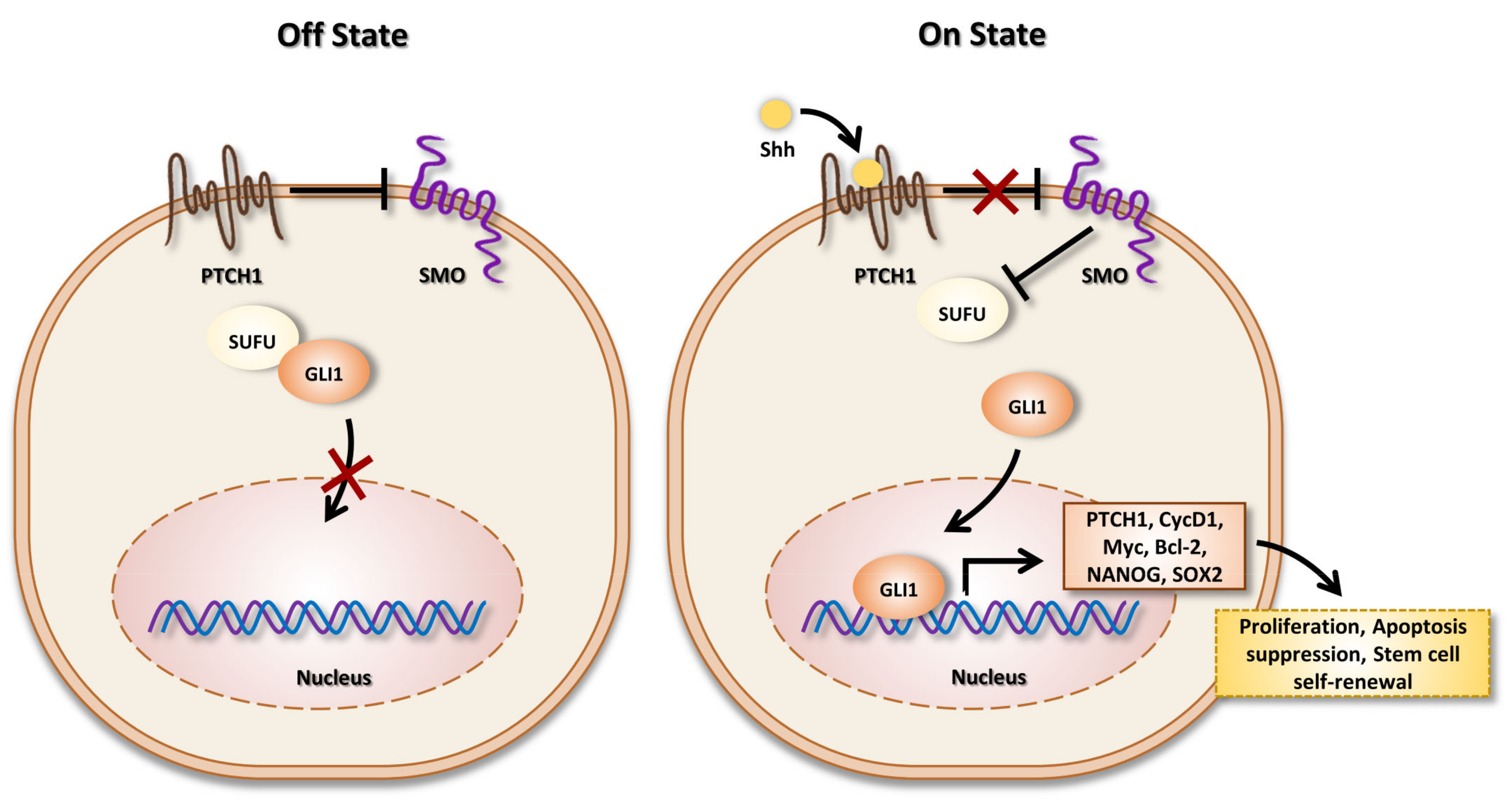
Figure 1.
Off state (left): In the absence of Sonic Hedgehog (SHH) ligand, Ptch inhibits signal transduction by SMO. Gli canonical SHH signaling pathway proteins are sequestered in the cytoplasm by interaction with Sufu. Therefore, the expression of SHH signaling targets turns off. On state (right): In the presence of ligand, SHH binds to Ptch and, therefore, induces the translocation of SMO to the primary cilium, a subcellular compartment essential for signal transduction to the positive forms of Gli proteins. Gli proteins accumulate into the nucleus and activate target genes transcription.

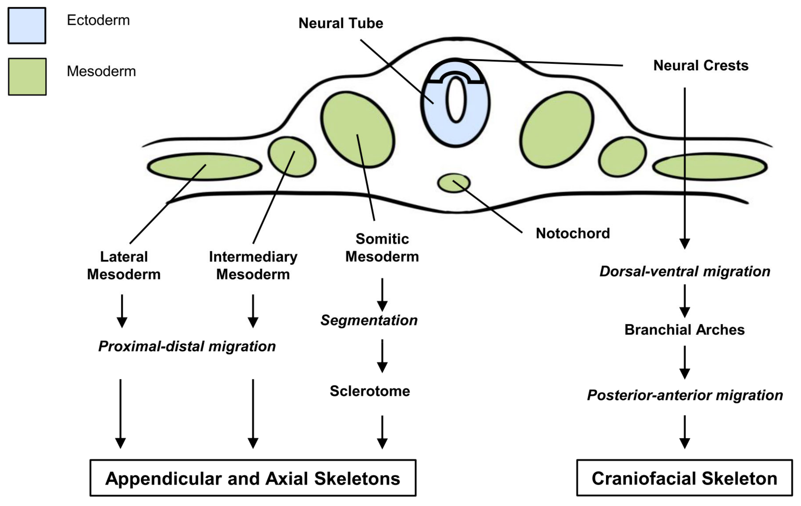
Figure 2.
Origins of appendicular, axial and craniofacial skeletons. The axial and appendicular skeletons derive from cells of the paraxial mesoderm following a proximal-distal migration of cells from the lateral and intermediary mesoderm or the segmentation of cells from the somatic mesoderm. The craniofacial skeleton derives from the ectoderm cells of the neural crests following a dorsal-ventral migration, enabling the formation of the branchial arches and a posterior-anterior migration.

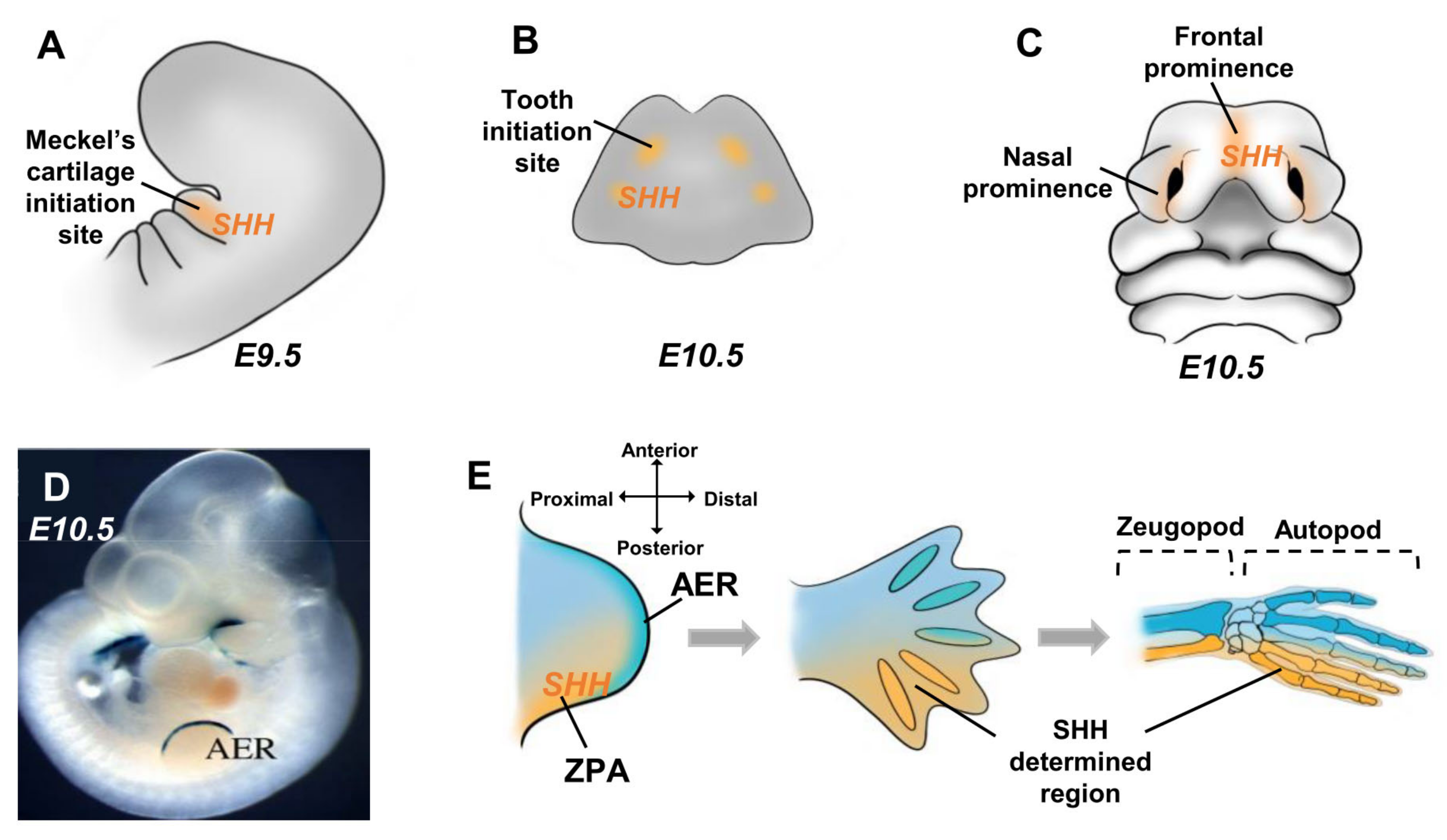
Figure 3.
Sonic Hedgehog (SHH) implications in the craniofacial and limb morphogenesis. (A) SHH expression pattern in the first branchial arch determines the Meckel cartilage initiation site on mouse embryonic day 9.5 (E9.5). (B) SHH expression on mouse embryonic day 10.5 mandible determines the tooth initiation site. (C) SHH expression in the frontonasal prominences of the mouse embryonic day 10.5 governs the morphogenesis of the upper facial structures. (D–E) In the mouse limb bud at embryonic day 10.5, SHH expression in the zone of polarizing activity (ZPA) determines the posterior region of the limb bud, where distal growth is controlled by the apical ectodermal ridge (AER) visualized by Dlx2/LacZ expression in (D) and the subjacent progression zone. The cells determined by the SHH signaling will give rise to the posterior bones of the autopod and zeugopod (E).

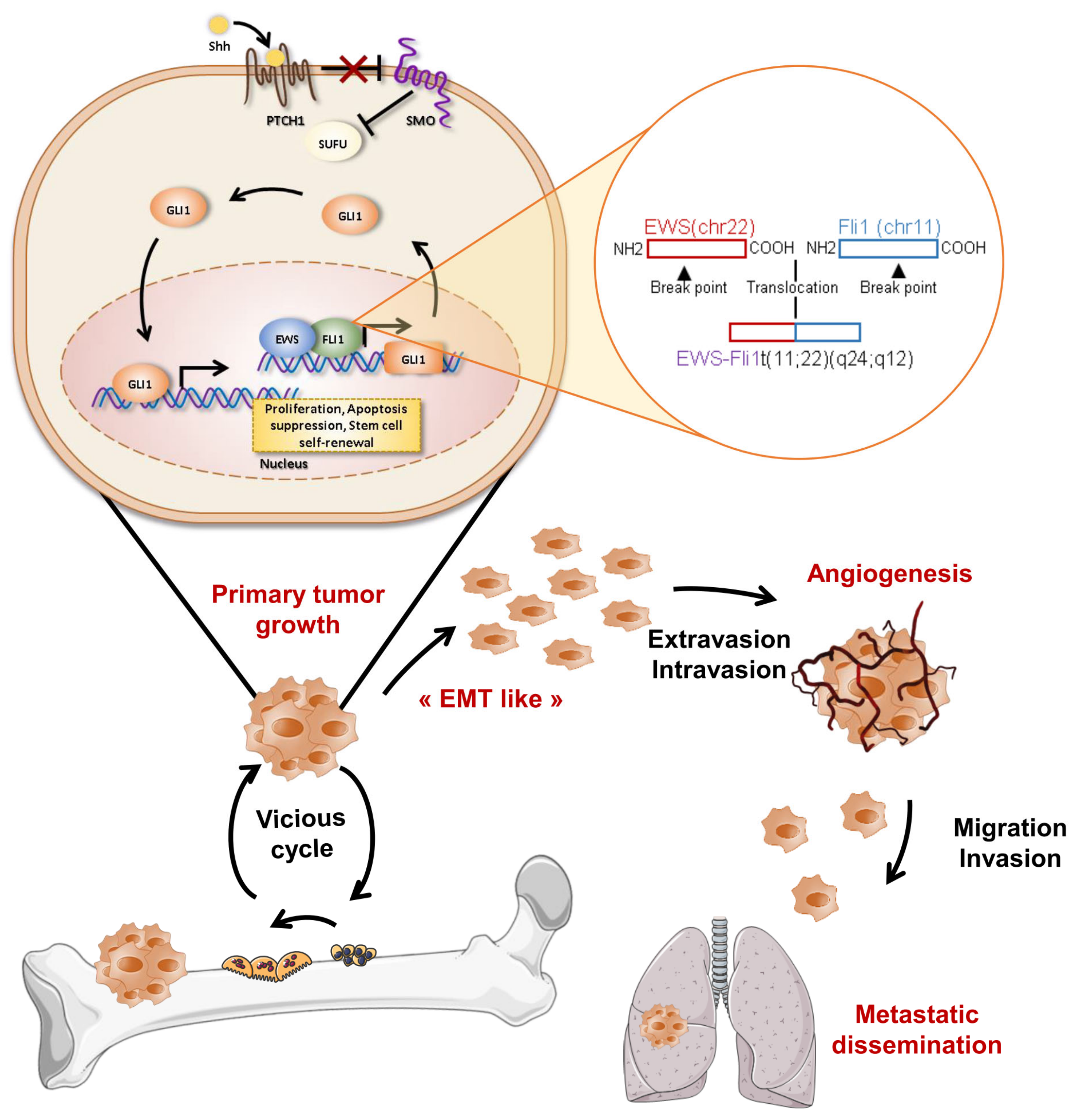
Figure 4.
Role of SHH/Gli cascade in Ewing sarcoma development. Upper panels: EWS-Fli1 protein regulates Gli1 gene expression at the transcriptional level and, therefore, promotes the expression of Gli gene targets implicated in various cellular events, such as cell proliferation and migration. Lower panels: progression of Ewing sarcoma development. During primary tumor growth, cancer cells produce soluble factors, such as growth factors or cytokines, that activate osteoclastogenesis and, in turn, bone degradation. This bone resorption then allows the release of trapped growth factors into the bone matrix able to stimulate tumor growth, “EMT-like” (epithelial mesenchymal transition) angiogenesis and, therefore, the metastatic process.

© 2020 by the authors. Licensee MDPI, Basel, Switzerland. This article is an open access article distributed under the terms and conditions of the Creative Commons Attribution (CC BY) license (http://creativecommons.org/licenses/by/4.0/).
Share and Cite
MDPI and ACS Style
Lézot, F.; Corre, I.; Morice, S.; Rédini, F.; Verrecchia, F. SHH Signaling Pathway Drives Pediatric Bone Sarcoma Progression. Cells 2020, 9, 536. https://doi.org/10.3390/cells9030536
AMA Style
Lézot F, Corre I, Morice S, Rédini F, Verrecchia F. SHH Signaling Pathway Drives Pediatric Bone Sarcoma Progression. Cells. 2020; 9(3):536. https://doi.org/10.3390/cells9030536
Chicago/Turabian StyleLézot, Frédéric, Isabelle Corre, Sarah Morice, Françoise Rédini, and Franck Verrecchia. 2020. "SHH Signaling Pathway Drives Pediatric Bone Sarcoma Progression" Cells 9, no. 3: 536. https://doi.org/10.3390/cells9030536
Note that from the first issue of 2016, this journal uses article numbers instead of page numbers. See further details here.