Electrochemical Signal Amplification Strategies and Their Use in Olfactory and Taste Evaluation

 ,
,  ,
, 
Abstract
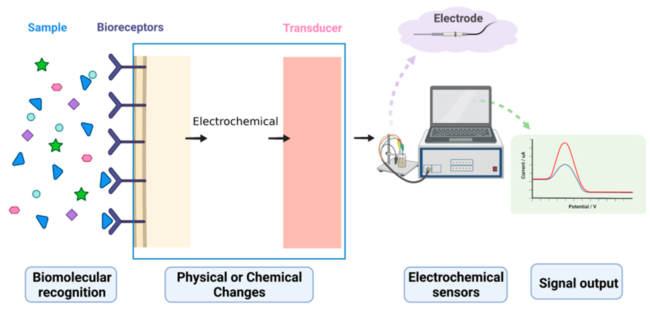
:1. Electrochemical Biosensors
2. Advances in Electrochemical Signal Amplification Strategies for Olfactory and Taste Measurements
2.1. Classical Analytical Techniques for Olfactory and Taste Detection
2.2. Olfactory and Taste Detection Based on Biosensor Technology
2.3. Taste Electrochemical Sensors Based on a Cellular Signal Cascade Amplification System
2.4. Olfactory Electrochemical Sensors Based on a Cellular Signal Cascade Amplification System
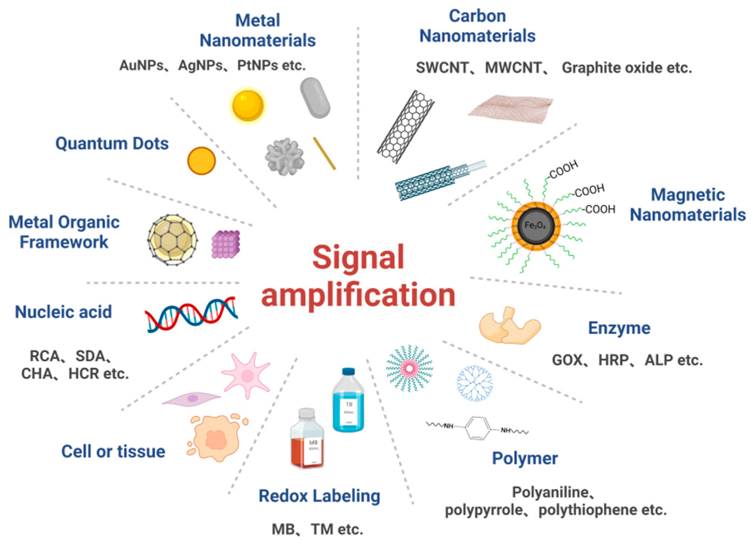
3. Commonly Used Signal Amplification Strategies for Electrochemical Biosensors
3.1. Signal Amplification Strategies Based on Nanomaterials
3.1.1. Metallic Nanomaterials
3.1.2. Carbon Nanomaterials
3.1.3. Quantum Dots
3.1.4. Magnetic Nanoparticles
3.1.5. Metal-Organic Framework Materials
3.2. Signal Amplification Strategies Based on Enzymes
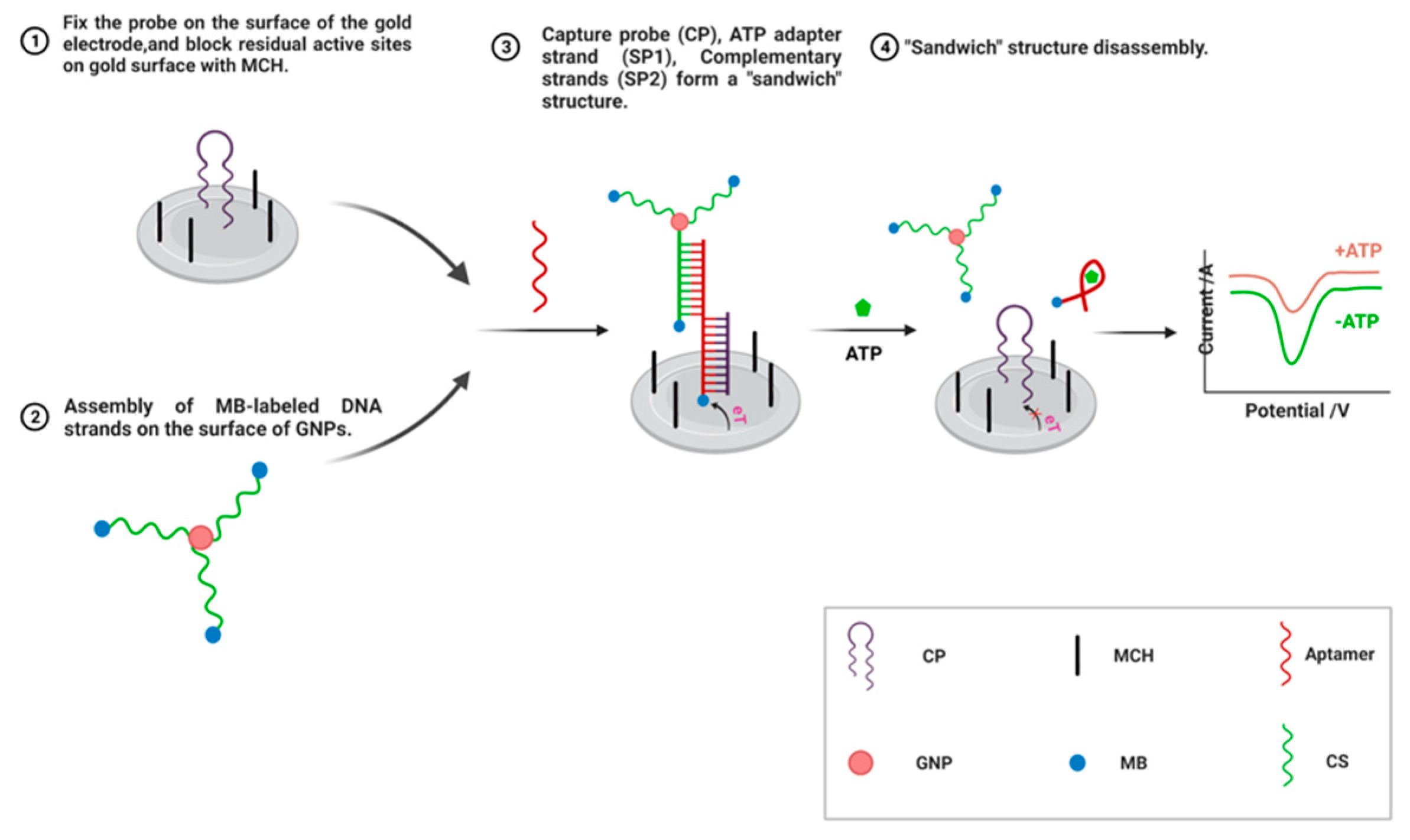
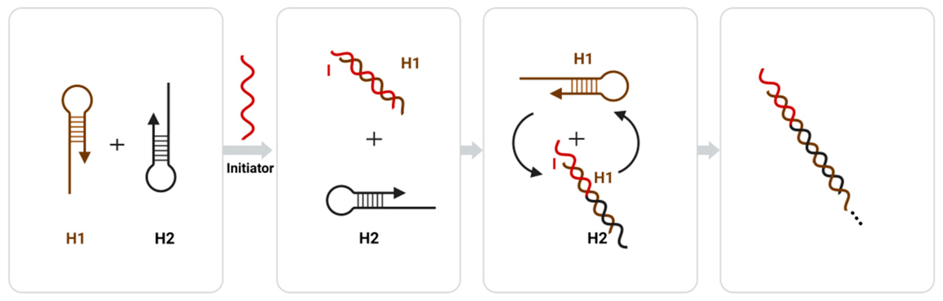
3.3. Signal Amplification Strategies Based on Nucleic Acid Amplification Techniques
3.3.1. Signal Amplification Strategies Based on Nuclease
3.3.2. Signal Amplification Strategies Based on Enzyme-Free Nucleic Acids
3.4. Signal Amplification Strategies Based on Polymers
3.5. Signal Amplification Strategies Based on Redox Markers
3.6. Signal Amplification Strategies Based on Cells or Tissue
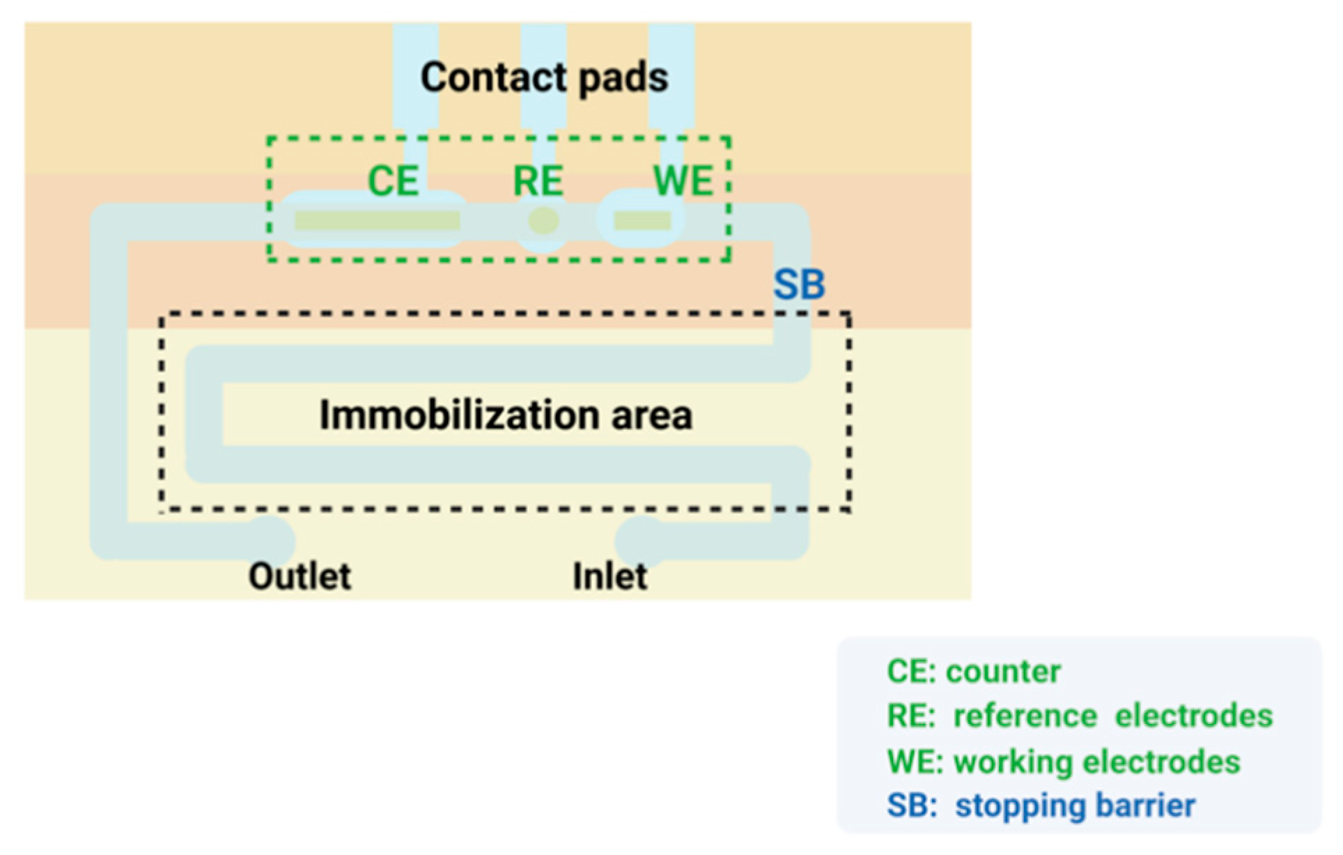
3.7. Signal Amplification Strategies Based on Microfluidics
3.8. Signal Amplification Strategy for the Combination of Multiple Materials
4. Summary and Outlook
Author Contributions
Funding
Data Availability Statement
Acknowledgments
Conflicts of Interest
References
- Clark, L.C., Jr.; Lyons, C. Electrode systems for continuous monitoring in cardinovascular surgery. Ann. N. Y. Acad. Sci. 1962, 102, 29–45. [Google Scholar] [CrossRef] [PubMed]
- Updike, S.J.; Hicks, G.P. Reagentless substrate analysis with immobilizsed enzymes. Science 1967, 158, 270–272. [Google Scholar] [CrossRef] [PubMed]
- Updike, S.J.; Hicks, G.P. The enzyme electrode. Nature 1967, 214, 986–988. [Google Scholar] [CrossRef] [PubMed]
- Zhang, X.-E. Biosensors: 50 Years Development and Future Perspectives. Bull. Chin. Acad. Sci. 2017, 32, 1271–1280. [Google Scholar]
- Homola, J.; Yee, S.S.; Gauglitz, G. Surface plasmon resonance sensors: Review. Sens. Actuators B Chem. 1999, 54, 3–15. [Google Scholar] [CrossRef]
- Anker, J.N.; Hall, W.P.; Lyandres, O.; Shah, N.C.; Zhao, J.; Van Duyne, R.P. Biosensing with plasmonic nanosensors. Nat. Mater. 2008, 7, 442–453. [Google Scholar] [CrossRef]
- Mohammadniaei, M.; Park, C.; Min, J.; Sohn, H.; Lee, T. Fabrication of Electrochemical-Based Bioelectronic Device and Biosensor Composed of Biomaterial-Nanomaterial Hybrid. Nat. Public Health Emerg. Collect. 2018, 1064, 263–296. [Google Scholar]
- Wasilewski, T.; Brito, N.F.; Szulczyński, B.; Wojciechowski, M.; Buda, N.; Melo, A.C.A.; Kamysz, W.; Gębicki, J. Olfactory receptor-based biosensors as potential future tools in medical diagnosis. TrAC Trends Anal. Chem. 2022, 150, 116599. [Google Scholar] [CrossRef]
- Zhang, M.; Ding, Q.; Zhu, M.; Yuan, R.; Yuan, Y. An ultrasensitive electrochemical biosensor with amplification of highly efficient triple catalytic hairpin assembly and tetris hybridization chain reaction. Sens. Actuators B Chem. 2022, 361, 131683. [Google Scholar] [CrossRef]
- Thevenot, D.R.; Toth, K.; Durst, R.A.; Wilson, G.S. Electrochemical biosensors: Recommended definitions and classification. Pure Appl. Chem. 1999, 71, 2333–2348. [Google Scholar] [CrossRef] [Green Version]
- Zouari, M.; Campuzano, S.; Pingarrón, J.M.; Raouafi, N. Femtomolar direct voltammetric determination of circulating miRNAs in sera of cancer patients using an enzymeless biosensor. Anal. Chim. Acta 2020, 1104, 188–198. [Google Scholar] [CrossRef]
- Salahandish, R.; Ghaffarinejad, A.; Omidinia, E.; Zargartalebi, H.; Majidzadeh-A, K.; Naghib, S.M.; Sanati-Nezhad, A. Label-free ultrasensitive detection of breast cancer miRNA-21 biomarker employing electrochemical nano-genosensor based on sandwiched AgNPs in PANI and N-doped graphene. Biosens. Bioelectron. 2018, 120, 129–136. [Google Scholar] [CrossRef]
- Li, G.; Qi, X.; Wu, J.; Xu, L.; Wan, X.; Liu, Y.; Chen, Y.; Li, Q. Ultrasensitive, label-free voltammetric determination of norfloxacin based on molecularly imprinted polymers and Au nanoparticle-functionalized black phosphorus nanosheet nanocomposite. J. Hazard. Mater. 2022, 436, 129107. [Google Scholar] [CrossRef]
- Li, G.; Qi, X.; Zhang, G.; Wang, S.; Li, K.; Wu, J.; Wan, X.; Liu, Y.; Li, Q. Low-cost voltammetric sensors for robust determination of toxic Cd(II) and Pb(II) in environment and food based on shuttle-like α-Fe2O3 nanoparticles decorated β-Bi2O3 microspheres. Microchem. J. 2022, 179, 107515. [Google Scholar] [CrossRef]
- Bakker, E.; Qin, Y. Electrochemical sensors. Anal. Chem. 2006, 78, 3965–3984. [Google Scholar] [CrossRef] [Green Version]
- Wang, Z.; Guo, H.; Gui, R.; Jin, H.; Zhang, F. Simultaneous and selective measurement of dopamine and uric acid using glassy carbon electrodes modified with a complex of gold nanoparticles and multiwall carbon nanotubes. Sens. Actuators B Chem. 2017, 255, 2069–2077. [Google Scholar] [CrossRef]
- El-Moghazy, A.Y.; Wisuthiphaet, N.; Yang, X.; Sun, G.; Nitin, N. Electrochemical biosensor based on genetically engineered bacteriophage T7 for rapid detection of Escherichia coli on fresh produce. Food Control 2022, 135, 108811. [Google Scholar] [CrossRef]
- El-Moghazy, A.Y.; Soliman, E.A.; Ibrahim, H.Z.; Marty, J.L.; Istamboulie, G.; Noguer, T. Biosensor based on electrospun blended chitosan-poly (vinyl alcohol) nanofibrous enzymatically sensitized membranes for pirimiphos-methyl detection in olive oil. Talanta 2016, 155, 258–264. [Google Scholar] [CrossRef]
- Ju, H. Signal Amplification for Highly Sensitive Immunosensing. J. Anal. Test. 2017, 1, 7. [Google Scholar] [CrossRef]
- Plaks, V.; Koopman, C.D.; Werb, Z. Circulating Tumor Cells. Science 2013, 341, 1186–1188. [Google Scholar] [CrossRef]
- Pantel, K.; Alix-Panabières, C. Circulating tumour cells and cell-free DNA in gastrointestinal cancer. Nat. Rev. Gastroenterol. Hepatol. 2017, 14, 73–74. [Google Scholar] [CrossRef]
- Rezaei, B.; Irannejad, N. Electrochemical Detection Techniques in Biosensor Applications[M]//Electrochemical Biosensors; Elsevier: Amsterdam, The Netherlands, 2019; pp. 11–43. [Google Scholar]
- Koyappayil, A.; Lee, M.H. Ultrasensitive Materials for Electrochemical Biosensor Labels. Sensors 2020, 21, 89. [Google Scholar] [CrossRef]
- Kazzy, M.E.; Weerakkody, J.S.; Hurot, C.; Mathey, R.; Hou, Y. An Overview of Artificial Olfaction Systems with a Focus on Surface Plasmon Resonance for the Analysis of Volatile Organic Compounds. Biosensors 2021, 11, 244. [Google Scholar] [CrossRef]
- He, C.; Liu, L.; Korposh, S.; Correia, R.; Morgan, S.P. Volatile Organic Compound Vapour Measurements Using a LocalisedSurface Plasmon Resonance Optical Fibre Sensor Decorated with a Metal-Organic Framework. Sensors 2021, 21, 1420. [Google Scholar] [CrossRef]
- Fabre, H.J.H.; Legros, G.V. Souvenirs Entomologiques: E’tude sur l’instinct et les Moeurs des Insectes; Robert Laffont: Paris, France, 1989; Volume 2, p. 1. [Google Scholar]
- Rau, P.; Rau, N. The Sex Attraction and Rhythmic Periodicity in Giant Saturnid Moths. Trans. Acad. Sci. St. Louis 1929, 26, 83. [Google Scholar]
- Fredriksson, R.; Lagerstrom, M.C.; Lundin, L.G.; Schiöth, H.B. The G-proteincoupled receptors in the human genome form five main families. Phylogenetic analysis, paralogon groups, and fingerprints. Mol. Pharmacol. 2003, 63, 1256–1272. [Google Scholar] [CrossRef] [Green Version]
- Rüffer, D.; Hoehne, F.; Bühler, J. New Digital Metal-Oxide (MOx) Sensor Platform. Sensors 2018, 18, 1052. [Google Scholar] [CrossRef] [Green Version]
- Wasilewski, T.; Kamysz, W.; Gebicki, J. Bioelectronic tongue: Current status and perspectives. Biosens. Bioelectron. 2020, 150, 111923. [Google Scholar] [CrossRef]
- Du, L.; Wu, C.; Liu, Q.; Huang, L.; Wang, P. Recent advances in olfactory receptor-based biosensors. Biosens. Bioelectron. 2013, 42C, 570–580. [Google Scholar]
- Wasilewski, T.; Gebicki, J.; Kamysz, W. Bio-inspired approaches for explosives detection. TrAC Trends Anal. Chem. 2021, 142, 116330. [Google Scholar] [CrossRef]
- Ren, X.; Sun, Y.; Wang, Z.; Barceló, D.; Wang, Q.; Zhang, Z.; Zhang, Y. Abundance and characteristics of microplastic in sewage sludge: A case study of Yangling, Shaanxi province, China. Case Stud. Chem. Environ. Eng. 2020, 2, 100050. [Google Scholar] [CrossRef]
- Lu, D.; Lu, F.; Geng, L.; Pang, G. Recent Advances in Olfactory Receptor Biosensors and Cell Signaling Cascade Amplification Systems. Sens. Mater. Int. J. Sens. Technol. 2018, 30, 67–87. [Google Scholar]
- Röck, F.; Barsan, N.; Weimar, U. Electronic Nose: Current Status and Future Trends. Chem. Rev. 2008, 108, 705–725. [Google Scholar] [CrossRef] [PubMed]
- Tahara, K.; Toko, K. Electronic tongues—A review. IEEE Sens. J. 2013, 13, 3001–3011. [Google Scholar] [CrossRef]
- Son, M.; Park, T.H. The bioelectronic nose and tongue using olfactory and taste receptors: Analytical tools for food quality and safety assessment. Biotechnol. Adv. 2017, 36, 371–379. [Google Scholar] [CrossRef]
- Buck, L.; Axel, R. A novel multigene family may encode odorant receptors: A molecular basis for odor recognition. Cell 1991, 65, 175–187. [Google Scholar] [CrossRef]
- Kotlowski, C.; Aspermair, P.; Khan, H.U.; Rozman, C.R.; Breu, J.; Szunerits, S.; Kim, J.J.; Bao, Z.; Kleber, C.; Pelosi, P. Electronic biosensing with flexible organic transistor devices. Flex. Print. Electron. 2018, 3, 034003. [Google Scholar] [CrossRef]
- D’Onofrio, C.; Zaremska, V.; Zhu, J.; Knoll, W.; Pelosi, P. Ligand-Binding Assays with OBPs and CSPs. Methods Enzymol. 2020, 642, 229–258. [Google Scholar]
- Park, S.J.; Kwon, O.S.; Lee, S.H.; Song, H.S.; Park, T.H.; Jang, J. Ultrasensitive flflexible graphene based field-effect transistor (FET)-type bioelectronic nose. Nano Lett. 2012, 12, 5082–5090. [Google Scholar] [CrossRef]
- Son, M.; Kim, D.; Ko, H.J.; Hong, S.; Park, T.H. A portable and multiplexed bioelectonic sensor using human olfactory and taste receptors. Biosens. Bioelectron. 2017, 87, 901–907. [Google Scholar] [CrossRef]
- Kwon, O.S.; Song, H.S.; Park, S.J.; Lee, S.H.; An, J.H.; Park, J.W.; Yang, H.; Yoon, H.; Bae, J.; Park, T.H.; et al. An Ultrasensitive, Selective, Multiplexed Superbioelectronic Nose That Mimics the Human Sense of Smell. Nano Lett. 2015, 15, 6559–6567. [Google Scholar] [CrossRef]
- Barbosa, A.J.M.; Oliveira, A.R.; Roque, A.C.A. Protein- and Peptide-Based Biosensors in Artifificial Olfaction. Trends Biotechnol. 2018, 36, 1244–1258. [Google Scholar] [CrossRef] [Green Version]
- El kazzy, M.; Hurot, C.; Weerakkody, J.S.; Buhot, A.; Hou, Y. Biomimetic Olfactory Biosensors and Bioelectronic Noses. In Advances in Biosensors: Reviews; Yurish, S.Y., Ed.; IFSA Publishing: Barcelona, Spain, 2020; Volume 3, pp. 15–54. [Google Scholar]
- Cave, J.W.; Wickiser, J.K.; Mitropoulos, A.N. Progress in the development of olfactory-based bioelectronic chemosensors. Biosens. Bioelectron. 2018, 123, 211–222. [Google Scholar] [CrossRef]
- Bohbot, J.D.; Vernick, S. The Emergence of Insect Odorant Receptor-Based Biosensors. Biosensors 2020, 10, 26. [Google Scholar] [CrossRef] [Green Version]
- Ward, R.J.; Jjunju, F.P.M.; Griffifith, E.J.; Wuerger, S.M.; Marshall, A. Artifificial Odour-Vision Syneasthesia via Olfactory Sensory Argumentation. IEEE Sens. J. 2021, 21, 6784–6792. [Google Scholar] [CrossRef]
- Lee, S.H.; Jun, S.B.; Ko, H.J.; Kim, S.J.; Park, T.H. Cell-based olfactory biosensor using microfabricated planar electrode. Biosens. Bioelectron. 2009, 24, 2659–2664. [Google Scholar] [CrossRef]
- Xu, Q.; Lu, D.; Pang, G. Comparative study of hGPR120 receptor self-assembled nano-gold sensor and tissue sensor. Sens. Actuators B Chem. 2020, 320, 128382. [Google Scholar] [CrossRef]
- Glatz, R.; Bailey-Hill, K. Mimicking nature’s noses: From receptor deorphaning to olfactory biosensing. Prog. Neurobiol. 2011, 93, 270–296. [Google Scholar] [CrossRef]
- Khadka, R.; Carraher, C.; Hamiaux, C.; Travas-Sejdic, J.; Kralicek, A. Synergisticimprovement in the performance of insect odorant receptor based biosensors in the presence of Orco. Biosens. Bioelectron. 2020, 153, 112040. [Google Scholar] [CrossRef]
- Lu, D.; Xu, Q.; Pang, G. A bombykol electrochemical receptor sensor and its kinetics. Bioelectrochemistry 2019, 128, 263–273. [Google Scholar] [CrossRef]
- Xiao, B.K.; Pang, G.C.; Xin, Y.L.; Meng, W.; Wei, M.Z. Study on a hydrogen peroxide biosensor based on horseradish peroxidase/GNPs-thionine/chitosan. Electrochim. Acta 2012, 62, 327–334. [Google Scholar]
- Kang, X.B.; Pang, G.C.; Chen, Q.S.; Liang, X.Y. Fabrication of Bacilluscereus electrochemical immunosensor based on double-layer gold nanoparticles and chitosan. Sens. Actuators B 2013, 177, 1010–1016. [Google Scholar] [CrossRef]
- Lu, D.; Lu, F.; Pang, G. A novel glutathione-S transferase immunosensor based on horseradish peroxidase and double-layer gold nanoparticles. Biomed. Microdevices 2016, 18, 1–9. [Google Scholar] [CrossRef]
- Qi, Y.; Zhang, T.; Jing, C.Y.; Liu, S.J.; Chen, W. Nanocrystal facet modulation to enhance transferrin binding and cellular delivery. Nat. Commun. 2020, 11, 1262. [Google Scholar] [CrossRef] [Green Version]
- Li, W.; Zhan, X.; Song, X.A. Review of Recent Applications of Ion Beam Techniques on Nanomaterial Surface Modification: Design of Nanostructures and Energy Harvesting. Small 2019, 15, 1901820. [Google Scholar] [CrossRef]
- Cheng, J. Electrochemical Biosensing of microRNA Based on Multiple Signal Amplification Strategy. Master’s Thesis, Nanjing University of Posts and Telecommunications, Nanjing, China, 2021. [Google Scholar] [CrossRef]
- Wang, X.; Pang, G. Amplification systems of weak interaction biosensors: Applications and prospects. Sens. Rev. 2015, 35, 30–42. [Google Scholar] [CrossRef]
- Nawaz, N.; Abu Bakar, N.K.; Mahmud, H.N.M.E.; Jamaludin, N.S. Molecularly imprinted polymers-based DNA biosensors. Anal. Biochem. 2021, 630, 114328. [Google Scholar] [CrossRef]
- Vilian, A.T.E.; Dinesh, B.; Kang, S.M.; Krishnan, U.M.; Huh, Y.S.; Han, Y.K. Recent advances in molybdenum disulfide-based electrode materials for electroanalytical applications. Microchim. Acta 2019, 186, 203.1–203.29. [Google Scholar]
- Li, S.; Yang, Z.; Chen, Y.; Chen, L.; Li, X. An ultrasensitive ATP electrochemical sensor for cells assay based on bio-nanoassembly and signal amplification. J. Guangxi Med. Univ. 2020, 37, 2276–2281. [Google Scholar]
- Mazloum-Ardakani, M.; Hosseinzadeh, L.; Taleat, Z. Synthesis and electrocatalytic effect of Ag@Pt core–shell nanoparticles supported on reduced graphene oxide for sensitive and simple label-free electrochemical aptasensor. Biosens. Bioelectron. 2015, 74, 30–36. [Google Scholar] [CrossRef]
- Wang, Z.; Si, L.; Bao, J.; Dai, Z. A reusable microRNA sensor based on the electrocatalytic property of heteroduplex-templated copper nanoclusters. Chem. Commun. 2015, 51, 6305–6307. [Google Scholar] [CrossRef] [PubMed]
- Posha, B.; Nambiar, S.R.; Sandhyarani, N. Gold atomic cluster mediated electrochemical aptasensor for the detection of lipopolysaccharide. Biosens. Bioelectron. 2018, 101, 199–205. [Google Scholar] [CrossRef] [PubMed]
- Li, Y.; Si, S.; Huang, F.; Wei, J.; Dong, S.; Yang, F.; Li, H.; Liu, S. Ultrasensitive label-free electrochemical biosensor for detecting linear microcystin-LR using degrading enzyme MlrB as recognition element. Bioelectrochemistry 2021, 144, 108000. [Google Scholar] [CrossRef] [PubMed]
- Bonanni, A.; Chua, C.K.; Zhao, G.; Sofer, Z.; Pumera, M. Inherently Electroactive Graphene Oxide Nanoplatelets as Labels for Single Nucleotide Polymorphism Detection. ACS Nano 2012, 6, 8546–8551. [Google Scholar] [CrossRef]
- Liu, X.; Cheng, H.; Zhao, Y.; Wang, Y.; Li, F. Portable electrochemical biosensor based on laser-induced graphene and MnO2 switch-bridged DNA signal amplification for sensitive detection of pesticide. Biosens. Bioelectron. 2021, 199, 113906. [Google Scholar] [CrossRef]
- Hu, T.; Zhang, L.; Wen, W.; Zhang, X.; Wang, S. Enzyme catalytic amplification of miRNA-155 detection with graphene quantum dot-based electrochemical biosensor. Biosens. Bioelectron. 2016, 77, 451–456. [Google Scholar] [CrossRef]
- Medina-Sánchez, M.; Miserere, S.; Morales-Narváez, E.; Merkoçi, A. On-chip magneto-immunoassay for Alzheimer’s biomarker electrochemical detection by using quantum dots as labels. Biosens. Bioelectron. 2014, 54, 279–284. [Google Scholar] [CrossRef]
- Zhu, L.; Yang, B.; Qian, K.; Qiao, L.; Liu, Y.; Liu, B. Sensitive electrochemical aptasensor for detecting EpCAM with silica nanoparticles and quantum dots for signal amplification. J. Electroanal. Chem. 2019, 856, 113655. [Google Scholar] [CrossRef]
- Lahcen, A.A.; Baleg, A.A.; Baker, P.; Iwuoha, E.; Amine, A. Synthesis and electrochemical characterization of nanostructured magnetic molecularly imprinted polymers for 17-beta-Estradiol determination. Sens. Actuators B Chem. 2017, 241, 698–705. [Google Scholar] [CrossRef]
- Yuan, Y.H.; Wu, Y.D.; Chi, B.Z.; Wen, S.H.; Liang, R.P.; Qiu, J.D. Simultaneously electrochemical detection of microRNAs based on multifunctional magnetic nanoparticles probe coupling with hybridization chain reaction. Biosens. Bioelectron. 2017, 97, 325–331. [Google Scholar] [CrossRef]
- Ye, Z.; Wang, Q.; Qiao, J.; Xu, Y.; Li, G. In situ synthesis of sandwich MOFs on reduced graphene oxide for electrochemical sensing of dihydroxybenzene isomers. Analyst 2019, 144, 2120–2129. [Google Scholar] [CrossRef]
- Ma, B.; Guo, H.; Wang, M.; Li, L.; Jia, X.; Chen, H.; Xue, R.; Yang, W. Electrocatalysis of Cu?MOF/Graphene Composite and its Sensing Application for Electrochemical Simultaneous Determination of Dopamine and Paracetamol. Electroanalysis 2019, 31, 1002–1008. [Google Scholar] [CrossRef]
- Peng, B.; Cui, J.; Wang, Y.; Liu, J.Q.; Zheng, H.M.; Jin, L.; Zhang, Y.; Wu, Y.C. CeO2-x/C/rGO nanocomposites derived from Ce-MOF and graphene oxide as robust platform for highly sensitive uric acid detection. Nanoscale 2018, 10, 1939–1945. [Google Scholar] [CrossRef]
- Russo, L.; Bueno, J.L.; Bergua, J.F.; Costantini, M.; Giannetto, M.; Puntes, V.; De La Escosura-Muñiz, A.; Merkoçi, A. Low-Cost Strategy for the Development of a Rapid Electrochemical Assay for Bacteria Detection Based on AuAg Nanoshells. ACS Omega 2018, 3, 18849–18856. [Google Scholar] [CrossRef]
- Xiong, W.; Wu, S.F.; Liao, F.S.; Hong, N.; Fan, H.; Wei, G.B. A ZnS-Nanoparticle-Label-Based Electrochemical Codeine Sensor. In Applied Mechanics and Materials; Trans Tech Publications Ltd.: Bach, Switzerland, 2017; pp. 173–177. [Google Scholar]
- Abi, A.; Mohammadpour, Z.; Zuo, X.; Safavi, A. Nucleic acid-based electrochemical nanobiosensors. Biosens. Bioelectron. 2018, 102, 479–489. [Google Scholar] [CrossRef]
- Xiang, J.; Pi, X.; Chen, X.; Xiang, L.; Yang, M.; Ren, H.; Shen, X.; Qi, N.; Deng, C. Integrated signal probe based aptasensor for dual-analyte detection. Biosens. Bioelectron. 2017, 96, 268–274. [Google Scholar] [CrossRef]
- Zhao, J.; Hu, S.; Zhong, W.; Wu, J.; Shen, Z.; Chen, Z.; Li, G. Highly sensitive electrochemical aptasensor based on a ligase-assisted exonuclease III-catalyzed degradation reaction. Acs Appl. Mater. Interfaces 2014, 6, 7070. [Google Scholar] [CrossRef]
- Bindra, D.S.; Wilson, G.S. Pulsed amperometric detection of glucose in biological fluids at a surface-modified gold electrode. Anal. Chem. 1989, 61, 2566–2570. [Google Scholar] [CrossRef]
- Spampinato, V.; Parracino, M.A.; la Spina, R.; Rossi, F.; Ceccone, G. Surface Analysis of Gold Nanoparticles Functionalized with Thiol-Modified Glucose SAMs for Biosensor Applications. Front. Chem. 2016, 4, 8. [Google Scholar] [CrossRef]
- Reimers, J.R.; Ford, M.J.; Halder, A.; Ulstrup, J.; Hush, N.S. Gold surfaces and nanoparticles are protected by Au(0)–thiyl species and are destroyed when Au(I)–thiolates form. Proc. Natl. Acad. Sci. USA 2016, 113, E1424–E1433. [Google Scholar] [CrossRef] [Green Version]
- Daniel, M.-C.; Astruc, D. Gold Nanoparticles: Assembly, Supramolecular Chemistry, Quantum-Size-Related Properties, and Applications toward Biology, Catalysis, and Nanotechnology. Chem. Rev. 2003, 104, 293–346. [Google Scholar] [CrossRef]
- Tian, B.; Shuang, Y.; Luyang, Z.; Min, W. Signal amplification strategy for electrochemical aptasensor based on gold nanostars and nitrogen-doped porous carbon derived from biomass. J. Henan Univ. Technol. (Nat. Sci. Ed.) 2021, 42, 85–91. [Google Scholar]
- Wei, M.; Feng, S. Amperometric determination of organophosphate pesticides using an acetylcholinesterase based biosensor made from nitrogen-doped porous carbon deposited on a boron-doped diamond electrode. Micro-Chim. Acta 2017, 184, 3461–3468. [Google Scholar] [CrossRef]
- Hui, L.; Xu, D. Silver nanoparticles as labels for applications in bioassays. TrAC Trends Anal. Chem. 2014, 61, 67–73. [Google Scholar]
- Dong, H.; Jin, S.; Ju, H.; Hao, K.; Xu, L.P.; Lu, H.; Zhang, X. Trace and label-free microRNA detection using oligonucleotide encapsulated silver nanoclusters as probes. Anal. Chem. 2012, 84, 8670–8674. [Google Scholar] [CrossRef]
- Xu, M.; Yadavalli, V.K. Flexible biosensors for the impedimetric detection of protein targets using silk-conductive polymer biocomposites. ACS Sens. 2019, 4, 1040–1047. [Google Scholar] [CrossRef]
- Cho, I.H.; Lee, J.; Kim, J.; Kang, M.S.; Paik, J.K.; Ku, S.; Cho, H.M.; Irudayaraj, J.; Kim, D.H. Current Technologies of Electrochemical Immunosensors: Perspective on Signal Amplification. Sensors 2018, 18, 207. [Google Scholar] [CrossRef] [Green Version]
- Chen, Y.X.; Huang, K.J.; Niu, K.X. Recent advances in signal amplification strategy based on oligonucleotide and nanomaterials for microRNA detection—A review. Biosens. Bioelectron. 2018, 99, 612–624. [Google Scholar] [CrossRef]
- Bourne, D.G.; Jones, G.J.; Blakeley, R.L.; Jones, A.; Negri, A.P.; Riddles, P. Enzymatic pathway for the bacterial degradation of the cyanobacterial cyclic peptide toxin microcystin LR. Appl. Environ. Microbiol. 1996, 62, 4086–4094. [Google Scholar] [CrossRef] [Green Version]
- Bo, X.; Zhou, M.; Guo, L. Electrochemical sensors and biosensors based on less aggregated graphene. Biosens. Bioelectron. 2017, 89, 167–186. [Google Scholar] [CrossRef]
- Kanagavalli, P.; Andrew, C.; Veerapandian, M.; Jayakumar, M. In-situ redox-active hybrid graphene platform for label-free electrochemical biosensor: Insights from electrodeposition and electroless deposition. TrAC Trends Anal. Chem. 2021, 143, 116413. [Google Scholar] [CrossRef]
- Ambrosi, A.; Chua, C.K.; Bonanni, A.; Pumera, M. Electrochemistry of Graphene and Related Materials. Chem. Rev. 2014, 114, 7150–7188. [Google Scholar] [CrossRef] [PubMed]
- Bonanni, A. Advances on the Use of Graphene as a Label for Electrochemical Biosensors. ChemElectroChem 2020, 7, 4177–4185. [Google Scholar] [CrossRef]
- Puetz, P.; Behrent, A.; Baeumner, A.J.; Wegener, J. Laser-Scribed Graphene (LSG) as New Electrode Material for Impedance-Based Cellular Assays. Sens. Actuators B Chem. 2020, 321, 128443. [Google Scholar] [CrossRef]
- Ramnani, P.; Saucedo, N.M.; Mulchandani, A. Carbon nanomaterial-based electrochemical biosensors for label-free sensing of environmental pollutants. Chemosphere 2016, 143, 85–98. [Google Scholar] [CrossRef] [Green Version]
- He, L.; Huang, R.; Xiao, P.; Liu, Y.; He, N. Current signal amplification strategies in aptamer-based electrochemical biosensor: A review. Chin. Chem. Lett. 2021, 32, 1593–1602. [Google Scholar] [CrossRef]
- Niemeyer, C.M. Nanoparticles, Proteins, and Nucleic Acids: Biotechnology Meets Materials Science. Angew. Chem. Int. Ed. 2001, 40, 4128–4158. [Google Scholar] [CrossRef]
- Farzin, M.A.; Abdoos, H. A critical review on quantum dots: From synthesis toward applications in electrochemical biosensors for determination of disease-related biomolecules. Talanta 2021, 224, 121828. [Google Scholar] [CrossRef]
- Li, C.; Hu, J.; Lu, M.; Zhang, C. Quantum dot-based electrochemical biosensor for stripping voltammetric detection of telomerase at the single-cell level. Biosens. Bioelectron. 2018, 122, 51–57. [Google Scholar] [CrossRef]
- Gidwani, B.; Sahu, V.; Shukla, S.S.; Pandey, R.; Joshi, V.; Jain, V.K.; Vyas, A. Quantum dots: Prospectives, toxicity, advances and applications. J. Drug Deliv. Sci. Technol. 2021, 61, 102308. [Google Scholar] [CrossRef]
- Wang, X.; Sun, X.; He, H.; Yang, H.; Lao, J.; Song, Y.; Xia, Y.; Xu, H.; Zhang, X.; Huang, F. A two-component active targeting theranostic agent based on graphene quantum dots. J. Mater. Chem. B 2015, 3, 3583–3590. [Google Scholar] [CrossRef]
- Justino, C.I.L.; Rocha-Santos, T.A.P.; Cardoso, S.; Duarte, A.C.; Cardosa, S. Strategies for enhancing the analytical performance of nanomaterial-based sensors. TrAC Trends Anal. Chem. 2013, 47, 27–36. [Google Scholar] [CrossRef]
- Yadav, S.; Masud, M.K.; Islam, M.N.; Gopalan, V.; Lam, A.K.; Tanaka, S.; Nguyen, N.T.; Hossain, M.S.; Li, C.; Yamauchi, Y. Gold-loaded nanoporous iron oxide nanocubes: A novel dispersible capture agent for tumor-associated autoantibody analysis in serum. Nanoscale 2017, 9, 8805–8814. [Google Scholar] [CrossRef]
- Ingerslev, F.; Vaclavik, E.; Halling-Sorensen, B. Pharmaceuticals, and personal care products—A source of endocrine disruption in the environment? Pure Appl. Chem. 2003, 75, 1881. [Google Scholar] [CrossRef]
- Wang, Z.; Wang, P.; Tu, X.; Wu, Y.; Zhan, G.; Li, C. A novel electrochemical sensor for estradiol based on nanoporous polymeric film bearing poly{1-butyl-3-[3-(N-pyrrole) propyl] imidazole dodecyl sulfonate}moiety. Sens. Actuators B 2014, 193, 190–197. [Google Scholar] [CrossRef]
- Zamora-Gálvez, A.; Morales-Narváez, E.; Mayorga-Martinez, C.C.; Merkoçi, A. Nanomaterials connected to antibodies and molecularly imprinted polymers as bio/receptors for bio/sensor applications. Appl. Mater. Today 2017, 9, 387–401. [Google Scholar] [CrossRef]
- Zhang, L.; Li, J.; Zeng, Y. Molecularly imprinted magnetic nanoparticles for determination of the herbicide chlorotoluron by gate-controlled electro-catalytic oxidation of hydrazine. Mikrochim. Acta 2014, 182, 249–255. [Google Scholar] [CrossRef]
- Villalonga, A.; Villalonga, R.; Vilela, D. Hybrid magnetic nanoparticles for electrochemical biosensors. In Magnetic Nanoparticle-Based Hybrid Materials; Woodhead Publishing: Thorston, UK, 2021; pp. 679–720. [Google Scholar]
- Chen, M.; Gan, N.; Zhou, Y.; Li, T.; Xu, Q.; Cao, Y.; Chen, Y. A novel aptamer-metal ions-nanoscale MOF based electrochemical biocodes for multiple antibiotics detection and signal amplification. Sens. Actuators B Chem. 2017, 242, 1201–1209. [Google Scholar] [CrossRef]
- Qiu, G.H.; Lu, W.Z.; Hu, P.P.; Jiang, Z.H.; Bai, L.P.; Wang, T.R.; Li, M.M.; Chen, J.X. A metal-organic framework based PCR-free biosensor for the detection of gastric cancer associated microRNAs. J. Inorg. Biochem. 2017, 177, 138–142. [Google Scholar] [CrossRef]
- Lei, X.; Zuo, G.; Wang, P.; Li, Z. Research and application of graphene/MOF composites in electrochemical biosensor. Transducer Microsyst. Technol. 2022, 41, 1–6. [Google Scholar]
- Chen, X.; Zhu, J.; Xi, Q.; Yang, W. A high performance electrochemical sensor for acetaminophen based on single-walled carbon nanotube–graphene nanosheet hybrid films. Sens. Actuators B Chem. 2012, 161, 648–654. [Google Scholar] [CrossRef]
- Wilson, S.P.; Kamin, D.L.; Fe Ldman, J.M. Acetaminophen administration interferes with urinary metanephrine (and catecholamine) determinations. Clin. Chem. 1985, 31, 1093–1094. [Google Scholar] [CrossRef]
- Zhang, Z.; Lou, Y.; Guo, C.; Jia, Q.; Song, Y.; Tian, J.-Y.; Zhang, S.; Wang, M.; He, L.; Du, M. Metal–organic frameworks (MOFs) based chemosensors/biosensors for analysis of food contaminants. Trends Food Sci. Technol. 2021, 118, 569–588. [Google Scholar] [CrossRef]
- Nangare, S.N.; Sangale, P.M.; Patil, A.G.; Boddu, S.H.; Deshmukh, P.K.; Jadhav, N.R.; Tade, R.S.; Patil, D.R.; Pandey, A.; Mutalik, S.; et al. Surface architectured metal organic frameworks-based biosensor for ultrasensitive detection of uric acid: Recent advancement and future perspectives. Microchem. J. 2021, 169, 106567. [Google Scholar] [CrossRef]
- Nemiwal, M.; Zhang, T.C.; Kumar, D. Enzyme immobilized nanomaterials as electrochemical biosensors for detection of biomolecules. Enzym. Microb. Technol. 2022, 156, 110006. [Google Scholar] [CrossRef]
- Nguyen, H.H.; Lee, S.H.; Lee, U.J.; Fermin, C.D. Immobilized Enzymes in Biosensor Applications. Materials 2019, 12, 121. [Google Scholar] [CrossRef] [Green Version]
- Cao, Q.; Xiao, Y.S.; Meng, Q.Y.; Yuan, X.Z.; Liu, H.; Cheng, L.J.; Dong, W.L. Research progress of enzyme-based biosensors in rapid detection. J. Food Saf. Qual. 2019, 10, 6902–6908. [Google Scholar]
- O’Sullivan, M.J.; Bridges, J.W.; Marks, V. Enzyme Immunoassay: A Review. Ann. Clin. Biochem. Int. J. Lab. Med. 1979, 16, 221–239. [Google Scholar] [CrossRef] [Green Version]
- Kang, C.; Kang, J.; Lee, N.-S.; Yoon, Y.H.; Yang, H. DT-Diaphorase as a Bifunctional Enzyme Label That Allows Rapid Enzymatic Amplification and Electrochemical Redox Cycling. Anal. Chem. 2017, 89, 7974–7980. [Google Scholar] [CrossRef]
- Zhao, S.; Zhou, T.; Khan, A.; Chen, Z.; Liu, P.; Li, X. A novel electrochemical biosensor for bisphenol A detection based on engineered Escherichia coli cells with a surface-display of tyrosinase. Sens. Actuators B Chem. 2021, 353, 131063. [Google Scholar] [CrossRef]
- Kurbanoglu, S.; Erkmen, C.; Uslu, B. Frontiers in electrochemical enzyme based biosensors for food and drug analysis. TrAC Trends Anal. Chem. 2020, 124, 115809. [Google Scholar] [CrossRef]
- Fernández, H.; Arévalo, F.J.; Granero, A.M.; Robledo, S.N.; Díaz Nieto, C.H.; Riberi, W.I.; Zon, M.A. Electrochemical biosensors for the determination of toxic substances related to food safety developed in South America: Mycotoxins and herbicides. Chemosensors 2017, 5, 23. [Google Scholar] [CrossRef] [Green Version]
- Nguyen, H.H.; Park, J.; Park, S.J.; Lee, C.S.; Kim, M. Long-term stability and integrity of plasmid-based DNA data storage. Polymers 2018, 10, 28. [Google Scholar] [CrossRef] [PubMed] [Green Version]
- Tomassetti, M.; Pezzilli, R.; Prestopino, G.; Natale, C.D.; Medaglia, P.G. Fabrication and characterization of a Layered Double Hydroxide based catalase biosensor and a catalytic sensor for hydrogen peroxide determination. Microchem. J. 2021, 170, 106700. [Google Scholar] [CrossRef]
- Niu, W.; Guo, J. Novel fluorescence-based biosensors incorporating unnatural amino acids. Methods Enzymol. 2017, 589, 191–219. [Google Scholar]
- Das, P.; Das, M.; Chinnadayyala, S.R.; Singha, I.M.; Goswami, P. Recent advances on developing 3rd generation enzyme electrode for biosensor applications. Biosens. Bioelectron. 2016, 79, 386–397. [Google Scholar] [CrossRef]
- Li, M.; Cheng, J.; Yuan, Z.; Zhou, H.; Zhang, L.; Dai, Y.; Shen, Q.; Fan, Q. Sensitive electrochemical detection of microRNA based on DNA walkers and hyperbranched HCR-DNAzyme cascade signal amplification strategy. Sens. Actuators B Chem. 2021, 345, 130348. [Google Scholar] [CrossRef]
- Sun, D.; Lu, J.; Luo, Z.; Zhang, L.; Liu, P.; Chen, Z. Competitive electrochemical platform for ultrasensitive cytosensing of liver cancer cells by using nanotetrahedra structure with rolling circle amplification. Biosens. Bioelectron. 2018, 120, 8–14. [Google Scholar] [CrossRef]
- Zhang, Y.; Xu, G.; Lian, G.; Luo, F.; Xie, Q.; Lin, Z.; Chen, G. Electrochemiluminescence biosensor for miRNA-21 based on toehold-mediated strand displacement amplification with Ru(phen)32+ loaded DNA nanoclews as signal tags. Biosens. Bioelectron. 2019, 147, 111789. [Google Scholar] [CrossRef]
- Yang, B.; Zhang, S.; Fang, X.; Kong, J. Double signal amplification strategy for ultrasensitive electrochemical biosensor based on nuclease and quantum dot-DNA nanocomposites in the detection of breast cancer 1 gene mutation. Biosens. Bioelectron. 2019, 142, 111544. [Google Scholar] [CrossRef]
- Yu, L.; He, P.; Xu, Y.; Kou, X.; Yu, Z.; Xie, X.; Miao, P. Manipulations of DNA four-way junction architecture and DNA modified Fe3O4@Au nanomaterials for the detection of miRNA. Sens. Actuators B Chem. 2020, 313, 128015. [Google Scholar] [CrossRef]
- Zhang, C.; Li, D.; Li, D.; Wen, K.; Yang, X.; Zhu, Y. Rolling circle amplification-mediated in situ synthesis of palladium nanoparticles for the ultrasensitive electrochemical detection of microRNA. Analyst 2019, 144, 3817–3825. [Google Scholar] [CrossRef]
- Yin, P.; Choi HM, T.; Calvert, C.R.; Pierce, N.A. Programming biomolecular self-assembly pathways. Nature 2008, 451, 318–322. [Google Scholar] [CrossRef] [Green Version]
- Wang, W.; Zhang, C.; Guo, J.; Li, G.; Zou, L. Sensitive electrochemical detection of oxytetracycline based on target triggered CHA and poly adenine assisted probe immobilization. Anal. Chim. Acta 2021, 1181, 338895. [Google Scholar] [CrossRef]
- Ikbal, J.; Lim, G.S.; Gao, Z. The hybridization chain reaction in the development of ultrasensitive nucleic acid assays. TrAC Trends Anal. Chem. 2015, 64, 86–99. [Google Scholar] [CrossRef]
- Augspurger, E.E.; Rana, M.; Yigit, M.V. Chemical and biological sensing using hybridization chain reaction. ACS Sens. 2018, 3, 878–902. [Google Scholar] [CrossRef]
- Ling, P.; Wang, L.; Cheng, S.; Gao, X.; Sun, X.; Gao, F. Ultrasensitive electrochemical biosensor for protein detection based on target-triggering cascade enzyme-free signal amplification strategy. Anal. Chim. Acta 2022, 1202, 339675. [Google Scholar] [CrossRef]
- Cheng, W.; Ma, J.; Cao, P.; Zhang, Y.; Li, J. Enzyme-free electrochemical biosensor based on double signal amplification strategy for the ultra-sensitive detection of exosomal microRNAs in biological samples. Talanta 2020, 219, 121242. [Google Scholar] [CrossRef]
- Fozooni, T.; Ravan, H.; Sasan, H. Signal Amplification Technologies for the Detection of Nucleic Acids: From Cell-Free Analysis to Live-Cell Imaging. Appl. Biochem. Biotechnol. 2017, 183, 1224–1253. [Google Scholar] [CrossRef]
- Hasanzadeh, M.; Shadjou, N.; de la Guardia, M. Electrochemical biosensing using hydrogel nanoparticles. TrAC Trends Anal. Chem. 2014, 62, 11–19. [Google Scholar] [CrossRef]
- Strehlitz, B.; Gründig, B.; Kopinke, H. Sensor for amperometric determination of ammonia and ammonia-forming enzyme reactions. Anal. Chim. Acta 2000, 403, 11. [Google Scholar] [CrossRef]
- Zhybak, M.T.; Vagin, M.Y.; Beni, V.; Liu, X.; Dempsey, E.; Turner, A.P.F.; Korpan, Y.I. Direct detection of ammonium ion by means of oxygen electrocatalysis at a copper- polyaniline composite on a screen-printed electrode. Microchim. Acta 2016, 183, 1981. [Google Scholar] [CrossRef] [Green Version]
- Uzunçar, S.; Meng, L.; Turner, A.P.F.; Mak, W.C. Processable and nanofibrous polyaniline: Polystyrene-sulphonate (nanoPANI: PSS) for the fabrication of catalyst-free ammonium sensors and enzyme-coupled urea biosensors. Biosens. Bioelectron. 2021, 171, 112725. [Google Scholar] [CrossRef] [PubMed]
- Singh, A.; Sharma, R.; Singh, M.; Verma, N. Electrochemical determination of L-arginine in leukemic blood samples based on a polyaniline-multiwalled carbon nanotube—Magnetite nanocomposite film modified glassy carbon electrode. Instrum. Sci. Technol. 2020, 48, 400–416. [Google Scholar] [CrossRef]
- Verma, N.; Singh, A.K.; Saini, N. Synthesis and characterization of ZnS quantum dots and application for development of arginine biosensor. Sens. BioSens. Res. 2017, 15, 41. [Google Scholar] [CrossRef]
- Li, C.; Wang, Y.; Jiang, H.; Wang, X. Biosensors based on advanced sulfur-containing nanomaterials. Sensors 2020, 20, 3488. [Google Scholar] [CrossRef]
- Zheng, J.; Zhao, H.; Ning, G.; Sun, W.; Wang, L.; Liang, H.; Xu, H.; He, C.; Zhao, H.; Li, C.-P. A novel affinity peptide–antibody sandwich electrochemical biosensor for PSA based on the signal amplification of MnO2 -functionalized covalent organic framework. Talanta 2021, 233, 122520. [Google Scholar] [CrossRef]
- Liu, Y.; Ai, K.; Lu, L. Polydopamine and Its Derivative Materials: Synthesis and Promising Applications in Energy, Environmental, and Biomedical Fields. Chem. Rev. 2014, 114, 5057–5115. [Google Scholar] [CrossRef]
- Hao, N.; Zhang, X.; Zhou, Z.; Hua, R.; Zhang, Y.; Liu, Q.; Qian, J.; Li, H.; Wang, K. AgBr nanoparticles/3D nitrogen-doped graphene hydrogel for fabricating all-solid-state luminol-electrochemiluminescence Escherichia coli aptasensors. Biosens. Bioelectron. 2017, 97, 377–383. [Google Scholar] [CrossRef]
- Huang, K.-J.; Wang, L.; Zhang, J.-Z.; Wang, L.-L.; Mo, Y.-P. One-step preparation of layered molybdenum disulfide/multi-walled carbon nanotube composites for enhanced performance supercapacitor. Energy 2014, 67, 234–240. [Google Scholar] [CrossRef]
- Zhang, W.; Zhang, P.; Su, Z.; Wei, G. Synthesis and sensor applications of MoS2-based nanocomposites. Nanoscale 2015, 7, 18364–18378. [Google Scholar] [CrossRef]
- Sun, Y.; Wu, X. Construction and Application of an Electrochemiluminescence Immunosensor Based on Gold Particles Modified Molybdenum Disulfide Nanocomposites. J. Instrum. Anal. 2021, 40, 363–369. [Google Scholar]
- Cui, F.; Zhou, Z.; Zhou, H.S. Molecularly Imprinted Polymers and Surface Imprinted Polymers Based Electrochemical Biosensor for Infectious Diseases. Sensors 2020, 20, 996. [Google Scholar] [CrossRef] [Green Version]
- Ates, M. A review study of (bio)sensor systems based on conducting polymers. Mater. Sci. Eng. C Mater. Biol. Appl. 2013, 33, 1853–1859. [Google Scholar] [CrossRef]
- Tian, L.; Qian, K.; Qi, J.; Liu, Q.; Yao, C.; Song, W.; Wang, Y. Gold nanoparticles superlattices assembly for electrochemical biosensor detection of microRNA-21. Biosens. Bioelectron. 2018, 99, 564–570. [Google Scholar] [CrossRef]
- Hong, C.-Y.; Chen, X.; Liu, T.; Li, J.; Yang, H.-H.; Chen, J.-H.; Chen, G.-N. Ultrasensitive electrochemical detection of cancer-associated circulating microRNA in serum samples based on DNA concatamers. Biosens. Bioelectron. 2013, 50, 132–136. [Google Scholar] [CrossRef]
- Cai, J.; Huang, H.; Li, Z.; Gao, Y.; Liang, Q.; Chen, X.; Chu, N.; Hao, W.; Wang, D.; Jiang, Y.; et al. A rechargeable microbial electrochemical sensor for water biotoxicity monitoring. Biosens. Bioelectron. X 2022, 10, 100132. [Google Scholar] [CrossRef]
- Xu, Q.; Lu, D.; Pang, G. Study on Bombykol Receptor Self-Assembly and Universality of G Protein Cellular Signal Amplification System. Acs Sens. 2019, 4, 257–264. [Google Scholar] [CrossRef]
- Seger, R.; Krebs, E.G. The MAPK signaling cascade. FASEB J. 1995, 9, 726–735. [Google Scholar] [CrossRef]
- Wei, L.; Qiao, L.; Pang, G.; Xie, J. A kinetic study of bitter taste receptor sensing using immobilized porcine taste bud tissues. Biosens. Bioelectron. 2017, 92, 74–80. [Google Scholar] [CrossRef]
- Jang, M.; Cai, L.; Udeani, G.O.; Slowing, K.V.; Thomas, C.F.; Beecher, C.W.W.; Fong, H.H.S.; Farnsworth, N.R.; Kinghorn, A.D.; Mehta, R.G.; et al. Cancer Chemopreventive Activity of Resveratrol, a Natural Product Derived from Grapes. Science 1997, 275, 218–220. [Google Scholar] [CrossRef] [Green Version]
- Tsai, H.-Y.; Ho, C.-T.; Chen, Y.-K. Biological actions and molecular effects of resveratrol, pterostilbene, and 3′-hydroxypterostilbene. J. Food Drug Anal. 2016, 25, 134–147. [Google Scholar] [CrossRef] [Green Version]
- Ahmad, I.; Hoda, M. Attenuation of diabetic retinopathy and neuropathy by resveratrol: Review on its molecular mechanisms of action. Life Sci. 2020, 245, 117350. [Google Scholar] [CrossRef]
- Diaz-Gerevini, G.T.; Repossi, G.; Dain, A.; Tarres, M.C.; Das, U.N.; Eynard, A.R. Beneficial action of res-veratrol: How and why? Nutrition 2016, 32, 174–178. [Google Scholar] [CrossRef]
- Kim, C.-W.; Hwang, K.-A.; Choi, K.-C. Anti-metastatic potential of resveratrol and its metabolites by the inhibition of epithelial-mesenchymal transition, migration, and invasion of malignant cancer cells. Phytomedicine 2016, 23, 1787–1796. [Google Scholar] [CrossRef]
- Ren, R.; Lu, D.; Liu, T. Development of a sandwich-type rat small intestine tissue sensor for detecting resveratrol and its receptors. Biomed. Microdevices 2021, 23, 1–8. [Google Scholar] [CrossRef]
- Elvira, K.S. Microfluidic technologies for drug discovery and development: Friend or foe? Trends Pharmacol. Sci. 2021, 42, 518–526. [Google Scholar] [CrossRef]
- Xie, Y.; Dai, L.; Yang, Y. Microfluidic technology and its application in the point-of-care testing field. Biosens. Bioelectron. X 2022, 10, 100109. [Google Scholar] [CrossRef]
- Xing, G.; Zhang, W.; Li, N.; Pu, Q.; Lin, J. Recent progress on microfluidic biosensors for rapid detection of pathogenic bacteria. Chin. Chem. Lett. 2021, 4, 1743–1751. [Google Scholar] [CrossRef]
- Liu, Y.; Jiang, D.; Wang, S.; Cai, G.; Xue, L.; Li, Y.; Liao, M.; Lin, J. A microfluidic biosensor for rapid detection of Salmonella typhimurium based on magnetic separation, enzymatic catalysis and electrochemical impedance analysis. Chin. Chem. Lett. 2021, 33, 3156–3160. [Google Scholar] [CrossRef]
- Shenoy, V.J.; Edwards, C.E.; Helgeson, M.E.; Valentine, M.T. Design and characterization of a 3D-printed staggered herringbone mixer. BioTechniques 2021, 70, 285–289. [Google Scholar] [CrossRef] [PubMed]
- Hadjigeorgiou, A.G.; Boudouvis, A.G.; Kokkoris, G. Thorough computational analysis of the staggered herringbone micromixer reveals transport mechanisms and enables mixing efficiency-based improved design. Chem. Eng. J. 2021, 414, 128775. [Google Scholar] [CrossRef]
- Schmidt-Speicher, L.M.; Lnge, K. Microfluidic Integration for Electrochemical Biosensor Applications. Curr. Opin. Electrochem. 2021, 29, 100755. [Google Scholar] [CrossRef]
- Kutluk, H.; Bruch, R.; Urban, G.A.; Dincer, C. Impact of assay format on miRNA sensing: Electrochemical microfluidic biosensor for miRNA-197 detection—ScienceDirect. Biosens. Bioelectron. 2020, 148, 111824. [Google Scholar] [CrossRef]
- Li, Y.; Zuo, S.; Ding, L.; Xu, P.; Wang, K.; Liu, Y.; Li, J.; Liu, C. Sensitive immunoassay of cardiac troponin I using an optimized microelectrode array in a novel integrated microfluidic electrochemical device. Anal. Bioanal. Chem. 2021, 412, 8325–8338. [Google Scholar] [CrossRef]
- Ansari, M.H.; Hassan, S.; Qurashi, A.; Khanday, F. Microfluidic-integrated DNA nanobiosensors. Biosens. Bioelectron. 2016, 85, 247–260. [Google Scholar] [CrossRef]
- Liu, J.; Geng, Z.; Fan, Z.; Liu, J.; Chen, H. Point-of-care testing based on smartphone: The current state-of-the-art (2017–2018). Biosens. Bioelectron. 2019, 132, 17–37. [Google Scholar] [CrossRef]
- Zhang, H.; Fan, M.; Jiang, J.; Shen, Q.; Cai, C.; Shen, J. Sensitive electrochemical biosensor for MicroRNAs based on duplex-specific nuclease-assisted target recycling followed with gold nanoparticles and enzymatic signal amplification. Anal. Chim. Acta 2019, 1064, 33–39. [Google Scholar] [CrossRef]
- Li, X.-M.; Wang, L.-L.; Luo, J.; Wei, Q.-L. A dual-amplified electrochemical detection of mRNA based on duplex-specific nuclease and bio-bar-code conjugates. Biosens. Bioelectron. 2014, 65, 245–250. [Google Scholar] [CrossRef]
- Chen, D.; Zhang, M.; Ma, M.; Hai, H.; Li, J.; Yang, S. A novel electrochemical DNA biosensor for transgenic soybean detection based on triple signal amplification. Anal. Chim. Acta 2019, 1078, 24–31. [Google Scholar] [CrossRef]
- Zhang, J.; Cui, H.; Wei, G.; Cheng, L.; Lin, Y.; Ma, G.; Hong, N.; Liao, F.; Fan, H. A double signal amplification electrochemical MicroRNA biosensor based on catalytic hairpin assembly and bisferrocene label. J. Electroanal. Chem. 2020, 858, 113816. [Google Scholar] [CrossRef]
- Xiao, S.; Song, P.; Bu, F.; Pang, G.; Xie, J. The investigation of detection and sensing mechanism of spicy substance based on human TRPV1 channel protein-cell membrane biosensor. Biosens. Bioelectron. 2021, 172, 112779. [Google Scholar] [CrossRef]
- Lim, S.A.; Ahmed, M.U. Electrochemical immunosensors and their recent nanomaterial-based signal amplification strate-gies: A review. RSC Adv. 2016, 6, 24995–25014. [Google Scholar] [CrossRef]


| Strategies | Examples | Limit of Detections | Linearity Ranges | Ref. |
|---|---|---|---|---|
| Metallic nanomaterials | Electrochemical aptasensors for ATP detection based on sulfhydryl chemistry and DNA self-assembly techniques and gold nanoparticles | 29.6 aM | ATP:10 fmol/L–1 mmol/L | [63] |
| Electrochemical biosensor based on gold nanoparticles and multi-walled carbon nanotubes for the detection of dichlorvos | 4 μg/L | 10–100 μg/L | [64] | |
| Reusable miRNA biosensor based on electrocatalytic properties of heterogeneous double template copper nanoclusters (CuNCs) | 8.2 fM | 25–300 fM | [65] | |
| Detection of lipopolysaccharide by aptasensor based on gold cluster | 7.94 × 10⁻³ amol/L | 0.01 amol/L–1 × 10−6 amol/L | [66] | |
| Carbon nanomaterials | MIrB is used as a recognition element, and the electrode modified with -COOH functionalized MWCNT to detect microcystin-LR | 0.127 pg/mL | 1 pg/mL–100 ng/mL | [67] |
| Electrochemical biosensor using graphene oxide (GO) as a direct marker for the detection of DNA polymorphs | - | OTA:310 fM–310 pM | [68] | |
| Based on laser-induced graphene and MnO2 switch-bridged DNA signal amplification for sensitive detection of pesticides | 1.2 ng/mL | OPs: 3–4000 ng/mL | [69] | |
| Quantum Dots | Electrochemical biosensor for detection of miRNA-155 based on graphene quantum dots and horseradish peroxidase (HRP) | 0.14 fM | miRNA-155: 1 fM–100 pM | [70] |
| Detection of Alzheimer’s disease biomarker ApoE by electrochemical biosensor based on cadmium-selenium/zinc sulfide quantum dots | ~12.5 ng/m L | 10–200 ng/m L | [71] | |
| An electrochemical aptasensor to detect epithelial cell adhesion molecules (EpCAM) using silica nanoparticles and quantum dots | 10 amol/L | 10 amol/L–1.0 × 108 amol/L | [72] | |
| Magnetic nanoparticles | An electrochemical biosensor to detect 17-b-estradiol using magnetic molecularly imprinted polymer nanocomposites (Fe3O4-MIP) modified on the surface of screen-printed carbon electrodes (SPCE) | 20 nM | 0.05–10 μM | [73] |
| Combining magnetic nanomaterials Fe3O4NPs and HCR for simultaneous signal-guided electrochemical detection of miRNAs | miR-141:0.28 fM miR-21:0.36 fM | 1 fM–1 nM | [74] | |
| Metal-organic framework materials | Sensitivity detection of three isomers of hydroquinone, catechol, and resorcinol based on M@Pt@M-RGO electrochemical biosensor | HQ:0.015 μmol/L CT:0.032 μmol/L RS:0.133 μmol/L | HQ:0.05–200 μmol/L CT:0.1–160 μmol/L RS:0.4–300 μmol/L | [75] |
| An electrochemical biosensor to detect simultaneously PA and DA using HKUST-1 (Cu-BTC) coupled with graphene oxide (ERGO) | PA:0.2–160 μM DA:0.2–300 μM | PA:0.016 μM DA:0.013 μM | [76] | |
| An electrochemical biosensor to detect UA using CeO2-x/C/RGO nanocomposites synthesized by MOF and graphene oxide | 2.0 μmol/L | 49.8–1050.0 μmol/L | [77] |
| Strategies | Examples | Limit of Detections | Linearity Ranges | Ref. |
|---|---|---|---|---|
| Enzyme | Electrochemical immunosensor based on DT-diaphorase (DT-D) as oxidoreductase labeling and 4-nitroso-1-naphthol (4-NO-1-N) as reaction substrate | PTH:2 pg/mL | 2 pg/mL–1 μg/mL | [125] |
| Electrochemical biosensor based on the display of tyrosinase on the surface of Escherichia coli cells for the detection of Bisphenol A | 0.01 nm | BPA:0.01 nm–100 nm | [126] | |
| Nucleic acid amplification | An electrochemical biosensor based on cyclic enzyme signal amplification (CESA) with DSN and 3-QD-DNA nanocomposites as cascade signal probes for hypersensitive detection of microRNA | 1.2 amol/L | 5 amol/L–5 fmol/L | [136] |
| An electrochemical biosensor using double-stranded specific nuclease (DSN) and cleavage endonuclease (NEase) catalyzed reactions to detect miRNA | 3 aM | 10 aM–10 fM | [137] | |
| Ultra-sensitive detection of microRNA by an electrochemical biosensor based on RCA-mediated palladium nanoparticles (PdNPs) | 8.6 amol/L | 50 amol/L–100 fmol/L | [138] | |
| Protein detection by electrochemical biosensors based on molecular recognition between aptamer and target | 0.17 pM | 0.5 pM–300 nM | [143] | |
| Efficient detection of exosomal microRNAs by strand displacement reaction (SDR) based electrochemical biosensor | 0.4 fM | miRNA-21:1 fM–200 pM | [144] | |
| Polymers | Electrochemical biosensor based on methylene blue (MB) containing MnO2-functionalized COF, and metallic gold-platinum nanoparticles (AuPbNPs) for ultra-sensitive detection of PSA | 16.7 fg mL−1 | 0.00005–10 ng mL−1 | [153] |
| Electrochemiluminescent immunosensor based on AMGMs nanocomposites for the detection of PSA in serum | 0.1 pg/mL | PSA:0.1 pg/mL–50 ng/mL | [158] | |
| Redox markers | An electrochemical biosensor to detect microRNA-21 using toluidine blue (TB) electrostatic adsorption aggregation signal amplification | 78 amol/L | 100 amol/L–1 nmol/L | [161] |
| An electrochemical biosensor based on RuHex and screen-printed gold electrodes (SPGEs) to detect microRNA | 100 amol/L | 100 amol/L–100 pmol/L | [162] | |
| Cell or tissue | The RSIT sensor by using rat small intestine tissue cells as a sensitive element and effector to detect resveratrol | 1 × 10⁻¹³ mol/L | - | [172] |
| Cell membrane biosensor with hTRPV1 immobilized directly on the HEK293T cell membrane to detect spicy substances | - | - | [188] |
Publisher’s Note: MDPI stays neutral with regard to jurisdictional claims in published maps and institutional affiliations. |
© 2022 by the authors. Licensee MDPI, Basel, Switzerland. This article is an open access article distributed under the terms and conditions of the Creative Commons Attribution (CC BY) license (https://creativecommons.org/licenses/by/4.0/).
Share and Cite
Wang, X.; Lu, D.; Liu, Y.; Wang, W.; Ren, R.; Li, M.; Liu, D.; Liu, Y.; Liu, Y.; Pang, G. Electrochemical Signal Amplification Strategies and Their Use in Olfactory and Taste Evaluation. Biosensors 2022, 12, 566. https://doi.org/10.3390/bios12080566
Wang X, Lu D, Liu Y, Wang W, Ren R, Li M, Liu D, Liu Y, Liu Y, Pang G. Electrochemical Signal Amplification Strategies and Their Use in Olfactory and Taste Evaluation. Biosensors. 2022; 12(8):566. https://doi.org/10.3390/bios12080566
Chicago/Turabian StyleWang, Xinqian, Dingqiang Lu, Yuan Liu, Wenli Wang, Ruijuan Ren, Ming Li, Danyang Liu, Yujiao Liu, Yixuan Liu, and Guangchang Pang. 2022. "Electrochemical Signal Amplification Strategies and Their Use in Olfactory and Taste Evaluation" Biosensors 12, no. 8: 566. https://doi.org/10.3390/bios12080566